Embed Size (px)
Citation preview
UNIVERSITI PUTRA MALAYSIA
ISOLATION AND CHARACTERISATION OF A LACTOCOCCAL PLASMID AND PRELIMINARY CONSTRUCTION OF A
LACTOCOCCUS - E. COLI SHUTILE VECTOR
ERNIE EILEEN RIZLAN ROSS
FSMB 2001 4
ISOLATION AND CHARACTERISATION OF A LACTOCOCCAL PLASMID AND PRELIMINARY CONSTRUCTION OF A
LACTOCOCCUS - E. COLI SHUTILE VECTOR
By
ERNIE En.EEN RIZLAN ROSS
Thesis Submitted in Fulfilment of the Requirement for the Degree of Master of Science in the Faculty of Food Science and Biotechnology
Universiti Patra Malaysia
f\ugust 2001
Abstract of thesis presented to the Senate ofUniversiti Puna Malaysia in fulfilment of the requirement for the degree of Master of Science.
ISOLATION AND CHARACTERISATION OF A LACTOCOCCAL PLASMID AND PRELIMINARY CONSTRUCTION OF A LACTOCOCCUS - E. COLI
SHU'ITLE VECTOR
By
ERNIE EILEEN RIZLAN ROSS
August 2001
Chairperson: Raha Abdul Rahim, Ph.D.
Faculty: Food Science and Biotechnology
A total of 38 isolates from the ceaca1 content of two weeks old chicks were positively
identified as Lactococcus lactis using API 50 CH Identification kit (bioMerieux,
France). Plasmid analysis was performed on all 38 isolates. Seven isolates (AI05, B61,
B 1 06, C5, C62, C119 and 041) were found to carry small sized plasmid Antibiogram of
the seven isolates against seven antibiotics showed that all of them being susceptible to
ampicillin (10 J.1g) and penicillin (10 J.1g). All isolates were found to be resistant to
streptomycin (10 J.1g), gentamycin (10 J.1g) and kanamycin (30 J.1g). Only isolate B61
showed susceptibility towards erythromycin whereas the others showed resistance. An
erythromycin resistant plasmid from isolate C5 was successfully electro-transformed
into a plasmidless L. lactis MG 1363. The plasmid, designated pAJ01, was characterized
by restriction endonuclease digestions. From the digestion results, a Lactococcus - E.
coli shuttle vector was constructed by the ligation of plasmid pAJOl with pUC19 at their
EcoRI site. The recombinant plasmid designated pAJ02 was shown to be able to
replicate well in both E. coli and Lactococcus. Both the plasmids pAJOl and pAJ02 were
2
found to be highly stable in Lactococcuv with the estimated stability of } 00% and 98%
respectively. The partial sequence of the plasmid pAJO} was obtained and analysed for
open reading frames (ORF). Two ORFs were identified and by using Basic Local
Alignment Search Tools (BLAST) programme provided by National Center for
Biotechnology Information (http://www.ncbi.nlm.nih.gov), the two ORFs were
identified as replication gene and erythromycin resistance gene. Full sequences of the
two genes were obtained.
3
Abstrak tesis yang dikemukakan kepada Senat Universiti Putra Malaysia sebagai memenuhi keperJuan untuk ijazah Master Sains.
PEMENCILAN DAN PENCIRIAN PLASMID LACTOCOCCUS DAN PEMBINAAN AW AL vEKTOR PENGANGKUT UCTOCOCCUS - E. COLI
Oleh
ERNIE Ell...EEN RIZLAN ROSS
Ogos 2001
Pengerusi: Raha Abdul Rahim, Ph.D.
Fakulti: Sains Makanan dan Bioteknologi
Sejumlah 38 pencilan bakteria dari sekum ayam berumur dua minggu telah dikenalpasti
sebagai Lactococcus iactis menggunakan kit pengenalan API 50 CH (bioMerieux,
France). Analisis plasmid telah dijalankan ke atas ke semua 38 pencilan bakteria Tujuh
pencilan bakteria (A105, B61, BI06, C5, C62, C1l9 and D41) didapati mempunyai
plasmid bersaiz kecil. Kerentanan terhadap tujuh jenis agen antimikrob menunjukkan
kesemua pencilan bakteria adalah sensitif kepada ampisilin (10 Jlg) dan penisilin (10
Jlg). Kesemua pencilan bakteria juga menunjukkan kerentanan terhadap streptomisin (10
Jlg), gentamisin (10 Jlg) dan kanamisin (30 Jlg). Hanya pencilan bakteria B61 sahaja
yang sensitif kepada eritromisin (15 Jlg). Plasmid yang rentan terhadap eritromisin,
dinamakan pAlO} dari pencilan C5 telah berjaya dimasukkan ke dalam sel perumah L.
iactis MG1363. Pencirian plasmid pAlO} telah dijalankan melalui kaedah penguraian
enzim pembatas. Sebuah vektor pengangkut Lactococcus - E. coli telah dibina dengan
pencantuman plasmid pAlO I dan pUCI9 untuk menghasilkan plasmid rekombinan yang
diberi nama pAl02. Plasmid rekombinan pAl02 didapati sangat stabil di dalam
4
Lactococcus dengan anggaran kestabilan masing-masing adalah lOO% dan 98%. Jujukan
separa plasmid pAlOl telah diperolehi. Jujukan separa tersebut menunjukkan kehadiran
dua rangkaan bacaan terbuka. Dengan menggunakan program 'Basic Local Alignment
Search Tools' (BLAST) yang disediakan oleh 'National Center for Biotechnology
Information' (http://www.ncbi.nJm.nih.gov), kedua-dua rangkaan bacaan terbuka
terse but didapati adalah gen replikasi dan gen kerentanan eritromisin masing-masing.
Jujukan penuh kedua-dua gen juga telah diperolehi.
5
ACKNOWLEDGEMENTS
First of all, I would like to express my utmost gratitude to Allah s. w. t for opening
doors of opportunity to me throughout my life and for giving me the strength and health
to achieve what I have achieved so far.
I would like to thank my supervisory committee for all their help throughout my
work in this project. Dr. Raha, I would not be able to gain this much knowledge if it was
not for you. Thank you for giving me the chance to prove to everyone and mostly to
myself that I can do it Dr. Khatijah, Dr. Yazid and Prof. Aini, thank you for your
guidance and patience throughout my course of study.
My deepest thanks to my family who has been patient with my tight schedule
throughout my study, who tried to support me though they don't understand my work.
To my younger siblings (my brother and my younger cousins), I dedicate this thesis to
all of you with the hope that you will work hard to achieve your dreams. Please
remember that there are no short cuts to success. You will never be too old to study.
Learning is a lifetime process. Remember, you can be whatever you dream to be and the
key for your success is your hard work. persistence and your doa to Allah.
To my dearest friends, thank you for bringing joy to my life. Cik Pin, you are more
like my sister than a friend Thank you for being there when I needed you To Sahak,
what can I say? lowe you A LOT! Thank you for listening to my non-stop babbling,
thank you for being patient in enduring my stubbornness and thank you for
6

understanding me. To Kak Ida, Cik Na, Kak DilIa, Kak Liza, Kak Siti and Kak Tipah,
my utmost respects for all of you, and thanks for helping me directly or indirectly. To
Bazli, Amin, Musa, Li Yen, Perk Tsong, Li Ling, Hooi Ling, Chyan Leong, Li Lung,
Yiap, Varma, Tin, Yanti, Madie, and all my friends in ATCL, MKT and Genetic Lab,
thank you for your friendships. Finally, I would like to thank all those thank have helped
me along the way, directly or indirectly. May God be with us always.
7
I certify that an Examination Committee met on 15th August 2001 to conduct the final examination of Ernie Eileen Rizlan Ross on her Master of Science thesis entitled "Isolation and Characterisation of a Lactococcal Plasmid and Preliminary Construction of a Lactococcus - E. coli Shuttle Vector" in accordance with Universiti Pertanian Malaysia (Higher Degree) Act 1980 and Universiti Pertanian Malaysia (Higher Degree) Regulations 1981. The Committee recommends that the candidate be awarded the relevant degree. Members of the Examination Committee are as follows:
Son Radu, Ph.D. Associate Professor Faculty of Food Science and Biotechnology Universiti Putra Malaysia (Chairperson)
Raha Abdul Rahim, Ph.D. Faculty of Food Science and Biotechnology, Universiti Putra Malaysia. (Member)
Khatijah Mohd Yusoff, Ph.D. Associate Professor, Faculty of Science and Environmental Studies, Universiti Putra Malaysia (Member)
Mohd Yazid Abdul Manap, Ph.D. Associate Professor, Faculty of Food Science and Biotechnology, Universiti Putra Malaysia. (Member)
Aini !deris, Ph.D. Professor, Faculty of Veterinary Medicine, Universiti Putra Malaysia (Member)
HAZ�I MORA YIDIN, Ph.D, Profe IDeputy Dean of Graduate School, Universiti Putra Malaysia
Date: 8 OCT 1.001 i
8
This thesis submitted to the Senate of Universiti Putra Malaysia has been accepted as fulfilment of the requirement for the degree of Master of Science.
9
AINI IDERIS, Ph.D. Professor, Dean of Graduate School, Universiti Putra Malaysia.
Date:
DECLARATION
I hereby declare that the thesis is based on my original work except for quotations and citations which have been duly acknowledged. I also declare that it has not been previously or concurrently submitted for any other degree at UPM or other institutions.
Ernie Eileen bt Rizlan Ross
Date:
10
TABLE OF CONTENTS
ABSTRACT ABSTRAK ACKNOWLEDGEMENTS APPROVAL DECLARATION LIST OF TABLES LIST OF FIGURES LIST OF ABBREVIATIONS
CHAPTER
I INTRODUCTION
II LITERATURE REVIEW Microtlora of Chicken Intestine Laclococcus
General Information Taxonomy
Antimicrobial Resistance in Lactococcus Lactococcal Hosts Gene Transfer System in Lactococcus Plasmids in Lactococcus
Importance ofLactococcal Plasmids Plasmid (In)stability
Lactococcal Plasmid Vectors General Cloning Vectors Special Purpose Vectors
Recent Works Using Laclococcus Systems
N MATERIALS AND METHODS Bacterial Isolation and Identification
Bacterial Isolation Gram Staining Catalase Test Lactose Fermentation API 50 CH Biochemical Test Bacterial Purification and Stock Preparation
Plasmid Analysis Plasmid Extraction from Lactococcus Agarose Gel Electrophoresis
Antibiotic Resistance Test 11
Page
2 4 6 8 10 13 14 15
17
20 20 23 23 24 24 25 27 30 30 31 33 33 37 39
42 42 42 42 43 43 44 45 45 45 47 47
Preparation of Competent Cells 48 Lactococcus lactis MG 1363 48 E. coli stIain XLI-Blue 49
Transformation of C5 Plasmids into L. lactis MG 1363 50 Restriction Enzyme Analysis of pAJO 1 5 1 Construction of Plasmid pAJ02 52
Plasmid Extraction ofpUCI9 from E. coli JM109 52 Cloning ofpAJOI into pUC19 53 Transformation of Ligation Mixture into E. coli XLI-Blue 53 Transformation of Ligation Mixture into L. lactis MG 1363 54
Growth Curve of Trans formants 55 Plasmid Stability 55 Southern Blot 56
Pre-treatment of Agarose Gel and Blotting 56 Probe Labelling 57 Hybridisation 58 Detection 59
Sequencing of pAJO 1 60 Sequencing of the Replication Gene and Erythromycin Resistance Gene 60
N RESULTS AND DISCUSSIONS Bacterial Isolation and Identification Plasmid Analysis Antibiotic Resistance Analysis Transformation of C5 Plasmids into MG 1363 Restriction Enzyme Analysis Construction of pAJ02 and Transformation into E. coli XLI-Blue and L.lactis MG1363 Growth Curve of Transfonnants in L. lactis Plasmid Stability Southern Blot Confirmation Sequencing of Plasmid pAJOI
V CONCLUSION
REFERENCES APPENDICES VITA
12
62 62 63 65 67 69
73 76 78 79 82
84
86 93 1 17
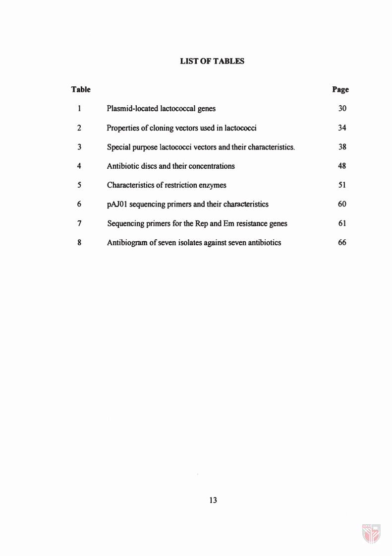
LIST OF TABLES
Table Page
1 Plasmid-located lactococcal genes 30
2 Properties of cloning vectors used in lactococci 34
3 Special purpose lactococci vectors and their characteristics. 38
4 Antibiotic discs and their concentrations 48
5 Characteristics of restriction enzymes 51
6 pAJO 1 sequencing primers and their characteristics 60
7 Sequencing primers for the Rep and Em resistance genes 61
8 Antibiogram of seven isolates against seven antibiotics 66
13
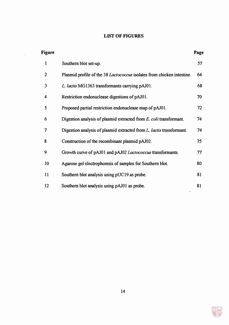
LIST OF FIGURES
Figure Page
1 Southern blot set-up. 57
2 Plasmid profile of the 38 Lactococcus isolates from chicken intestine. 64
3 L. lactis MG1363 transfonnants carrying pAlOl. 68
4 Restriction endonuclease digestions of pAlO 1. 70
5 Proposed partial restriction endonuclease map of pAlO 1. 72
6 Digestion analysis of plasmid extracted from E. coli transformant. 74
7 Digestion analysis of plasmid extracted from L. lactis transfonnant. 74
8 Construction of the recombinant plasmid pAl02. 75
9 Growth curve of pAlO 1 and pAl02 Lactococcus transfonnants. 77
10 Agarose gel electrophoresis of samples for Southern blot. 80
11 Southern blot analysis using pUC19 as probe. 81
12 Southern blot analysis using pAlO 1 as probe. 81
14
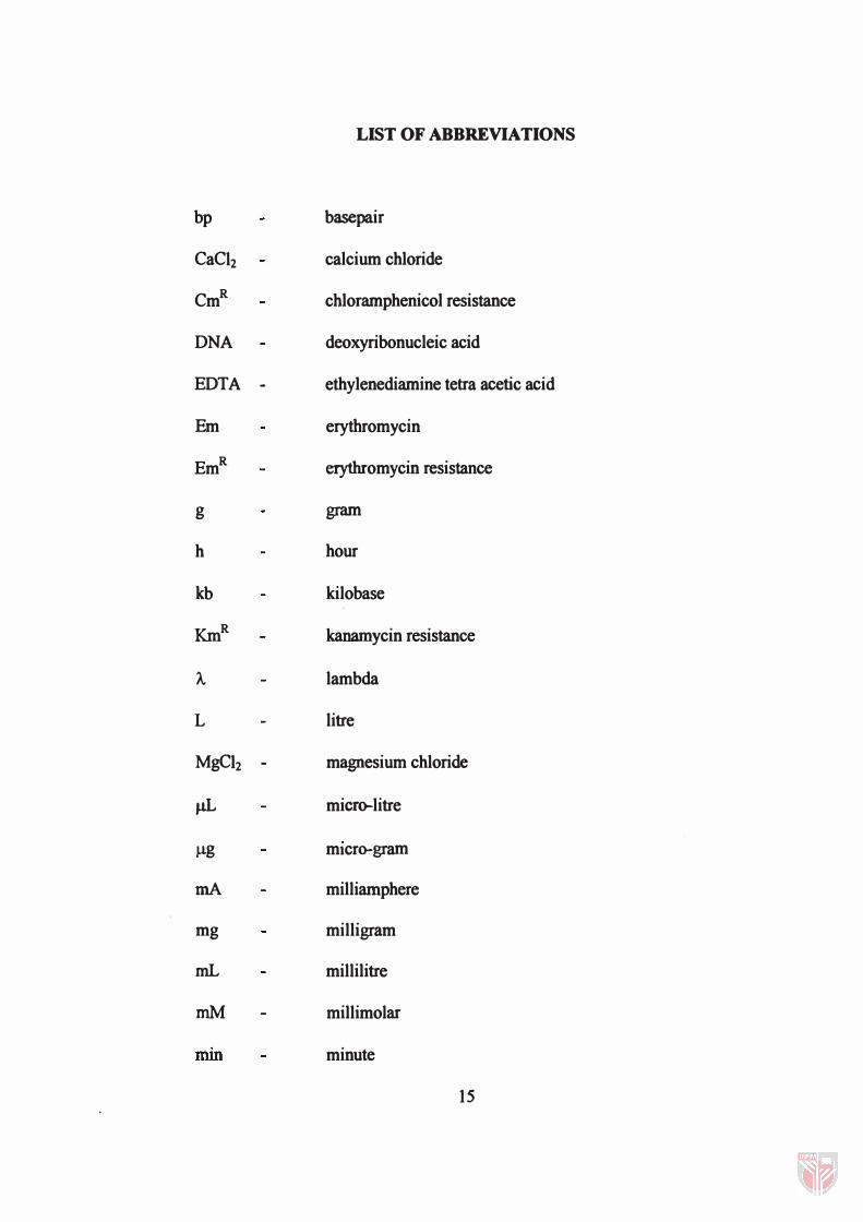
LIST OF ABBREVIATIONS
bp basepair
CaCh calcium chloride
CmR chloramphenicol resistance
DNA deoxyribonucleic acid
EDTA - ethylenediamine tetra acetic acid
Em erythromycin
EmR erythromycin resistance
g gram
h hour
kb kilobase
KmR kanamycin resistance
A. lambda
L litre
MgCh - magnesium chloride
j.1L micf(rlitre
J,lg mIcro-gram
rnA milliamphere
mg milligram
mL millilitre
mM millimolar
mm minute
15
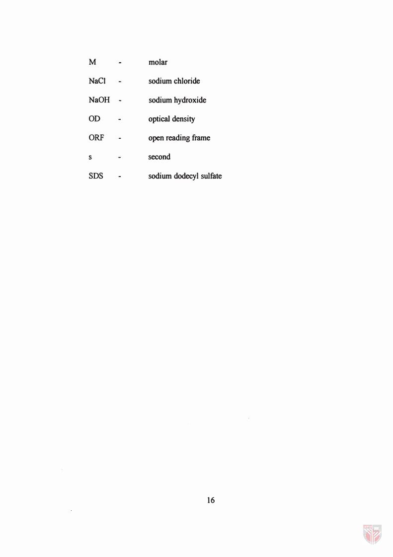
M molar
NaCI sodium chloride
NaOH - sodium hydroxide
OD optical density
ORF open reading frame
s second
SDS sodium dodecyl sulfate
16
CHAPTER I
INTRODUCTION
Lactic acid bacteria, including the members of the genera lactobacillus, Lactococcus,
Leuconostoc, Pediococcus and Streptococcus have long been used in food fermentation
processes. This includes a broad range of products derived from a variety of raw
materials such as vegetables, cereals, meat and milk. The fermentation not only serves as
preservation of the food but they also add to the development of flavour and texture of
the products. Additionally, the Lactococcus and Lactobacillus have been reported to
possess probiotic effects. These features explain the major economic importance of the
lactic acid bacteria The genus Lactococcus however is mainly used in dairy
fermentation such as cheese and buttermilk production.
The genus Lactococcus itself has been studied extensively and is the genetically best
characterised species of the lactic acid bacteria. Nonetheless, molecular studies on the
Lactococcus are relatively new when compared to Escherichia co/i. This is because the
cell membrane of Gram-positive bacteria with its thick layer of peptidoglycan provides
an effective barrier for DNA extractions and gene manipulations. In the late 1970s and
early 1980s, researchers began to develop ways for gene transfer in Gram-positive
bacteria including Lactococcus (De Vos and Simons, 1994). The development of DNA
extraction protocols and gene transfer systems have opened doors for genetic
manipulations in the Lactococcus.
1 7
Molecular studies on the Lactococcus have been focused on improving their abilities in
dairy fermentation especially in cheese and buttermilk production (De Vos and Simons,
1988). These include improvement of starter cultures in order to improve the taste,
texture and odour of cheese (Haandrikman et al. , 1989; Kondo, 1989; Rijnen et al.
2000). Other than that, efforts have been made to produce starter cultures that are
insensitive to bacteriophages (Forde et al. , 1999).
Recently, the usage of the Lactococcus in molecular field has diversified (De Vos and
Simons, 1994; Robinson et aI., 1997; Drouault et al. , 2000) giving new perspectives of
the bacteria. They are generally regarded as safe (GRAS) organisms and together with
the new advances in molecular field, Lactococcus are now being used in various fields
from food fermentation to the synthesis of fine chemicals, pharmaceuticals, and other
products. The bacteria are not only non-pathogenic but they also do not elicit any
immune response and have been ingested throughout history placing it into the group of
bacteria that have the potential to be used for vaccine delivery. Lactococcus exhibits the
ability to express several homogeneous and heterogeneous proteins from both
prokartotic and eukaryotic genes. However, one drawback in the molecular studies of
the Lactococcus is that presently there are no commercially available plasmid vectors
that can be obtained in the market. Therefore, there is a need to develop new plasmid
vectors.
This study looks at a selected number of naturally occurring plasmids of Lactococcus
isolated from chicken intestine, specifically the chicken caeca. The major objective of
1 8
this study is to construct a Laclococcus - E. coli shuttle vector. In order to achieve this
objective, the following steps have to be undertaken:
• to isolate and identify Laclococcus spp. from the chicken intestine,
• to study the naturally occurring plasmid(s) from these isolates,
• to characterise a plasmid pAlO 1 by restriction endonuclease digestion for the
preliminary construction of a Laclococcus - E. coli shuttle vector, and
• to identify and analyse the replication gene and erythromycin resistant gene on the
plasmid pAlO 1 .
19
CHAPTER D
LITERATURE REVIEW
Microflora of Chicken Intestine
The study of the microflora of chicken intestine has long been done since the early
1900s, unfortunately we still have very little understanding about it. One of the major
problems frequently faced in many of these studies was in the recovery of the whole
bacterial population from the intestine (Smith, 1965; Salanitro et al., 1974a). It is
generally known that the intestine is a source of a wide range of microbial species with
various growth requirements. This fact complicates the selection of recovery media used
in the isolation of microbes from the intestine. The recovery media has to be able to
support the growth of most if not all of the bacterial population present in the intestine
(Kelley, 1983). Isolation conditions also play a critical role in microflora studies of the
intestine (Salanitro et al. , 1974b). In general, most of the bacterial species present in the
intestine are mostly facultatively anaerobes and strict anaerobes. This is due to the low
level of oxygen present in the gut intestinal tract. Thus, to look at the microbial
composition of the intestinal tract, the isolation condition should be suitable in order to
recover both the facultatively anaerobes and strict anaerobes bacterial species.
There are several reasons why scientists study the microflora of the chicken intestine.
First, to study the development of intestinal microflora of healthy chickens from the
moment the chicks hatched from their eggs until the chicks reached their age of maturity 20
where the intestinal microflora has stabilised (Barnes et al., 1972� Salanitro et al.,
1974b).
Once the information has been gathered, then changes in the microflora of the chicken
can be monitored especially in comparative studies such as the use of different types of
animal feed and the addition of antimicrobial agents in the feed as growth promoters
(Sarra et al., 1992).
Since the intestine comprises of a wide range of bacterial species, it can be used as a
natural source of certain bacterial species such as Lactobacillus and Lactococcus. Most
of the studies on intestinal microflora of chickens were done actively during 1960s and
1970s. Unfortunately, the genus Lactococcus was only established in 1985 (Schleifer et
al. , 1985). Prior to that the genus Lactococcus was placed in the Streptococcus family
under the Lancefield serological Group N making it difficult to trace any early studies
on the lactococci.
Lev and Briggs (l956ab) were one of the earliest successful researchers to study the
microflora development in chicken intestine. They looked at newly hatched chicks taken
directly from the incubator and found that these chicks hardly had any microorganism in
the crop, gizzard duodenum and ileum as what they have expected However, they were
able to detect dense microflora in the caeca of the chicks, which was mainly dominated
by the Clostridium sp. Only an hour after hatching, Lev and Briggs (1956a) observed a
rapid establishment of microorganisms in all parts of the intestinal tract. Interestingly,
after 12 - 48 h post hatching, it was reported that Escherichia coli and Streptococcus sp.
have started to dominate while the number of Clostridia spp. was observed to decrease
21
even though the total bacterial count increased through time. Nevertheless, with age, E.
coli and Streptococcus spp. counts decreased in all parts of the intestine with exception
of the caeca.
Salanitro et al. (1974a,b), using a non-selective medium developed for isolating rumen
anaerobic bacteria, isolated at least 11 groups of bacteria from the caeca of chickens.
They found that 90 % of the 298 isolates represented species of anaerobic Gram-
negative cocci, facultatively anaerobic cocci and streptococci, Peptostreptococcus,
Propionibacterium, Eubacterium, Bacteroides and Clostridium. A total of 17.5 %
represents two types of facultatively anaerobic bacteria (Gram-positive cocci and E.
coli).
In a subsequent study, Salanitro et al. (1978) found that the streptococci, lactobacilli and
E. coli accounted for about 60 - 90 % of the bacteria in the duodenum, and upper and
lower ileum. Predominant anaerobes recovered from the caeca included Gram-positive
cocci, Eubacterium, Clostridium, Gemmiger, Fusobacterium and Bacteroides species.
Unfortunately, microflora studies on the chicken intestines have dramatically reduced to
almost none in the 1980s. Be that as it may, from the studies presented, we can observe
that the results on the microflora of the chicken intestine vary in some cases. This is due
to many factors such as isolation techniques and recovery medium used. However, most
studies confirmed that the intestinal tract of newly hatched chicks is fairly sterile except
for the caeca and that as soon as the chicks started feeding, the microflora rapidly
developed. Through the findings from the studies discussed, it was also agreed that the
22
caeca of the chicken intestinal tract has the highest bacterial count compared to any
other regions of the intestine.
Lactococcus
General Information
The genus Lactococcus was established by Schleifer et al. (1985) for the lactic
streptococci, S. iactis and S. cremoris. Bergey's Manual® of Determinative
Bacteriology (1975) described this genus as spherical or ovoid in shape with the size of
0.5 - 1.2 X 0.5 - 1.5 J.1IIl. Lactococci usually appears in pairs or short chains in liquid
media. The lactococci are non-motile, catalase negative, oxidase negative, facultatively
anaerobic Gram-positive cocci that do not form endospores. This genus is
chemoorganotroph, meaning that it relies on chemical compounds for energy and uses
organic compounds as a source of electrons. The lactococci are able to ferment a number
of carbohydrates but produces mainly L (+) - lactic acid without any gas production.
The optimum growth temperature for this genus is at 30°C. Another characteristic of
lactococci is that they can grow between 10°C and 40°C but the cells rapidly lose their
viability if they are subjected to temperatures greater than 45°C. They are also of
Lancefield serological Group N. Generally, the lactococci are usually found in dairy and
plant products.
23
Taxonomy
Due to the similarities between S. lactis and s. cremoris, the 9th edition of Bergey's
Manual® of Systematic Bacteriology (1986) grouped s. lactis, S. lactis ssp.
diacetylactis, and S. cremoris into one species; S. lactis. Garvie and Farrow (1982)
suggested the subspecies designation of S. lactis ssp. lactis, S. lactis ssp. cremoris, and
S. lactis ssp. diacetylactis. However, based on nucleic acid hybridisation studies
(Ludwig et. al. , 1985), immunological relationships of superoxide dismutase,
lipoteichoic acid structures, lipid patterns, and fatty acids and menaquinone composition,
Schleifer et al. (1985) proposed that the lactic streptococci be classified within a new
genus, Lactococcus. The International Union of Microbiology Society approved the
Lactococcus genus in 1986 (Anonymous, 1986). The new nomenclature now designates
S. lactis and S. lactis ssp. diacetylactis as Lactococcus lactis ssp. lactis and S. cremoris
as Lactococcus lactis ssp. cremoris. Sandine (1988) suggested that strains of
Lactococcus lactis ssp. lactis, which utilise citmte to for diacetyl, to be termed
Lactococcus lactis ssp. lactis var. diacetylous. The proposed terminology would be quite
beneficial because citrate-fermenting lactococci are so widely used by the dairy industry.
Antimicrobial Resistance in Lactococcus
Antibiotic susceptibility tests have been extensively used as a method of characterisation
of the bacterial species. This method was however mainly used on pathogens with the
24