Embed Size (px)
Citation preview
u n i ve r s i t y o f co pe n h ag e n
Københavns Universitet
Investigation of the physiology of genetically modified strains of Lactococcus lactis,and their potential for accelerated ripening of cheeseRyssel, Mia
Publication date:2010
Document versionPublisher's PDF, also known as Version of record
Citation for published version (APA):Ryssel, M. (2010). Investigation of the physiology of genetically modified strains of Lactococcus lactis, and theirpotential for accelerated ripening of cheese. Department of Food Science, University of Copenhagen.
Download date: 13. okt.. 2019

Preface
i
Preface The present thesis was carried out by Mia Ryssel at the Department of Food Science, Food
Microbiology, Faculty of Life Sciences, University of Copenhagen as well as the Department of
System Biology, Centre for Systems Microbiology at the Technical University of Denmark. The
work fulfils the requirements for a Ph.D. degree at the Research School FOOD, Centre for
Advanced Food Studies (LMC), Frederiksberg, Denmark. The work has partly been financed by
the the Danish Dairy Research Foundation.
I would like to thank my supervisor Associate Professor Henrik Siegumfeldt for excellent
guidance and many fruitful discussions throughout this project. I would also like to thank
Professor Mogens Kilstrup for letting me work with his group and for the many hours he spend
on discussing results obtained in the laboratory.
My thanks also to my colleagues at the Food Microbiology Section for a pleasant and friendly
working atmosphere, a special thanks to Jacqueline, Mette, Line T. and Line O. whom I have
also spent a great deal of time with outside of work. Thanks to all my friends and my soccer
team for taking my mind of the thesis and providing me with new energy.
Warm thanks to my family; Jan, Lonnie, Heidi and Sarah, for always supporting and believing in
me.

Summary
ii
Summary The overall objective of the present Ph.D. thesis was to investigate the physiology of genetically
modified strains of Lactococcus lactis and investigate whether these could be used to accelerate
the ripening time of cheese. This was done by examining the growth of mutants on a solid
surface, examining the stress phenotype of different mutants and finally examining how
proteome and transcriptome analysis could be performed on cells embedded in a matrix.
Autolysis of the starter culture is essential in the ripening of cheese and it is desirable to have a
high amount of autolysis at the ripening stages. As the cells will be present in the cheese matrix
as discrete colonies, rather than dispersed individual cells, we developed a method to quantify
growth and death of single cells on a surface. It was first attempted to use the measurements of
intracellular pH to determine the status of the cells, but this did was unsuccessful because of the
very large variations in the measurements within replicates. Instead we used a combination of
propidium iodide, which penetrates only dead cells with a permeabilised membrane, and bright
field, for visualizing all cells. When we examined the growth by microscopy, very large
variations were found at different fields of view within the same specimen. This underlines the
difficulty in quantitatively interpreting individual microscopic images of cell growth. By taking
the average of many positions, in this case 16, we were able to clearly differentiate between the
growth behaviour of different strains. Growth and death of Lactococcus lactis strains, with
different mutations in the purine biosynthesis pathway, were examined by this method. The
mutants had different levels of guaB (IMP dehydrogenase) expression, and it was shown that
reduced levels of guaB resulted in a decrease of growth rate. The death of the cells was also
influenced by the guaB expression level but not in the same linear way as for the cell growth and
a strain with an intermediate expression of guaB demonstrated the slowest death rate.
Previously it has been reported that Lactococcus lactis can acquire multiple stress resistance
preferentially by mutations in genes involved in purine nucleotide metabolism. A hypothesis was
therefore that purine nucleotides have an influence on the stress phenotype of Lactococcus lactis.
To examine this, genetically engineered mutants with deletion in genes involved in the purine
metabolism were constructed; the genes involved were hpt, hprT, guaA, and guaB. Also a mutant
with an inactive pup gene was examined. Stress phenotypes for L. lactis MG1363 were found to
Summary
iii
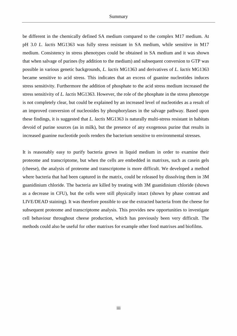
be different in the chemically defined SA medium compared to the complex M17 medium. At
pH 3.0 L. lactis MG1363 was fully stress resistant in SA medium, while sensitive in M17
medium. Consistency in stress phenotypes could be obtained in SA medium and it was shown
that when salvage of purines (by addition to the medium) and subsequent conversion to GTP was
possible in various genetic backgrounds, L. lactis MG1363 and derivatives of L. lactis MG1363
became sensitive to acid stress. This indicates that an excess of guanine nucleotides induces
stress sensitivity. Furthermore the addition of phosphate to the acid stress medium increased the
stress sensitivity of L. lactis MG1363. However, the role of the phosphate in the stress phenotype
is not completely clear, but could be explained by an increased level of nucleotides as a result of
an improved conversion of nucleosides by phosphorylases in the salvage pathway. Based upon
these findings, it is suggested that L. lactis MG1363 is naturally multi-stress resistant in habitats
devoid of purine sources (as in milk), but the presence of any exogenous purine that results in
increased guanine nucleotide pools renders the bacterium sensitive to environmental stresses.
It is reasonably easy to purify bacteria grown in liquid medium in order to examine their
proteome and transcriptome, but when the cells are embedded in matrixes, such as casein gels
(cheese), the analysis of proteome and transcriptome is more difficult. We developed a method
where bacteria that had been captured in the matrix, could be released by dissolving them in 3M
guanidinium chloride. The bacteria are killed by treating with 3M guanidinium chloride (shown
as a decrease in CFU), but the cells were still physically intact (shown by phase contrast and
LIVE/DEAD staining). It was therefore possible to use the extracted bacteria from the cheese for
subsequent proteome and transcriptome analysis. This provides new opportunities to investigate
cell behaviour throughout cheese production, which has previously been very difficult. The
methods could also be useful for other matrixes for example other food matrixes and biofilms.

Sammendrag
iv
Sammendrag
Det overordnede formål med dette Ph.D. projekt har været at undersøge fysiologien for genetisk
modificerede stammer af Lactococcus lactis, og hvordan disse stammer kan anvendes til at øge
modningshastigheden af ost. Dette blev gjort ved at undersøge væksten af forskellige mutanter
på en fast overflade, undersøge stress fænotyper af forskellige mutanter og endeligt ved at
undersøge, hvordan proteom- og transcriptomeanalyser kan foretages på celler indkapslet i en
matrice.
Autolyse af starterkulturen er af afgørende betydning for modning af ost, og høj grad af autolyse
er ønskværdigt. I ost er bakterierne tilstede som små kolonier og ikke spredt som enkelte celler.
Vi udviklede en metode til at kvantificere vækst og død af enkelte celler på en fast overflade.
Der blev forsøgt at bruge målinger af intracellulær pH til at bestemme cellernes status, dette var
dog ikke succesfuldt, da der var store variationer indenfor målinger i den samme prøve. I stedet
blev propidium iodid brugt til at farve døde celler, da dette kun trænger ind i celler med
permeabel cellemembran, og lys mikroskopi blev brugt til at visualisere alle celler. Indenfor
samme bakteriestamme gav vækstundersøgelserne ved mikroskopi store variationer i hvert felt
der blev observeret. Dette understreger problemet ved kvantitativt at undersøge cellevækst ved
enkelte billeder taget ved mikroskopi. Ved at følge vækst i flere felter, i dette tilfælde 16, var det
muligt, klart at se forskel på vækst af forskellige stammer. Denne metode blev brugt til at følge
vækst og død af Lactococcus lactis stammer med forskellige mutationer i purinbiosynthesen.
Mutanterne havde forskellig ekspression af guaB (IMP dehydrogenase), hvor lavere udtryk af
guaB resulterede i nedsat væksthastighed. Hastigheden, hvor med cellerne døde, var også
afhængig af ekspressionen af guaB, men ikke i samme lineære grad som væksten. Mutanten med
den største overlevelse havde et middelsvagt udtryk af guaB.
Det er tidligere vist, at Lactococcus lactis kan blive multiresistent overfor stress ved
genmutationer, der er involveret i purinbiosyntesen. Hypotesen var derfor, at purinnukleotider
har en indflydelse på stressfænotypen af Lactococcus lactis. For at undersøge dette, blev der
konstrueret genetisk modificerede mutanter med deletioner i purinmetabolismen. Generne, der
blev undersøgt, var hpt, hprT, guaA og guaB. En mutant med inaktivt pup gen blev også
undersøgt. Det blev vist, at stressfænotypen af L. lactis MG1363 var anderledes i kemisk

Sammendrag
v
defineret SA medie sammenlignet med det komplekse M17 medie. Ved pH 3.0 var L. lactis
MG1363 stress resistent i SA medie, mens den var sensitiv i M17 medie. Det var muligt at
gentage bestemmelser af stressfænotyper i SA medie, og det blev vist, at når optag af puriner
(tilsætning til mediet) og videreomdannelse til GTP var muligt, blev L. lactis MG1363 og dets
mutanter sensitive overfor syrestress. Endvidere blev stresssensitiviteten af L. lactis MG1363
øget, når fosfat blev tilsat til syrestressmediet. Hvilken rolle fosfat har i forhold til
stressfænotypen, er imidlertid ikke helt afklaret. En mulig forklaring kan være, at fosfat øger
omdannelsen af nukleosider til nukleotider og derved hæver mængden af nukleotider i cellen.
Vores resultater indikerer, at L. lactis MG1363 naturligt er multi-stressresistent i miljøer uden
purinkilder (så som mælk), mens tilstedeværelsen af purinkilder omvendt resulterer i en øget
mængde guaninnukleotider i cellen og dermed øget sensitivitet overfor stress.
Det er forholdsvis let at oprense bakterier fra et flydende medie og anvende disse til proteom- og
transkriptomanalyser, men når bakterierne er indkapslet i en matrice, som f.eks. en kaseingel
(ost), bliver analysen af proteomet og transkriptomet meget besværlig. Vi udviklede en metode,
hvor celler indkapslet i en matrice kan blive frigivet ved opløsning i 3M guanidinium klorid.
Bakterierne dør ved behandling med 3M guanidinium klorid, hvilket er vist som fald i CFU, men
de beholder en fysisk intakt membran, hvilket kunne ses mikroskopisk ved fasekontrast og
LIVE/DEAD farvning. Det var derfor muligt at bruge de ekstraherede celler fra ost til proteom-
og transkriptomanalyser. Dette giver mange nye muligheder for at undersøge bakteriel adfærd i
alle stadier i osteproduktionen, hvilket tidligere har været meget besværligt. Metoden kan
desuden bruges til andre matricer f.eks. andre fødevarer matricer eller biofilm.
List of Abbreviations
vi
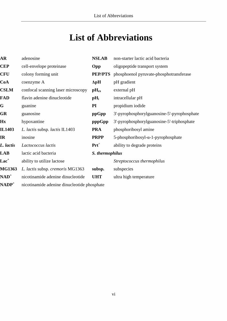
List of Abbreviations
AR adenosine
CEP cell-envelope proteinase
CFU colony forming unit
CoA coenzyme A
CSLM confocal scanning laser microscopy
FAD flavin adenine dinucleotide
G guanine
GR guanosine
Hx hypoxantine
IL1403 L. lactis subsp. lactis IL1403
IR inosine
L. lactis Lactococcus lactis
LAB lactic acid bacteria
Lac+ ability to utilize lactose
MG1363 L. lactis subsp. cremoris MG1363
NAD+ nicotinamide adenine dinucleotide
NADP+ nicotinamide adenine dinucleotide phosphate
NSLAB non-starter lactic acid bacteria
Opp oligopeptide transport system
PEP/PTS phosphoenol pyruvate-phosphotransferase
ΔpH pH gradient
pHex external pH
pHi intracellular pH
PI propidium iodide
ppGpp 3'-pyrophosphorylguanosine-5'-pyrophosphate
pppGpp 3'-pyrophosphorylguanosine-5'-triphosphate
PRA phosphoribosyl amine
PRPP 5-phosphoribosyl-α-1-pyrophosphate
Prt+ ability to degrade proteins
S. thermophilus
Streptococcus thermophilus
subsp. subspecies
UHT ultra high temperature

Contents
1
Contents Preface ................................................................................................................................................... i
Summary .............................................................................................................................................. ii
Sammendrag........................................................................................................................................ iv
List of Abbreviations .......................................................................................................................... vi
1 Introduction ............................................................................................................................ 3
2 Lactic Acid Bacteria .............................................................................................................. 5
2.1 Lactococcus lactis ..................................................................................................................... 10
2.2 Intracellular pH (pHi) ............................................................................................................... 12
3 Cheese Production ............................................................................................................... 16
3.1 Cheese Manufacturing .............................................................................................................. 17
3.2 Cheese Ripening ....................................................................................................................... 22
3.3 Autolysis of Lactococcus in Cheese ......................................................................................... 27
4 Acid Stress in Lactococcus .................................................................................................. 32
5 Nucleotide Metabolism in Lactococcus ............................................................................... 35
5.1 Purine Metabolism .................................................................................................................... 36
6 Conclusion and Perspectives ............................................................................................... 44
7 References ............................................................................................................................ 47
Appendix 1:
M. Ryssel, Z. Duan, and H. Siegumfeldt. (2010) Quantitative Examination of Cell Growth and
Death of Lactococcus lactis by Microscopy. International Dairy Journal. Submitted.
Appendix 2:
M. Ryssel, J. Haaber, M. Liu, M.S. Dawish, K. Hammer, J. Martinussen & M. Kilstrup. (2010)
Purine Induced Stress Sensitivity in Lactococcus lactis. Molecular Microbiology. Submitted.
Contents
2
Appendix 3:
M. Ejby, M. Ryssel, L. Nesic & M. Kilstrup. Fractionation of physically intact bacteria from
casein gels for proteome and RNA analysis. In preparation.
Introduction
3
1 Introduction Lactic acid bacteria (LAB) have been used for fermenting foods for thousands of years.
Fermentation is a way to conserve otherwise perishable foods. A large number of foods that are
eaten every day, e.g. cheese, yoghurt, sausages and sauerkraut, obtain their characteristics from
fermentation with LAB. Nowadays, a starter culture is usually added and the fermentation is
carried out under highly controlled conditions. Especially in the dairy sector large amounts of
different starter cultures are used, and these are crucial for the quality of the final product.
In 2008 a total of 320 tons of cheese were produced in Denmark and from this 240 tons were
exported for a value of 7 billion DKK (www.danishdairyboard.dk), showing the importance of
the cheese industry in Denmark. Cheese production is a complex process, which is dependent on
the microbial activities, during both cheese manufacturing and ripening. Therefore there is a
need to strictly control the whole cheese process. Research within starter cultures and the
development of new starter cultures are therefore important areas of investigation. Better
understanding of the behaviour of starter cultures implemented in production could lead to more
uniform and thereby higher quality products and also reduce loss from failed fermentation.
During cheese production the role of the starter culture changes. Initially the acidification of the
milk depends on the starter culture, at a later stage the autolysis of the starter culture releases
intracellular enzymes, which are essential for the aroma and texture of the cheese (ripening). A
thorough knowledge on cell physiology during the different steps of cheese production helps the
interpretation of how the cells are influenced by the processes and how the processes influence
the cells.
The nature of the cheese fermentation process causes phage problems to continuously appear in
the dairy industry, resulting in slow or dead “vats” and substantial economic losses (Walker et
al., 2000). The key problems in the process are that (i) the milk cannot be heat treated properly to
kill phages without spoiling the cheese properties of the milk, and therefore the fermentation is
not sterile, (ii) the fermentation is conducted under a stringent schedule and the efficiency of the
process is easily disrupted, (iii) the number and diversity of starter cultures available are limited,

Introduction
4
since the products rely on very specialized strains and (iv) continuous use of the same starter
cultures provides an ever present host for phage attack (Klaenhammer et al., 1994). Although the
problems of phages in cheese production are an important issue it will not be dealt with in this
thesis.
This thesis aims to review the LAB and how these affect the manufacturing and ripening of
cheese. Additionally, the ability of LAB to cope with acid stress and the influence of nucleotide
metabolism on the stress phenotype are being described.
Lactic Acid Bacteria
5
2 Lactic Acid Bacteria Lactic Acid Bacteria (LAB) represent a group of bacteria that are functionally related by their
ability to produce lactic acid during fermentation. Other characteristics for LAB are: Gram
positive, katalase negative (pseudokatalase does exist), obligate fermentative, non spore-formers,
acid-tolerant, usually non motile and they have extensive growth requirements. There have been
some controversy about which genera to include in LAB, but the following are included from a
practical and food-technological point of view: Aerococcus, Carnobacterium, Enterococcus,
Lactobacillus, Lactococcus, Leuconostoc, Oenococcus, Pediococcus, Streptococcus,
Tetragenococcus, Vagococcus and Weissella (Axelsson, 2004; Walstra et al., 2006b).
A variety of fermented foods are reliant on LAB to provide the correct flavour, texture and
preservative qualities. The most important genus/species for industrial application are:
Lactococcus (milk), Lactobacillus (milk, meat, vegetables, cereal), Leuconostoc (vegetables,
milk), Pediococcus (vegetables, meat), Oenococcus oeni (wine) and Streptococcus thermophilus
(milk) (Klaenhammer et al., 2002).
Due to their practical significance in fermentation, bioprocessing, food, and more recently,
medicine, LAB have been subject to considerable research and commercial development. One
effort has been to determine the genome sequence of a representative collection of LAB species
and strains (Klaenhammer et al., 2002). Until now 73 LAB have been sequenced completely
(www.ncbi.nlm.nih.gov/genomes/genlist.cgi) and more are in the pipeline. The size of the
chromosome for LAB ranges from 1.8 to 3.4 Mbp (Davidson et al., 1996).
For LAB used in the production of cheese the metabolism of lactose and the degradation of
proteins (caseins) are important properties. These properties will be described below.
Transport and Metabolism of Sugars
There are two different mechanisms in LAB for actively transporting sugar across the cell
membrane: The permease and the phosphoenol pyruvate-phosphotransferase system (PEP/PTS).
Thermophilic LAB species and leuconoctocs use the permease, whereas the PEP/PTS system is

Lactic Acid Bacteria
6
the mechanism used in Lactococci. The permease system transports the lactose across the
membrane at the expense of ATP. Inside the cell, lactose is hydrolysed to glucose and galactose
by β-galactosidase. In the PEP/PTS system, the transport of lactose into the cell is mediated via a
complex system where lactose is phosphorylated and transported across the cell membrane.
Inside the cell, the lactose phosphate is hydrolyzed to glucose and galactose-6-phosphate by
phospho-β-galactosidase (McKay et al., 1970; Hickey et al., 1986; Thompson, 1987; Fox et al.,
1990; Teuber, 1995; Mayra-Makinen et al., 2004).
Figure 1. Major fermentation pathways of glucose. A) homolactic fermentation (glycolysis, Embden-
Meyerhof-Parnas pathway). B) heterolactic fermentation (6-phosphogluconate/phosphoketolase pathway)
(Axelsson, 2004).
Lactic Acid Bacteria
7
LAB can be either homo- or heterofermentative. Glycolysis (Embden-Meyerhof-Parnas
pathway) is the homolactic fermentation where pyruvate is produced. Pyruvate can further be
converted to lactic acid. In homofermentation, 1 mole glucose results in 2 moles of lactic acid
and 2 mole of ATP. The heterolactic fermentation (6-phosphogluconate/phosphoketolase
pathway) results in lactic acid and a significant amount of other end products such as ethanol,
acetate and CO2 (Figure 1). In heterofermentation, 1 mole glucose results in 1 mole of lactic
acid, ethanol, CO2 and ATP (Axelsson, 2004; Walstra et al., 2006b). Glycolysis is used by all
LAB except the leuconostoc, oenococci, weissella and some lactobacilli (group III) (Axelsson,
2004).
In cheese production the pathway for lactose metabolism is dependent on the type of starter used
for the cheese production. Lactococcus lactis produces L-lactate from lactose. Streptococcus
thermophilus hydrolyzes lactose to glucose and galactose, and glucose is further metabolized to
L-lactate, while galactose cannot be metabolized and is secreted in antiport with lactose, which
constitutes a different transport system altogether. Some strains of lactobacilli are galactose-
positive and are able to convert galactose (secreted by e.g. Streptococcus) to DL-lactate (Hickey
et al., 1986; Fox et al., 1997; Axelsson, 2004).
The fermentation of galactose depends on the uptake system. If the PEP/PTS system is used,
galactose-6-phosphate is converted to pyruvate by the tagatose-6-phosphate pathway. When the
permease is used for the uptake of lactose, the Leloir pathway is used for converting galactose to
pyruvate (Figure 2) (Axelsson, 2004).

Lactic Acid Bacteria
8
Figure 2. Galactose metabolism in lactic acid bacteria. A) tagatose-6-phosphate pathway. B) Leloir pathway
(Axelsson, 2004).
The proteolytic system of LAB Milk is rich in nitrogen, but most of it is present as proteins in the form of caseins. The
breakdown of caseins by LAB plays an important role in generating peptides and amino acids for
bacterial growth and in the formation of metabolites that contribute to flavour formation of
fermented products. The proteolytic system of LAB comprises three major components: (i) cell-
envelope proteinase (CEP) that initiates the degradation of extracellular casein into
oligopeptides, (ii) peptide transporters that transport the peptides into the cell, and (iii) various
intracellular peptidases that degrade the peptides into shorter peptides and amino acids (Figure 3)
(Teuber, 1995; Parente et al., 2004; Savijoki et al., 2006; Liu et al., 2010)
Lactic Acid Bacteria
9
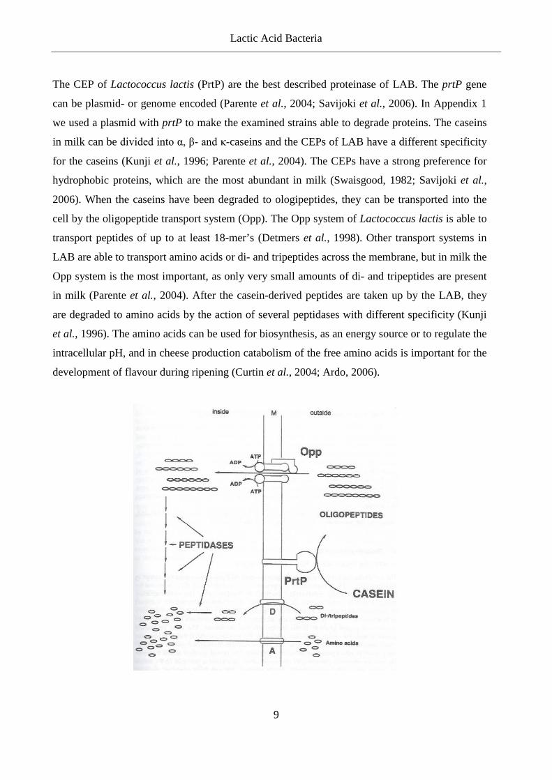
The CEP of Lactococcus lactis (PrtP) are the best described proteinase of LAB. The prtP gene
can be plasmid- or genome encoded (Parente et al., 2004; Savijoki et al., 2006). In Appendix 1
we used a plasmid with prtP to make the examined strains able to degrade proteins. The caseins
in milk can be divided into α, β- and κ-caseins and the CEPs of LAB have a different specificity
for the caseins (Kunji et al., 1996; Parente et al., 2004). The CEPs have a strong preference for
hydrophobic proteins, which are the most abundant in milk (Swaisgood, 1982; Savijoki et al.,
2006). When the caseins have been degraded to ologipeptides, they can be transported into the
cell by the oligopeptide transport system (Opp). The Opp system of Lactococcus lactis is able to
transport peptides of up to at least 18-mer’s (Detmers et al., 1998). Other transport systems in
LAB are able to transport amino acids or di- and tripeptides across the membrane, but in milk the
Opp system is the most important, as only very small amounts of di- and tripeptides are present
in milk (Parente et al., 2004). After the casein-derived peptides are taken up by the LAB, they
are degraded to amino acids by the action of several peptidases with different specificity (Kunji
et al., 1996). The amino acids can be used for biosynthesis, as an energy source or to regulate the
intracellular pH, and in cheese production catabolism of the free amino acids is important for the
development of flavour during ripening (Curtin et al., 2004; Ardo, 2006).

Lactic Acid Bacteria
10
Figure 3. Model of the proteolytic pathway in Lactococcus lactis. Included is transport of di- and tripeptides
and free amino acids, even though they contribute very little to the proteolytic system when the cells are
grown in milk. PrtP, cell-envelope proteinase; Opp, oligopeptide transport system; D, di-/tripeptide transport
systems; A, amino acid transport systems; M, cytoplasmic membrane (Axelsson, 2004).
2.1 Lactococcus lactis Lactococci are homofermentative, they are cocci and occur singly, in pairs or in chains. The cells
are often elongated in the direction of the chain. They grow at 10°C and 30°C, but not at 45°C.
Usually they can grow in 4% (w/v) NaCl (although Lactococcus lactis subsp. cremoris only
tolerates 2% (w/v) NaCl) (Teuber, 1995). Lactococci are closely associated with the
fermentation of dairy products, but only one species, L. lactis, is actually used in the dairy
industry. Two subspecies of L. lactis are important in the dairy industry; L. lactis subsp. lactis
and L. lactis subsp. cremoris, (Schleifer et al., 1985; Axelsson, 2004). The two subspecies have
been intensely studied, mainly because of their industrial interest, and they have become
excellent models for research in metabolism, physiology, genetics, and molecular biology of
LAB (Bolotin et al., 2001). Some phenotypic characteristics are different for the two subspecies
and can be used to differentiate between them; L. lactis subsp. lactis produces ammonia from
arginine and can grow at 40°C, whereas L. lactis subsp. cremoris does neither of those things
(Davidson et al., 1996). Also sequencing of 16S rRNA and DNA hybridization can differentiate
between the subspecies (Salama et al., 1991; Godon et al., 1992). Liu et al. (2010) found that the
pattern of proteolytic enzymes differs between the two subspecies, this again can be used as a
method to identify the subspecies.
L. lactis are anaerobic bacteria, but are aerotolorant (Walstra et al., 2006b). They possess certain
oxygen metabolizing enzymes like superoxide dismutase or NADH oxidases, which enable them
to grow in the presence of oxygen. Therefore they have an aerotolerant phenotype, which has the
advantage that their medium, e.g. milk, does not have to be completely free of oxygen. This is
convenient when used in large scale industrial application (Teuber, 1995).
L. lactis often contains plasmids, and most strains contain between 4 and 7 different plasmids per
cell. Certain plasmids carry important properties for the industrial process (McKay, 1983;
Teuber, 1995). The ability to utilize lactose (Lac+) and degrade proteins (Prt+) is essential for

Lactic Acid Bacteria
11
growth in milk. These abilities, along with others, e.g. the ability to utilise citrate, are associated
with plasmids and IS elements (McKay, 1983; Romero et al., 1993; Liu et al., 2010). This
suggests that, in the adaptation of L. lactis to its predominance in the dairy environment,
plasmids and IS elements have played a crucial role (Davidson et al., 1996).
LAB are nutritionally fastidious and are generally auxotrophic for the amino acids isoleucine,
valine, leucine, histidine, methionine, arginine and proline (Chopin, 1993; Teuber, 1995). For
exponential growth Lactococcus lactis needs a complement of 19 amino acids (Jensen et al.,
1993). In milk the content of free amino acids is too low to support growth of Lactococci to more
than about 107 CFU/ml. For sufficient acidification of the milk for cheese production, the cell
number needs to reach more than 109 CFU/ml (Teuber, 1995). This can only be accomplished by
use of the proteolytic system of the dairy Lactococci, described in the previous section.
The first LAB to be sequenced was Lactococcus lactis subsp. lactis IL1403 by Bolotin et al.
(2001). The genome size was 2.4 Mb. Some unexpected sequences were found; biosynthetic
pathways for all 20 amino acids, a complete set of late competence genes, five complete
prophages and partial components for aerobic metabolism. Some of these systems are not
functional or complete, but they may indicate an evolutionary trend towards minimizing the
chromosome and deactivating unnecessary systems during adaptation to nutritionally complex
but specific environments, such as milk (Chopin, 1993; Bolotin et al., 2001). Comparison of the
genomes of L. lactis subsp. lactis IL1403 and L. lactis subsp. cremoris MG1363 showed that
MG1363 had a larger genome (160 kb) than IL1403 and contained several additional genes for
carbohydrate metabolism and transport, thereby enabling MG1363 able to grow on various
carbohydrates from plants (Wegmann et al., 2007). This could indicate that the original habitat
of Lactococci is plants, and actually many strains of Lactococci are still found in plant
environments.
When genomes from plant-associated strains, L. lactis subsp. lactis KF147 and KF282 (Siezen et
al., 2008) were compared with genomes of L. lactis subsp. lactis IL1403 (Bolotin et al., 2001)
and L. lactis subsp. cremoris SK11 (Makarova et al., 2006) from the dairy environment, it was
found that the plant isolates carried genes e.g. for growth on plant carbohydrates (as was also the
case for MG1363), but these were not seen in the dairy isolates (Siezen et al., 2008). Analysis of
Lactic Acid Bacteria
12
the “unique” genes in the plant isolates, on the basis of the G+C content, indicates that these
were ancient genes that were lost from the dairy isolates, supporting the hypothesis that dairy
strains have adapted to a rich environment and not the other way around (Siezen et al., 2008).
Efficient use of Lactococci by the dairy industry requires understanding of the many aspects of
bacterial physiology, such as the use of sugars and proteins in milk for growth, conversion of
sugars to lactate and synthesis of substances involved in flavours (Bolotin et al., 2001). Not
surprisingly, many studies on Lactococci have been performed, and still more research will have
to be performed before we understand the nature of these mechanisms.
2.2 Intracellular pH (pHi) Intracellular pH (pHi) is an important aspect of cell physiology, and the cells exercise relatively
tight regulation of the pHi. Cells of growing Lactococcus have an initial pH gradient (ΔpH) of
0.8 to 1.0 (alkaline inside), which causes accumulation of acids inside the cell and thereby a
reduction in pHi (Booth, 1985). The lowering of pHi by dissociation of intracellular protons
affects ΔpH, which contributes to the proton motive force. The proton motive force is used as an
energy source in numerous transports across the membrane (Slonczewski et al., 1996; van de
Guchte et al., 2002). A decrease in pHi also changes the enzymatic activity and may denature
proteins and damage nucleic, thereby having a great effect on the cell (Champomier-Verges et
al., 2002).
LABs are able to maintain a ΔpH over a wide range of low external pH (pHex) values (Hutkins et
al., 1993). A pHi of 7.5 to 6.0 (pHex > 5.0) is the optimal pH for growth of LAB and a pHi of 5.0
is critical for growth (Nannen et al., 1991). Cook and Russell (1994) showed that Lactococcus
lactis ML3 were able to grow in MRS media until pHex reached 5.3, pH decreased as a
consequence of lactate accumulation. The pHi decreased as the pHex decreased, so an almost
constant ΔpH was maintained. If the cells maintained a high pHi it would cause a large
accumulation of anions from the fermentation. It therefore appears that for fermentative bacteria
it is better to decrease pHi when pHex is decreased, resulting in less accumulation of anions.
Hutkins and Nannen (1993) and Poolman et al. (1987) observed that some LAB, including
Lactococci, maintain an almost neutral internal pH until the pHex drops to a certain threshold
Lactic Acid Bacteria
13
value and then the pHi starts to decrease. This is in contrast with results reported from other
groups as described above.
Lowering the pHex by HCl or lactate has a different effect on the pHi. A higher pHi could be
maintained when the pHex was decreased by HCl compared to lactate (Cook et al., 1994). This
suggests that when comparing experimental results, it is important to note if the pH is
manipulated by adding organic or inorganic acid, and that organic acid should be used if the
experiment should resemble a milk fermentation.
Measuring pHi
In the early stages, the distribution of radio labelled weak acids or weak bases was used to
measure pHi (Kashket, 1985) but this method has some temporal limitations. Also the pHi can
only be measured under steady-state conditions or if the changes in pH occur slowly (Molenaar
et al., 1991). More convenient methods have now been developed.
Carboxyfluorescein is a ratiometric pH probe that can be used for pHi measurements in the cells.
When excited at 435 nm it shows no pH sensitivity, but excitation at 490 nm results in emissions
that are pH dependent. A ratio, which is independent of the concentration of the probe, can be
obtained from the intensities of the two wavelengths. This ratio can be converted to a pHi-value
(Siegumfeldt et al., 2000). Siegumfeldt et al. (2000) showed that when the pHex was lowered
from 7.0 to 5.0, the pHi for Lactococcus lactis decreased over a time span of 20 min. L. lactis
was able to maintain a ΔpH of 0.8 at both 7.0 and 5.0. For four strains of Streptococcus
thermophilus the authors showed that they were able to maintain a ΔpH of 0.5 at pHex of 7.0 to
5.0. The decrease in pHi for S. thermophilus was faster than for L. lactis.
The method of measuring pHi by carboxyfluorescein is a method that could be used in the dairy
industry to evaluate the physiological status of the starter culture both before addition to the
cheese milk and also within the cheese. If the pHex is acidic and the pHi is the same as pHex, it
indicates that the cells can be dead, whereas cells with a higher pHi are metabolic active, and
therefore more likely to be viable. An intermediate pHi will indicate that the cells are stressed,
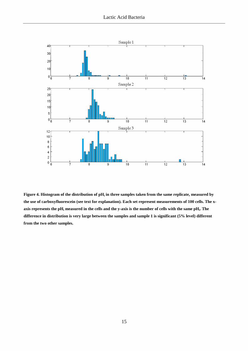
but trying to counter the stress. Early in this project, we tried to use the measurements of pHi to
determine the condition of single cells in a starter culture. After trying to optimize the
Lactic Acid Bacteria
14
experimental procedure it was recognized that the variation in the measurements was very large
and even replicates within the same sample were significantly different (Figure 4). As we have
shown in Appendix 1, large variations within a sample can still lead to useful results, but when
the average pHi of three replicas showed statistical differences, it was decided to abandon the use
of pHi to predict the status of the starter culture in this project, although this method has proven
very useful in other experiments. Another problem with this method was autofluorescence,
especially when measurements were performed in milk or cheese, the milk proteins seemed to
fluoresce to such a degree that it was difficult to observe the single cells and measure their pHi
with a sufficient precision.
If these methods are to be used for measuring pHi within a solid product e.g. cheese it is very
important to obtain results that are representative of the whole sample. In Appendix 1 we showed
that large variations in the growth of cells were observed in different spots on the solid surface.
The variation could be levelled out by performing many (in this case 16) observations. The
observations were chosen automatically, thereby reducing any bias from the researcher who
might prefer to choose a specific area for examination.
Lactic Acid Bacteria
15
Figure 4. Histogram of the distribution of pHi in three samples taken from the same replicate, measured by
the use of carboxyfluorescein (see text for explanation). Each set represent measurements of 100 cells. The x-
axis represents the pHi measured in the cells and the y-axis is the number of cells with the same pHi. The
difference in distribution is very large between the samples and sample 1 is significant (5% level) different
from the two other samples.
Cheese Production
16
3 Cheese Production Many hundreds of different cheeses exist. They differ due to the starter culture, the composition
of the milk used, the manufacturing steps (degree of heating, cutting, stirring, water addition,
salting, moulding and pressing), the ripening time and temperature. All these factors affect the
texture, flavour, colour etc., which gives each cheese its characteristics.
The production of cheese includes different steps that are principally the same for most cheese
varieties; I) acidification, II) coagulation, III) syneresis, IV) moulding, V) salting and VI)
ripening (Fox et al., 1990; Fox et al., 1996b). Cheese production is basically a dehydration
process. Fat and casein in milk are concentrated 6-12 times depending on variety. The hydration
is dependent on the cheese manufacturing steps and on the milk composition (Fox et al., 1990;
Fox et al., 1996b).
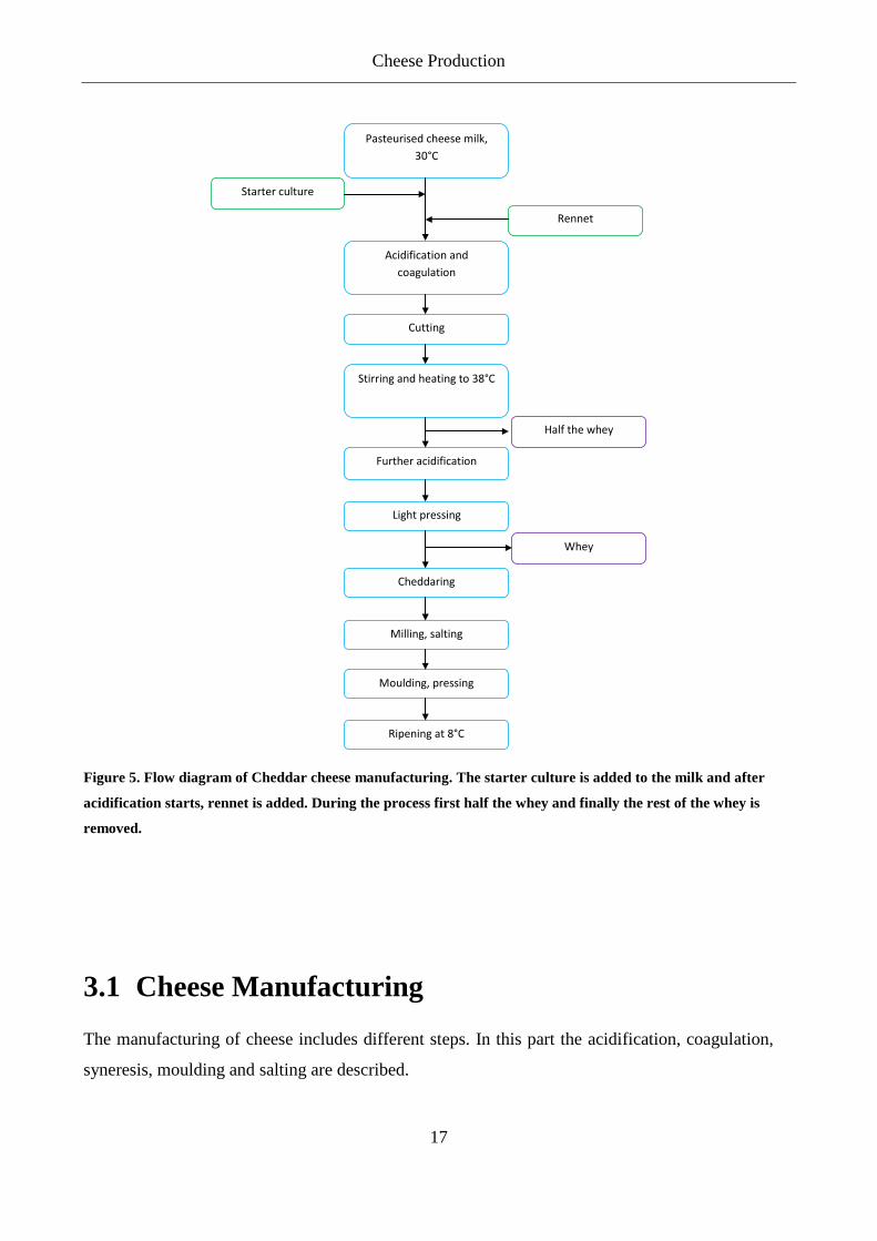
The production of cheese comprises manufacturing and ripening. The manufacturing of Cheddar
cheese, for example, contains the following steps: acidification of cheese milk by the starter,
coagulation by the rennet and cutting. The curd is then cooked, drained, cheddared, milled, salted
and pressed (Figure 5). The cheese is then left to ripen at around 8°C. During ripening,
hydrolysis of proteins and polypeptides takes place as well as the hydrolysis of fats to free fatty
acids and glycerol. The ripening affects the flavour and texture of the cheese. Modification of
amino acids and free fatty acids, which contribute to the flavour, take place late in the ripening
process. Cheddar is a common cheese worldwide and is special from many other cheeses in the
way that it is salted before moulding. Danbo, which is the most common cheese in Denmark, is
moulded and then salted in brine.
In the following sections the manufacture and ripening of cheese are described, with emphasis on
the Cheddar cheese.
Cheese Production
17
Pasteurised cheese milk,
30°C
Starter culture
Rennet
Cutting
Acidification and coagulation
Stirring and heating to 38°C
Half the whey
Further acidification
Light pressing
Whey
Milling, salting
Moulding, pressing
Ripening at 8°C
Cheddaring
Figure 5. Flow diagram of Cheddar cheese manufacturing. The starter culture is added to the milk and after
acidification starts, rennet is added. During the process first half the whey and finally the rest of the whey is
removed.
3.1 Cheese Manufacturing The manufacturing of cheese includes different steps. In this part the acidification, coagulation,
syneresis, moulding and salting are described.
Cheese Production
18
Acidification
One of the primary processes in cheese manufacturing is the acidification of the milk. The
acidification has a huge impact on cheese production and the final quality of the cheese (Fox et
al., 1996b). The starter culture is responsible for the acidification of the milk and the rate of
acidification. E.g. L. lactis subsp. lactis acidifies faster than L. lactis subsp. cremoris (Dawson et
al., 1957). The pH of the cheese curd reaches pH 5.2 to 4.5 depending on the variety of cheese
(Fox et al., 1990). Cheddar cheese is salted in an amount that retards further acidification and
therefore the pH of the Cheddar curd at salting (pH~5.3) is close to the final pH (~5.1) in the
cheese. For cheeses salted in brine the acidification continues after the salting and these cheeses
can be salted at a higher pH (Fox et al., 1996b; Shakeel et al., 2004).
Acidification is important because the lower pH raises the activity of the rennet and inhibits the
growth of unwanted organisms. The pH also affects the texture of the cheese. Acid production
has an effect on almost all steps in the manufacture of cheese and therefore affects both the
composition and quality of the final cheese (Fox et al., 1990).
Coagulation
After the starter culture has started to grow, thereby lowering the pH of the milk, the rennet is
added. Rennet is able to degrade the caseins of the milk, which in the end leads to coagulation.
The rennet coagulation of milk comprises two stages that overlap: The primary phase consists of
the enzymatic breakdown of κ-casein to produce para-κ-caseins and glycomacropeptides, the
second phase consists of aggregation of para-κ-casein Ca2+ (Visser, 1976; Fox, 1988; Dalgleish,
1992; Dalgleish, 1993; Leaver et al., 1995). When 60-90% of the κ-casein has been hydrolyzed,
the micelles are destabilized and start to coagulate (Figure 6) (Dalgleish, 1979; Carlson et al.,
1986; Van Hooydonk et al., 1986). The coagulation of micelles is critically dependent on the
Ca2+ concentration and temperature. The coagulation will not take place if the temperature is too
low (<18°C for bovine milk) (Fox et al., 1996b). Calcium ions might contribute to crosslinking
of the micelles or neutralization of the micelles charges and thereby influences the coagulation of
the micelles.
Cheese Production
19
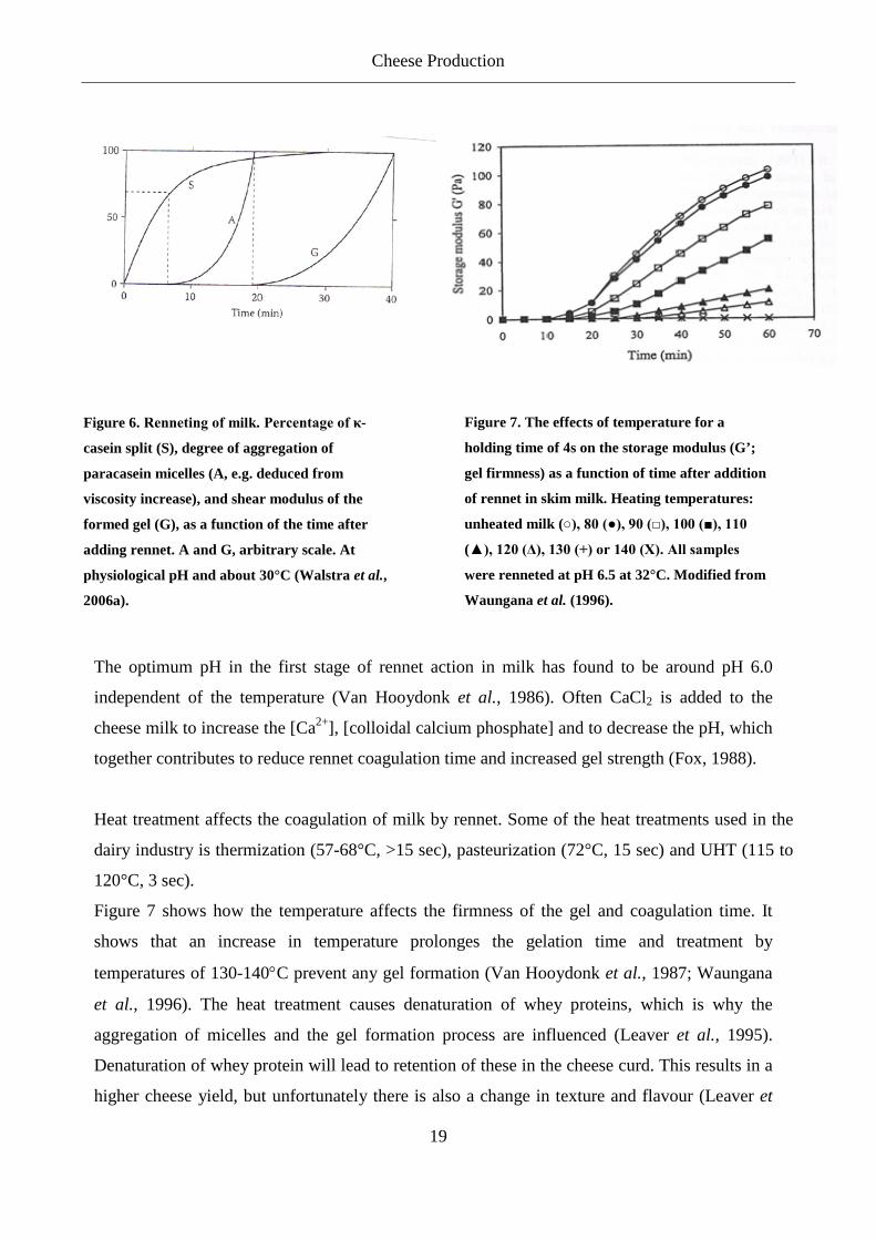
Figure 6. Renneting of milk. Percentage of κ-
casein split (S), degree of aggregation of
paracasein micelles (A, e.g. deduced from
viscosity increase), and shear modulus of the
formed gel (G), as a function of the time after
adding rennet. A and G, arbitrary scale. At
physiological pH and about 30°C (Walstra et al.,
2006a).
Figure 7. The effects of temperature for a
holding time of 4s on the storage modulus (G’;
gel firmness) as a function of time after addition
of rennet in skim milk. Heating temperatures:
unheated milk (○), 80 (●), 90 (□), 100 (■), 110
(▲), 120 (Δ), 130 (+) or 140 (X). All samples
were renneted at pH 6.5 at 32°C. Modified from
Waungana et al. (1996).
The optimum pH in the first stage of rennet action in milk has found to be around pH 6.0
independent of the temperature (Van Hooydonk et al., 1986). Often CaCl2 is added to the
cheese milk to increase the [Ca2+], [colloidal calcium phosphate] and to decrease the pH, which
together contributes to reduce rennet coagulation time and increased gel strength (Fox, 1988).
Heat treatment affects the coagulation of milk by rennet. Some of the heat treatments used in the
dairy industry is thermization (57-68°C, >15 sec), pasteurization (72°C, 15 sec) and UHT (115 to
120°C, 3 sec).
Figure 7 shows how the temperature affects the firmness of the gel and coagulation time. It
shows that an increase in temperature prolonges the gelation time and treatment by
temperatures of 130-140°C prevent any gel formation (Van Hooydonk et al., 1987; Waungana
et al., 1996). The heat treatment causes denaturation of whey proteins, which is why the
aggregation of micelles and the gel formation process are influenced (Leaver et al., 1995).
Denaturation of whey protein will lead to retention of these in the cheese curd. This results in a
higher cheese yield, but unfortunately there is also a change in texture and flavour (Leaver et
Cheese Production
20
al., 1995). If the milk is acidified or CaCl2 is added the effect of the heat treatment can be
reversed (Van Hooydonk et al., 1987).
Syneresis
When the coagulation of the milk has led to a desired firmness, the gel is cut in order to
promote syneresis. At syneresis whey are expelled from the cheese curd. The syneresis does not
occur spontaneously but handling of the rennet gel will cause the syneresis to occur (Walstra et
al., 2006a).
The rate and extent of the syneresis depends on temperature, pH, degree of stirring, protein
concentration and Ca2+ concentration (Fox et al., 1990). The syneresis will be enhanced by
increasing temperature, decreasing pH, addition of CaCl2, fine cutting of the gel, vigorous
stirring and high casein concentration, whereas heat treatment of the milk and increasing fat
content will retard syneresis (Fox et al., 1996b; Dejmek et al., 2004). The pH at the time of the
syneresis has a great influence on the mineral content of the cheese. The loss of calcium and
phosphate from the casein micelles determines the disruption of the micelles and this has a
considerable influence on the structure and texture of the cheese (Lawrence et al., 1983). Low
pH in the curd gives a crumbly texture (Cheshire) whereas a higher pH gives a more elastic
cheese (Emmental) (Fox et al., 1990). Another effect of low pH is that more rennet is retained
in the curd and being more active than at higher pH, meaning increased proteolysis occurs in
cheeses with low pH (Holmes et al., 1977; Dalgleish, 1979; Creamer et al., 1985). The
knowledge of the influence of these factors can help the cheesemaker to control the syneresis of
the curd and thereby control the final water content of the cheese, which affects both the texture
and ripening of the cheese (Fox et al., 1996b).
Moulding
The composition of the curd greatly affects the deformation of the cheese grains. At pH 5.2-5.3
the highest deformability is obtained, at higher or lower pH the deformability of the cheese
grains decreases. With higher water content and higher temperature the deformability also
increases. If the curd has a low pH, and a low water content and the moulding is done at low

Cheese Production
21
temperature, holes can remain in the cheese even if it is heavily pressed (Walstra et al., 2006a).
In Cheddar cheese where the curd is more acidic than for most other cheeses and the curd has a
low water content, it is necessary to apply a large press when the curd is shaped into the
moulds, in order to avoid holes in the cheese (Walstra et al., 2006a).
The size of the mould may have an influence on the final composition of the cheese e.g. for
unsalted cheese curds a larger cheese will end up with lower water content than a smaller one
(Figure 8) (Geurts, 1978; Walstra et al., 2006a). At salting, by brining, the salt gradient will be
levelled out faster for smaller cheeses than for larger cheeses. Again this has an influence on
the ripening and final quality of the cheese.
Figure 8. Distribution of moisture throughout unsalted spherical loaves of cheese of 1 and 6 kg. They have
been moulded from the same curd, lightly pressed and left for three days. The broken lines represent the
average moisture content in the different size cheese (Geurts, 1978).
Salting
Salting is an important step in cheese production. Salt preserves the cheese, affects the ripening
process, the final flavour and the texture. Most cheeses contain about 4-5% salt-in-water
(Walstra et al., 2006b). Dry salting, rubbing and brining are the most common methods used
for salting cheeses (Guinee et al., 2004).
Schroeder et al. (1988) have shown that cheeses with lower NaCl levels have a higher moisture
content. By salting the cheese it absorbs salt, but simultaneously some water is lost from the
Cheese Production
22
cheese and results in a weight loss of approximately 3% for brined cheeses (Walstra et al.,
2006a).
The water content and the microbiology in cheeses changes with different levels of NaCl.
Cheeses with the lowest levels of NaCl generally contain higher numbers of LAB. A NaCl
level of less than 0.85% supports a significantly higher LAB population than cheeses
containing higher concentrations (Schroeder et al., 1988).
In an experiment made by Schroeder et al. (1988) cheeses were produced with 1.44, 1.12, 0.73,
0.37 and 0.07% NaCl. The 1.44% NaCl level being the typical salt level for Cheddar cheeses
on the market. Trained judges in a taste panel were able to differentiate the NaCl levels in the
cheeses and rated them from highest to lowest NaCl content. No other discriminating flavours
were found between the cheese with 1.44% and 1.12% NaCl. Below 1.12% NaCl flavour
scores decreased, higher acidity, lower Cheddar intensity, more intense bitterness and an
unpleasant aftertaste were noted by the taste panel. Another result of the lower NaCl was a
reduction in firmness, while adhesiveness and cohesiveness increased. This is consistent with
the results obtained by Ryssel et al (unpublished), which show that a reduction in salt of 25%
did not have any effect on the texture or sensory properties of Cheddar cheese. These results
indicate that a reduction of salt in cheese is possible without changing the flavour significantly.
This can be an advantage for the many consumers who have a too high intake of salt, which is
detrimental to their health.
3.2 Cheese Ripening The manufacturing steps have a very large influence on the quality of the final cheese.
However, it is during ripening that the flavour and texture develop, which characterise the
different varieties of cheese (Fox et al., 1996b; McSweeney, 2004). The biochemical changes
that take place during ripening are very complex. The composition of the curd has an influence
on the ripening process, as well as the rennet and indigenous milk enzymes (plasmin,
lipoprotein lipase). Starter cultures and their enzymes, non-starter lactic acid bacteria (NSLAB)
and their enzymes also participate in the ripening process (Fox, 1989; Fox et al., 1996b; Fox et

Cheese Production
23
al., 1996c; Fox et al., 2004). The focus in this section will be primarily on the influence of the
starter culture on cheese ripening.
The ripening time for cheeses ranges from 2 weeks (Mozzarella) to more than 2 years (e.g.
Parmesan or extra-mature Cheddar) (Fox et al., 1996a; Fox et al., 1996b). In the ripening of
cheese different reaction take place: (1) metabolism of residual lactose and catabolism of
lactate, (2) lipolysis and catabolism of free fatty acids and (3) proteolysis and further break
down of amino acids (Fox et al., 1996a; Fox et al., 1996b; McSweeney, 2004).
In the production of cheese the primary starter culture have different roles. Initially, they are
responsible for the rapid acidification of the milk through efficient conversion of lactose into
lactic acid. In this later stage of the process, the proteolytic, peptidolytic, and amino acid-
converting enzymes of the starter bacteria play a crucial role in the generation of flavour
components. Most of these enzymes are located in the cytoplasm, while their substrates are
mostly present outside the cells in the cheese matrix. Therefore, the effect of lysis of the starter
culture, whereby the enzymes are released, is generally considered an essential part of the
ripening process (Crow et al., 1995a; deRuyter et al., 1997; Cibik et al., 2000; Ouzari et al.,
2002). At the start of the ripening, the starter cultures have reached a level of 109 CFU/g, but
after 2-3 months of ripening the viability is below 1% of this level (Martley et al., 1972;
Wilkinson et al., 1994b). Although starter culture autolysis is usually beneficial, undesirable
consequences, such as insufficient acid production and removal of residual lactose, can occur if
autolysis occurs too soon. In practice, a balance in autolysis is necessary for optimal Cheddar
cheese ripening and flavour development (Crow et al., 1995a). The autolysis of LAB in cheese
will be discussed further later.
Fate of Lactose in Cheese
Most of the lactose (98%) from the cheese milk is removed with the whey, while the remaining
lactose is metabolised to lactate early in the ripening process (Huffman et al., 1984; Fox et al.,
1996b; Fox et al., 1996c). Fermentation of lactose by homo- and heterofermentation is
described earlier. The starter primarily converts the lactose into L-lactate, but if a high
population of NSLAB are present a considerable amount of D-lactate will be formed. Calcium

Cheese Production
24
D-lactate is less soluble than calcium-L-lactate and can crystallize especially on the surface of
the cheese (Fox et al., 2000).
Lactate may be oxidized to acetate, but this will depend on the NSLAB population and the
availability of oxygen (Fox et al., 1996b). Very extensive metabolism of lactose is seen in
surface mould-ripened varieties, e.g. Camembert and Brie (Fox et al., 1996b; McSweeney et
al., 2004). The metabolism of lactate by Clostridium spp. to butyrate, H2 and CO2 leads to “late
blowing” and off-flavours that are common defects in many cheeses (Fox et al., 1997).
Lipolysis
LAB have low lipolytic activities, but when present in high numbers for long periods, as in the
cheese ripening, the milk fat will be hydrolyzed to a significant degree (Fryer et al., 1967; Fox
et al., 1997). Lipolysis only occurs to a low degree in most cheese varieties, and is considered
undesirable in most varieties because it may impart a rancid taste to the cheese. Exceptions are
Blue cheeses and a few other varieties where lipolysis is considered a part of the characteristic
taste of the cheese (Fox et al., 1996b; Fox et al., 1996c; Fox et al., 1997).
Proteolysis
Proteolysis is an important process in all cheese varieties and is often the rate-limiting process
in the ripening of cheese. The indigenous milk enzymes, rennet, starter culture and NSLAB
contribute to the proteolysis in the cheese (Fox et al., 1996c). Rennet and to some degree
plasmin are mainly responsible for the initial proteolysis of caseins leading to large and
intermediate sized peptides, which can be further degraded by the rennet and other enzymes
from the starter culture and NSLAB. Further breakdown to small peptides and free amino acids
are due to enzymes from the starter culture and NSLAB (Reiter et al., 1969; Okeeffe et al.,
1976; Visser et al., 1977; Okeeffe et al., 1978; Fox, 1989; Lane et al., 1996; Lynch et al.,
1997). Small peptides and amino acids are very important components in cheese as they
contribute to the flavour (Engels et al., 1994). The flavours of different amino acids are very
distinctive and a combination of these flavours is believed to give each cheese its characteristic
taste (Visser, 1993; Fox et al., 1996b). The proteolysis does indeed vary considerably between
different cheese varieties due mainly to differences in the manufacturing process. In cheeses
with a high cooking temperature (e.g. Mozzarella, Emmental) the plasmin activity is increased

Cheese Production
25
by increased conversion of plasminogen to plasmin (Visser, 1993). High cooking temperatures
may also lead to inactivation of the rennet leaving plasmin as the main contributor to the
primary proteolysis (Matheson, 1981; Singh et al., 1990). In other cheeses, e.g. Cheddar, with
low cooking temperatures the rennet contributes to the primary proteolysis of these types of
cheeses (Fox et al., 1996b). About 10-20% of the rennet added to the cheese milk will be
retained in the cheese curd of e.g. Cheddar and Gouda (Visser, 1993; Bansal et al., 2009) and
thus contribute to the proteolysis in the cheese curd.
During ripening proteolysis modifies the texture of the cheese. In Cheddar with its low pH the
caseins are hydrolyzed faster than in cheeses with a higher pH, because the colloidal calcium
phosphate is solubilised and therefore micelles are dissociated and more susceptible to
proteolysis. Additionally, more rennet is retained in the curd and this is more active at low pH
(O’Keffe et al 1975, Holmes et al 1977, Creamer et al 1985).
The rate of secondary proteolysis in cheese depends on the autolysis of the starter culture. If
strains with a high rate of autolysis are used, higher levels of intracellular enzymes are present
and thereby higher secondary proteolysis are observed in the cheese (O'Donovan et al., 1996).
This is further described in section 3.3 Autolysis of LAB in Cheese.
Non-Starter Lactic Acid Bacteria
The NSLAB found in cheese can either be contaminant bacteria entering the milk from the
environment or originates from the milk. These NSLAB include mainly homofermentative and
heterofermentative mesophilic lactobacilli and pediococci and are present at low numbers in the
milk. During the acidification of the milk the starter culture dominates, but during ripening the
NSLAB will grow and reach a maximal number. NSLAB population in Cheddar cheese ranges
from 101 to 104 CFU/g during the first 10 days of ripening and after a few weeks the level ends
at approximately 108 CFU/g (Crow et al., 1993; Wilkinson et al., 1994b; Folkertsma et al.,
1996; Fox et al., 1996c; Crow et al., 2001). It has been shown that NSLAB contributes to the
cheese flavour development. Especially in cheeses with extended ripening time, their
intracellular content are released in the cheese curd after inactivation of enzymes from the
starter culture (Crow et al., 1995a).

Cheese Production
26
Proteinases and peptidases from NSLAB play an important role in proteolysis in some cheese
varieties (Broome et al., 1990; Broome et al., 1991; Fox et al., 1996b). E.g. for Cheddar the
NSLAB seems to supplement positively to the peptidolytic activity of the starter and especially
for the production of amino acids (Lynch et al., 1997). Addition of both heat treated lactobacilli
(Ardo et al., 1988; Ardo et al., 1989) and live lactobacilli (Lee et al., 1990b; Lynch et al.,
1997) appear to accelerate the ripening and are closely related to the higher concentration of
free amino acids in cheese (Broome et al., 1990; Lee et al., 1990a).
Acceleration of Ripening
For the dairy industry the ripening of cheese is a time consuming process and thereby an
expensive process. It would obviously be beneficial for the industry to accelerate ripening. The
problem is how to maintain the characteristic flavour and texture of the cheese varieties while
decreasing the ripening time. Of the biochemical processes involved in ripening, proteolysis
seems to be the rate limiting process (Fox et al., 1996b). Elevated temperature, exogenous
enzymes or modified starter culture are some of the ways the ripening can be accelerated
(Wilkinson, 1993; Fox et al., 1996b). Folkertsma et al. (1996) and Aston et al. (1983; 1985)
have shown that the ripening of Cheddar cheese can be accelerated by raising the temperature
from 8°C to 12-16°C or to 20°C for the first four weeks and then left at 13°C for the rest of the
ripening time. Addition of proteinases and peptidases have been shown to increase ripening
(Law et al., 1982; Law et al., 1983; Hayashi et al., 1990). Also starter cultures that autolyse
easier than normal have shown to increase the ripening activity (Chapot-Chartier et al., 1994;
Wilkinson et al., 1994a).
Sheehan et al. (2009) examined a relative small block of Cheddar (12 kg) and found that there
were considerable variations throughout the block in some factors that influence cheese quality.
E.g. the number of NSLAB was greater in the interior of the block compared to the outer layer,
and this can lead to a variable concentration of released intracellular enzymes when the cells
lyse. This could have an influence on the pH and salt-in-moisture, which also varied throughout
the block. These regional differences in the interior and outer layer of a cheese block are
important in understanding the production of a uniform high quality cheese.
Cheese Production
27
It is in general difficult to examine microbial activity in cheese because the cells are embedded
in the cheese matrix, but in Appendix 3 we have demonstrated a way to purify the cells from
the cheese. This gives us the ability to analyse the proteome and transcriptome of bacterial cells
during cheese manufacturing and ripening. Changes in gene expression can be followed during
cheese production and this could provide novel knowledge on which genes that are important
during the different processes in cheese.
3.3 Autolysis of Lactococcus in Cheese In the early stages of ripening the starter culture enters the stationary phase, become nonviable
and the cell wall and membrane are disintegrated in varying degrees (autolysis) (Crow et al.,
1995a). Therefore, cells in the cheese may be found in many different stages where some are
viable, whereas others are nonviable and may be fully disintegrated.
The autolysis of the starter culture has a prominent effect on biochemical reactions involved in
flavour development (Law et al., 1974; Chapot-Chartier et al., 1994; Crow et al., 1995a; Crow
et al., 1995b; Gatti et al., 1999; Cibik et al., 2000; Ouzari et al., 2002). Intracellular enzymes
access their substrates in the cheese curd via autolysis of the cells (Chapot-Chartier et al., 1994;
Niven et al., 1998; Bunthof et al., 2001). In particular, it has been shown that the release of
intracellular peptidases accelerates amino acid production and results in lower bitterness by
hydrolysis of large hydrophobic peptides. In addition, the amino acid catabolism leading to
aroma formation can also be stimulated by autolysis (Gatti et al., 1999; Bourdat-Deschamps et
al., 2004).
Autolysis of the starter culture in cheese depends on several different factors. The strain has an
influence, but also the cheese-processing conditions such as pH, temperature and salt
concentrations have a big influence (Chapot-Chartier et al., 1994; Wilkinson et al., 1994b;
Crow et al., 1995a; Crow et al., 1995b; O'Donovan et al., 1996; Buist et al., 1998; Meijer et al.,
1998; Hickey et al., 2004; Kozakova et al., 2010). Flavour development may be enhanced
during ripening if strains that lyse rapidly are selected, and/or if processes that favour lysis are
used (Crow et al., 1993; Fox et al., 1996c). A difference in autolysis can be seen between the
two subspecies of Lactococcus lactis; L. lactis subsp. lactis survives better in cheese than L.

Cheese Production
28
lactis subsp. cremoris, this suggests that the level of autolysis of the latter is highest (Dawson et
al., 1957; Martley et al., 1972; Crow et al., 1993; Chapot-Chartier et al., 1994).
Kozakova et al. (2010) examined an autolytic strain (L. lactis subsp. lactis) and a non-autolytic
strain (L. lactis subsp. cremoris) and how different factors influenced their autolysis. The
authors found in their setup that both of them had the highest degree of autolysis when they
were exposed to 200 mM sodium citrate, 6.5 g/l NaCl, pH 6.0, temperatures between 13 and
30°C and when cells in early exponential phase were used. The study also demonstrated that a
medium with 50 mM sodium citrate, 15 g/l NaCl, pH 5.0 and cells cultivated for 6 hours
showed the highest differences in autolysis of the autolytic and non-autolytic strain after 12
days of incubation at 13°C. This medium can be used to screen strains for their autolytic
abilities (Kozakova et al., 2010). Another way to determine differences in autolysis is reported
in appendix 1. It was found that mutants with a lower level of guaB expression (IMP
dehydrogenase) lead to a lower degree of autolysis.
As already mentioned, the caseins of the milk are initially broken down by rennet and the
lactococcal cell envelope proteinase, hereafter the autolysis of the starter culture releases
enzymes beneficial for the cheese ripening (Crow et al., 1995a; Fox et al., 1996c). In cheese,
lysis may be slow because of the stabilizing effect of the matrix and this can leave many cells
in a permeable stage of cell disruption. Peptides from the cheese curd may freely diffuse inside
permeable cells where they are hydrolyzed by intracellular enzymes, thus contributing to the
protein degradation and flavour formation in the cheese (Bunthof et al., 2001).
Lysis of the starter is important for the ripening of cheese, but it is a balance. Intact cells are
required for the utilization of lactose and maybe for the formation of certain aroma compounds.
On the other hand, significant autolysis of the starter is necessary for the proteolysis. The
cheese manufacturing conditions, the composition of the cheese, the inherent characteristics of
the starter with respect to their proteolytic and flavour potentials, and the susceptibility of the
starters to autolysis, will in the end influence the result of the cheese (Crow et al., 1995a; Crow
et al., 1995b).

Cheese Production
29
Monitoring Autolysis in Cheese
Cell viability can be assessed rapidly and directly by fluorescence microscopy (Mcfeters et al.,
1995). This is an advantage when compared to measuring the cell viability by plate counting,
but a limitation is that particular strains cannot be identified (Auty et al., 2001). Fluorescent
indicators for the viability of cells can be based on membrane integrity, enzymes activity,
membrane potential, respiration or pH gradient (Stubberfield et al., 1990; Molenaar et al.,
1991; Rodriguez et al., 1992; Gant et al., 1993; Mcfeters et al., 1995).
The BacLight LIVE/DEAD is a commercial kit from the Invitrogen corporation. In the kit two
nucleic acid stains are used; Propidium iodide (PI) and SYTO 9. PI does not penetrate intact
cell membranes in contrast to SYTO 9 that diffuses through the plasma membrane. SYTO 9
therefore stains all cells regardless of the membrane integrity, whereas PI only stains cells with
damaged membranes. However, PI competes for nucleic acid binding sites with SYTO 9 and
SYTO 9 may be replaced by PI. This means that live cells will fluoresce green (SYTO 9) and
dead cells will fluoresce red (PI). By using fluorescence microscopy with a suitable optical
filter set, the live and dead cells can be viewed separately or simultaneously. The LIVE/DEAD
staining has several advantages compared to other staining methods. It is reliable, rapid and
both viable and total counts are obtained in one step (Boulos et al., 1999).
Figure 9. CSLM analysis of cheese curd for the lytic strain L. lactis AM1 (top) and nonlytic L. lactis 320
(bottom). The images are taken from a section of curd after overnight pressing. AM1 shows that a high
proportion of cells stain red (indicating the majority of cells are nonviable). In contrast, the image taken
from a section of curd manufactured with L. lactis 320 illustrates high numbers of viable (i.e., the
membrane is intact) cells that stain green as shown by CSLM observation (O'Sullivan et al., 2000).
Autolysis of the starter culture can be monitored by staining cells and, at different time points,
counting the number of intact and permeable cells (Bunthof et al., 2001). O'Sullivan et al.
(2000) showed that different strains have different rates of autolysis and this could be seen by
in situ confocal scanning laser microscopy (CSLM) where the cells were stained by
LIVE/DEAD Bacligth (Figure 9). Also Bunthof et al. (2001) have shown that it is possible to
stain cells with LIVE/DEAD staining in a cheese (Figure 10). Many researchers have used
Cheese Production
30
LIVE/DEAD staining as a method for analysing the viability of a bacterial population (Virta et
al., 1998; Boulos et al., 1999; Berney et al., 2007).
Figure 10. CSLM image of 2-week-old Gouda cheese stained with SYTO 9 and PI. Bar, 10 mm (Bunthof et
al., 2001).
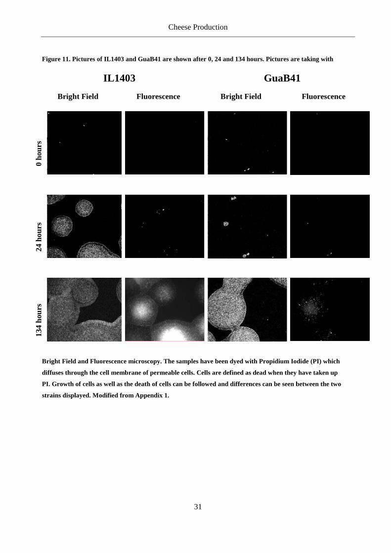
In Appendix 1 we have used PI for staining dead cells, while total cells are estimated in bright
field. When the media used are transparent, staining with SYTO 9 is not required, as cells can
be visualized by bright field. We were able to show a difference in the growth and death rate of
different mutants. It was shown that, within each strain, there were large variations in each field
of view of growth. The large variations that would normally prevent a differentiation of the
mutant, could be overcome by taking the average of several spots. Figure 11 shows an example
of growth of L. lactis subsp. lactis IL1403 and L. lactis subsp. lactis GuaB41 (Appendix 1).
IL1403 have a shorter generation time than GuaB41 in a synthetic minimal media (SA) and this
is consistent with the pictures in Figure 11.
Cheese Production
31
Figure 11. Pictures of IL1403 and GuaB41 are shown after 0, 24 and 134 hours. Pictures are taking with
Bright Field and Fluorescence microscopy. The samples have been dyed with Propidium Iodide (PI) which
diffuses through the cell membrane of permeable cells. Cells are defined as dead when they have taken up
PI. Growth of cells as well as the death of cells can be followed and differences can be seen between the two
strains displayed. Modified from Appendix 1.
IL1403 GuaB41
Bright Field Fluorescence Bright Field Fluorescence
0 ho
urs
24 h
ours
134
hour
s

Acid Stress in Lactococcus
32
4 Acid Stress in Lactococcus In cheese production, the first challenge that the starter culture encounters is acid stress. The
cells thus have to grow at low pH, in order for the production of cheese to succeed. Lactococci
are acid tolerant and their optimal pH conditions for growth are 6.3-6.9 (Harvey, 1965; Bibal et
al., 1988; Andersen et al., 2009). During the fermentation of milk the pH drops because of the
production of lactic acid. At external pH values higher than 5, the Lactococci are able to
maintain a ΔpH of around 1, with alkaline interior. However, by the end of the fermentation the
pH in the milk is around 4.5. At this pH, the cells can maintain a ΔpH of around 0.6 for at least
72 hours. It is the low pHi that causes the arrest in growth, while the nutrients are still in excess
(Nannen et al., 1991). It has been shown that for L. lactis subsp. cremoris 712, pHi values
between 7.2 and 5.9 enables growth, but with decreasing growth rate at low pHi (O'Sullivan et
al., 1997).
Most Lactoccoci appear to have several inducible responses to low pH. Carbon starvation and
mildly acidic media induce resistance to acid stress (Hartke et al., 1994; Rallu et al., 1996;
Hartke et al., 1996; Kim et al., 1999). Exposure to sublethal concentrations of ethanol, H2O2 and
NaCl did not increase tolerance to potentially lethal acid concentration in L. lactis subsp.
cremoris 712. On the other hand, heat and acid did increase acid tolerance (O'Sullivan et al.,
1997), thus indicating that heat and acid stress responses are somehow overlapping (Frees et al.,
2003). A physical maintenance of pH homeostasis might be involved when acid-adapted cells,
during an acid challenge, maintain a slightly higher pHi (around 0.2) than non-adapted cells
(O'Sullivan et al., 1997).
A large number of proteins are induced in LAB during acid adaptive response. Some of the
proteins induced at acid stress adaptation are heat shock proteins (mostly chaperons) and
subunits of the H+-ATPase (O'Sullivan et al., 1999; Champomier-Verges et al., 2002). In L.
lactis subsp. lactis IL1403 and L. lactis subsp. cremoris MG1363, acid adaptation induced 33
and 23 proteins, respectively (Hartke et al., 1996; Frees et al., 2003). Surprisingly, de novo
synthesis of proteins did not seem to be necessary in L. lactis subsp. lactis IL1403 for survival of
acid stress (Hartke et al., 1996), whereas L. lactis subsp. cremoris MG1363, was dependent on

Acid Stress in Lactococcus
33
de novo synthesis of proteins for acid adaptation (Rallu et al., 1996) and this also resulted in
resistance from other stresses (heat, ethanol, H2O2 and NaCl) (O'Sullivan et al., 1997; O'Sullivan
et al., 1999).
Budin-Verneuil et al. (2005) showed that the acid tolerance response of L. lactis subsp. cremoris
MG1363 was different when the cells were grown in M17 compared to SA medium. This might
explain the results from Rallu et al. (1996) where it was found that de novo synthesis of proteins
was necessary for the acid tolerance response in L. lactis subsp. cremoris MG1363 in SA,
whereas Hartke et al. (1996) found that the acid tolerance response in L. lactis subsp. lactis
IL1403 was not depending on de novo synthesis of proteins in M17. This was confirmed by
Budin-Verneuil et al. (2005) using the same strain MG1363 in the two media showing that the
results were not related to difference in subspecies, but a difference in media used. MG1363 was
dependent on de novo protein synthesis for acid tolerance response in SA medium but not in
M17 medium. This should be taken into consideration when results from different experiments
are being compared. In appendix 2 we also confirmed that acid tolerance is different in M17 and
SA medium. At pH 3.0 MG1363 survived 100% in GSA medium but only 0.1% survived in
GM17 medium after 25 min. This shows a big difference between the two media and how the
rich medium GM17 renders the strain sensitive to acid stress.
Exponential growing cells are much more sensitive to acid stress than cells in the stationary
phase (Rallu et al., 2000). During exponential growth there were only small changes in pHi,
suggesting that other factors than pHi were responsible for the acid resistance in the exponential
phase (Alemayehu et al., 2000). The level of autolysis of Lactococcus lactis greatly decreases
when the cells have entered stationary phase before they are exposed to e.g. acid stress. Also
within the exponential phase there is a difference in the level of autolysis. By examining strains
in the exponential phase Kozakova et al. (2010) showed that early exponential phase cells
survived poorly due to autolysis compared to cells in a later growth stage when exposed to acid
stress. This emphasizes the importance of controlling the growth phase of the cells when stress
response is examined.
Rallu et al. (2000) tested 14 mutants that were resistant to acid stress which were constructed by
insertional mutagenesis and selecting survivors at pH 5 and 37.5°C. 10 of these mutants grew

Acid Stress in Lactococcus
34
with the same rate as the wild type (L. lactis subsp. cremoris MG1363) with a doubling time of
around 32 min in M17 media while the last 4 mutants grew slower (doubling time ~43 min).
Auxotrophy in some of the mutants might be correlated to their resistance to acid stress, although
it does not necessarily mean that they grow slow. Six of the mutants survived 120 to 1000-fold
better than MG1363 after 2 days at pH 4.3. Four of these mutants were affected in purine
metabolism. These mutants were also 20 to 110-fold better at surviving carbon starvation at pH
6.8. Since carbon starvation often involves global stress resistance, this could suggest multistress
resistance. This was confirmed, as they were resistant to heat shock and three of them to
oxidative stress (Rallu et al., 2000).
Nucleotide Metabolism in Lactococcus
35
5 Nucleotide Metabolism in Lactococcus Nucleotides are vital for living cells because they are needed for the synthesis of DNA, RNA and
several coenzymes. Therefore the synthesis and salvage of nucleobases, nucleosides and
nucleotides are some of the most important biochemical pathways in the cell (Martinussen et al.,
2003).
Nucleotides play a role in nearly all biochemical processes. They are precursors of DNA and
RNA, and serve as cofactors in sugar and lipid metabolism, in methylation reactions and as
currency of energy in biological systems. They are also constituents of the redox coenzymes
NAD+, NADP+, FAD and CoA (Stryer, 1999). Nucleotides are found as either ribo- or
deoxyribonucleotides, and these are either mono- di- or triphosphates. The triphosphates are
substrates for DNA and RNA polymerases. The mono- and diphosphates are intermediates in the
biosynthetic reactions and they may also arise from hydrolytic or phosphorylytic breakdown of
nucleic acids or other metabolic activities in the cell. Nucleosides and nucleobases may be
converted to nucleotides by the salvage pathways (Kilstrup et al., 2005). Figure 14 shows the
purine biosynthesis and salvage pathways found in Lactococci.
Some LAB are able to produce nucleotides de novo, whereas others require access to either
purines or pyrimidines for growth. After transport into the cell, salvage pathways in auxotrophic
bacteria are used for conversion of the nucleobases or nucleosides to nucleotides. Nucleotides
cannot be transported into the cell but need to be dephosphorylated to nucleosides by
extracellular nucleotidases before entering the cell (Kilstrup et al., 2005). In prototrophic
bacteria, salvage systems for the uptake and utilization of nucleobases and nucleosides, are also
present, and under conditions with excess of purine or pyrimidine sources the de novo pathway
can be shut down (Kilstrup et al., 2005).
5-phosphoribosyl-α-1-pyrophosphate (PRPP) is a common precursor in both purine and
pyrimidine metabolism. PRPP is used for the biosynthesis of nucleotides, nicotinamide
coenzymes (NAD+, NADP+), the amino acids histidine and tryptophane and for the salvage of
nucleobases (Figure 12). Other roles for PRPP are described in the purine metabolism section.
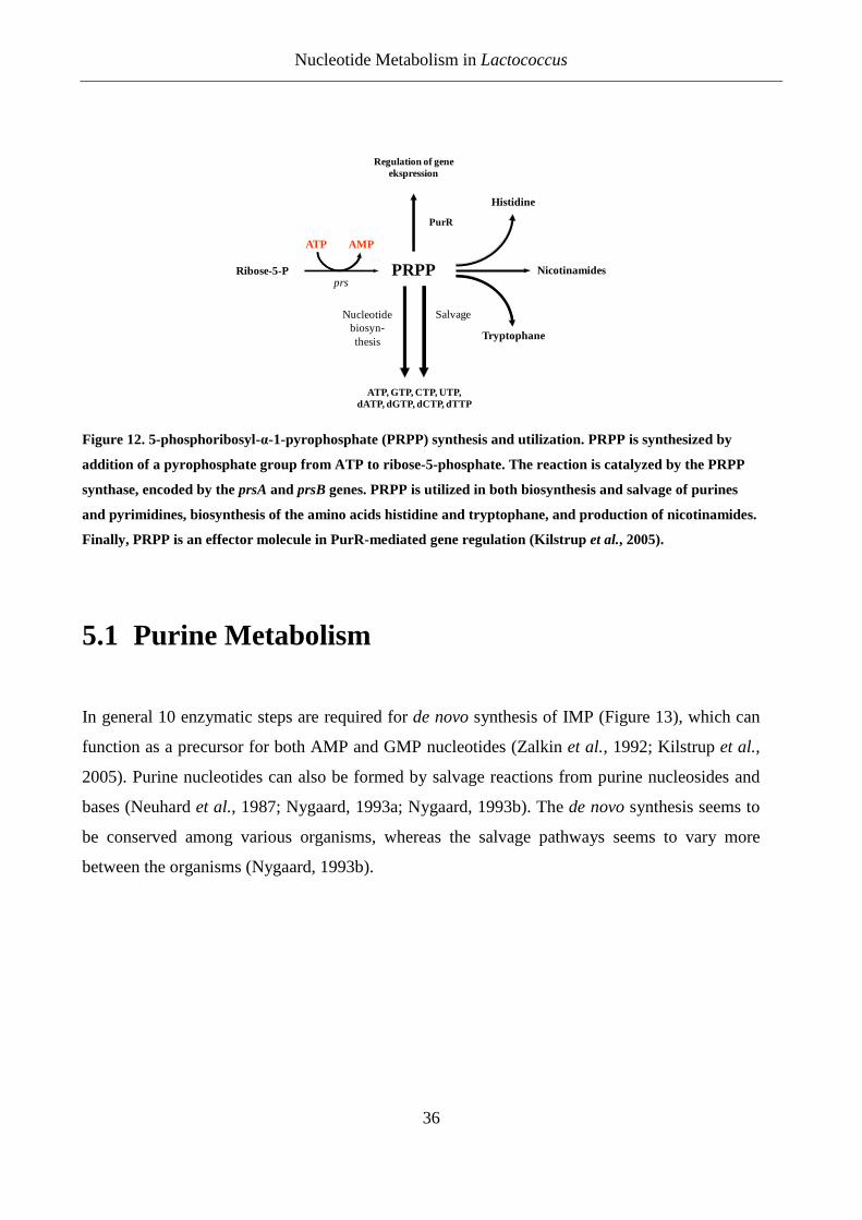
Nucleotide Metabolism in Lactococcus
36
Ribose-5-P
ATP AMP
prsPRPP
ATP, GTP, CTP, UTP,dATP, dGTP, dCTP, dTTP
Nucleotide biosyn-thesis
Regulation of geneekspression
Nicotinamides
Tryptophane
Histidine
Salvage
PurR
Figure 12. 5-phosphoribosyl-α-1-pyrophosphate (PRPP) synthesis and utilization. PRPP is synthesized by
addition of a pyrophosphate group from ATP to ribose-5-phosphate. The reaction is catalyzed by the PRPP
synthase, encoded by the prsA and prsB genes. PRPP is utilized in both biosynthesis and salvage of purines
and pyrimidines, biosynthesis of the amino acids histidine and tryptophane, and production of nicotinamides.
Finally, PRPP is an effector molecule in PurR-mediated gene regulation (Kilstrup et al., 2005).
5.1 Purine Metabolism
In general 10 enzymatic steps are required for de novo synthesis of IMP (Figure 13), which can
function as a precursor for both AMP and GMP nucleotides (Zalkin et al., 1992; Kilstrup et al.,
2005). Purine nucleotides can also be formed by salvage reactions from purine nucleosides and
bases (Neuhard et al., 1987; Nygaard, 1993a; Nygaard, 1993b). The de novo synthesis seems to
be conserved among various organisms, whereas the salvage pathways seems to vary more
between the organisms (Nygaard, 1993b).
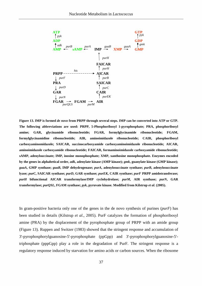
Nucleotide Metabolism in Lactococcus
37
purH
FAICAR
AICAR
SAICAR
CAIR
AIRFGAM
PRPP
PRA
GAR
FGAR
purF
purD
purN
purQLS purM
purEK
purC
purB
purHhis
ATP
ADP
AMP
GTP
GDP
GMPsAMP IMP XMPguaB guaApurApurB
gmk
pykpyk
adk
Figure 13. IMP is formed de novo from PRPP through several steps. IMP can be converted into ATP or GTP.
The following abbreviations are used: PRPP, 5-Phosphoribosyl 1-pyrophosphate; PRA, phosphoribosyl
amine; GAR, glycinamide ribonucleotide; FGAR, formylglycinamide ribonucleotide; FGAM,
formylglycinamidine ribonucleotide; AIR, aminoimidazole ribonucleotide; CAIR, phosphoribosyl
carboxyaminomidazole; SAICAR, succinocarboxyamide carboxyaminoimidazole ribonucleotide; AICAR,
aminoimidazole carboxyamide ribonucleotide; FAICAR, formaminoimidazole carboxyamide ribonucleotide;
sAMP, adenylsuccinate; IMP, inosine monophosphate; XMP, xanthosine monophosphate. Enzymes encoded
by the genes in alphabetical order, adk, adenylate kinase (AMP kinase); gmk, guanylate kinase (GMP kinase);
guaA, GMP synthase; guaB, IMP dehydrogenase; purA, adenylosuccinate synthase; purB, adenylosuccinate
lyase; purC, SAICAR synthase; purD, GAR synthase; purEK, CAIR synthase; purF PRPP amidotransferase;
purH bifunctional AICAR transformylase/IMP cyclohydrolase; purM, AIR synthase; purN, GAR
transformylase; purQSL, FGAM synthase; pyk, pyruvate kinase. Modified from Kilstrup et al. (2005).
In gram-positive bacteria only one of the genes in the de novo synthesis of purines (purF) has
been studied in details (Kilstrup et al., 2005). PurF catalyzes the formation of phosphoribosyl
amine (PRA) by the displacement of the pyrophosphate group of PRPP with an amide group
(Figure 13). Ruppen and Switzer (1983) showed that the stringent response and accumulation of
3'-pyrophosphorylguanosine-5'-pyrophosphate (ppGpp) and 3'-pyrophosphorylguanosine-5'-
triphosphate (pppGpp) play a role in the degradation of PurF. The stringent response is a
regulatory response induced by starvation for amino acids or carbon sources. When the ribosome

Nucleotide Metabolism in Lactococcus
38
encounters uncharged tRNA’s, the stringent factor (RelA) become activated and further activate
the synthesis of the two nucleotides, ppGpp and pppGpp (Haseltine et al., 1973; Wendrich et al.,
2002). This high level of (p)ppGpp accumulation that is induced under amino acid starvation
(Ruppen et al., 1983), is known to specifically inhibit the IMP dehydrogenase enzyme, resulting
in limited synthesis of XMP and GMP (Gallant et al., 1971; Ochi et al., 1982; Inaoka et al.,
2002; Budin-Verneuil et al., 2007). Some mutants in the relA gene have shown stress resistant,
as described more in the next section.
L. lactis rely on phosphoribosylation of bases in purine salvage since they cannot directly
phosphorylate purine ribonucleosides. PRPP is essential in these organisms because it is used in
the biosynthesis of nucleotides and the salvage of nucleobases (Kilstrup et al., 2005).
Kilstrup and Martinussen (1998) showed, as previously found in Bacillus subtilis (Weng et al.,
1995), that PRPP in L. lactis is not only a substrate in the biosynthetic pathway, but also induce
gene expression of genes in the de novo synthesis of purines. The expression of the purine
biosynthesis genes in Lactococcus is dependent on an activator (PurR), which stimulates
transcription under high PRPP concentrations. The regulation of the de novo pathway is
therefore feed-forward by its precursor PRPP, which ensures that the pathway is only fully active
when the substrate is found in excess. PRPP is under allosteric regulation from ADP, which has
the effect that a high concentration of ADP will inhibit the catalytic effect of PRPP (Eriksen et
al., 2000) and prevent activation of PurR regulated genes. Martinussen et al. (2003) showed that
in the presence of exogenous purines the PRPP pool was low. This again indicates that PRPP
functions as a reporter of the purine nucleoside status in the cell.
Purine-requiring mutants of L. lactis cannot grow in milk, as the wild type strain (Dickely et al.,
1995). This means de novo synthesis of purine nucleotides are required for L. lactis when
growing in milk (Nilsson et al., 1998). Martinussen et al. (2003) showed that addition of
exogenous purines in GSA media led to an increase in both growth rate and size of the purine
nucleotide (triphosphate) pools in L. lactis subsp. cremoris MG1363, which implies that the
strain has a partial purine requirement. It appears that the capacity of de novo synthesis of both
purines and pyrimidine in L. lactis are limited (Kilstrup et al., 2005). Reduced growth rates were
obtained by growing strains with mutations in nucleotide biosynthesis genes under conditions
Nucleotide Metabolism in Lactococcus
39
where the availability of nucleotide precursors was limited (Martinussen et al., 2003) (Appendix
1).
Two different nucleoside transporters have been found in L. lactis. One enables the uptake of
uridine and the other is responsible for the uptake of different purine nucleosides and cytidine
(Martinussen et al., 2003). This means that adenosine (AR), guanosine (GR), inosine (IR) and
cytidine will be competing for the same transporter and that high concentration of one nucleoside
will diminish the uptake of the other nucleosides. In this way it is possible to modulate the
intracellular nucleotide pools. This is a principle we have used in Appendix 2.
Purines and Stress Resistance
Rallu et al. (2000) obtained 21 different insertional mutants of L. lactis subsp. cremoris MG1363
that are resistant to acid stress. Some of these mutants were only resistant to acid stress and some
were multi stress resistant. Analysing these mutant indicated that intracellular pools of phosphate
and guanine nucleotides (both GMP/GTP and (p)ppGpp) could be involved in the regulation of
acid stress resistance. Some of the mutants examined by Rallu et al. (2000) was guaA, hpt, deoB,
pstS and relA*. It is important to know the function of these genes in relation to the purine
metabolism, in order to understand how they can lead to stress resistance, therefore a short
description of these genes follows (also see Figure 14):
guaA
The GuaA enzyme catalyzes the formation of GMP from XMP, and inactivation of the guaA
gene will block GMP synthesis and render GMP formation dependent upon the salvage of GR or
G (guanine) from the medium.
Hpt and hprT
The genes hpt and hprT encode guanine and hypoxantine phosphoribosyl tranferases that
catalyses the formation of GMP and IMP from G and Hx (hypoxanthine), respectively (Nilsson
et al., 1992) (Appendix 2). Inactivation of hpt and hprT will block GMP formation by salvage
and render GMP formation dependent upon de novo synthesis.
Nucleotide Metabolism in Lactococcus
40
deoB/pup
The gene deoB is part of en operon containing deoB, orfC (unidentified open reading frame) and
deoD (purine-nucleoside phosphorylase (pup)). Insertion in deoB is believed to inactivate the
whole operon and therefore also the pup gene (Duwat et al., 1999). The Pup enzyme catalyzed
the convertion of GR, IR and AR to G, Hx and A, respectively (Martinussen et al., 1995). An
inactivation of the (deoB or orfC or) pup gene will therefore prevent salvage of purines from
nucleosides.
pstS
pstS encode the substrate binding component of the high affinity ABC transporter of phosphate.
Inactivation of pstS leads to a reduction in the intracellular phosphate concentration. Phosphate is
needed for salvage of purines and a lowering of the phosphate concentration might therefore lead
to a lowering of the GMP/GTP pool.
relA
The relA mutant, which has been found to be multi-stress resistant (Rallu et al., 2000) has by a
more complicated pathway to stress resistance. The wild type RelA protein is closely bound to
the ribosome and produces (p)ppGpp, from GTP and GDP, when the ribosome encounters
uncharged tRNA’s during the stringent response (Haseltine et al., 1973; Wendrich et al., 2002).
It was found that the multi-stress resistant mutation resulted in truncated RelA proteins, called
RelA*, which synthesized (p)ppGpp in an uncontrolled fashion (Rallu et al., 2000; Budin-
Verneuil et al., 2007). Since high concentrations of (p)ppGpp is known to specifically inhibit the
IMP dehydrogenase enzyme, the synthesis of XMP and thereby GTP was proposed to be limited
in the relA* mutant (Ochi et al., 1982; Rallu et al., 2000; Inaoka et al., 2002; Budin-Verneuil et
al., 2007). Besides limiting the production of GTP the high production of (p)ppGpp drains the
GDP and GTP pools and results in even lower levels. In either case a RelA* mutant is expected
to have low levels of GMP and GTP.

Nucleotide Metabolism in Lactococcus
41
G
ATP
ADP
AR
AMP
A
GTP
GDP
GMP
GR
sAMP IMP XMP
IR XR
Hx X
PRPP
Purine de novo pathway
guaB guaA
guaC
purApurB
add xpt hpthprT
apt
puppuppup
gmk
pykpyk
adk
hpthprT
pppGpprelAppGpp
relA Nucleotideinterconversionreactions
Salvagereactions
De novosynthesisreactions
Figure 14. Purine metabolic network in L. lactis. The metabolic network is devided into three types of
reactions, de novo synthesis reactions, nucleotide interconversion reactions, and salvage reactions. IMP is
formed de novo from PRPP through several steps. Only bases and nucleosides, the substrates for the salvage
reactions can be supplied from the medium. The following abbreviations are used: PRPP, 5-Phosphoribosyl
1-pyrophosphate; A, adenine; Hx, hypoxanthine; X, xanthine; G, guanine; AR, adenosine; IR, inosine; XR,
xanthosine; GR, guanosine; sAMP, adenylsuccinate; IMP, inosine monophosphate; XMP, xanthosine
monophosphate. Enzymes encoded by the genes in alphabetical order, add, adenosine deaminase; adk,
adenylate kinase (AMP kinase); apt, adenine phosphoribosyltransferase; gmk, guanylate kinase (GMP
kinase); guaA, GMP synthase; guaB, IMP dehydrogenase; guaC, GMP reductase; hpt, hypoxanthine/guanine
phosphoribosyltransferase; hprT, hypoxanthine/guanine phosphoribosyltransferase; pup, purine nucleoside
phosphorylase; purA, adenylosuccinate synthase; purB, adenylosuccinate lyase; pyk, pyruvate kinase; relA,
(p)ppGpp synthase and xpt, xanthine phosphoribosyltransferase. Modified from Kilstrup et al. (2005).
Rallu et al (2000) showed that addition of GR/G did not have any influence on the survival of
the hpt mutant in acid stress experiments, but addition of Hx made the mutant die out at the same
rate as the wild type. The guaA mutant stayed resistant with the addition of Hx, but was not
resistant when GR or G was added. The results for the guaA mutant are consistent with the result
we got (see Appendix 2) and with the hypothesis that a low GMP/GTP pool induces stress
resistance. Multi-stress resistant guaA mutants have been shown to grow slower than the wild
type (Rallu et al., 2000), which indicates that the supply of GMP from purine salvage was
suboptimal. The guaA gene is shown to be inhibited by the addition of decoyinine, and addition
of decoyinine to the wild type before acid challenge gives the cells 100 fold better survival than

Nucleotide Metabolism in Lactococcus
42
the non treated cells (Rallu et al., 2000). This again indicates that the intracellular nucleotide
pool have to be low for the cell to be resistant to acid stress.
Three of the acid resistant mutant from Rallu et al (2000) were further examined by Budin-
Verneuil et al. (2007) for their protein expression compared to the wild type. The three mutants
had insertions in the guaA, relA or pstS gene (see Figure 14). When Budin-Verneuil et al. (2007)
examined the expression of proteins in the mutants compared to the wild type they found
significantly different patterns. Of a total of 58 upregulated proteins in the three mutants, but few
of these genes were upregulated in all three mutants. The three mutants had similar effects on the
concentration of GTP. As mentioned, insertion in guaA leads to a decrease in GMP/GTP
because the formation of GMP from XMP is inhibited. The truncated RelA* protein promotes
the accumulation of (p)ppGpp, which leads to inhibition GuaB (IMP dehydrogenase) and this
again leads to a decrease in GMP/GTP. The lower intracellular phosphate concentration, as a
result of the insertion in pstS gene, probably affects the concentration of GMP/GTP by
decreasing it (Budin-Verneuil et al., 2007). This means that these acid resistant mutants have a
low concentration of GMP/GTP, this is consistent with our result from Appendix 2. Here we also
show a better survival at acid challenging by cells with low intracellular GMP/GTP
concentration.
Duwat et al. (1999) examined temperature resistant mutants found in a recA strain of L. lactis
subsp. cremoris MG1363. Out of 18 obtained mutants 10 were involved in guanine metabolism.
They were affected in the genes deoB, guaA and tktA. The function of deoB and guaA were
explained above. tktA encodes a transketolase that catalyses the transformation of xylose-5-
phosphate to ribose-5-phosphate (a precursor in the nucleotide biosynthesis). All these three
genes are therefore likely to affect the guanine nucleotide pools in the cell by lowering it. The
temperature resistance of the recA-guaA mutant could be lost by addition of GR or G to the
media, which would properly increase the guanine nucleotide pool. For the recA-deoB mutant
the resistance was lost by addition of G and Hx and for the recA-tktA mutant it was the addition
of GR, G, Hx and IR that made the strain sensitive (Duwat et al., 1999). These results indicate
that the guanine nucleotide pools also have an impact on the temperature sensitivity in L. lactis
subsp. cremoris MG1363, this was also the case for acid resistance.
Nucleotide Metabolism in Lactococcus
43
In appendix 1 it is shown that when lowering the expression of guaB the generation time in GSA
is increased. This tendency is illustrated in Figure 11. L. lactis subsp. lactis IL1403 is the wild
type and L. lactis subsp. lactis SGJ126 is a mutant that expresses only 41% of guaB compared to
IL1403. The cell death of SGJ126 is much lower than IL1403, which could be explained by the
low level of guanine nucleotides in SGJ126. Starvation for guanine nucleotides in Lactococcus
makes it more stress resistance (appendix 2). All mutants examined in appendix 1 with a lower
expression of guaB compared to the wild type dies in a lower rate (Figure 11). Again this points
out the significance of the GMP/GTP level on the survival and stress resistance of the cells.
It is shown in Appendix 2 and also shown by Rallu et al (2000) that the GMP pools may have an
important influence on the acid stress resistance of MG1363. Also for temperature resistance the
GMP pools was shown to have an influence (Duwat et al., 1999). Of special interest for studying
stress resistance are the genes guaA, guaB, hpt, hprT, pup and relA and the way their gene
products are related to purine nucleotide metabolism (Figure 14). These mutants are able to
prevent de novo synthesis of purine nucleotides (guaA, guaB and relA) or salvages of purine
bases and nucleosides (hpt, hprT and pup) and lead to low GMP/GTP pools, which again lead to
stress resistance as shown Appendix 2. The relA* mutant was not examined in Appendix 2, but
we are convinced that it is the low GMP/GTP pool in the mutant that gives it its stress resistance
phenotype.
Conclusion and Perspectives
44
6 Conclusion and Perspectives Results from Appendix 1 show that large variations are seen when examining growth of
Lactococcus lactis on a solid surface. We showed it was necessary to make multiple observations
in order to lower the standard error means, so that differences in growth between strains could be
determined. The developed method could be further developed to examine growth and death of
cells on other solid surfaces, for example in cheese. This could provide us with a greater
knowledge of how cells behave in microcolonies on solid surfaces or within a food matrix.
The mutants used in Appendix 1 had different levels of guaB expression. The level of gene
expression influenced the growth rates as well as the death rates of the mutants. Both the wild
type and the mutant with wild type expression of guaB, grew faster and to a higher number
compared to the mutants with a lower expression of guaB. Furthermore, they had the highest
ratio of dead cells. A higher expression of guaB would probably lead to a higher amount of
GMP/GTP and this leads to stress sensitivity as shown in Appendix 2. However, additional
studies on mutants with higher expressions of guaB would be needed to confirm this. Since
stress sensitive strains have a higher level of autolysis, this could accelerate the ripening of
cheese. However, further studies are needed in order to shed light on this theory.
In Appendix 1 we also observed that cells from the same strain stopped growing at different
stages, visualized as microcolonies of different sizes. As mentioned in the Appendix, we do not
have an explanation for this. Further studies are needed to examine the background for this
phenomenon, and could be a subject for future studies.
In Appendix 2 we have shown that Lactococcus lactis were intrinsically resistant to acid stress,
but became sensitive when grown in media containing purines and where conversion in
Lactococcus lactis to GMP/GTP was possible. We were puzzled by the fact that the double
deletion mutant Δhpt ΔhprT was stress sensitive in the GSAM17 medium that we had designed.
The double mutant was not expected to be able to convert nucleobases (Hx, G) into nucleosides
(IMP, GMP), but it appeared to retain this ability under certain conditions. The results in
Appendix 2 suggest that the apt gene product could be responsible for conversion of Hx and G
Conclusion and Perspectives
45
into IMP and GMP, and for future studies a Δapt Δhpt ΔhprT triple mutant could be used to
elucidate this issue.
It is possible to measure the intracellular levels of GMP/GTP, and by combining these
measurements with the stress experiments, it might provide us with even deeper insight into
stress sensitivity. The addition of phosphate appeared to increase stress sensitivity in the
experiments from Appendix 2. The role of phosphate is not quite clear, but we do consider that
phosphate addition could speed up the conversion of nucleosides and thereby increase the level
of nucleotides. By determining the GMP/GTP levels before and after phosphate addition, the
effect of phosphate addition on the level of GMP/GTP could be determined.
In the future, further investigation on the role of guanine nucleotides in the acid stress sensitivity
of Lactococcus lactis (and maybe other LAB) is required. In my view, it is of the utmost
importance to the dairy industry to widen their knowledge about the background for stress
resistance and sensitivity in LAB. If acidification to a low pH is desired, the Lactococcus should
grow with a low amount of G-nucleotides, whereas adding precursors for the G-nucleotide will
inhibit the Lactococcus because of stress sensitivity. More knowledge of this balance would be
of great importance in regulating the process and creating high quality products.
The results from Appendix 3 show, that we are able to fractionate cells from a cheese (casein)
matrix by the use of guanidinium chloride. The cells remained intact, we could subsequently
purify RNA from the cells that can be used further for transcriptome analysis. Proteome analysis
of cells after guanidinium chloride extraction from acidified milk and from a Cheddar cheese
was performed and showed promising results. Until now it has been rather difficult to examine
the proteome and transcriptome expression of LAB as soon as they are embedded in the
coagulated milk or cheese. The ability to fractionate the cells from a casein matrix permits us to
follow the behaviour of the LAB during the whole cheese production and thereby enabling us to
regulate and control the process more elaborately.
The method developed in Appendix 3, might be used for other food matrixes, and would be a
great advantage in allowing examination of microbial development, proteome and transcriptome
analysis in many kinds of food matrixes. We know that cells behave differently in a synthetic

Conclusion and Perspectives
46
laboratory medium compared to a complex food matrix, and by extracting cells from these
complex matrixes we will get a better understanding of microbial behaviour in the actual
products.
Rererences
47
7 References Alemayehu,D., O'Sullivan,E., and Condon,S. (2000) Changes in acid tolerance of Lactococcus lactis
during growth at constant pH. Int J Food Microbiol 55: 215-221.
Andersen,A.Z., Carvalho,A.L., Neves,A.R., Santos,H., Kummer,U., and Olsen,L.F. (2009) The metabolic pH response in Lactococcus lactis: An integrative experimental and modelling approach. Computational Biology and Chemistry 33: 71-83.
Ardo,Y. (2006) Flavour formation by amino acid catabolism. Biotechnology Advances 24: 238-242.
Ardo,Y., Mansson,H.L., Hedenberg,A., and Larsson,P.O. (1989) Studies of Peptidolysis During Early Maturation and Its Influence on Low-Fat Cheese Quality. Milchwissenschaft-Milk Science International 44: 485-495.
Ardo,Y., and Pettersson,H.E. (1988) Accelerated Cheese Ripening with Heat-Treated Cells of Lactobacillus-Helveticus and A Commercial Proteolytic-Enzyme. J Dairy Res 55: 239-245.
Aston,J.W., Fedrick,I.A., Durward,I.G., and Dulley,J.R. (1983) The Effect of Elevated Ripening Temperatures on Proteolysis and Flavor Development in Cheddar Cheese .1. Higher Initial Storage Temperatures. New Zealand Journal of Dairy Science and Technology 18: 143-151.
Aston,J.W., Giles,J.E., Durward,I.G., and Dulley,J.R. (1985) Effect of Elevated Ripening Temperatures on Proteolysis and Flavor Development in Cheddar Cheese. J Dairy Res 52: 565-572.
Auty,M.A., Gardiner,G.E., McBrearty,S.J., O'Sullivan,E.O., Mulvihill,D.M., Collins,J.K. et al. (2001) Direct in situ viability assessment of bacteria in probiotic dairy products using viability staining in conjunction with confocal scanning laser microscopy. Appl Environ Microbiol 67: 420-425.
Axelsson,L. (2004) Lactic Acid Bacteria: Classification and Physiology. In Lactic Acid Bacteria. Salminen,S., Wright,A.v., and Ouwehand,A. (eds). New York: Marcel Dekker, pp. 1-66.
Bansal,N., Fox,P.F., and McSweeney,P.L.H. (2009) Comparison of the level of residual coagulant activity in different cheese varieties. J Dairy Res 76: 290-293.
Berney,M., Hammes,F., Bosshard,F., Weilenmann,H.U., and Egli,T. (2007) Assessment and interpretation of bacterial viability by using the LIVE/DEAD BacLight Kit in combination with flow cytometry. Appl Environ Microbiol 73: 3283-3290.
Bibal,B., Goma,G., Vayssier,Y., and Pareilleux,A. (1988) Influence of pH, lactose and lactic acid on the growth of Streptococcus cremoris: a kinetic study. Appl Microbiol Biotechnol 28: 340-344.
Bolotin,A., Wincker,P., Mauger,S., Jaillon,O., Malarme,K., Weissenbach,J. et al. (2001) The complete genome sequence of the lactic acid bacterium Lactococcus lactis ssp lactis IL1403. Genome Res 11: 731-753.
Booth,I.R. (1985) Regulation of cytoplasmic pH in bacteria. Microbiol Rev 49: 359-378.

Rererences
48
Boulos,L., Prevost,M., Barbeau,B., Coallier,J., and Desjardins,R. (1999) LIVE/DEAD (R) BacLight (TM): application of a new rapid staining method for direct enumeration of viable and total bacteria in drinking water. J Microbiol Methods 37: 77-86.
Bourdat-Deschamps,M., Le Bars,D., Yvon,M., and Chapot-Chartier,M.P. (2004) Autolysis of Lactococcus lactis AM2 stimulates the formation of certain aroma compounds from amino acids in a cheese model. International Dairy Journal 14: 791-800.
Broome,M.C., Krause,D.A., and Hickey,M.W. (1990) The Use of Nonstarter Lactobacilli in Cheddar Cheese Manufacture. Australian Journal of Dairy Technology 45: 67-73.
Broome,M.C., Krause,D.A., and Hickey,M.W. (1991) The Use of Proteinase Negative Starter and Lactobacilli in Cheddar Cheese Manufacture. Australian Journal of Dairy Technology 46: 6-11.
Budin-Verneuil,A., Pichereau,V., Auffray,V., Ehrlich,D., and Maguin,E. (2007) Proteome phenotyping of acid stress-resistant mutants of Lactococcus lactis MG1363. Proteomics 7: 2038-2046.
Budin-Verneuil,A., Pichereau,V., Auffray,Y., Ehrlich,D.S., and Maguin,E. (2005) Proteomic characterization of the acid tolerance response in Lactococcus lactis MG1363. Proteomics 5: 4794-4807.
Buist,G., Venema,G., and Kok,J. (1998) Autolysis of Lactococcus lactis is influenced by proteolysis. J Bacteriol 180: 5947-5953.
Bunthof,C.J., van,S.S., Meijer,W., Abee,T., and Hugenholtz,J. (2001) Fluorescent method for monitoring cheese starter permeabilization and lysis. Appl Environ Microbiol 67: 4264-4271.
Carlson,A., Hill,G.C., and Olson,N.F. (1986) The Coagulation of Milk with Immobilized Enzymes - A Critical-Review. Enzyme Microb Technol 8: 642-650.
Champomier-Verges,M.C., Maguin,E., Mistou,M.Y., Anglade,P., and Chich,J.F. (2002) Lactic acid bacteria and proteomics: current knowledge and perspectives. Journal of Chromatography B-Analytical Technologies in the Biomedical and Life Sciences 771: 329-342.
Chapot-Chartier,M.P., Deniel,C., Rousseau,M., Vassal,L., and Gripon,J.C. (1994) Autolysis of two strains of Lactococcus lactis during cheese ripening. International Dairy Journal 4: 251-269.
Chopin,A. (1993) Organization and Regulation of Genes for Amino-Acid Biosynthesis in Lactic-Acid Bacteria. FEMS Microbiol Rev 12: 21-38.
Cibik,R., and Chapot-Chartier,M.P. (2000) Autolysis of dairy leuconostocs and detection of peptidoglycan hydrolases by renaturing SDS-PAGE. J Appl Microbiol 89: 862-869.
Cook,G.M., and Russell,J.B. (1994) The Effect of Extracellular Ph and Lactic-Acid on Ph Homeostasis in Lactococcus-Lactis and Streptococcus-Bovis. Curr Microbiol 28: 165-168.
Creamer,L.K., Lawrence,R.C., and Gilles,J. (1985) Effect of Acidification of Cheese Milk on the Resultant Cheddar Cheese. New Zealand Journal of Dairy Science and Technology 20: 185-203.

Rererences
49
Crow,V., Curry,B., and Hayes,M. (2001) The ecology of non-starter lactic acid bacteria (NSLAB) and their use as adjuncts in New Zealand Cheddar. International Dairy Journal 11: 275-283.
Crow,V.L., Coolbear,T., Gopal,P.K., Martley,F.G., Mckay,L.L., and Riepe,H. (1995a) The role of autolysis of lactic acid bacteria in the ripening of cheese. International Dairy Journal 5: 855-875.
Crow,V.L., Martley,F.G., Coolbear,T., and Roundhill,S.J. (1995b) The Influence of Phage-Assisted Lysis of Lactococcus-Lactis Subsp Lactis Ml8 on Cheddar Cheese Ripening. International Dairy Journal 5: 451-472.
Crow,V.L., Coolbear,T., Holland,R., Pritchard,G.G., and Martley,F.G. (1993) Starters as finishers: Starter properties relevant to cheese ripening. International Dairy Journal 3: 423-460.
Curtin,A.C., and McSweeney,P. (2004) Catabolism of Amino Acids in Cheese during Ripening. In Cheese: Chemistry, Physics and Microbiology. Vol 1: General Aspects. Fox,P., McSweeney,P., Cogan,T., and Guinee,T.P. (eds). London: Elsevier Academic Press, pp. 435-454.
Dalgleish,D.G. (1979) Proteolysis and Aggregation of Casein Micelles Treated with Immobilized Or Soluble Chymosin. J Dairy Res 46: 653-661.
Dalgleish,D.G. (1992) The Enzymatic Coagulation of Milk. In Advanced Dairy Chemistry. Fox,P.F. (ed). London: Elsevier Applied Science, pp. 579-619.
Dalgleish,D.G. (1993) The Enzymatic Coagulation of Milk. In Cheese: Chemistry Physics and Microbiology. Vol 1: General Aspects. Fox,P.F. (ed). London: Chapman & Hall, pp. 69-100.
Davidson,B.E., Kordias,N., Dobos,M., and Hillier,A.J. (1996) Genomic organization of lactic acid bacteria. Antonie Van Leeuwenhoek International Journal of General and Molecular Microbiology 70: 161-183.
Dawson,D.J., and Feagan,J.T. (1957) Bacteriology of Cheddar Cheese - A Study of Starter Organisms in Manufacture and Maturing. J Dairy Res 24: 210-224.
Dejmek,P., and Walstra,P. (2004) The Syneresis of Rennet-coagulated Curd. In Cheese: Chemistry, Physics and Microbiology. Vol 1: General Aspects. Fox,P., McSweeney,P., Cogan,T., and Guinee,T.P. (eds). London: Elsevier Academic Press, pp. 71-104.
deRuyter,P.G.G.A., Kuipers,O.P., Meijer,W.C., and deVos,W.M. (1997) Food-grade controlled lysis of Lactococcus lactis for accelerated cheese ripening. Nat Biotechnol 15: 976-979.
Detmers,F.J.M., Kunji,E.R.S., Lanfermeijer,F.C., Poolman,B., and Konings,W.N. (1998) Kinetics and specificity of peptide uptake by the oligopeptide transport system of Lactococcus lactis. Biochemistry (Mosc) 37: 16671-16679.
Dickely,F., Nilsson,D., Hansen,E.B., and Johansen,E. (1995) Isolation of Lactococcus lactis nonsense suppressors and construction of a food-grade cloning vector. Mol Microbiol 15: 839-847.
Duwat,P., Ehrlich,S.D., and Gruss,A. (1999) Effects of metabolic flux on stress response pathways in Lactococcus lactis. Mol Microbiol 31: 845-858.
Rererences
50
Engels,W.J.M., and Visser,S. (1994) Isolation and Comparative Characterization of Components That Contribute to the Flavor of Different Types of Cheese. Netherlands Milk and Dairy Journal 48: 127-140.
Eriksen,T.A., Kadziola,A., Bentsen,A.K., Harlow,K.W., and Larsen,S. (2000) Structural basis for the function of Bacillus subtilis phosphoribosylpyrophosphate synthetase. Nat Struct Biol 7: 303-308.
Folkertsma,B., Fox,P.F., and McSweeney,P.L.H. (1996) Accelerated ripening of Cheddar cheese at elevated temperatures. International Dairy Journal 6: 1117-1134.
Fox,P.F. (1988) Rennets and Their Action in Cheese Manufacture and Ripening. Biotechnol Appl Biochem 10: 522-535.
Fox,P.F. (1989) Proteolysis During Cheese Manufacture and Ripening. J Dairy Sci 72: 1379-1400.
Fox,P.F., Guinee,T.P., Cogan,T.M., and McSweeney,P.L. (2000) Fundamentals of Cheese Science Gaithersburg, Maryland: Aspen Publisher.
Fox,P.F., Lucey,J.A., and Cogan,T.M. (1990) Glycolysis and Related Reactions During Cheese Manufacture and Ripening. Crit Rev Food Sci Nutr 29: 237-253.
Fox,P.F., and McSweeney,P. (2004) Cheese: An Overview. In Cheese: Chemistry, Physics and Microbiology. Vol 1: General Aspects. Fox,P., McSweeney,P., Cogan,T., and Guinee,T.P. (eds). London: Elsevier Academic Press, pp. 1-18.
Fox,P.F., and McSweeney,P.L.H. (1996a) Proteolysis in cheese during ripening. Food Reviews International 12: 457-509.
Fox,P.F., O'Connor,T.P., McSweeney,P.L., Guinee,T.P., and O'Brien,N.M. (1996b) Cheese: physical, biochemical, and nutritional aspects. Adv Food Nutr Res 39: 163-328.
Fox,P.F., and Wallace,J.M. (1997) Formation of flavor compounds in cheese. Adv Appl Microbiol 45: 17-85.
Fox,P.F., Wallace,J.M., Morgan,S., Lynch,C.M., Niland,E.J., and Tobin,J. (1996c) Acceleration of cheese ripening. Antonie Van Leeuwenhoek International Journal of General and Molecular Microbiology 70: 271-297.
Frees,D., Vogensen,F.K., and Ingmer,H. (2003) Identification of proteins induced at low pH in Lactococcus lactis. International Journal of Food Microbiology 87: 293-300.
Fryer,T.F., Reiter,B., and Lawrence,R.C. (1967) Lipolytic Activity of Lactic Acid Bacteria. J Dairy Sci 50: 388-&.
Gallant,J., Irr,J., and Cashel,M. (1971) The mechanism of amino acid control of guanylate and adenylate biosynthesis. J Biol Chem 246: 5812-5816.
Gant,V.A., Warnes,G., Phillips,I., and Savidge,G.F. (1993) The Application of Flow-Cytometry to the Study of Bacterial Responses to Antibiotics. J Med Microbiol 39: 147-154.

Rererences
51
Gatti,M., Fornasari,M.E., Mucchetti,G., Addeo,F., and Neviani,E. (1999) Presence of peptidase activities in different varieties of cheese. Lett Appl Microbiol 28: 368-372.
Geurts,T.J. (1978) Some Factors Which Affect Moisture-Content of Cheese Before Salting. Netherlands Milk and Dairy Journal 32: 112-124.
Godon,J.J., Delorme,C., Ehrlich,S.D., and Renault,P. (1992) Divergence of Genomic Sequences Between Lactococcus-Lactis Subsp Lactis and Lactococcus-Lactis Subsp Cremoris. Appl Environ Microbiol 58: 4045-4047.
Guinee,T.P., and Fox,P.F. (2004) Salt in Cheese: Physical, Chemical and Biological Aspects. In Cheese: Chemistry, Physics and Microbiology. Vol 1: General Aspects. Fox,P., McSweeney,P., Cogan,T., and Guinee,T.P. (eds). London: Elsevier Academic Press, pp. 207-260.
Hartke,A., Bouche,S., Gansel,X., Boutibonnes,P., and Auffray,Y. (1994) Starvation-Induced Stress Resistance in Lactococcus lactis subsp. lactis IL1403. Appl Environ Microbiol 60: 3474-3478.
Hartke,A., Bouche,S., Giard,J.C., Benachour,A., Boutibonnes,P., and Auffray,Y. (1996) The Lactic Acid Stress Response of Lactococcus lactis subsp. lactis. Curr Microbiol 33: 194-199.
Harvey,R.J. (1965) Damage to Streptococcus lactis resulting from growth at low pH. J Bacteriol 90: 1330-1336.
Haseltine,W.A., and Block,R. (1973) Synthesis of guanosine tetra- and pentaphosphate requires the presence of a codon-specific, uncharged transfer ribonucleic acid in the acceptor site of ribosomes. Proc Natl Acad Sci U S A 70: 1564-1568.
Hayashi,K., Revell,D.F., and Law,B.A. (1990) Effect of Partially Purified Extracellular Serine Proteinases Produced by Brevibacterium-Linens on the Accelerated Ripening of Cheddar Cheese. J Dairy Sci 73: 579-583.
Hickey,M.W., Hillier,A.J., and Jago,G.R. (1986) Transport and Metabolism of Lactose, Glucose, and Galactose in Homofermentative Lactobacilli. Appl Environ Microbiol 51: 825-831.
Hickey,R.M., Ross,R.P., and Hill,C. (2004) Controlled autolysis and enzyme release in a recombinant lactococcal strain expressing the metalloendopeptidase enterolysin A. Appl Environ Microbiol 70: 1744-1748.
Holmes,D.G., Duersch,J.W., and Ernstrom,C.A. (1977) Distribution of Milk Clotting Enzymes Between Curd and Whey and Their Survival During Cheddar Cheese Making. J Dairy Sci 60: 862-869.
Huffman,L.M., and Kristoffersen,T. (1984) Role of Lactose in Cheddar Cheese Manufacturing and Ripening. New Zealand Journal of Dairy Science and Technology 19: 151-162.
Hutkins,R.W., and Nannen,N.L. (1993) Ph Homeostasis in Lactic-Acid Bacteria. J Dairy Sci 76: 2354-2365.
Inaoka,T., and Ochi,K. (2002) Re1A protein is involved in induction of genetic competence in certain Bacillus subtilis strains by moderating the level of intracellular GTP. J Bacteriol 184: 3923-3930.
Rererences
52
Jensen,P.R., and Hammer,K. (1993) Minimal Requirements for Exponential Growth of Lactococcus lactis. Appl Environ Microbiol 59: 4363-4366.
Kashket,E.R. (1985) The Proton Motive Force in Bacteria - A Critical-Assessment of Methods. Annu Rev Microbiol 39: 219-242.
Kilstrup,M., Hammer,K., Jensen,P.R., and Martinussen,J. (2005) Nucleotide metabolism and its control in lactic acid bacteria. FEMS Microbiol Rev 29: 555-590.
Kilstrup,M., and Martinussen,J. (1998) A transcriptional activator, homologous to the Bacillus subtilis PurR repressor, is required for expression of purine biosynthetic genes in Lactococcus lactis. J Bacteriol 180: 3907-3916.
Kim,W.S., Ren,J., and Dunn,N.W. (1999) Differentiation of Lactococcus lactis subspecies lactis and subspecies cremoris strains by their adaptive response to stresses. FEMS Microbiol Lett 171: 57-65.
Klaenhammer,T., Altermann,E., Arigoni,F., Bolotin,A., Breidt,F., Broadbent,J. et al. (2002) Discovering lactic acid bacteria by genomics. Antonie Van Leeuwenhoek 82: 29-58.
Klaenhammer,T.R., and Fitzgerald,G.F. (1994) Bacteriophage and Bacteriophage Resistance. In Genetics and Biotechnology of Lactic Acid Bacteria. Gasson,M.J., and de Vos,W.M. (eds). Blackie Academic and Professional, Chapman and Hall, Glasgow, United Kingdom, pp. 106-168.
Kozakova,D., Solichova,K., Ondrackova,I., Svirakova,E., and Plockova,M. (2010) Effect of some environmental factors on autolysis of lactococci used for cheese production. Journal of Food and Nutrition Research 49: 1-9.
Kunji,E.R.S., Mierau,I., Hagting,A., Poolman,B., and Konings,W.N. (1996) The proteolytic systems of lactic acid bacteria. Antonie Van Leeuwenhoek International Journal of General and Molecular Microbiology 70: 187-221.
Lane,C.N., and Fox,P.F. (1996) Contribution of starter and adjunct lactobacilli to proteolysis in Cheddar cheese during ripening. International Dairy Journal 6: 715-728.
Law,B.A., Sharpe,M.E., and Reiter,B. (1974) Release of Intracellular Dipeptidase from Starter Streptococci During Cheddar Cheese Ripening. J Dairy Res 41: 137-146.
Law,B.A., and Wigmore,A. (1982) Accelerated Cheese Ripening with Food Grade Proteinases. J Dairy Res 49: 137-146.
Law,B.A., and Wigmore,A.S. (1983) Accelerated Ripening of Cheddar Cheese with A Commercial Proteinase and Intracellular Enzymes from Starter Streptococci. J Dairy Res 50: 519-525.
Lawrence,R.C., Gilles,J., and Creamer,L.K. (1983) The Relationship Between Cheese Texture and Flavor. New Zealand Journal of Dairy Science and Technology 18: 175-190.
Leaver,J., Law,A.J.R., Horne,D.S., and Banks,J.M. (1995) Influence of Heating Regime and Ph on the Primary Phase of Renneting of Whole Milk. International Dairy Journal 5: 129-140.
Rererences
53
Lee,B.H., Laleye,L.C., Simard,R.E., Munsch,M.H., and Holley,R.A. (1990a) Influence of Homofermentative Lactobacilli on the Microflora and Soluble Nitrogen Components in Cheddar Cheese. Journal of Food Science 55: 391-397.
Lee,B.H., Laleye,L.C., Simard,R.E., Munsch,M.H., Holley,R.A., Emmons,D.B., and Giroux,R.N. (1990b) Influence of Homofermentative Lactobacilli on Physicochemical and Sensory Properties of Cheddar Cheese. Journal of Food Science 55: 386-390.
Liu,M.J., Bayjanov,J.R., Renckens,B., Nauta,A., and Siezen,R.J. (2010) The proteolytic system of lactic acid bacteria revisited: a genomic comparison. Bmc Genomics 11.
Lynch,C.M., McSweeney,P.L.H., Fox,P.F., Cogan,T.M., and Drinan,F.D. (1997) Contribution of starter lactococci and non-starter lactobacilli to proteolysis in Cheddar cheese with a controlled microflora. Lait 77: 441-459.
Makarova,K., Slesarev,A., Wolf,Y., Sorokin,A., Mirkin,B., Koonin,E. et al. (2006) Comparative genomics of the lactic acid bacteria. Proc Natl Acad Sci U S A 103: 15611-15616.
Martinussen,J., and Hammer,K. (1995) Powerful Methods to Establish Chromosomal Markers in Lactococcus-Lactis - An Analysis of Pyrimidine Salvage Pathway Mutants Obtained by Positive Selections. Microbiology-Uk 141: 1883-1890.
Martinussen,J., Wadskov-Hansen,S.L., and Hammer,K. (2003) Two nucleoside uptake systems in Lactococcus lactis: competition between purine nucleosides and cytidine allows for modulation of intracellular nucleotide pools. J Bacteriol 185: 1503-1508.
Martley,F.G., and Lawrence,R.C. (1972) Cheddar Cheese Flavour. II. Characteristics of Single Strain Starters Associated with Good or Poor Flavour Development. New Zealand Journal of Dairy Science and Technology 7: 38-44.
Matheson,A.R. (1981) The Immunochemical Determination of Chymosin Activity in Cheese. New Zealand Journal of Dairy Science and Technology 16: 33-41.
Mayra-Makinen,A., and Bigret,M. (2004) Industrial Use and Production of Lactic Acid Bacteria. In Lactic Acid Bacteria. Salminen,S., von Wright,A., and Ouwehand,A. (eds). New York: Marcel Dekker, pp. 175-198.
Mcfeters,G.A., Yu,F.P.P., Pyle,B.H., and Stewart,P.S. (1995) Physiological Assessment of Bacteria Using Fluorochromes. J Microbiol Methods 21: 1-13.
McKay,L. (1983) Functional-Properties of Plasmids in Lactic Streptococci. Antonie Van Leeuwenhoek Journal of Microbiology 49: 259-274.
McKay,L., Miller,A., Sandine,W.E., and Elliker,P.R. (1970) Mechanisms of Lactose Utilization by Lactic Acid Streptococci - Enzymatic and Genetic Analyses. J Bacteriol 102: 804-&.
McSweeney,P. (2004) Biochemistry of Cheese Ripening: Introduction and Overview. In Cheese: Chemistry, Physics and Microbiology. Vol 1: General Aspects. Fox,P., McSweeney,P., Cogan,T., and Guinee,T.P. (eds). London: Elsvier Academic Press, pp. 347-360.
Rererences
54
McSweeney,P., and Fox,P.F. (2004) Metabolism of Residual Lactose and of Lactate and Citrate. In Cheese: Chemistry, Physics and Microbiology. Vol 1: General Aspects. Fox,P., McSweeney,P., Cogan,T., and Guinee,T.P. (eds). London: Elsvier Academic Press, pp. 361-371.
Meijer,W., Dobbelaar,C., and Hugenholtz,J. (1998) Thermoinducible lysis in Lactococcus lactis subsp. cremoris SK110: Implications for cheese ripening. International Dairy Journal 8: 275-280.
Molenaar,D., Abee,T., and Konings,W.N. (1991) Continuous measurement of the cytoplasmic pH in Lactococcus lactis with a fluorescent pH indicator. Biochimica et Biophysica Acta (BBA) - General Subjects 1115: 75-83.
Nannen,N.L., and Hutkins,R.W. (1991) Intracellular Ph Effects in Lactic-Acid Bacteria. J Dairy Sci 74: 741-746.
Neuhard,J., and Nygaard,P. (1987) Purines and Pyrimidines. In Escherichia coli and salmonella typhimurium: cellular and molecular biology. Neidhart,F., Ingraham,J., Low,K., Magasanik,B., Schaechter,M., and Umbarger,H. (eds). Washington, D.C.: American society for microbiology, pp. 445-473.
Nilsson,D., and Kilstrup,M. (1998) Cloning and expression of the Lactococcus lactis purDEK genes, required for growth in milk. Appl Environ Microbiol 64: 4321-4327.
Nilsson,D., and Lauridsen,A.A. (1992) Isolation of purine auxotrophic mutants of Lactococcus lactis and characterization of the gene hpt encoding hypoxanthine guanine phosphoribosyltransferase. Mol Gen Genet 235: 359-364.
Niven,G.W., and Mulholland,F. (1998) Cell membrane integrity and lysis in Lactococcus lactis: the detection of a population of permeable cells in post-logarithmic phase cultures. J Appl Microbiol 84: 90-96.
Nygaard,P. (1993a) Purine and pyrimidine salvage pathways. In Bacillus subtilis and other gram-positive bacteria: biochemistry, physiology, and molecular genetics. Sonenshein,A., Hoch,J., and Losick,R. (eds). Washington, D. C.: American Society for Microbiology, pp. 359-378.
Nygaard,P. (1993b) Utilization of preformed purine bases and nucleotides. In Metabolism of nucleotids, nucleosides and nucleobases in microorganisms. Munch-Petersen,A. (ed). New York: Academic Press, pp. 27-93.
O'Donovan,C.M., Wilkinson,M.G., Guinee,T.P., and Fox,P.F. (1996) An investigation of the autolytic properties of three lactococcal strains during cheese ripening. International Dairy Journal 6: 1149-1165.
O'Sullivan,D., Ross,R.P., Fitzgerald,G.F., and Coffey,A. (2000) Investigation of the relationship between lysogeny and lysis of Lactococcus lactis in cheese using prophage-targeted PCR. Appl Environ Microbiol 66: 2192-2198.
O'Sullivan,E., and Condon,S. (1997) Intracellular pH is a major factor in the induction of tolerance to acid and other stresses in Lactococcus lactis. Appl Environ Microbiol 63: 4210-4215.
Rererences
55
O'Sullivan,E., and Condon,S. (1999) Relationship between acid tolerance, cytoplasmic pH, and ATP and H+-ATPase levels in chemostat cultures of Lactococcus lactis. Appl Environ Microbiol 65: 2287-2293.
Ochi,K., Kandala,J., and Freese,E. (1982) Evidence That Bacillus-Subtilis Sporulation Induced by the Stringent Response Is Caused by the Decrease in Gtp Or Gdp. J Bacteriol 151: 1062-1065.
Okeeffe,A.M., Fox,P.F., and Daly,C. (1978) Proteolysis in Cheddar Cheese - Role of Coagulant and Starter Bacteria. J Dairy Res 45: 465-477.
Okeeffe,R.B., Fox,P.F., and Daly,C. (1976) Contribution of Rennet and Starter Proteases to Proteolysis in Cheddar Cheese. J Dairy Res 43: 97-107.
Ouzari,H., Cherif,A., and Mora,D. (2002) Autolytic phenotype of Lactococcus lactis strains isolated from traditional Tunisian dairy products. J Appl Microbiol 92: 812-820.
Parente,E., and Cogan,T.M. (2004) Starter Cultures: General Aspects. In Cheese: Chemistry, Physics and Microbiology. Vol 1: General Aspects. Fox,P., McSweeney,P., Cogan,T., and Guinee,T.P. (eds). London: Elsevier Academic Press, pp. 123-147.
Poolman,B., Driessen,A.J.M., and Konings,W.N. (1987) Regulation of Solute Transport in Streptococci by External and Internal pH Values. Microbiol Rev 51: 498-508.
Rallu,F., Gruss,A., Ehrlich,S.D., and Maguin,E. (2000) Acid- and multistress-resistant mutants of Lactococcus lactis: identification of intracellular stress signals. Mol Microbiol 35: 517-528.
Rallu,F., Gruss,A., and Maguin,E. (1996) Lactococcus lactis and stress. Antonie Van Leeuwenhoek International Journal of General and Molecular Microbiology 70: 243-251.
Reiter,B., Sorokin,Y., Pickerin,A., and Hall,A.J. (1969) Hydrolysis of Fat and Protein in Small Cheeses Made Under Aseptic Conditions. J Dairy Res 36: 65-&.
Rodriguez,G.G., Phipps,D., Ishiguro,K., and Ridgway,H.F. (1992) Use of A Fluorescent Redox Probe for Direct Visualization of Actively Respiring Bacteria. Appl Environ Microbiol 58: 1801-1808.
Romero,D.A., and Klaenhammer,T.R. (1993) Transposable Elements in Lactococci - A Review. J Dairy Sci 76: 1-19.
Ruppen,M.E., and Switzer,R.L. (1983) Involvement of the Stringent Response in Degradation of Glutamine Phosphoribosylpyrophosphate Amidotransferase in Bacillus-Subtilis. J Bacteriol 155: 56-63.
Salama,M., Sandine,W., and Giovannoni,S. (1991) Development and application of oligonucleotide probes for identification of Lactococcus lactis subsp. cremoris. Appl Environ Microbiol 57: 1313-1318.
Savijoki,K., Ingmer,H., and Varmanen,P. (2006) Proteolytic systems of lactic acid bacteria. Appl Microbiol Biotechnol 71: 394-406.
Rererences
56
Schleifer,K.H., Kraus,J., Dvorak,C., Kilpperbalz,R., Collins,M.D., and Fischer,W. (1985) Transfer of Streptococcus-Lactis and Related Streptococci to the Genus Lactococcus Gen-Nov. Syst Appl Microbiol 6: 183-195.
Schroeder,C.L., Bodyfelt,F.W., Wyatt,C.J., and Mcdaniel,M.R. (1988) Reduction of Sodium-Chloride in Cheddar Cheese - Effect on Sensory, Microbiological, and Chemical-Properties. J Dairy Sci 71: 2010-2020.
Shakeel,U.R., Waldron,D., and Fox,P.F. (2004) Effect of modifying lactose concentration in cheese curd on proteolysis and in quality of Cheddar cheese. International Dairy Journal 14: 591-597.
Sheehan,A., O'Cuinn,G., Fitzgerald,R.J., and Wilkinson,M.G. (2009) Distribution of microbial flora, intracellular enzymes and compositional indices throughout a 12 kg Cheddar cheese block during ripening. International Dairy Journal 19: 321-329.
Siegumfeldt,H., Rechinger,B.K., and Jakobsen,M. (2000) Dynamic changes of intracellular pH in individual lactic acid bacterium cells in response to a rapid drop in extracellular pH. Appl Environ Microbiol 66: 2330-2335.
Siezen,R.J., Starrenburg,M.J.C., Boekhorst,J., Renckens,B., Molenaar,D., and Vlieg,J.E.T.V. (2008) Genome-scale genotype-phenotype matching of two Lactococcus lactis isolates from plants identifies mechanisms of adaptation to the plant niche. Appl Environ Microbiol 74: 424-436.
Singh,H., and Creamer,L.K. (1990) A Sensitive Quantitative Assay for Milk Coagulants in Cheese and Whey Products. J Dairy Sci 73: 1158-1165.
Slonczewski,J.L., and Foster,J.W. (1996) pH-regulated Genes and Survival at Extreme pH. In Escherichia coli and Salmonella: Cellular and Molecular Biology. Neidhardt,F.C. (ed). Washington, DC.: ASM Press, pp. 1539-1549.
Stryer,L. (1999) Biosynthesis of Nucleotides. In Biochemestry. Stryer,L. (ed). New York: W. H. Freeman and Company, pp. 739-762.
Stubberfield,L.C.F., and Shaw,P.J.A. (1990) A Comparison of Tetrazolium Reduction and Fda Hydrolysis with Other Measures of Microbial Activity. J Microbiol Methods 12: 151-162.
Swaisgood,H.E. (1982) Chemistry of Milk Protein. In Developments in Dairy Chemistry, Vol.1. Fox,P.F. (ed). London: Applied Science Publisher, pp. 1-59.
Teuber,M. (1995) The genus Lactococcus. In The Genera of Lactic Acid Bacteria. Wood,B.J.B., and Holzapfel,W.H. (eds). Glasgow: Blackie Academic and Professional, pp. 173-234.
Thompson,J. (1987) Regulation of Sugar-Transport and Metabolism in Lactic-Acid Bacteria. FEMS Microbiol Rev 46: 221-231.
van de Guchte,M., Serror,P., Chervaux,C., Smokvina,T., Ehrlich,S.D., and Maguin,E. (2002) Stress responses in lactic acid bacteria. Antonie Van Leeuwenhoek International Journal of General and Molecular Microbiology 82: 187-216.

Rererences
57
Van Hooydonk,A.C.M., Boerrigter,I.J., and Hagedoorn,H.G. (1986) Ph-Induced Physicochemical Changes of Casein Micelles in Milk and Their Effect on Renneting .2. Effect of Ph on Renneting of Milk. Netherlands Milk and Dairy Journal 40: 297-313.
Van Hooydonk,A.C.M., de Koster,P.G., and Boerrigter,I.J. (1987) The Renneting Properties of Heated Milk. Netherlands Milk and Dairy Journal 41: 3-18.
Virta,M., Lineri,S., Kankaapaa,P., Karp,M., Peltonen,K., Nuutila,J., and Lilius,E.M. (1998) Determination of complement-mediated killing of bacteria by viability staining and bioluminescence. Appl Environ Microbiol 64: 515-519.
Visser,F.M.W. (1976) Method for Manufacture of Rennet-Free Cheese. Netherlands Milk and Dairy Journal 30: 41-54.
Visser,F.M.W., and de Groot-Mostert,A.E.A. (1977) Contribution of Enzymes from Rennet, Starter Bacteria and Milk to Proteolysis and Flavor Development in Gouda Cheese .4. Protein Breakdown - Gel Electrophoretical Study. Netherlands Milk and Dairy Journal 31: 247-264.
Visser,S. (1993) Symposium - Proteolytic-Enzymes and Cheese Ripening - Proteolytic-Enzymes and Their Relation to Cheese Ripening and Flavor - An Overview. J Dairy Sci 76: 329-350.
Walker,S.A., and Klaenhammer,T.R. (2000) An explosive antisense RNA strategy for inhibition of a lactococcal bacteriophage. Appl Environ Microbiol 66: 310-319.
Walstra,P., Wouters,J., and Geurts,T. (2006a) Cheese Manufacture. In Dairy Science and Technology, Second Edition. Barbosa-Canovas,G., Davidson,P., Dreher,M., Hartel,R., Juneja,L., Karel,M. et al. (eds). Boca Raton: CRC Press, pp. 583-640.
Walstra,P., Wouters,J., and Geurts,T. (2006b) Lactic Fermentations. In Dairy Science and Technology, Second Edition. Barbosa-Canovas,G., Davidson,P., Dreher,M., Hartel,R., Juneja,L., Karel,M. et al. (eds). Boca Raton: CRC Press, pp. 357-398.
Waungana,A., Singh,H., and Bennett,R.J. (1996) Influence of denaturation and aggregation of beta-lactoglobulin on rennet coagulation properties of skim milk and ultrafiltered milk. Food Research International 29: 715-721.
Wegmann,U., O'Connell-Motherwy,M., Zomer,A., Buist,G., Shearman,C., Canchaya,C. et al. (2007) Complete genome sequence of the prototype lactic acid bacterium Lactococcus lactis subsp cremoris MG1363. J Bacteriol 189: 3256-3270.
Wendrich,T.M., Blaha,G., Wilson,D.N., Marahiel,M.A., and Nierhaus,K.H. (2002) Dissection of the Mechanism for the Stringent Factor RelA. Mol Cell 10: 779-788.
Weng,M.L., Nagy,P.L., and Zalkin,H. (1995) Identification of the Bacillus-Subtilis Pur Operon Repressor. Proc Natl Acad Sci U S A 92: 7455-7459.
Wilkinson,M.G. (1993) Acceleration of Cheese Ripening. In Cheese: Chemistry, Physics and Microbiology. Vol 1: General Aspects. Fox,P.F. (ed). London: Chapman & Hall, pp. 523-555.
Rererences
58
Wilkinson,M.G., Guinee,T.P., Ocallaghan,D.M., and Fox,P.F. (1994a) Autolysis and Proteolysis in Different Strains of Starter Bacteria During Cheddar Cheese Ripening. J Dairy Res 61: 249-262.
Wilkinson,M.G., Guinee,T.P., and Fox,P.F. (1994b) Factors which may influence the determination of autolysis of starter bacteria during cheddar cheese ripening. International Dairy Journal 4: 141-160.
Zalkin,H., and Dixon,J.E. (1992) De novo purine nucleotide biosynthesis. Prog Nucleic Acid Res Mol Biol 42: 259-287.
www.ncbi.nlm.nih.gov/genomes/genlist.cgi ; NCBI. Accessed 26/3-2010.
www.danishdairyboard.dk ; Danish Dairy Board. Accessed 31/4-2010.