Embed Size (px)
Citation preview


Ana Sofia Rodrigues dos Santos Azevedo
Universidade de Coimbra
Tese apresentada para prestação de provas de
doutoramento (PhD) 2007 – 2011
Coimbra, Fevereiro, 2012
Regeneração da barbatana
caudal do peixe zebra: o
efeito de amputações repetidas e
envelhecimento


Dissertation presented to obtain de PhD degree in Biology at Faculdade de Ciências e
Tecnologia, Universidade de Coimbra. This work was carried out under the tutorage of the
Center for Neuroscience and Cell Biology of Coimbra, in the context of the PhD programme for
Experimental Biology and Biomedicine (2007), under the supervision of Professor Carlos
Duarte. The practical work was performed under the supervision of Professor Leonor Saúde and
Professor António Jacinto at the Instituto de Medicina Molecular, Faculdade de Medicina,
Universidade de Lisboa. This work was supported by the grant SFRH/BD/33179/2007 from
Fundação para a Ciência e a Tecnologia.
Dissertação apresentada para obtenção do grau de Doutoramento em Biologia pela Faculdade
de Ciências e Tecnologia da Universidade de Coimbra. Este trabalho foi efectuado sob a tutela
do Centro de Neurociências e Biologia Celular de Coimbra ao abrigo do Programa Doutoral de
Biologia Experimental e Biomedicina (2007), sob a orientação do Professor Carlos Duarte. O
trabalho prático foi realizado sob a orientação da Professora Leonor Saúde e do Professor
António Jacinto, no Instituto de Medicina Molecular, Faculdade de Medicina, Universidade de
Lisboa. Este trabalho foi financiado pela bolsa SFRH/BD/33179/2007 da Fundação para a
Ciência e a Tecnologia.


Acknowledgments
Estes últimos anos poderiam ter sido muito mais difíceis sem o contributo,
absolutamente essencial, das pessoas que me acompanharam até aqui.
Começo por onde tudo começou, a agradecer ao jurí do PDBEB por ter
acreditado em mim e por me ter dado a oportunidade fantástica de pertencer
a um programa doutoral com tanta qualidade! Também aos meus colegas
BEB6 por sermos tão unidos, proporcionarem sempre um excelente ambiente
de trabalho, discussões e amizade. Espero que tenham todo o sucesso que
merecem!
Leonor, é impossível que haja melhor chefe no mundo! As longas
discussões científicas que tive contigo foram uma verdadeira inspiração
durante o meu doutoramento, aprendi mais contigo do que possas imaginar!
Queria agradecer-te por todo o apoio, compreensão, paciência, positivismo
que me transmitiste durante estes 3 anos. Acima de tudo, obrigada por
confiares e acreditares em mim. Não consigo expressar de melhor forma a
minha gratidão!
Antonio, queria agradecer o teu envolvimento neste trabalho e as longas
e incansáveis discussões científicas que tivemos. Obrigada por teres sempre
uma perspectiva positiva acerca de um resultado negativo e uma perspectiva
crítica acerca de um resultado positivo.
Ao grupo pelo fantástico ambiente de trabalho. Foi um previlégio
pertencer à UDEV. Rita, obrigada pela força e por tantas vezes pensares no
meu projecto e contribuires com ideias, sei que vais ter muito sucesso!
Susana P., és a alegria do lab! Obrigada pelos mimos e desabafos nos dias
mais difíceis. Raquel L. obrigada pela tua generosidade e por colocares
sempre boa música no lab. Raquel M. obrigada pela tua disponibilidade
sempre que precisei de ajuda. Joaninha, obrigada pelo teu companheirismo e
a tua permanente boa disposição. Susana L., obrigada pelas críticas
construtivas durante as minhas apresentações. Andreia, Patrícia e Ana
Margarida, obrigada por toda a ajuda no lab, que foi muito importante
durante a realização deste trabalho.
Agradeço às meninas zebrafish da UMO, Mariana, Sara e Rita pelas ideias
para novas experiências. Foi muito bom poder contar com a vossa ajuda para
discutir o meu projecto. Boa sorte na conclusão dos vossos doutoramentos!
Aos Professores do CNC que estiveram, de uma forma ou de outra,
envolvidos no meu doutoramento. Professor Carlos Duarte, obrigada pelos

conselhos, paciência, disponibilidade e por ter aceite ser o meu supervisor da
UC. Sukalyan, thank you for helping me to find my way, several times.
To Gilbert Weidinger, for triggering an efficient and productive
collaboration. It became an important part of the thesis.
Ao Miguel Godinho por me ter recebido no seu lab e ter ensinado várias
técnicas. Queria agradecer também a todos os elementos do grupo Telomere
and Genome Stability: Tiago, Maria, Clara, Hugo e Ana Teresa por serem
pessoas fantásticas e que me ajudaram com as técnicas que utilizei para
realizar as minhas experiências.
Agradeço à Professora Leonor Cancela por me ter recebido no seu grupo
em Faro. Anabela, obrigada por me teres ajudado durante as maratonas de
recolhas diurnas e noturnas e por me teres recebido tão bem em Faro. Um dia
ainda vamos voltar a colaborar! Ricardo, obrigada por animares os nossos
longos dias de trabalho!
Agradeço ao meu comité de tese, Nuno Afonso, Catarina Certal e João
Barata, por se terem mostrado disponíveis para me ajudarem e discutirem o
meu trabalho.
À Unidade de Zebrafish. Lara, foste fundamental desde o início até ao
final do meu doutoramento. Obrigada por seres tão dedicada e prestável. Por
tratares tão bem dos zebrafish, por toda a ajuda experimental, pelos
desabafos, e por tentares tornar os dias difíceis um pouco menos difíceis.
Fábio, a tua ajuda foi preciosa. Obrigada por ajudares nas recolhas a meio da
noite e pelo empenho, eficiência e carinho com que sempre cuidaste dos
zebrafish.
Ao José Rino e António T. da Unidade de Imagiologia e à Andreia da
Unidade de Histologia, que foram incansáveis na ajuda que me deram durante
todo o doutoramento.
A todos os meus amigos pelo apoio que me deram durante a realização
do meu doutoramento. Malino, obrigada pela a tua boa disposição e por seres
tão activo em organizar tantas saídas. Mariana, obrigada pela companhia nas
idas ao Algarve e nas actividades culturais em Lisboa. Ana Luísa C., obrigada
pelas idas à praia, pela tua alegria natural e por me apoiares quando mais
precisei. Rui F. (Erre Pê), já lá vão mais de 11 anos de amizade e espero que
muitos ainda para vir, obrigada por tudo! Boa sorte para o teu novo projecto.
Sofia O., o que seria de mim sem a presença da tua alegria, força e amizade?
Obrigada pelos jantares cozinhados maravilhosamente por ti no final de um
dia de trabalho. És a generosidade em pessoa e este trabalho também é um

bocadinho teu. Dani, obrigada pelas longas conversas pelo telemóvel entre
Lisboa e Porto, estiveste sempre presente. À minha família lisboeta, Anita,
Ana I., minhas grandes amigas e companheiras de casa, Gonçalo O. e
Catarina. Obrigada pela amizade e força.
Queria agradecer à minha família (sem esquecer o Tyson). À minha mãe
por achar que faço sempre tudo tão bem, ao meu pai por achar que podia
fazer sempre melhor e pela ajuda com as figuras da tese e ao meu irmão.
Obrigada pelo apoio incondicional e por sempre me incentivarem na procura
de mais conhecimento. Mesmo quando estão ausentes, estão e estarão
sempre presentes!
E ao Gonçalo, obrigada pela força que me deste durante o meu
doutoramento, pela enorme paciência que tiveste que ter em alguns
momentos e por todas as críticas e sugestões que ajudaram a melhorar a tese
e o artigo. Sem ti, não teria sido a mesma coisa.


TABLE OF CONTENTS
Abstract i
Resumo v
Abbreviations xi
Index of Figures xii
CHAPTER I
Introduction 1
I.1. Regeneration 3
I.1.1. The importance of studying the mechanisms of regeneration 3
I.1.2. Regeneration Vs Repair Vs Homeostasis 4
I.1.3. The ability to regenerate declined during evolution 5
I.1.4. Evolutionary loss of regenerative capacity and its relation to cancer 9
I.1.5. Different model organisms used to study regeneration 10
I.1.5.1. Invertebrates 10
I.1.5.1.1. Hydra 11
I.1.5.1.1. Planarian 12
I.1.5.2. Vertebrates 14
I.1.5.2.1. Zebrafish 14
I.1.5.2.2. Anuran amphibians 14
I.1.5.2.3. Urodele amphibians 15
I.1.5.2.4. Mammals 15
I.1.6. The different phases of zebrafish caudal fin regeneration 16
I.1.6.1. Wound healing 16
I.1.6.2. Blastema formation 19
I.1.6.3. Regenerative Outgrowth 20
I.1.7. Signalling centers involved in caudal fin regeneration 21
I.1.7.1. Wnt/β-catenin signalling regulates fin regeneration 21
I.1.7.2. ActivinβA signalling is required during the three phases of fin
regeneration 26
I.1.7.3. IGF signalling is activated and necessary during fin regeneration 28
I.1.7.4. RA signalling is essential throughout the different regeneration
phases 30
I.1.7.5. Fgf signalling plays a key role in blastema formation and
regenerative outgrowth 32
I.1.7.6. Hh signalling is necessary for the fin outgrowth 34
I.1.7.7. Bmp signalling is induced and required in the outgrowth phase 37

I.1.8. Cellular sources of regeneration 38
I.1.8.1. Stem/progenitor cell based regeneration 38
I.1.8.2. Dedifferentiation/transdifferentiation based regeneration 39
I.2. Aims and outline of the thesis 41
CHAPTER II
The regenerative capacity of the zebrafish caudal fin is not affected by
repeated amputations 45
Abstract 48
Introduction 49
Results 52
II.1. The caudal fin maintains its original size after consecutive repeated amputations 52
II.2. Blastema formation is not impaired after consecutive repeated amputations 54
II.3. Consecutive repeated amputations affect the non-regenerated bone 57
II.4. Regenerative capacity is not affected after repeated inhibition of caudal fin
regeneration following Wnt/β-catenin signalling inhibition 59
Discussion 61
Materials and methods 64
References 69
CHAPTER III
An amputation resets positional memory to a proximal identity in the regenerating zebrafish
caudal fin 73
Abstract 75
Introduction 76
Results 78
III.1. Repeated amputations progressively shift the bifurcation position distally 78
III.2. The bifurcation position is only shifted distally when the amputations are
performed near the bifurcation 80
III.3. shh expression pattern is independent of the place of amputation 82
III.4. Fgf signalling does not play a role in the determination of the boy ray
bifurcation position 85
Discussion 87
Materials and methods 91

CHAPTER IV
Discussion 95
IV.1. The potential of the zebrafish caudal fin as regeneration model 97
IV.2. Zebrafish caudal fin regeneration does not decline with consecutive repeated
amputations and aging 98
IV.3. Stem-cell niches maintained by Wnt signalling do not contribute to the robust
regeneration capacity of the zebrafish caudal fin 100
IV.4. Dedifferentiation and implications for regenerative medicine 101
IV.5. Positional memory in regenerating appendages 103
IV.6. Positional memory of the caudal fin bifurcation is influenced by the
amputation plane 105
IV.7. Shh is not the signal for the formation of a bony ray bifurcation 106
IV.8. shh expression in two separate epidermal domains might be required for bone
alignment during regeneration 107
IV.9. Fgf signalling does not seem to be involved in the determination of the
bifurcation position 107
IV.10. Central questions in the field of regeneration 108
IV.11. Future perspectives in the regenerative medicine field 109
References 113


i
Abstract
Whilst all organisms developed schemes to respond to injury and illness, their
capacity to recover from severe loss or damage of organs and appendages
diverge quite a lot. A vertebrate organism that retained regenerative capacity
is the zebrafish (Danio rerio). Its amenability to molecular and genetic
manipulation turned it into a powerful regeneration model. In particular,
zebrafish caudal fin regeneration has emerged as an ideal model to further
study vertebrate regeneration due its accessibility and simple anatomical
structure. The caudal fin is composed of several segmented bony rays. Each
bony ray, with the exception of the most lateral, is bifurcated in the distal
region of the fin.
Regarding the caudal fin regeneration process, it is commonly believed
that regeneration efficiency is lost upon repeated amputations. The aim of my
thesis was to characterize in detail whether there is a decrease in
regeneration efficiency and to identify the signalling pathways that are
altered, in response to repeated injuries. To this end, we designed a protocol
of consecutive repeated amputations in which the same caudal fins were
subjected to three consecutive amputations every month. This protocol was
repeated 10 times and resulted in a total of 29 amputations in the end of the
protocol.
Our results show that the size of the blastema, which is a structure
comprised of progenitor cells that direct regeneration, and of the fully
regenerated fin remains unchanged. Thus, consecutive repeated amputations
of the zebrafish caudal fin do not reduce its regeneration capacity and do not
compromise any of the successive regeneration steps: wound healing,
blastema formation and regenerative outgrowth.
The inhibition of Wnt/β-catenin signalling using heat-shock-mediated
overexpression of Dickkopf1 (Dkk1) completely blocks fin regeneration. We
overexpressed dkk1-gfp twice daily starting shortly before fin amputation and
until 4 days-post-amputation (dpa) to completely inhibit fin regeneration.

ii
Once these fish were relieved from the heat-shock treatment, spontaneous
regeneration did not occur. However, when fins were re-amputated at the
non-inhibitory temperature, the caudal fin regenerated and reached its
original length. To further challenge the regenerative capacity we performed
repeated cycles of amputation, inhibition of Wnt/β-catenin signalling, recovery
and second amputation. Remarkably, repeated blockage of blastema
formation and fin regeneration after inhibition of Wnt/β-catenin signalling, did
not diminish the regenerative capacity after a new amputation stimulus. We
conclude that, blastema formation and regenerative outgrowth do not depend
on a biological process that is permanently disrupted or depleted by loss of
Wnt/β-catenin signalling.
In spite of this amazing capacity to regenerate, we observed that, while
the bone distal to the amputation plane (new bone) regenerated with a
normal morphology, the bone proximal to the amputation plane (old bone)
became progressively thickened with the repeated cycles of amputations. We
suggest that this progressive bone thickening can be due to an inappropriate
activation of osteoblasts that secrete matrix far away from the amputation
plane or, alternatively, an unbalanced ratio of bone-forming and bone-
degrading cells.
Moreover, we detected an alteration in the original pattern of pigment
cells and a distal shift in the position of the bony ray bifurcations in the
regenerated caudal fins.
We wanted to further investigate how the positional information is
established during fin regeneration and whether it is altered by repeated
amputations at different proximo-distal (PD) places along the fin. Our results
show that upon a first amputation at 4 segments of the bony ray from the
base of the fin (proximal amputation), the bifurcation position was
immediately distalized when compared to its previous position in the uncut
fin. Following the second, third and fourth amputation, the bifurcation position
was maintained in the regenerated fin. On the other hand, the bifurcation
position was progressively distalized when the amputations were done at 1
iii
segment proximal to the bifurcation (near bifurcation – distal amputation).
Thus, we show that while amputations performed at a long distance from the
bifurcation do not change its PD position in the regenerated fin (after a first
amputation), consecutive distal amputations induce a positional reset and
progressively shift its position distally. Therefore, it is possible that an
amputation proximally near the bifurcation will inhibit the signal responsible
to initiate the formation of a bifurcation and consequently delay this process.
We aimed to determine the signals involved in the control of the
bifurcation position by the amputation place. To this end, we analyzed in
detail the role of Sonic hedgehog (Shh), since previous reports propose that,
preceding the formation of a bony ray bifurcation, shh duplicates its single
domain. However, in contrast, our analysis shows that the dynamics of shh
expression does not change in response to different amputation places, being
always two domains of expression throughout the regeneration process. Thus,
Shh does not seem to be the factor that modulates the bifurcation position
during fin regeneration.
Given the fact that it has been proposed that Shh might play a role in the
osteoblasts patterning and/or differentiation during fin regeneration we
analyzed Zns5 expression, an osteoblast marker in a shh-gfp transgenic
reporter line. We observed that soon after the detection of shh expression,
the bone alters its growing tip, and the forming osteoblasts start to be aligned
close to the basal layer of the epidermis next to shh expressing cells. This
leads to the hypothesis that shh expression in two separate domains might be
important to align and direct the growth of the regenerating bone.
Finally, we analyzed the implication of Fibroblast growth factor (Fgf)
signalling in the modulation of the bifurcation position by the amputation
place, since it was previously reported that the levels of Fgf signalling
activation vary according to the PD place of amputation. This reveals the
existence of positional memory in the regenerating fin that can be mediated
or act through Fgf signalling. In order to investigate whether Fgf signalling
would determine the PD position of the bifurcation in the regenerated fin, we

iv
made use of the hsp70:dn-fgfr1 zebrafish transgenic. This transgenic contains
a dominant-negative fgfr1-egfp fusion gene (dn-fgfr1) driven by a heat-
inducible zebrafish hsp70 promoter and efficiently attenuates Fgf signalling
during fin regeneration in a dose dependent manner. However, Fgf signalling
attenuation did not alter the position of the bony ray bifurcation, when
compared to the controls, indicating that Fgf signalling may not be the trigger
signal for the formation of a bifurcation in zebrafish fin regeneration.
The establishment of positional memory during vertebrate regeneration
has been mainly investigated in the amphibian limb. Nevertheless, the signals
involved in the maintenance of positional memory remain poorly understood.
The better understanding of this process in model organisms will be of great
importance in the regenerative medicine field, namely to achieve the proper
tridimensional structure for a successful and functional integration of the in
vitro generated organs into patients.
Additionally, we believe that better understanding of the cellular
mechanisms underlying the virtually unlimited regenerative capacity of fish
caudal fin regeneration will be informative for efforts to improve repair in
humans.

v
Resumo
Apesar de todos os organismos terem desenvolvido mecanismos de resposta
a um ferimento ou doença, a sua capacidade de recuperar de uma perda ou
dano de órgãos ou apêndices é muito variada. Um organismo vertebrado que
mantém a capacidade regenerativa é o peixe zebra (Danio rerio). A facilidade
de manipulação molecular e genética, tornou este organismo num poderoso
modelo de estudo da regeneração. Em particular, a barbatana caudal do peixe
zebra devido à sua acessibilidade e a uma estrutura anatómica simples,
tornou-se um modelo ideal para aprofundar o estudo de regeneração em
vertebrados. A barbatana caudal é constituída por vários ossos segmentados.
Cada osso, com a excepção dos ossos mais laterais, é bifurcado na parte
distal da barbatana.
Relativamente ao processo de regeneração da barbatana caudal é, na
generalidade aceite, que haja uma perda de eficiência de regeneração após
amputações repetidas. O objectivo da minha tese foi caracterizar em detalhe
a hipótese de amputações repetidas provocarem uma diminuição da eficiência
de regeneração e identificar as vias de sinalização envolvidas nessa resposta.
Para isso, estabelecemos um protocolo de amputações repetidas, no qual as
mesmas barbatanas caudais foram submetidas a três amputações
consecutivas todos os meses. Este protocolo foi repetido 10 vezes, resultando
num total de 29 amputações no final do protocolo.
Os nossos resultados mostram que o tamanho do blastema, estrutura
constituída por células progenitoras essenciais no processo de regeneração, e
o tamanho final da barbatana caudal completamente regenerada, não são
alterados. Desta forma, amputações consecutivas repetidas da barbatana
caudal do peixe zebra não diminuem a sua capacidade de regeneração e não
afectam qualquer um dos passos sucessivos de regeneração: cicatrização,
formação do blastema e crescimento regenerativo.
A inibição da via de sinalização Wnt/β-catenin através da sobre-expressão
de Dickkopf1 (Dkk1) por método de choque térmico causa um bloqueio

vi
completo da regeneração da barbatana. Iniciámos a sobre-expressão de
dkk1-gfp imediatamente antes da amputação da barbatana, duas vezes por
dia até aos 4 dias-após-amputação (dpa), para inibir completamente a
regeneração da barbatana. Uma vez não sendo mais expostos ao tratamento
de choque térmico verificou-se que não ocorreu regeneração espontânea
nestes peixes. Contudo, quando as suas barbatanas foram novamente
amputadas a uma temperatura não inibitória, a barbatana caudal regenerou e
atingiu o seu tamanho original. A fim de colocar ainda mais à prova a
capacidade de regeneração realizámos ciclos repetidos de amputação, inibição
da sinalização Wnt/β-catenin, recuperação e segunda amputação.
Notavelmente, o bloqueio repetido da formação do blastema e da regeneração
da barbatana após inibição da via de sinalização Wnt/β-catenin não diminuiu a
capacidade regenerativa após o estímulo de uma nova amputação. Estes
resultados permitem-nos concluir que a formação do blastema e o
crescimento regenerativo não dependem de um processo biológico que é
destruído permanentemente ou esgotado pela perda da via de sinalização
Wnt/β-catenin.
Apesar desta surpreendente capacidade de regenerar, observámos que,
enquanto o osso distal em relação ao plano de amputação (osso novo)
regenerou com a morfologia normal, o osso proximal em relação ao plano de
amputação (osso velho) ficou progressivamente mais espesso com os ciclos
repetidos de amputações. Sugerimos que este espessamento progressivo do
osso possa ser devido a uma activação inapropriada de osteoblastos que
secretaram matriz longe do plano de amputação ou, alternativamente, a um
desequilíbrio no rácio de células que formam e degradam osso.
Além disso, detectámos uma alteração no padrão original de células de
pigmento e uma distalização na posição das bifurcações dos ossos das
barbatanas caudais regeneradas.
De seguida, investigámos como é estabelecida a informação posicional
durante a regeneração da barbatana caudal e se é alterada por amputações
repetidas a diferentes níveis proximo-distais (PD) ao longo da barbatana. Os

vii
nossos resultados revelam que após uma primeira amputação a 4 segmentos
da base da cauda (amputação proximal) a bifurcação é imediatamente
distalizada quando comparada com a sua posição prévia na barbatana não
amputada. Após a segunda, terceira e quarta amputação, a posição da
bifurcação foi mantida na barbatana regenerada. Por outro lado, a posição da
bifurcação foi progressivamente distalizada quando as amputações foram
efectuadas a 1 segmento proximal da bifurcação (perto da bifurcação –
amputação distal). Deste modo, mostramos que, enquanto amputações
efectuadas a uma grande distância da bifurcação não alteram a sua posição
PD (após uma primeira amputação), amputações distais consecutivas
induzem um “reset” posicional e alteram a sua posição para progressivamente
mais distal. Assim, é possível que uma amputação perto da bifurcação iniba o
sinal responsável por iniciar a formação da bifurcação e consequentemente
atrase esse processo.
Procurámos determinar os sinais envolvidos no controlo da posição da
bifurcação pelo plano de amputação. Para este fim, analisámos em detalhe o
papel de Sonic hedgehog (Shh) uma vez que, estudos anteriores propõem
que, antes da formação de uma bifurcação de um osso, shh duplica o seu
único domínio de expressão. Contudo, a nossa análise mostra que a dinâmica
de expressão de shh não é alterada em resposta aos diferentes planos de
amputação, estando sempre em dois domínios de expressão durante todo o
processo de regeneração.
Dado que foi proposto que Shh poderá ter um papel na padronização ou
diferenciação de osteoblastos durante a regeneração da barbatana,
procedemos à análise da expressão de Zns5, um marcador de osteoblastos,
numa linha reporter transgénica shh-gfp. Observámos que logo depois da
detecção da expressão de shh, o osso altera a forma da sua extremidade de
crescimento e os pré-osteoblastos começam a alinhar-se perto da camada
basal da epiderme junto às células que expressam shh. Isto conduz à
hipótese de que a expressão de shh em dois domínios separados poderá ser
importante para alinhar e direccionar o crescimento do osso a regenerar.

viii
Por fim, analisámos o envolvimento da via de sinalização Fibroblast
growth factor (Fgf) na regulação da posição da bifurcação pelo plano de
amputação, uma vez que já foi demonstrado que os níveis de activação da
sinalização Fgf variam de acordo com o nível PD da amputação. Este dado
revela a existência de memória posicional na barbatana durante a
regeneração que pode ser mediada ou actuar através da via de sinalização
Fgf. Com o intuito de investigar se a sinalização Fgf determina a posição PD
da bifurcação na barbatana regenerada, utilizámos a linha transgénica de
peixe zebra hsp70:dn-fgfr1. Este transgénico contém uma fusão genética
fgfr1-gfp dominante-negativa (dn-fgfr1) sob influência do promotor induzido
por choque térmico hsp70 de peixe zebra e atenua com uma eficácia dose-
dependente a via de sinalização Fgf durante a regeneração da barbatana.
Contudo, a atenuação da sinalização Fgf não afectou a posição da bifurcação
do osso quando comparada com os controlos, indicando que a sinalização Fgf
parece não ser o sinal activador para a formação da bifurcação na
regeneração da barbatana caudal do peixe zebra.
O estabelecimento de memória posicional durante a regeneração em
vertebrados tem sido maioritariamente investigada no membro do anfíbio.
Porém, os sinais envolvidos na manutenção da memória posicional continuam
mal compreendidos. Uma melhor compreensão deste processo em organismos
modelo terá uma grande importância na área da medicina regenerativa,
nomeadamente para obter a estrutura tridimensional correcta dos orgãos
criados in vitro,.de modo a assegurar com sucesso a integração funcional nos
pacientes
Adicionalmente, acreditamos que uma maior compreensão dos
mecanismos celulares que suportam a capacidade regenerativa virtualmente
ilimitada da barbatana caudal do peixe zebra será informativa para as
tentativas de aumento da capacidade de reparação de tecidos em humanos.

ix
ABBREVIATIONS
AEC Apical epidermal cap
AER Apical ectodermal ridge
Bmp Bone morphogenetic protein
Dkk1 Dikkopf1
Dpa Days-post-amputation
Fgf Fibroblast growth factor
Fgfr1 Fibroblast growth factor receptor 1
Hh Hedgehog
Hpa Hours-post-amputation
Hsp Heat-shock protein
Igf Insulin-like growth factor
PCP Planar cell polarity
PD Proximo-distal
Ptc1 Patched1
qPCR Quantitative polymerase chain reaction
RAR Retinoic acid receptor
RXR Retinoic X receptor
Shh Sonic Hedgehog
Smo Smoothened
Tgf-β Transforming growth factor beta
Wpa Weeks-post-amputation


xi
INDEX OF FIGURES
CHAPTER I
Introduction
Figure 1.1. Inverse correlation between evolutionary complexity and regeneration
capacity 6
Figure 1.2. Zebrafish caudal fin architecture 17
Figure 1.3. Zebrafish caudal fin regeneration steps represented in longitudinal
sections 18
Figure 1.4. Signalling centers present during the regenerative outgrowth phase
represented through a longitudinal section of the caudal fin 22
Figure 1.5. Timeline of activation of different players during zebrafish caudal fin
regeneration 23
Figure 1.6. Canonical Wnt signalling pathway, Planar cell polarity transduction
pathway and Wnt/Ca2+ signal transduction cascade 24
Figure 1.7. Tgf-β signalling pathway 27
Figure 1.8. Igfr1 signalling pathway 29
Figure 1.9. Overview of the RA function in the cell 31
Figure 1.10. Fgf signalling pathway 33
Figure 1.11. Hh signalling pathway 35
CHAPTER II
The regenerative capacity of the zebrafish caudal fin is not affected by repeated amputations
Figure 2.1. Outline of the consecutive repeated amputations performed every
month over an 11-month period 53
Figure 2.2. Consecutive repeated amputations maintain the original size of the
fully regenerated caudal fin 55
Figure 2.3. The 72 hpa regenerate size of the caudal fin is maintained with
consecutive repeated amputations 56
Figure 2.4. Consecutive repeated amputations affect the structure of the bone
proximal to the amputation plane (old bone) 58
Figure 2.5. Repeated inhibition of fin regeneration by interference with Wnt/β-catenin
signalling does not diminish regenerative capacity 60
xii
CHAPTER III
An amputation resets positional information to a proximal identity in the regenerating zebrafish
caudal fin
Figure 3.1. The bifurcation position is distalized with repeated amputations 79
Figure 3.2. The distalization of the bifurcation is dependent on the PD level of
amputation 81
Figure 3.3. The expression pattern of shh during regeneration does not change
with the PD level or the number of amputations 83
Figure 3.4. Fgf signalling does not seem to play a role in the determination of the
PD position where the bifurcation is formed 86
Figure S3.5. Fgf signalling does not seem to play a role in the determination of the
PD position of the bifurcation. 90

1
CHAPTER I
Introduction


CHAPTER I - Introduction
3
I.1. Regeneration
I.1.1 The importance of studying the mechanisms of regeneration
Regeneration is the ability to completely restore tissue architecture and
function after injury and is one of the most elaborate processes that occur
during adult life. Regeneration happens in organisms from distant phyla and
with different levels of biological complexity, can be triggered by a variety of
insults, can take place at different developmental stages and can proceed
through a variety of cellular and molecular processes that are activated upon
injury. Humans have only a limited capacity to regenerate their tissues and
organs. In contrast, some other vertebrates present an amazing capacity to
fully regenerate complex structures and organs as the limbs, the eye, the
spinal cord or even the heart. These organisms are excellent models to
understand the cellular and molecular mechanisms that could be used to
develop regenerative strategies in humans and push forward the field of
regenerative medicine. The ultimate goal is to have the knowledge to be able
to restore cells, tissues and structures that are lost or damaged after injury,
disease or as a consequence of aging. The field of regenerative medicine has
brought hope with key achievements: the identification of stem and progenitor
cells in most human organs holds promise for a tissue specific activation to
induce regeneration; in vitro culturing of stem and progenitor cells and their
differentiation into specific cell types suitable for implanting into patients; and
in vitro growing of organs and tissues for transplantation into patients (Jopling
et al., 2011; Poss, 2010; Stoick-Cooper et al., 2007a). However, in spite of
these major achievements, there are still many limitations to overcome before
we are able to successfully replace an organ. Some of these limitations have
been related to the difficulty of efficiently control differentiation of stem cells
into the target cell type and the isolation of the differentiated cells to obtain a
pure population, in order to avoid the formation of teratomas upon
transplantation into the host. In addition, it has been a major issue, to
successfully and functionally integrate the in vitro generated

CHAPTER I - Introduction
4
organ/differentiated cells into the patients’ tissues (Koh and Atala, 2004).
Therefore, even though the current strategies are promising, they will certainly
benefit from continued regeneration studies in the different model organisms.
I.1.2. Regeneration Vs Repair Vs Homeostasis
The recovery of the damaged tissue upon injury can be viewed as a process of
regeneration or repair. Regeneration refers to the complete restitution of lost
or damaged tissues or organs, such as the re-growth of an amputated limb in
amphibians. Conversely, repair leads to a partial recovery of the original
tissues or organs and involves collagen deposition and the formation of scar
tissue, which invariably results in impaired organ function (an example of this
is seen in the mammalian cardiac muscle). Homeostasis is another form of
tissue regeneration, which is transversal to all tissues and common to all
animals. It occurs in a physiologic manner, regularly replacing cells lost by
apoptosis and aging, through the activity of self-renewing stem cells. Examples
of this type of regulation are observed in tissues like the mammalian skin,
gastrointestinal epithelium and hematopoietic tissues. However, as opposed to
the other forms of regeneration, it does not need to be activated by a stimulus
like an injury (Krafts, 2010).
Even though the outcome of a regenerative response may be similar
between species, the mechanisms used to accomplish such response can vary
among them. Therefore, regeneration complexity as been classically divided
into two main categories: morphallactic and epimorphic. As defined by Thomas
Hunt Morgan in 1901, morphallactic regeneration takes place when the repair
of lost or damaged structures does not dependent on cellular proliferation and
relies on remodelling of the remaining tissues. This is the case of hydra head
regeneration since, upon amputation, a new head will form from the existing
tissue. Once the regeneration program is completed, the regenerated organism
will be smaller and will grow to reach the original size through a proliferation-
dependent mechanism. In contrast, epimorphic regeneration depends on

CHAPTER I - Introduction
5
cellular proliferation and on the formation of a regeneration-specific structure
named blastema, which comprises proliferative cells that will differentiate and
lead to the complete recovery of the lost body structures (as seen for example,
in the amphibian limb, tail and even spinal cord) (Galliot and Ghila, 2010).
One could see these distinct mechanisms of regeneration as two opposing
categorizations with several intermediate levels of contributions of each of
them in the different species. This could be the reason why it has been difficult
to describe a global mechanism including the different species-specific
response (Galliot and Ghila, 2010).
Repair is the most frequent type of healing in mammals. Indeed, mammals
have a limited capacity to regenerate whole organs and complex tissues after
injury being the term regeneration applied usually to processes such as liver
growth after partial resection, a process that consists of compensatory growth
rather than true regeneration. In most cases the repair mechanism consists of
a combination of two processes: replacement of the damaged tissue by new
cells (often viewed as a true regeneration mechanism) and deposition of
collagen. The contribution of each process depends on the rate of the tissue-
specific cell turnover and on the extent of injury. Therefore, the repair of a
damaged tissue with a high turnover rate will consist on a greater regeneration
contribution, whereas a larger wound will result in a more extensive collagen
deposition (Krafts, 2010).
I.1.3.The ability to regenerate declined during evolution
Key questions regarding the evolution of regeneration have been debated for
more than a century. However, it is still not understood why the ability to
replace lost body parts varies widely among animals. Examples that reflect this
amazing variation are cnidarians and flatworms that can regenerate an entire
individual from a small body fragment, whereas birds and mammals are largely
or completely incapable of regenerating any structure (Figure 1.1). Even
though it has been an old aspiration to identify the cause for regeneration

CHAPTER I - Introduction
6
Figure 1.1. Inverse correlation between the evolutionary complexity and regeneration
capacity. Whereas mammals have only a limited capacity to regenerate their tissues and
organs, lower vertebrates, such as certain urodeles (salamander) and teleosts (zebrafish),
present an elevated regenerative spectrum being able to regenerate complex structures
and organs like the brain, spinal cord, retina and heart. Additionally, the invertebrates
hydra and planarian can even regenerate an entire individual from a small body fragment.
Salamander, hydra e planarian images were taken fom Poss, 2010.

CHAPTER I - Introduction
7
variation, it has become increasingly evident that regeneration is shaped by a
diversity of ecological and evolutionary factors.
Based on the phylogenetic distribution of regeneration, it seems likely that
regeneration first arose in primordial animals, possibly coincident with the
origin of multicellularity. Once regeneration ability evolved, it could be
maintained by mechanisms other than those responsible for its origin and most
likely associated with the ecological context. Certain species experience high
frequencies of structure loss in nature. When a structure that is frequently lost
results in a decreased fitness, it indicates that regeneration of this structure is
important for the ecology of the organism (namely limb regeneration in
urodeles or the lizard’s tail). It also falls in this hypothesis, species that lose
and regenerate a structure that is unimportant at the time of loss but that
becomes important in a later stage of development (for example the anuran
limb regeneration as larvae). Importantly, the benefits of replacing the
structure should compensate the cost of its regeneration (Reichman, 1984).
Other theories considered to explain the retention of regeneration are the
pleiotropy and phylogenetic inertia. The pleiotropy theory, considers that the
ability to regenerate a structure was retained because it is tightly coupled with
a related phenomenon, such as asexual reproduction or embryogenesis. In
other words, the ability to regenerate a particular structure would not be part
of an adaptation to a certain biological context, since it would take advantage
of a shared developmental process. According to this theory, the high
regenerative capacity of cnidarians could have been maintained due to the
overlap of the cellular and molecular mechanisms used in regeneration and
normal growth (Bely and Nyberg, 2010). On the other hand, the phylogenetic
inertia hypothesis suggests that regeneration in certain species is an ancestral
trait that is neither important for the ecology of the animal nor retained by
pleiotropy. In this case, regeneration ability has simply not been eliminated but
can still be in the future (Bely and Nyberg, 2010).

CHAPTER I - Introduction
8
The hypotheses described above attempt to explain the maintenance of
regeneration. However, the opposite, restriction or loss of regeneration ability
has been a common feature across animal phylogeny. Why would species lose
such an apparent beneficial trait? One possibility could be that regeneration
becomes ecologically irrelevant due to an adaptive change in the species
(namely, increased defence ability from predators) or a particular structure or
body part could become essential for the immediate survival of the animal.
Loosing such structure would lead to the organisms’ death before it could be
properly regenerated, resulting in a lower frequency of tissue loss (Bely and
Nyberg, 2010). An example of this is the non-regenerating central nervous
system (CNS) of higher vertebrates versus the regeneration of the rudimentary
nervous system present in some invertebrates. Another additional difficulty
common to birds and mammals is the fact that they are homeothermic. The
maintenance of a constant body temperature increases the metabolic rate,
which consequently increases the blood flow to the organs and the need of
feeding. This will increase the chances of starving or bleeding to death upon a
severe injury. Indeed, it has been suggested that throughout evolution these
organisms have developed higher degrees of wound healing abilities to stop
the life-threatening loss of blood. Importantly, the factors associated with
wound healing in these organisms may inhibit regeneration (Reichman, 1984).
Another important factor to consider is the level of amputation. Generally,
during evolution, more proximal amputations became less likely to regenerate
(Reichman, 1984). While hydra and planarian regenerate upon an amputation
at any level, zebrafish regenerates the fins until a certain proximal limit, and
mammals are only able to regenerate the distal digit tip. Therefore, with
increased complexity a more proximal injury is more likely to trigger a severe
lesion, leading to death before regeneration can occur.
In the case of redundant structures, these might not be important enough
to worth the cost of a regeneration process. An example of this is the loss of a
leg that does not result in a detectable impairment or reproductive cost in

CHAPTER I - Introduction
9
some arachnids, possibly because of the functional redundancy that results
from having many legs (Bely and Nyberg 2010; Reichman, 1984).
Finally, loss of regenerative capacity could also occur if pleiotropic
interactions between regeneration and other developmental processes
dissociated during evolution (Bely and Nyberg, 2010).
I.1.4. Evolutionary loss of regenerative capacity and its relation to cancer
In mammals, the ability to restore complex structures such as limbs is lost
towards the end of embryonic development. The capacity of complete
regeneration persists during adulthood in rare cases such as the deer antlers,
the cartilage of the rabbit ear, the membrane of bat wings, or the human and
mouse digit tip distal to the terminal phalangeal joint. However, before aiming
to enhance this limited regenerative capacity in mammals, one should fully
understand the stem cell system involved, since regeneration usually relies on
a large accumulation of proliferating cells sharing potentially dangerous
similarities with cancer. Like in regeneration, cancer develops from an initial
injury (physical, chemical or biological) that leads to a permanent inflammatory
response. In a regenerative process, an injury is followed by controlled cell
migration, proliferation and functional integration within the pre-existing
tissue, while in cancer, the proliferation and migration events are abnormal,
resulting in the formation of a tumour (Oviedo and Beane, 2009). Importantly,
the molecular pathways involved in cell migration and proliferation are the
same during regeneration and carcinogenesis.
Mammals require an extended period of time to develop a complex body,
exposing proliferating cells to an increased risk of damage. Moreover, during
adulthood, tissues with a high cell turnover are supplied by a larger pool of
activated stem cells, which increases the risk of malignant transformation.
This might explain the overall higher incidence of cancer in the digestive,
respiratory, genital and urinary systems (Meng and Riordan, 2006). Thus, as
evolutionary complexity increased, it is likely that more regulatory checkpoints

CHAPTER I - Introduction
10
were introduced to control pluripotency in development, homeostasis and
repair. However, in addition to preventing the excessive proliferation that can
lead to tumours, the increased number of regulatory checkpoints might have
contributed to a progressive loss of the regenerative ability (Beachy et al.,
2004; Egger, 2008; Gardiner, 2005; Sanchez Alvarado, 2000).
Urodeles are a remarkable example of a model organism that is able to
regenerate and is also resistant to cancer. In these animals, not only
spontaneous tumours are not found, but also carcinogen application in the
regeneration-competent tissues results in normal morphogenesis and
differentiation (Oviedo and Beane, 2009; Tsonis, 2000). In the near future,
examples like this will require further investigation to better understand the
(most likely small) differences between regeneration and cancer and to
hopefully use this knowledge to treat cancer as a naturally healing wound.
I.1.5. Different model organisms used to study regeneration
In this section, I will discuss the classic regeneration model organisms: from
the amazing invertebrate regenerators, hydra and planarian, to the poorly
regenerating mammals. Anuran amphibians, urodele amphibians and zebrafish
are also briefly described as powerful vertebrate models to use in regeneration
studies. The mechanisms of zebrafish regeneration are further characterized,
since it was the model organism used for the work presented in this thesis.
I.1.5.1. Invertebrates
Hydra and planarian regeneration has been explored for over a century.
Initially, surgical manipulations and cellular observations were the methods
used to study the regeneration of these organisms. However, more recently,
the development of new tools such as reverse genetics through RNAi or, in the
case of hydra, the sequenced genome and the possibility of producing
transgenics, has allowed molecular and genetic studies. This has helped to
uncover the cellular and molecular mechanisms of regeneration in these
CHAPTER I - Introduction
11
organisms (Bosch, 2007; Reddien and Sanchez Alvarado, 2004).The
advantages of using invertebrates such as hydra and planarian as models for
morphological and molecular studies of regeneration include: optical
transparency facilitating in vivo tracking of cells within the intact animal; rapid
growth rate and mass culturing of clonally derived animals (Bosch, 2007).
I.1.5.1.1. Hydra
Hydras live as freshwater polyps with a body axis containing two poles
separated by a body column: in one side the head with tentacles and on the
opposite side a foot. These metazoans from the phylum Cnidaria possess two
cell layers, the ectoderm and the endoderm, separated by an extracellular
matrix, the mesoglea. Hydra presents an incredible capacity to regenerate and
was the first animal model used in regeneration experiments. A whole
organism can regenerate from a fragment with only a few hundred cells and
even dissociated hydra cells can re-aggregate and produce a new animal. This
ability is connected to the continuous tissue renewal and pluripotency that
involves the contribution of stem cells present in the ectodermal, endodermal
and interstitial tissue layers (Bosch, 2007; Bosch et al., 2010; Tanaka and
Reddien, 2011).
A regenerating hydra fragment is polarized, which is likely based on
gradients of molecules that provide positional information in a regenerating
fragment, determining the formation of a head in the apical end and of a foot
at the basal end (Bosch, 2007).
So far, a few pathways have been identified in the regulation of hydra
regeneration. Wnt signalling is among those factors, previously shown to be
necessary in hydra head regeneration. Curiously, its contribution varies
according to the level of amputation. Upon head amputation, Wnt3 is strongly
upregulated in interstitial epithelial cells driving morphollaxis-type of
regeneration. On the other hand, after an amputation at mid-gastric level Wnt3
is first detected and released from a subset of apoptotic interstitial cells leading
to the synchronous division of cycling interstitial cells. The latter mechanism of

CHAPTER I - Introduction
12
Wnt signalling is required for this epimorphic-like response, which is specifically
triggered in hydra head regeneration upon amputation at mid-gastric level
(Chera et al., 2009; Galliot and Ghila, 2010).
Other pathways that have been identified in hydra regeneration are the
mitogen activated protein pathway (MAPK), which plays a role in head
regeneration (Bosch, 2006) and Bmp, demonstrated to be implicated in axial
patterning and tentacle regeneration (Galliot and Chera, 2010; Reinhardt et
al., 2004).
I.1.5.1.2. Planarian
Planarians are bilaterally symmetrical metazoans of the phylum
Platyhelminthes. Its internal anatomy includes a nervous system, musculature,
excretory system, epidermis, eyes, and intestine (Reddien and Sanchez
Alvarado, 2004). Planarians are known for their capacity to produce all the
organ systems and cell types in the adult as they can regenerate complete
individuals from very small body parts. In a transverse amputation, muscle
cells, nerve tracts, intestine and mesenchymal cells are usually affected. This
extraordinary ability has been proposed to depend on a population of adult
somatic stem cells called neoblasts. These cells are distributed throughout the
planarian body in the parenchyma, which is beneath the basement membrane
and body wall musculature, and surrounds the intestine and nervous system.
The population of neoblasts constitutes ~25-30% of all the cells and are
thought to be able to replace all the different tissues that constitute an adult
planarian as they are the only mitotically active cells. Therefore, they are
involved in the replacement of cells lost in homeostatic events and also give
rise to the regeneration blastema in amputated animals. Evidence for the role
of neoblasts in the formation of the regeneration blastema came from
irradiation experiments, which lead to neoblast degeneration and blocked
regeneration. Regeneration capacity was rescued after transplanting normal
tissue into irradiated hosts. In addition, BrdU-labelling experiments
demonstrate that dividing cells with undifferentiated morphology contribute to

CHAPTER I - Introduction
13
blastema formation. However, in spite of these results strongly pointing to
neoblasts as a crucial source for regeneration, the possibility of the
contribution of processes such as dedifferentiation or transdifferentiation
cannot be excluded (Reddien and Sanchez Alvarado, 2004; Tanaka and
Reddien, 2011).
After wounding there is a strong muscular contraction to reduce the
surface area of the wound and a protective mucus with possible immunological
functions is released by specialized cells. Within 30 minutes a thin layer of
epithelial cells covers the wound, a process that relies on cell migration and
does not require cell proliferation. The blastema is originated from neoblasts
that can migrate from long distances to the wound site, where they are
induced to proliferate and differentiate to give rise to the new tissues (Reddien
and Sanchez Alvarado, 2004; Tanaka and Reddien, 2011).
Regardless of whether there is an amputation of the head, removal of the
head and midbody or even a greater body part, there is an identical outcome
of the regenerative response, which is the formation of a new head. This
means that the blastema tissue is not always able to fully recover the lost body
parts. Thus, when more than the head is amputated the proportion of
width/length of the regenerated animal is greater than the original. This is in
most of the cases compensated by the lengthening and thinning of the pre-
existing tissues (morphollaxis) (Reddien and Sanchez Alvarado, 2004).
Several players of signalling pathways, such as Bone morphogenetic
protein (Bmp), Hedgehog (Hh) and Wnt, have been shown to be conserved in
planarians and, more importantly, implicated in the establishment and
maintenance of planarian axial polarity during the regeneration process.
Wnt/b-catenin signalling pathway determines where head and tail will form
after an amputation. While low levels of Wnt signalling will lead to the
formation of a head, the upregulation of this signalling pathway will result in
tail formation (Adell et al., 2010; Tanaka and Weidinger, 2008). This
differential anterior-posterior expression of wnt was recently shown to be

CHAPTER I - Introduction
14
controlled by Hh signalling. Similarly to Wnt, reduced Hh signalling is required
for head formation and elevated Hh signalling is required for tail formation
(Rink et al., 2009).
On the other hand, Bmp signalling has been shown to be necessary for the
establishment of a correct dorso-ventral axis, promoting dorsal and inhibiting
ventral tissue regeneration (Adell et al., 2010; Reddien, 2011).
I.1.5.2.Vertebrates
I.1.5.2.1. Zebrafish
Zebrafish (Danio rerio) has emerged as a powerful model organism to study
the process of regeneration. This teleost fish has the ability to regenerate
various tissues and organs like the heart, the spinal cord, the retina and the
fins. Due to its accessibility, its fast and robust regeneration and its simple
architecture, the zebrafish caudal fin is currently one of the most powerful
models for regenerative studies. The advantage of using the zebrafish is that,
in contrast to what happens in amphibians, it is amenable for standard
molecular and genetic manipulations. Other advantages of this model organism
include a short generation time, the ability to raise and maintain a large
number of animals and the availability of reagents and technology generated
by zebrafish embryologists (Poss et al., 2003).
I.1.5.2.2. Anuran amphibians (frogs, toads)
Due to their permeable skin, anuran amphibians can be found in semi-aquatic
or humid regions, but move easily on land and are able to regenerate limbs,
tails and lens only as tadpoles. This ability declines during differentiation and
metamorphosis, such that tadpoles can only regenerate complex structures
while they are going through a period of morphological change. This suggests
that regeneration in anuran amphibians may depend on the presence of
undifferentiated cells, which are no longer present once differentiation has set
in. This stage-dependent regenerative ability enables the gain and loss of

CHAPTER I - Introduction
15
function studies to better understand the progressive loss of regeneration
capacity.
Important tools, such as transgenic overexpression, were developed in the
field of development biology in the frog and currently allow a detailed
molecular understanding of the regeneration process in this model organism
(Beck et al., 2009).
I.1.5.2.3. Urodele amphibians (salamanders, newts, axolotl)
Urodele amphibians can be fully aquatic, both terrestrial and aquatic or even
entirely terrestrial. Among vertebrates, they are the true champions of
regeneration. When injured, these animals regenerate several body parts
anytime during their life cycle, including the upper and lower jaw, lens, retina,
limb, tail, spinal cord, and intestine. In fact, limb regeneration in salamander,
represents one of the best examples of complex vertebrate regeneration.
Regeneration is a local response of the cells of the stump and results in a
perfect replacement of the original structure (Brockes and Kumar, 2005; Han
et al., 2005). The greater disadvantages of using urodele amphibians in
regeneration studies, when compared to some of the previous model
organisms described, is the lack of a sequenced genome and well-developed
molecular and genetic tools (Poss, 2010; Poss et al., 2003). This becomes a
major limitation in the dissection of the cellular and molecular mechanisms of
vertebrate regeneration.
I.1.5.2.4. Mammals
In mammals, throughout adult life, the only part of the mature limb that is able
to regenerate is the digit tips. Thus, digit tip regeneration has been the main
model system used to study mammalian regeneration. It was found in humans
as a result of fingertip amputation being a common injury, treated simply by
preventing infection of the wound and allowing it to heal without suturing
(Gardiner, 2005). However, the successful regeneration is dependent on the
level of amputation and it is only observed when the digit is amputated

CHAPTER I - Introduction
16
through the distal phalanx. Interestingly, while regeneration of bone is
common following fracture, its regeneration from a free surface, such as the
amputated distal phalanx, is a unique regenerative response in mammals (Han
et al., 2005).
I.1.6. The different phases of zebrafish caudal fin regeneration
The caudal fin is composed of several segmented bony rays and inter-ray
mesenchymal tissue. Each bony ray consists of 2 concave hemirays that define
an inner space filled with intra-ray mesenchymal cells and, with the exception
of the most lateral rays, is bifurcated in a distal position within the fin (Poss et
al., 2003) (Figure 1.2). These bifurcations are responsible for generating the
characteristic shape of the caudal fin and ultimately for increasing swimming
efficiency. Blood vessels and nerve axons are found in both intra- and inter-ray
tissues (Poss et al., 2003). Bony rays are produced and maintained by the
osteoblasts, skeletogenic cells that secrete bone matrix (Hall, 2005). When a
caudal fin is amputated, a regenerative program with stereotypic successive
steps is activated and it takes approximately 2 weeks to fully regenerate all the
tissues and structures that compose a functional fin. These steps include the
closure of the wound by the epidermis to form the regeneration epidermis and
the migration of the stump cells distally to form the blastema, which is a
structure comprised of proliferating cells. The blastema cells proliferate, go
through morphogenesis, pattern formation, and differentiation (Figure 1.3).
During the regeneration process, important interactions take place between the
blastema mesenchymal cells and the regeneration epidermis.
I.1.6.1. Wound healing
Upon amputation of the zebrafish caudal fin, there is little bleeding or
inflammation and within the first 1-3 hours-post-amputation (hpa) the
epithelial cells migrate to cover and close the wound. In the next 12 to 18
hours, the wound epidermis matures and accumulates additional layers,
commonly referred as apical epidermal cap (AEC), which is thought to be

CHAPTER I - Introduction
17
Figure 1.2. Zebrafish caudal fin architecture. The caudal fin is composed of
segmented bony fin rays. Each ray is comprised of concave, facing hemirays
(consisting of several hemisegments) and is bifurcated in the distal part of the
fin (with the exception of the most lateral rays) originating the sister rays.
(Adapted from Quint et al., 2002)

CHAPTER I - Introduction
18
Figure 1.3. Zebrafish caudal fin regeneration steps represented in longitudinal
sections. a. Wound healing. During the first 12 hr-post-amputation (hpa)
epidermal cells migrate to cover the wound. b. Blastema formation. In the
next 12 hpa, the wound epidermis thickens while the tissue proximal to the
amputation plane disorganizes and cells migrate distally. c. Blastema
formation. The blastema, a mass of proliferative cells, is formed distal to the
amputation plane. d. Regenerative outgrowth. During this stage, blastema
cells proliferate and differentiate to replace the missing structures. (Adapted
from Poss, 2000b)

CHAPTER I - Introduction
19
similar in function to the apical ectodermal ridge (AER) that forms in the limb
bud during embryonic development.These processes are only dependent on
migration events and do not involve cell proliferation. Around 18 – 24 hpa,
when the blastema starts being formed, there is the arrangement of an
epidermal basal layer of cells adjacent to the forming blastemal tissue. This
basal epidermal layer of cells expresses several important markers throughout
regeneration and is thought to interact with the blastema playing a key role in
the fin growth and pattern formation (Poss et al., 2003).
Little is still known about the signals that trigger the formation of the AEC.
The signalling pathways already identified to be important in this phase of
regeneration are the Wnt, Activin βA, Insulin-like growth factor (IGF) and
Retinoic acid (RA) signalling.
I.1.6.2. Blastema formation
The second regeneration step starts between 18 - 24 hpa when a mass of
proliferative cells, accumulates underneath the AEC via migration to form a
structure, at the top of each injured bony ray, called the blastema. The
blastema cells are the cellular source for the replacement of the lost
structures. The epidermis adjacent to the blastema cells is thought to
influence position, size and mitotic activity of the blastema. Indeed, it has
been known for a long time in newts, the importance of the wound
epidermis in blastema formation. Once the wound epidermis is removed
from a regenerating limb, regeneration is blocked until a new wound
epidermis is formed. It is likely that the wound epidermis plays the same
role in the zebrafish fin. It has also been demonstrated in newt that,
blastema formation is dependent on innervation. In teleosts, data has
similarly, provided evidence for the existence of nerve-derived factors that
simulate blastema proliferation. However, similar evidences are still missing
in zebrafish (Poss et al., 2003).
The formation of the blastema is a hallmark of epimorphic regeneration,
an event that distinguishes regeneration from embryogenesis, even though

CHAPTER I - Introduction
20
it displays embryonic characteristics and shares many of the
developmentally signalling pathways including the Wnt, Activin βA, IGF, RA
and Fibroblast growth factor (Fgf).
I.1.6.3. Regenerative Outgrowth
The transition to the regenerative outgrowth phase occurs by 48 hpa. At this
time-point, the proximal regenerate starts to present differentiated tissue,
namely osteoblasts, and the length of the cell cycle becomes shorter than
during blastema formation. The blastema cells segregate into two
morphologically indistinct compartments: a slowly proliferating distal
blastema and a rapidly proliferating proximal blastema. The distal blastema
seems to contain a pool of progenitors, contributing with daughter cells to
the proximal blastema, which is a population of cells that migrate to new
positions and differentiate to replace the lost tissues. At the molecular level,
the transition from blastema formation to the regenerative outgrowth
involves changes in the expression pattern of certain genes as well as
upregulation of new genes. An example of this is the change in the pattern
of expression of the blastema marker msxb. It starts by presenting a
diffused mesenchymal expression during blastema formation that becomes
limited to the distal blastema (in the slow proliferative cells) in the
regenerative outgrowth (Poss et al., 2003).
Throughout outgrowth, the temporal and spatial regulation of epidermal
signals, are crucial to regenerate the correct pattern and function. In fact, it
has been demonstrated that the basal layer of the epidermis contains two
spatially and functionally distinct cellular subtypes. While the distal domain
expresses wnt5b and pea3, the proximal domain expresses lef1 and sonic
hedgehog (shh). Wnt and Fgf signallings are likely involved in the activation
and maintenance of the markers of the two distinct cell populations within
the basal epidermal layer. Wnt5b inhibits distal shh and lef1, restricting their
expression to proximal domains while Fgf signalling induces the distal
expression wnt5b. Thus, Fgf signalling inhibits distal shh and lef1 expression
through Wnt5b and, additionally, induces proximal shh and lef1 expression
CHAPTER I - Introduction
21
through a Wnt5b independent mechanism. These different epidermal
compartments are important to signal throughout regenerative outgrowth to
the adjacent blastema tissue (Lee et al., 2009).
Different signalling centers are necessary for the regenerative
outgrowth phase, including Wnt, Activin βA, IGF, RA, Fgf, Bmp and
Hedgehog (Hh) (Figure 1.4 and Figure 1.5).
I.1.7.Signalling centers involved in caudal fin regeneration
I.1.7.1. Wnt/β-catenin signalling regulates fin regeneration
An extracellular Wnt signal activates transduction pathway cascades in the cell,
which includes the canonical or Wnt/β-catenin dependent pathway and non-
canonical or β-catenin independent pathways. The non-canonical pathway can
be divided into the Planar Cell Polarity pathway (PCP) and the Wnt/Ca2+
pathway (Komiya and Habas, 2008)(Figure 1.6). The Wnt ligands signal
through binding to cell-surface receptors of the Frizzled (Fz) family and
activate Dishevelled (Dsh). In the canonical Wnt pathway, Dsh activation will
result in the accumulation and translocation of β-catenin to the nucleus where
it complexes to the Lef/Tcf family members to mediate transcriptional induction
of target genes (Figure 1.6a). On the other hand, Dsh recruitment in the non-
canonical PCP pathway activates a downstream cascade that ultimately results
in the remodeling of the cytoskeleton (Figure 1.6b) while in the non-canonical
Wnt/Ca2+ pathway it modulates the intracellular calcium levels (Figure 1.6c).
Through these pathways, Wnt signalling plays a determinant role during
embryonic development, in cell differentiation and polarity (Komiya and Habas,
2008).
During zebrafish fin regeneration, Wnt signalling was shown to be
activated and to play an essential role. Upon caudal fin amputation there is a
rapid upregulation of β-catenin (Poss et al., 2003) . β-catenin expression is

CHAPTER I - Introduction
22
Figure 1.4. Signalling centers present during the regenerative outgrowth phase
represented in a longitudinal section of the caudal fin. The tissue of expression is color-
coded to match the corresponding color of the different players grouped according to their
expression domains. The dashed line represents the amputation plane. References: (1)
Poss et al., 2000a; (2) Stoick-Cooper et al., 2007; (3) Jazwinska et al., 2007; (4) Chablais
and Jazwinska, 2010; (5) Blum and Begemann, 2012; (6) Whitehead et al., 2005; (7) Poss
et al., 2000b; (8) Laforest et al., 1998; (9) Smith et al., 2006.

CHAPTER I - Introduction
23
Fig
ure
1.5
. Tim
eline o
f activation o
f diffe
rent
pla
yers
during z
ebra
fish c
audal fin r
egenera
tion.
Ple
ase n
ote
that
the t
ime o
f in
itia
tion o
f th
ese p
layers
has b
een b
ased o
n t
he a
vailable
data
(except
for
shh,
whic
h w
as b
ased o
n o
ur
ow
n d
ata
). I
t does n
ot
mean t
hat
it is t
he a
bsolu
te initia
tion t
ime.
Refe
rences:
(a)
Jazw
inska e
t al.,
2007;
(b)
Sto
ick-C
ooper
et
al.,
2007;
(c)
Blu
m a
nd B
egem
ann,
2012;
(d)
Whitehead e
t al.,
2005;
(e)
Chabla
is a
nd J
azw
inska,
2010;
(f)
Poss K
et
al.,
2000a;
(g)
Poss e
t al.,
2000b;
(h)
Lafo
rest
et
al.,
1998

CHAPTER I - Introduction
24
Figure 1.6. Canonical Wnt signalling pathway (a), Planar Cell Polarity transduction
cascade (b) and Wnt/Ca 2+ signal transduction cascade (c). (a) Upon Wnt stimulation,
stabilization of β-catenin is induced. β-catenin translocates into the nucleus where it
mediates the transcriptional induction of targets. (b) Wnt signalling transduction leads
to the regulation of the cytoskeleton through c-Jun N-terminal kinases (Jnk), Profilin
and Rho kinase (ROCK). (c) Wnt signaling transduction through the modulation of Ca2+
levels can inhibit β-catenin/TCF function and regulate ventral cell fates, tissue
separation and cell movements. Adapted from Komiya and Habas, 2008.

CHAPTER I - Introduction
25
induced in the external-most layers of the regeneration epidermis and also in
the epidermal regions several segments proximal to the amputation plane. This
expression pattern is maintained throughout regeneration (Poss et al., 2000a)
and could be important to maintain cell-cell interactions and facilitate migration
(Poss et al., 2003).
wnt10a is the earliest Wnt ligand detectable already at 3 hpa by
quantitative PCR (qPCR), possibly playing a role in the early activation of the β-
catenin pathway (Stoick-Cooper et al., 2007b) (Figure 1.5). At 12 hpa lef1
starts to be expressed in wound epidermal cells just distal to the amputation
plane, before the formation of the epidermal basal layer. During these early
stages, Lef1 might be involved in the formation of the basal epidermal layer
and/or in blastema induction. Later, during blastema formation, lef1 marks the
basal epidermal layer surrounding the forming blastema and in the
regenerative outgrowth phase, lef1 expression is localized in the proximal
region of the basal epidermal layer and in the distal blastema (Poss et al.,
2000a) (Figure 1.4). Both wnt5a and wnt5b are expressed in the basal
epidermal layer of the epidermis and in the distal blastema, with wnt5a
extending further proximally in the basal epidermal layer (Stoick-Cooper et al.,
2007b) (Figure 1.4).
Blocking Wnt signalling shortly before amputation, using a heat-shock
inducible transgenic for Dickkopf1 (Dkk1), an inhibitor of the Wnt/β catenin
signalling pathway, reveals that cells are still able to successfully migrate and
cover the wound. However, lef1 expression is lost, indicating that the basal
layer of the wound epidermis is not specified correctly (Stoick-Cooper et al.,
2007b). Moreover inhibition of Wnt/β-catenin signaling pathway severely
impairs formation of the regeneration blastema and its subsequent proliferation
in the outgrowth phase. On the other hand, it is also possible to enhance
Wnt/β-catenin signalling during fin regeneration using a transgenic zebrafish
line that overexpresses Wnt8 after heat shock. Wnt8 overexpression increases
CHAPTER I - Introduction
26
the expression of the Wnt target axin2, proliferation of the blastema
mesenchyme and overlying epithelium 6 hours after induction of the
transgene. In spite of presenting an increased proliferation, the regenerated fin
length is unaffected even after repeated pulses of activation of the transgene.
However, an increase in the fin length after 10 days of regeneration is
observed in a zebrafish mutant that has a mutation in one copy of axin1, an
inhibitor of the Wnt/β-catenin signalling pathway. The faster regeneration in
the axin1+/- zebrafish could be explained due to a more prolonged and
consistent activation of the pathway (Stoick-Cooper et al., 2007b).
On the other hand, the activation of the β-catenin independent pathway
using a transgenic line carrying a heat-shock inducible wnt5b-gfp, causes
defects similar to the inhibition of Wnt/β-catenin signaling pathway through
Dkk1 overexpresion and blocks regeneration. In fact, Wnt5b overexpression
leads to a reduced proliferation of the blastema mesenchyme and overlying
epithelium 6 hours after induction. Conversely, the homozygous wnt5b
(pipetail) mutant zebrafish had longer regenerates than wild-type siblings at 4
and 7 dpa, showing that wnt5b mutant fins regenerate faster, without
presenting any patterning defects or inappropriate growth (Stoick-Cooper et
al., 2007b).
I.1.7.2 Activin βA signalling is required during the three phases of fin
regeneration
Activin βA is a secreted ligand that belongs to the Tgf-β protein superfamily
and signals through serine/threonine kinase cell surface transmembrane
receptors, regulating a large variety of genes during embryogenesis as well as
in mature tissues (Shi and Massague, 2003) (Figure 1.7).
In the zebrafish caudal fin regeneration activin-βA is detected as early as 1
hpa by qPCR (Figure 1.5) and at 6 to 12 hpa by in situ hybridization, in
mesenchymal cells at the wound margin of the interrays. At 24 hpa, activin-βA
is additionally induced in the mesenchyme underlying the wound epidermis of

CHAPTER I - Introduction
27
Figure 1.7. Tgf-β signalling pathway. A Tgf-β ligand initiates signalling by binding to
and bringing together type I and type II receptor serine/threonine kinases on the cell
surface. This allows receptor II to phosphorylate the receptor I kinase domain, which
then propagates the signal through phosphorylation of the Smad proteins. The
activated Smad complexes translocate to the nucleus and, together with other nuclear
cofactors, regulate the transcription of target genes. Adapted from Shi and Massague,
2003.

CHAPTER I - Introduction
28
the rays, where the blastema is formed and at 72 hpa the expression is
strongly detected in the blastema (Jazwinska et al., 2007) (Figure 1.4).
Activin-βA signalling is required in the three regeneration phases: wound
healing, blastema formation and regenerative outgrowth. Its pharmacological
inhibition during wound healing results in retraction of the interrays from the
amputation plane. During blastema formation, pharmacological inhibition of
Activin-βA signalling reveals its involvement in mesenchymal remodelling,
mesenchymal proliferation and specification of blastema cells. In the
regenerative outgrowth phase, the pharmacological blockage of this signalling
demonstrates its requirement for the maintenance of the blastema proliferative
potential and, in addition, the MO-mediated knockdown of activin-βA and of its
receptor alk4 impairs normally initiated regeneration (Jazwinska et al., 2007).
I.1.7.3. IGF signalling is activated and necessary during fin regeneration
The IGF signalling consists of two cell surface receptors (Igf1r and Igf2r), two
ligands (Igf-1 and Igf-2), a family of six high-affinity Igf-binding proteins
(Igfbp), as well as a range of Igfbp degrading proteases (Edmondson et al.,
2003) (Figure 1.8).
IGF signalling has been considered required for mammalian skin
homeostasis and wound healing (Edmondson et al., 2003; Semenova et al.,
2008; Werner and Grose, 2003). However, only recently the contribution of Igf
signalling in fin regeneration was addressed (Chablais and Jazwinska, 2010).
When the zebrafish caudal fin is amputated, igf2b expression starts to be
detected during the wound healing phase, at 8 hpa by qPCR, and progressively
increases its expression levels in the subsequent phases of regeneration
(Figure 1.5). By in situ hybridization, igf2b is detected in the the blastema at
24, 48 and 72 hpa (Figure 1.4). In addition, igf1 receptors expression is
ubiquitous in the uncut and regenerating fin and the phosphorylated form of
Igf1r is induced at the wound margin upon amputation, indicating the
activation of this signalling during the regeneration process (Chablais and
Jazwinska, 2010).

CHAPTER I - Introduction
29
Figure 1.8. Igf1r signalling pathway. Igf1 or Igf2 binding to Igfr1 results in the
phosphorylation of the insulin receptor substrate (IRS), initiating a cascade of
events that will ultimately lead to protein synthesis. Signalling through the Igf1r
also activates the adaptor proteins Shc and Grb2, leading to the activation of
MAPK, which will interfere with gene expression and result in cell proliferation.
Adapted from Scartozzi et al., 2011.

CHAPTER I - Introduction
30
Importantly, using a pharmacological approach, it was addressed the
requirement of IGF signalling in all phases of fin regeneration: wound healing,
blastema formation and blastema function and maintenance during the
regenerative outgrowth phase. During the wound healing phase it acts as a
survival factor, is implicated in the formation of a well-structured wound
epidermis and in the maintenance of intrinsic molecular properties of the basal
epidermal layer (Chablais and Jazwinska, 2010). During blastema formation,
IGF signalling has a mitogenic role and regulates the expression of the
blastema markers msxb and fgf20a. The pharmacological inhibition at the
beginning of the outgrowth phase, affects the expression of molecular markers
and proliferation of the blastema cells resulting in the impairment of fin
regeneration (Chablais and Jazwinska, 2010).
I.1.7.4. RA signalling is essential throughout the different regeneration phases
RA is the biologically active form of vitamin A and is an important molecule
during growth and development. RA signalling is mediated by the retinoic acid
receptors (RAR) and retinoid X receptors (RXR). Binding of RA ligand to
receptors alters the conformation of the receptor, which affects the binding of
other proteins that either induce or repress transcription (Vilhais-Neto and
Pourquie, 2008) (Figure 1.9).
Only recently it was shown the importance of RA in fin regeneration (Blum
and Begemann, 2012). Following amputation, the RA-synthesizing enzyme
aldh1a2 expression is detected at 6 hpa by qPCR (Figure 1.5). At 18 hpa, by in
situ hybridization, aldh1a2 expression is observed within one segment proximal
do the amputation plane in the ray and inter-ray mesenchyme (Figure 1.4).
Using a transgenic line that allows heat shock-inducible degradation of
endogenous RA, it was demonstrated the requirement of RA signalling in the
three regeneration phases. RA is involved in the formation of a well-structured
and specified wound epidermis, controls cell cycle entry during blastema
formation and also subsequent proliferation in the regenerative outgrowth
phase. Importantly, RA regulates Fgf, Wnt and Igf signallings in the fin stump

CHAPTER I - Introduction
31
Figure 1.9. Overview of the RA function in the cell. In the absence of RA,
RAR/RXR heterodimers recruit the co-repressor complex NCOR/Sin3A/HDAC
(left nucleus). Upon retinoic acid binding to the RAR/RXR heterodimers, co-
activator complex HAT is recruited and transcription is initiated in the DNA
regions called retinoic acid response elements (RAREs) (right nucleus). Adapted
from Vilhais-Neto and Pourquie, 2008.

CHAPTER I - Introduction
32
and mediates a pro-survival mechanism in the blastema cells through
upregulation of bcl2 expression (Blum and Begemann, 2012).
I.1.7.5. Fgf signalling plays a key role in blastema formation and regenerative
outgrowth
Fgfs are key regulators of several developmental processes in which cell fate
and differentiation to various tissue lineages are determined. The Fgf ligands
signal via a family of tyrosine kinase receptors and, depending on the cell type
or stage of maturation, produce diverse biological responses that include
proliferation, growth arrest, differentiation or apoptosis (Ornitz and Itoh, 2001)
(Figure 1.10).
A few studies have addressed the role of Fgf signalling in fin regeneration.
Soon after amputation, at 6 hpa, fgf20a is detected (Figure 1.5) in
mesenchymal cells adjacent to the epidermis. During blastema formation,
fgf20a expression is observed in the blastema cells, where it colocalizes with
msxb. This overlap is maintained in the regenerative outgrowth phase in a
distal subset of msxb expressing cells (Whitehead et al., 2005) (Figure 1.4).
The fgf receptor 1 (fgfr1) expression is detected at 18 hpa in cells that
seem to be in the process of forming a blastema (Figure 1.5). The onset and
pattern of expression of fgfr1 is coincident with the blastema markers msxb
and msxc, expressed in the cycling cells during blastema formation At 48 hpa
fgfr1 is expressed in the mesenchymal cells of distal blastema and bilaterally in
the basal layer of the epidermis (Poss et al., 2000b) (Figure 1.4).
At the onset of regenerative outgrowth, 48 hpa, fgf24 (Figure 1.5) starts to
be expressed in the distal regeneration epidermis overlying the distal blastema
where fgfr1 and msxb/c are expressed (Poss et al., 2000b) (Figure 1.4).
The absence of Fgf20a, in the Fgf20a zebrafish mutant, does not lead to
primary defects in the wound closure, but results in an abnormally structured
basal epidermal layer and the lack of expression of basal epithelium markers
(lef1 and sparc1). In addition, the mesenchymal disorganization and the

CHAPTER I - Introduction
33
Figure 1.10. Fgf signalling pathway. Ligand binding leads to receptor dimerization, which
results in a conformational shift in the receptor structure activating the intracellular kinase
domain. This is followed by the activation of several intracellular signalling pathways that
ultimately result in a cellular response to regulate morphology, migration, survival,
proliferation and cell fate determination. Adapted from Dorey and Amaya, 2010.

CHAPTER I - Introduction
34
subsequent blastema formation are impaired (Whitehead et al., 2005).
Likewise, specifically inhibiting Fgfr1 does not affect formation of the wound
epidermis. However, treatment with a specific Fgfr1 inhibitor, decreases
msxb/c expression levels, impairs blastema formation and consequently blocks
the regeneration process. The reduction of msx genes expression, suggests
that msxb and msxc might be downstream targets of Fgf signalling pathway in
the induction of blastema formation. This hypothesis suggests a molecular and
cellular mechanism for the contribution of this pathway to the process of
blastema formation and outgrowth (Poss et al., 2000b). Furthermore,
treatment with an Fgfr1 inhibitor decreases the expression of the patterning
gene shh. Thus, in addition to its essential role in proliferation, Fgf might
directly or indirectly regulate shh transcription. Furthermore, the levels of
fgf20a transcripts are suppressed already at 3 hpa in Dkk1 overexpressing fins
suggesting a direct regulation of fgf20a expression by Wnt/β-catenin signalling
pathway (Stoick-Cooper et al., 2007b).
In addition, depletion of Fgf signalling during regenerative outgrowth,
using a heat-shock inducible transgenic for the dominant negative form of
Fgfr1, affects blastema proliferation. Importantly, the decrease in cell
proliferation is only observed in the distal blastema, the region flanked by the
epidermal expression of Fgf target genes (Lee et al., 2005).
I.1.7.6. Hh signalling is necessary for the fin outgrowth
The Hh signalling pathway is one of the key regulators of animal development
and is present in all bilaterians. The Hh ligand signals through the binding to
Patched-1 (Ptc1) receptor. Ptc1 inhibits Smoothened (Smo), a downstream
protein in the pathway, in the absence of ligand. Thus, binding of Hh will
relieve Smo inhibition, leading to activation of Gli transcription factors, which
then accumulate in the nucleus and regulate the transcription of Hh target
genes (Huangfu and Anderson, 2006) (Figure 1.11). During fin regeneration,
shh starts to be expressed around 36 hpa (Figure 1.5) in a subset of cells

CHAPTER I - Introduction
35
Figure 1.11. Hh signalling pathway. (a) In the absence of ligand,
Ptc1 inhibits Smo, preventing Smo accumulation in cilia and the
downstream events of the pathway. (b) In the presence of
Hedgehoh, Smo inhibition is relieved and Smo is targeted to cilia,
activating Gli proteins in a cilia-dependent manner. Adapted from
Huangfu and Anderson, 2006.

CHAPTER I - Introduction
36
on the proximal compartment of the basal layer of the epidermis, adjacent to
the newly formed and aligned osteoblasts (Figure 1.4). The Hh membrane
receptor ptc1 starts to be expressed around 40 hpa (Laforest et al., 1998)
(Figure 1.5) in the basal epidermal layer and in adjacent newly formed
osteoblasts at a similar proximo-distal (PD) position along the fin ray as shh
(Figure 1.4). However, while shh expression is observed in two groups of cells,
ptc1 transcripts occupy the whole width of the fin ray (Laforest et al., 1998).
Disruption of Hh signalling by inhibiting its receptor Smo, after treatment
with cyclopamine, causes a decrease in cell proliferation and cessation of fin
outgrowth. Conversely, the ectopic expression of the ligand Shh leads to
additional bone deposition, suggesting a role in proliferation and differentiation
of osteoblasts. Interestingly, this bone deposition is inhibited by coinjection
with chordin, an inhibitor of Bmp signalling, indicating that Bmp signalling
pathway is required for the bone formation induced by Shh. On the other hand,
cyclopamine treatments do not arrest bone matrix deposition by already
differentiated osteoblasts, suggesting that Shh has no effect on bone matrix
synthesis and release (Quint et al., 2002).
Treatment with SU5402, an inhibitor of Fgf signalling, declines shh
expression. Conversely, fgfr1 expression decreases after cyclopamine
treatment. This suggests the existence of a relationship between Fgf and Hh
signalling pathways which requires further investigation (Akimenko et al.,
2003). Moreover, RA treatment downregulates shh expression after 1 hour of
treatment and delays deposition of bone matrix after 24 hours of treatment.
The rapid downregulation of shh expression suggests that RA may directly
regulate this gene (Laforest et al., 1998). In fact, the zebrafish shh promoter
contains a RA response element, which was already shown to be regulated by
RA receptors in HeLa cells (Chang et al., 1997).

CHAPTER I - Introduction
37
I.1.7.7. Bmp signalling is induced and required in the outgrowth phase
The Bmps are soluble proteins belonging to the Tgf-β superfamily. Bmp ligands
signal through binding to a complex of specific receptors on the cell surface
consisting of the Bmp receptor type I and Bmp receptor type II. This leads to
the phosphorylation of the receptor type I that subsequently phosphorylates
the Bmp-specific Smads, which will translocate to the nucleus to act as
transcriptional enhancers (Figure 1.7). The Bmp signalling is essential during
embryonic development, patterning and early skeletal formation (Bleuming et
al., 2007).
In the zebrafish fin regeneration, Bmp signalling was already shown to play
a role during the regenerative outgrowth phase. bmp6 is expressed in the
differentiating osteoblasts, basal layer of the epidermis and proliferating
blastema, bmp4 is expressed in the distal blastema and bmp2b expression is
detected at 24 hpa (Figure 1.5) in the differentiating osteoblasts (Smith et al.,
2006), as well as in the adjacent cells of the basal epidermis where it overlaps
with shh (Laforest et al., 1998) (Figure 1.4). Importantly, ectopic expression of
Bmp2b in the inter-ray tissue induces bone matrix deposition leading to the
fusion of the bony rays (Quint et al., 2002). This suggests that Bmp2b might
play a role in the differentiation of osteoblasts or in the correct patterning of
the bone, possibly through interactions with the Hedgehog signalling pathway.
On the other hand, ectopic expression of chordin, a Bmp inhibitor, induces a
transient arrest of fin outgrowth, decreasing msxb expression and cell
proliferation, possibly through the inhibition of Bmp4 and/or Bmp6 signalling in
the distal blastema. In addition, ectopic expression of chordin also
downregulates runx2a and runx2b expression in the osteoblasts ultimately
resulting in a delayed bone matrix deposition. This phenotype is likely related
to the inhibition of Bmp2b and/or Bmp6 signalling in the differentiating
osteoblasts (Smith et al., 2006).

CHAPTER I - Introduction
38
I.1.8. Cellular sources of regeneration
Until recently, little was known about the source that supplies new cells for the
regeneration process. This has been an intriguing question that has for long
raised interest in the field of regenerative medicine. Additionally, another
major question has been to uncover whether all cells of the blastema are
equally potent or lineage restricted. Uncovering these cellular and molecular
mechanisms is an important step towards the development of regenerative
strategies in humans.
The main mechanisms providing the cellular sources for regeneration have
been generally classified as relying in stem/progenitor cells or in cell
dedifferentiation/transdifferentiation (Jopling et al., 2011; Poss, 2010; Tanaka
and Reddien, 2011).
I.1.8.1. Stem/progenitor-cell based regeneration
The stem/progenitor-cell-based regeneration requires the maintenance of a
population of undifferentiated cell types which is used to regenerate tissue
after injury. The identification of such population has often been limited due to
the absence of undifferentiated cell markers and lack of tools for lineage-
tracing studies (Poss, 2010; Tanaka and Reddien, 2011).
Well-understood examples of model organisms which have been shown to
present a stem-cell-based regeneration are the invertebrates hydra and
planarian, as previously described. In hydra, there is the contribution of three
stem cell types (ectodermal and endodermal epithelial cells, and interstitial
stem cells) while in planarian, a population of adult dividing cells, called
neoblasts, is responsible for new tissue formation during regeneration (Tanaka
and Reddien, 2011).
In vertebrates, many tissues maintain a stem cell population, including
blood, skin, brain, lung, gut epithelium and skeletal muscle (Poss, 2010). The
adult stem cells present in these tissues are known to be mainly involved in
homeostasis and repair. Notably, it has so far been unknown whether these

CHAPTER I - Introduction
39
cells participate in the regeneration of complex vertebrate tissues and organs
in classic model organisms such as salamanders, frogs or zebrafish.
I.1.8.2. Dedifferentiation and transdifferentiation based regeneration
Dedifferentiation or transdifferentiation based regeneration occurs through
mechanisms that do not require a population of multipotent stem cell or
undifferentiated progenitors. Dedifferentiation refers to a reduction in the
molecular and functional properties of a differentiated cell type and might lead
to a multipotent state. On the other hand, transdifferentiation is the conversion
from one cell type to another, sometimes through an undifferentiated
intermediate (Jopling et al., 2011; Poss, 2010).
Recent studies in zebrafish suggest that a dedifferentiation mechanism is
present in heart regeneration. Using Cre/loxP-based genetic labeling to track
cardiomyocytes, these studies show that cardiomyocyte dedifferentiation and
proliferation is the primary source for heart regeneration (Jopling et al., 2010;
Kikuchi et al., 2010). In another study, it was also demonstrated that
epicardial cell lineage do not contribute to cardiomyocyte formation during
heart regeneration, demonstrating the existence of lineage restriction (Kikuchi
et al., 2011).
Similar fate mapping studies in the zebrafish fin regeneration show that
mature osteoblasts dedifferentiate to form part of the blastema (Knopf et al.,
2011; Sousa et al., 2011). Osteoblast-derived blastema cells remain lineage
restricted and give rise only to osteoblasts in the regenerating fin (Knopf et al.,
2011).
Altogether, the heart and fin regeneration studies in zebrafish provide
strong evidence for mature cells as the source for vertebrate organ and tissue
regeneration.
The regeneration of the salamander limb represents one of the most
complex vertebrate regeneration examples. For this reason, it has been one of
the most extensively studied models over the last century. However, the
various experiments performed since 1961, led to many possible

CHAPTER I - Introduction
40
interpretations about the cellular sources of the limb blastema. Indeed, it is still
lacking in vivo evidence for the contribution of mature differentiated cells to
limb regeneration based on molecular markers of cellular differentiation status
and genetic lineage tracing (Poss, 2010; Tanaka and Reddien, 2011).
Importantly, previous work has shown that the different cell lineages retain
their fate when they go through a regenerative process. This was
demonstrated for vessel/artery, osteoblast, fibroblast, glial,
melanophore/xanthophore, iridiphore, epidermis and lateral line cell lineages in
the zebrafish fin (Tu and Johnson, 2011) and Schwann cells, muscle and
cartilage/connective tissue in the salamander limb (Kragl et al., 2009). More
recently, a similar fate restriction was documented in neonatal (Lehoczky et
al., 2011) and adult (Rinkevich et al., 2011) mouse digit tip regeneration.
Thus, the mechanism of cellular transdifferentiation does not seem to be
involved in the regeneration process in these models.
A well studied, and possibly the only reported example of a
transdifferentiation mechanism in a regeneration model organism, is the newts
lens regeneration. Upon removal of the lens, pigmented epithelial cells from
the dorsal iris undergo transdifferentiation events and regenerate a new
functional lens (Jopling et al., 2011; Poss, 2010).
CHAPTER I - Introduction
41
I.2. Aims and outline of the thesis
The aims of my PhD work were to address the regenerative capacity limit of
the zebrafish caudal fin with a detailed characterization of the morphology,
molecular markers and positional information.
In Chapter 1, I review the literature in the regeneration field. The reviewed
topics include: classic model organisms used in the regeneration studies, the
several hypothesis to explain the loss of regenerative ability during evolution,
the main signaling pathways involved in the successive steps of zebrafish
caudal fin regeneration and the cellular sources of regeneration in several
contexts/animal models.
In Chapter 2, I present the data of the paper Azevedo et al., 2011
published in Plos One. We show that consecutive repeated amputations of
zebrafish caudal fin do not reduce its regeneration capacity and do not
compromise any of the successive regeneration steps: wound healing,
blastema formation and regenerative outgrowth. Even after inhibition of
regeneration caused by the loss of Wnt/β−catenin signalling, a new amputation
resets the regeneration capacity within the caudal fin, suggesting that
blastema formation does not depend on a pool of stem/progenitor cells that
require Wnt/β-catenin signalling for their survival.
In Chapter 3, in an unpublished manuscript format, we demonstrate that
positional information of the bony ray bifurcation is affected with repeated
amputations at different levels. We show that there is a progressive
distalization of the position of this structure in the regenerated fin when the
repeated amputations are done proximally near the bifurcation. On the other
hand, its position is maintained with repeated amputations at a more proximal
level. By using a transgenic containing a dominant-negative fgfr1-egfp fusion
gene and a transgenic line expressing GFP under the control of shh promoter,
we have analyzed the role of Fgf and Shh in the determination of the

CHAPTER I - Introduction
42
bifurcation position. Using these tools we could observe that they do not seem
to be the instructive signals.
In Chapter 4, I summarize the main findings of the 3 year work presented
in the thesis, discussing and integrating them with the literature. I also
propose the follow up experiments to go further in the understanding of the
main unresolved questions.
45
CHAPTER II
The regenerative capacity of
the zebrafish caudal fin is not
affected by repeated
amputations
The work presented was published
in Azevedo, A. S., Grotek, B.,
Jacinto, A., Weidinger, G. and
Saude, L. (2011). "The regenerative
capacity of the zebrafish caudal fin
is not affected by repeated
amputations." PLoS One 6(7):
e22820.


CHAPTER II – The regenerative capacity of the zebrafish caudal fin is not affected by repeated amputations
47
The regenerative capacity of the zebrafish caudal fin is not affected by
repeated amputations
Ana Sofia Azevedo1,2,3, Bartholomäus Grotek4, António Jacinto1,2, Gilbert
Weidinger4 and Leonor Saúde1,2
1Instituto de Medicina Molecular e Instituto de Histologia e Biologia do
Desenvolvimento, Faculdade de Medicina da Universidade de Lisboa, 1649-
028 Lisboa, Portugal
2Instituto Gulbenkian de Ciência, P-2780-156 Oeiras, Portugal
3Centro de Neurociências e Biologia Celular, Universidade de Coimbra, 3004-
517 Coimbra, Portugal
4Biotechnology Center & Center for Regenerative Therapies, University of
Technology Dresden, 01307 Dresden, Germany
Corresponding authors:
Leonor Saúde (email: [email protected])
Gilbert Weidinger (email: [email protected])

CHAPTER II – The regenerative capacity of the zebrafish caudal fin is not affected by repeated amputations
48
Abstract
Background
The zebrafish has the capacity to regenerate many tissues and organs. The
caudal fin is one of the most convenient tissues to approach experimentally
due to its accessibility, simple structure and fast regeneration. In this work we
investigate how the regenerative capacity is affected by recurrent fin
amputations and by experimental manipulations that block regeneration.
Methodology/Principal Findings
We show that consecutive repeated amputations of zebrafish caudal fin do not
reduce its regeneration capacity and do not compromise any of the successive
regeneration steps: wound healing, blastema formation and regenerative
outgrowth. Interfering with Wnt/ß-catenin signalling using heat-shock-
mediated overexpression of Dickkopf1 completely blocks fin regeneration.
Notably, if these fins were re-amputated at the non-inhibitory temperature,
the regenerated caudal fin reached the original length, even after several
rounds of consecutive Wnt/ß-catenin signalling inhibition and re-amputation.
Conclusions/Significance
We show that the caudal fin has an almost unlimited capacity to regenerate.
Even after inhibition of regeneration caused by the loss of Wnt/ß-catenin
signalling, a new amputation resets the regeneration capacity within the
caudal fin, suggesting that blastema formation does not depend on a pool of
stem/progenitor cells that require Wnt/ß-catenin signalling for their survival.

CHAPTER II – The regenerative capacity of the zebrafish caudal fin is not affected by repeated amputations
49
Introduction
In contrast to humans, some organisms retain the extraordinary capacity to
regenerate throughout adult life. One of such organisms is the zebrafish, a
vertebrate that is able to regenerate fins, scales, retina, spinal cord and
heart among other internal organs [1].
Due to its accessibility, its fast and robust regeneration and its simple
architecture, the zebrafish caudal fin is one of the most powerful models for
regenerative studies. The caudal fin is composed of several segmented bony
rays and inter-ray mesenchymal tissue, all enclosed by an epidermis. Each
bony ray consists of 2 concave hemirays that define an inner space filled
with intra-ray mesenchymal cells. Blood vessels and nerve axons are found
in both intra- and inter-ray tissues [2]. Bony rays are produced and
maintained by osteoblasts (also called scleroblasts), skeletogenic cells that
secrete bone matrix [3].
When a caudal fin is amputated, a regenerative program with
stereotypic successive steps is activated and it takes approximately 2 weeks
to fully regenerate all the tissues and structures that compose a functional
fin. Within 1-3 hours-post-amputation (hpa), epithelial cells migrate to cover
and close the wound. By 18-24 hpa, an apical epidermal cap (AEC) is formed
and a mass of undifferentiated mesenchymal cells called the blastema
accumulates underneath the AEC [2]. At 24 hpa the blastema cells
segregate into two morphologically indistinct compartments: a slowly
proliferating distal blastema and a rapidly proliferating proximal blastema.
The distal blastema contributes with daughter cells to the proximal
blastema, which is a population of cells that migrate to new positions and
differentiate to replace the lost tissues. After 48 hpa the regeneration
program is installed and the regenerative outgrowth continues until the
original tissue architecture is reconstituted [4].
The capacity to make and organize a blastema is a shared feature of all
organisms that are able to efficiently regenerate upon appendage

CHAPTER II – The regenerative capacity of the zebrafish caudal fin is not affected by repeated amputations
50
amputation. Although the active cell proliferation of the blastema is required
for the progression of regeneration, little is known about the origin and fate
of the blastema cells in the fish fin. Regarding the origin of blastema cells,
we could consider two hypotheses. One possibility is that stem/progenitor
cells become activated upon amputation and migrate distally to form the
blastema. While stem cells are the source of regenerating tissues in
invertebrates such as planarians and annelids among others [5], little
evidence for the contribution of resident stem cells to the formation of the
blastema has been obtained in vertebrate appendage regeneration, with the
exception of a potential role of muscle satellite cells in salamander limb
regeneration [6]. Another possibility that has been proposed to occur in
urodele amphibians is that blastema cells originate from a process of
dedifferentiation of adult differentiated cells [7]. Lineage tracing analysis
using injection of dyes has suggested that muscle fibers disintegrate and
that cells containing the dye are found in the forming blastema in
regenerating urodele limbs [8,9]. However, whether muscle-derived cells
contribute to the forming regenerate has not been shown. Thus, in vivo
evidence for the contribution of mature differentiated cells to appendage
regeneration based on molecular markers of the cellular differentiation
status and genetic lineage tracing is lacking for the salamander. We have
recently used such tools to address the cellular mechanism of bone
regeneration in the zebrafish caudal fin [10]. Interestingly, we found that
mature osteoblasts dedifferentiate to form part of the appendage blastema.
Osteoblast-derived blastema cells remain lineage restricted and give rise
only to osteoblasts in the regenerating fin. Thus, strong evidence for mature
cells as the source of regenerating vertebrate appendages is starting to
accumulate. Other recent studies have shown that other cell lineages also
retain their fate when they go through a regenerative process in the
zebrafish fin [11] and in the salamander limb [12]. Therefore,
transdifferentiation from one lineage into another does not occur during
vertebrate appendage regeneration and blastema cells, whether they form

CHAPTER II – The regenerative capacity of the zebrafish caudal fin is not affected by repeated amputations
51
by dedifferentiation or from progenitor cells, do not appear to be
multipotent.
Regeneration of a complex organ must involve a number of signalling
pathways to coordinate blastema formation, cell proliferation, differentiation
and patterning events. Although we are beginning to understand the
molecular mechanisms of regeneration, it is becoming clear that signalling
pathways such as Hedgehog (Hh), Fibroblast growth factor (Fgf) and Wnt
among other molecules are activated upon amputation and control different
aspects of caudal fin regeneration in zebrafish [1,13]. Fin regeneration is
impaired due to a reduction in cell proliferation when Hh signalling is
disrupted by inhibiting its receptor Smoothened using cyclopamine.
Conversely, the ectopic overexpression of sonic hedgehog (shh) leads to
excessive bone deposition in regenerating fins, suggesting a role in
proliferation and differentiation of bone-secreting cells [14]. The formation of
the blastema is impaired in fgf20a mutants, when Fgfr1 is pharmacologically
inhibited and in a transgenic line expressing a dominant-negative Fgfr1,
[15,16,17]. The Wnt signalling pathway also plays a role during appendage
regeneration in zebrafish. Increasing canonical Wnt/ß-catenin signalling,
either by overactivating wnt8 or in axin1 heterozygous mutants, is sufficient
to augment regeneration while inhibition of Wnt/ß-catenin signalling by
overactivating the specific inhibitor Dkk1 leads to failure to form the blastema
and to a block in regeneration [13]. In contrast, overexpression of non-
canonical wnt5b inhibits fin regeneration, possibly by interfering with Wnt/ß-
catenin signalling. In agreement, fin regeneration is accelerated in wnt5b
homozygous mutants [13]. Therefore, a balance between canonical and non-
canonical Wnt signalling seems to be required for successful fin regeneration.
A big challenge now is to understand the interplay between these signalling
pathways and to uncover the ways by which they are modulated during
regeneration.

CHAPTER II – The regenerative capacity of the zebrafish caudal fin is not affected by repeated amputations
52
In this study, we have evaluated the robustness of the regenerative
capacity of zebrafish caudal fins. We show that consecutive repeated
amputations over a long period of time do not compromise blastema
formation and outgrowth. This reveals an almost unlimited capacity to
reconstitute a complex structure, possibly only limited by the life span of the
fish. In addition, we challenged the regenerative capacity even further, by
asking whether fin regeneration could occur normally after it has been
repeatedly blocked with cycles of amputation and inhibition of Wnt/ß-catenin
signalling. Once again we found that even in this extreme situation, the
permanent block of regeneration caused by overexpression of Dkk1 can be
relieved by a subsequent re-amputation, which then leads to normal
regeneration.
Results
II.1. The caudal fin maintains its original size after consecutive repeated
amputations
We designed a consecutive repeated amputation experiment to evaluate
whether caudal fin regeneration is limited (Figure 2.1). The caudal fin of
initially 24 adult zebrafish siblings was subjected to three amputations every
month. During the first 6 months the first amputation (1st amp) was done one
bone segment below the most proximal bony ray bifurcation. In the following
months, the first amputation (1st amp) was done 6 segments distally to the
base of the fin. After 8 hours (8hpa), a second amputation (2nd amp) was
performed to collect the regenerate portion (RP) together with stump tissue of
one bone segment in length (the non-regenerate portion, NRP). After 72
hours (72 hpa), a third amputation (3rd amp) was performed to collect
separately the RP and the NRP to evaluate the effect of consecutive repeated
amputations on regenerative outgrowth. Thereafter, we allowed the caudal fin
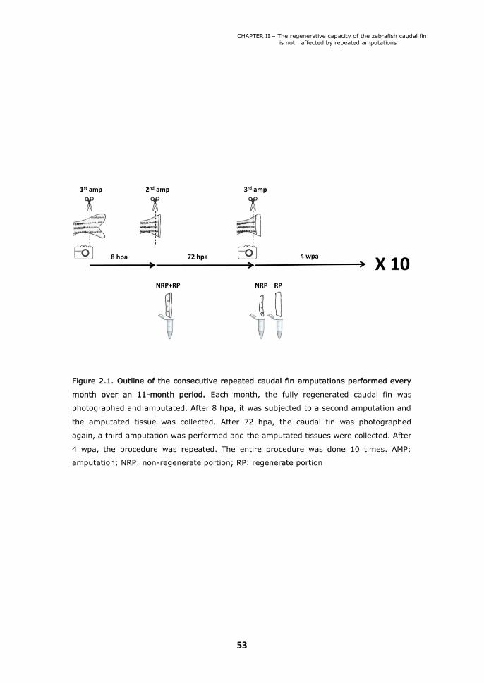
CHAPTER II – The regenerative capacity of the zebrafish caudal fin is not affected by repeated amputations
53
Figure 2.1. Outline of the consecutive repeated caudal fin amputations performed every
month over an 11-month period. Each month, the fully regenerated caudal fin was
photographed and amputated. After 8 hpa, it was subjected to a second amputation and
the amputated tissue was collected. After 72 hpa, the caudal fin was photographed
again, a third amputation was performed and the amputated tissues were collected. After
4 wpa, the procedure was repeated. The entire procedure was done 10 times. AMP:
amputation; NRP: non-regenerate portion; RP: regenerate portion
CHAPTER II – The regenerative capacity of the zebrafish caudal fin is not affected by repeated amputations
54
to regenerate for 4 weeks (4 wpa) to ensure a complete regeneration. This
amputation protocol was repeated 10 times spanning a period of
approximately 11 months. To evaluate the regenerative outgrowth state
following consecutive repeated amputations, we measured every month the 4
wpa full caudal fin area of each fish. As a control, we also measured the uncut
caudal fin area of each fish just before initiating the consecutive repeated
amputation experiment. The area of the 4 wpa full caudal fin did not change
when we compared the uncut caudal fin area (n=24) with the one obtained
after 27 cuts (n=14) (Figure 2.2A, B). To control for possible influence of fish
age, we also measured the caudal fin area of zebrafish siblings (n=10) that
were never amputated but were maintained over the experimental period in
the exact same conditions. Again, we found no differences in the caudal fin
area of these age-matched zebrafish siblings (Figure 2.2C). These results
show that the regenerative outgrowth of the zebrafish caudal fin does not
decline with repeated amputations.
II.2. Blastema formation is not impaired after consecutive repeated
amputations
We next asked whether early events after amputation, in particular wound
healing and blastema formation, might be affected by repeated amputations.
To this end, we measured the size of the regenerate (RP) at 72 hpa. When we
correct these values for the overall individual caudal fin size by dividing the RP
area by the 4 wpa full caudal fin area on each month, we found that the
relative area of the 72 hpa RP did not decrease significantly even when we
compared the 72 hpa RP obtained after 2 cuts (n=24) with the one obtained
after 29 cuts (n=14) (Figure 2.3A, B). To complement this data with a
molecular analysis, we quantified the expression levels of the wound healing
marker, mmp9 [18] and the blastema cell marker, msxb [4]. Although the
level of mmp9 expression in 8 hpa NRP+RP showed a decrease after 14 cuts,
this level was maintained in subsequent amputations (Figure 2.3C). The levels
of msxb also slightly decreased, even though not significantly, with increasing
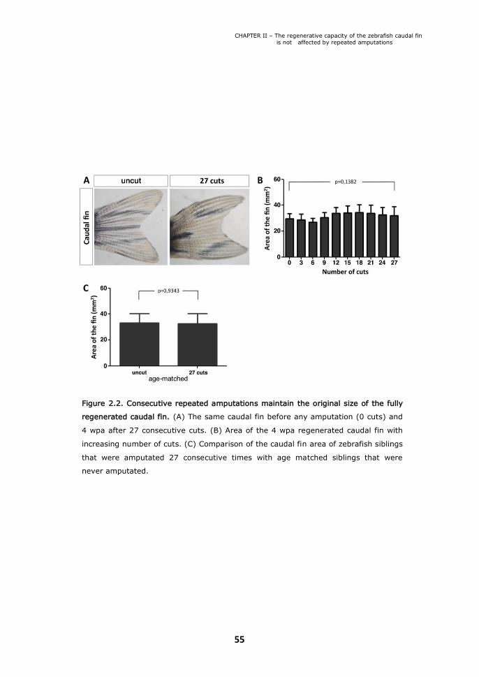
CHAPTER II – The regenerative capacity of the zebrafish caudal fin is not affected by repeated amputations
55
Figure 2.2. Consecutive repeated amputations maintain the original size of the fully
regenerated caudal fin. (A) The same caudal fin before any amputation (0 cuts) and
4 wpa after 27 consecutive cuts. (B) Area of the 4 wpa regenerated caudal fin with
increasing number of cuts. (C) Comparison of the caudal fin area of zebrafish siblings
that were amputated 27 consecutive times with age matched siblings that were
never amputated.

CHAPTER II – The regenerative capacity of the zebrafish caudal fin is not affected by repeated amputations
56
Figure 2.3. The 72 hpa regenerate size of the caudal fin is maintained with
consecutive repeated amputations over an 11-month period. (A) A 72 hpa caudal
fin obtained after the second consecutive amputation and after the twenty-seventh
consecutive amputation. (B) Area of the 72 hpa regenerate over the area of the
fully regenerated caudal fin immediately before the amputation measured with
increasing number of cuts. (C) mmp9 expression levels at 8 hpa with increasing
number of cuts. (D) msxb expression levels at 72 hpa in both non-regenerate
portions (NRP) and regenerate portions (RP) with increasing number of cuts.

CHAPTER II – The regenerative capacity of the zebrafish caudal fin is not affected by repeated amputations
57
number of amputations (Figure 2.3D). Since msxb is a blastema marker, it is
not surprising that the levels of expression were higher in the 72 hpa RP when
compared with the 72 hpa NRP (Figure 2.3D). These results reveal that, even
if the expression of these markers slightly decreases with repeated
amputations, these changes do not result in a decline of the fin’s ability to
successfully accomplish wound healing and blastema formation.
II.3. Consecutive repeated amputations affect the non-regenerated bone
A closer look at the bony rays present in caudal fins obtained after 27
consecutive amputations revealed a clear difference between the bone
segments located proximal to the amputation plane (bone that was never
amputated or old bone) and bone segments located distally to the amputation
plane (regenerated or new bone). Overall, old bony rays got wider and bone
segment boundaries became less defined along the entire proximal-distal axis
(Figure 2.4B). This phenotype is not age dependent since the bony rays of
uncut age-matched siblings did not change bone width and segment
boundaries definition with time (Figure 2.4A).
To be able to characterize and quantify the bone phenotype, we
performed an independent consecutive repeated amputation experiment
where two amputations were performed every other week. The first
amputation of the week was always done 6 segments distally to the base of
the fin and the second amputation was always done one segment below the
previous one. We observed that the old bone got progressively thicker after
an increased number of amputations and a clear difference between the old
and the new bone was already visible after 7 cuts (Figure 2.4C-E). Histological
longitudinal sections of bony rays stained with Masson’s trichrome expose the
collagen content. This staining showed that the amount of collagen was
increased in old bone (Figure 2.4G) when compared with new bone
regenerated after 14 cuts (Figure 2.4H). Interestingly, the new bone showed

CHAPTER II – The regenerative capacity of the zebrafish caudal fin is not affected by repeated amputations
58
Fig
ure
2.4
. Consecutiv
e re
peate
d a
mputa
tions a
ffect th
e s
tructu
re o
f the b
one p
roxim
al to
the a
mputa
tion p
lane.P
ictu
re o
f
the d
ors
al lo
be o
f an u
ncut c
audal fin
(A) a
nd its
age-m
atc
hed s
iblin
g a
fter 2
7 c
uts
(B). P
ictu
re o
f the d
ors
al lo
be o
f an
uncut c
audal fin
(C) a
nd a
caudal fin
afte
r 7 (D
) and 1
4 c
uts
(E). M
asson´s tric
hro
me s
tain
ing o
f longitu
din
al s
ectio
ns o
f an
uncut
bony ra
y (F
) and of
an old
(G
) and re
genera
ted (H
) re
gio
ns of
a bony ra
y afte
r 14 cuts
. Confo
cal
images of
transvers
e sectio
ns o
f a Z
ns5 im
munosta
ined p
roxim
al re
gio
n o
f an uncut c
audal fin
(I) and of th
e o
ld (J) a
nd new
(K
)
tissue o
f a c
audal fin
afte
r 14 c
uts
. Quantific
atio
n o
f the b
one th
ickness, in
ter- a
nd in
tra-ra
y tis
sue a
nd fin
thic
kness in
the
old
(L, N
, P) a
nd n
ew
(M, O
, Q).

CHAPTER II – The regenerative capacity of the zebrafish caudal fin is not affected by repeated amputations
59
a similar amount of collagen when compared to the one present in the control
uncut caudal fin (compare Figure 2.4H with Figure 2.4F). To determine if the
increase in collagen content was accompanied by an increase in the number
of osteoblasts, we analysed transverse sections of caudal fins immunostained
with Zns5 by confocal microscopy. A single layer of Zns5+ cells was found to
line the bone matrix in uncut controls and in old and new bone of fins after 14
cuts (Figure 2.4I-K), indicating that the number of osteoblasts lining the
hemirays did not increase with repeated amputations. Quantification of the
bone thickness, the space between the hemirays (intra-ray) and the space
between rays (inter-ray) showed that the thickness of old bone increased
significantly after 14 cuts, while the intra- and inter-ray space decreased
concomitantly (Figure 2.4I,J,L,N). In contrast, the regenerated new tissue
presented a slight decrease in the bone thickness and a mild reduction of the
inter-ray space, while the amount of intra-ray tissue is slightly increased
although not significantly when compared to the uncut caudal fins (Figure
2.4I,K,M,O). However the overall fin thickness, which is the sum of the bone
thickness and the intra-ray space, was not affected proximally (old tissue) or
distally (regenerated tissue) after 14 cuts. (Figure 2.4P,Q). We conclude that
repeated amputations result in abnormal remodelling of the bone and
mesenchymal tissue proximal to the amputation plane.
II.4. Regenerative capacity is not affected after repeated inhibition of caudal
fin regeneration following Wnt/ß-catenin signalling perturbation
When Wnt/ß-catenin signalling is inhibited immediately after fin amputation, a
wound epidermis forms, but blastema formation does not occur and
regeneration is completely blocked [13,19,20]. We analyzed whether fin
regeneration could occur normally after it has been previously perturbed.

CHAPTER II – The regenerative capacity of the zebrafish caudal fin is not affected by repeated amputations
60
Figure 2.5. Repeated inhibition of fin regeneration by interference with Wnt/b-catenin
signalling does not diminish regenerative capacity. (A) Schematic illustration of the
experimental scheme. Red shaded areas indicate periods in which fish were heat-shocked
twice daily, green areas indicate periods in which fish were allowed to regenerate in the
absence of heat-shock. amp = amputation, phot = photo of the tail fin. (B) Wild-type and
hsp70l:dkk1-gfp transgenic tail fins heat-shocked until 4 dpa and photographed 7 days after
amputation 1 (left column) and photographed after amputation 2 without heat-shocks (right
column). Note that heat-shocked wild-type fins regenerated, while dkk1-gfp expressing fins
did not, yet both fins regenerated in the absence of heat-shocks in response to amputation 2.
(C) The average regenerate length 7 days post amputation number 2, 4, 6, and 8 were
normalized to the length of wild-type fish. Note that there are no significant differences in
regenerate length between wild-type and hsp70l:dkk1-gfp fish.

CHAPTER II – The regenerative capacity of the zebrafish caudal fin is not affected by repeated amputations
61
To inhibit fin regeneration, we overexpressed the Wnt pathway inhibitor
Dkk1 using heat-shock inducible transgenic hsp70l:dkk1-gfp fish.
Overexpression of dkk1-gfp twice daily starting shortly before fin amputation
and continuing until 4 days-post-amputation (dpa) was sufficient to
completely inhibit fin regeneration (amputation 1 in Figure 2.5B) [13]. When
fish were relieved from the heat-shock treatment, spontaneous regeneration
did not occur. In contrast, when these fins that did not regenerate were re-
amputated and fish were kept at non-inducing standard temperatures, fins
completely regenerated (amputation 2 in Figure 2.5B). Thus, the ability to
regenerate after Wnt signalling inhibition requires a novel amputation
stimulus. Importantly, this also shows that inhibition of Wnt/ß-catenin
signalling does not permanently block the regenerative capacity of the
zebrafish caudal fin. To test whether repeated cycles of regenerative inhibition
caused by blockage of Wnt signalling can diminish the regenerative capacity,
we repeated the cycle of amputation, heat-shocking, recovery and second
amputation 4 times (Figure 2.5A). We measured the length of the regenerate
formed after every other amputation (in the absence of heat-shock) and
plotted the length of the hsp70l:dkk1-gfp transgenic regenerates normalized
to the one of their wild-type siblings. As shown in Figure 2.5C, no significant
difference between the two groups could be detected. Thus, repeated
blockage of blastema formation and fin regeneration by interference of Wnt/ß-
catenin signalling did not diminish the regenerative capacity after a new
amputation stimulus. We conclude that blastema formation and regenerative
outgrowth do not depend on a biological process that is permanently
disrupted or depleted by loss of Wnt/ß-catenin signalling.

CHAPTER II – The regenerative capacity of the zebrafish caudal fin is not affected by repeated amputations
62
Discussion
Repeated amputation experiments are fundamental to uncover the
regenerative capacity limit of lower vertebrates. Some reports reveal a
progressive increase of defects in the regenerated limb with an increasing
number of amputations in both larval Bufo regularis and adult Notophthalmus
viridescens newts [21,22]. In contrast, regeneration is successfully
accomplished with only minor defects after 16 tail amputations in adult
Triturus carnifex newts [23,24]. This led the authors to propose that
regeneration of the spinal cord in Triturus carnifex relies on differentiated cells
present in the stump that dedifferentiate contributing to the regenerate.
Whether the difference in capacity to repeatedly regenerate these structures
completely without defects is due to differences between newt species or
whether tails have a higher capacity to regenerate than limbs is unsolved.
Only very recently, the regeneration limit of the zebrafish caudal fin was
investigated [25]. In this report, it was shown that the regenerative capacity
of the zebrafish caudal fin does not decline when amputated up to 9 times.
This conclusion was based on the amount of regenerated tissue at 7 dpa and
on analysis of expression of msxb and fgf20a at 48 hpa. In our study, we
extended these results by showing that repeated amputations up to 29 times
over a period of 11 months do not alter regenerative capacity. However, in
contrast to this recent report, we observed a slight decrease of expression
levels of the wound healing marker mmp9 and the blastema marker msxb
with repeated cycles of regeneration (Figure 2.3C,D). Nonetheless, these
levels are still enough to accomplish a successful regeneration since the size
of the 72 hpa regenerate and 4 wpa full caudal fin did not significantly change
(Figure 2.2). Altogether, these data show that wound healing, blastema
formation and regenerative outgrowth are not affected when the caudal fin is
challenged with repeated amputations. Interestingly, it was recently
demonstrated that telomere length is not maintained upon 3 repeated
amputations in fish older than 3 months [26]. In this scenario, one could

CHAPTER II – The regenerative capacity of the zebrafish caudal fin is not affected by repeated amputations
63
speculate that consecutive amputations could lead to cell senescence.
However, our results demonstrate the amazing regenerative potential of the
zebrafish caudal fin even when challenged with a severe protocol of repeated
amputations in older fish. Therefore, cell senescence can not be a limiting
factor.
This almost unlimited capacity to regenerate that we have uncovered in
our study could be due to either the presence of stem cells, dedifferentiation
of mature cells or the contribution of both. In principle, each amputation could
activate the pool of putative stem cells that might be present in different fin
tissues, leading to the differentiation of all the missing structures.
Importantly, the decision between self-renewal and the initiation of
differentiation is controlled by signals provided by the tissue
microenvironment, or niche, where stem cells are believed to reside. The Wnt
signalling pathway plays a fundamental role in the control of maintenance and
proliferation initiation of adult stem cells reservoirs in the intestine [27] and
skin [28]. We made use of the heat-shock inducible transgenic hsp70l:dkk1-
gfp fish, to efficiently and in a time-controlled manner inhibit Wnt signalling.
Inhibition of Wnt signalling twice daily shortly before fin amputation and until
4 dpa completely impaired fin regeneration. However, if the fins that did not
regenerate were re-amputated and allowed to have an intact Wnt signalling
by keeping them at a non-inducing temperature, fins regenerated completely
(Figure 2.5). This reveals that there is a time window for the initiation of
regeneration that is triggered soon after each amputation and that is
absolutely dependent on Wnt/ß-catenin signalling. Importantly, these
experiments also indicate that blastema formation does not depend on a pool
of progenitor cells that requires Wnt for its maintenance. While these data do
not completely rule out a contribution of progenitor cells, it is more
compatible with the alternative model of regeneration based on
dedifferentiation. In fact, this model is now supported by recent findings
showing that mature osteoblasts dedifferentiate to form part of the blastema
and regenerate bone in the zebrafish caudal fin [10]. According to these

CHAPTER II – The regenerative capacity of the zebrafish caudal fin is not affected by repeated amputations
64
findings, Wnt signalling could be required for dedifferentiation and/or
expansion of the dedifferentiated cells to form a blastema.
In spite of this amazing capacity to regenerate, the bone proximally to
the amputation plane becomes thickened with repeated cycles of amputations
(Figure 2.4). Interestingly, we could not detect a clear difference in Zns5
staining, indicating that the number of osteoblasts did not change with
increased amputations (Figure 2.4I-K). Progressive bone thickening might be
a consequence of inappropriate activation of osteoblasts to secrete matrix far
away from the amputation plane. In fact there is strong evidence that
osteoblasts enter the cell cycle following amputation [10,29] and that
differentiated cells can be induced to proliferate even far from the amputation
plane [10,30]. Thus, while some dedifferentiated osteoblasts migrate distally
to form the blastema, it is unlikely that newly formed osteoblasts that far
from the amputation plane would participate in blastema formation. Rather,
they likely represent a source of cells replacing those moving into the
blastema. It is possible that activation of proliferation also causes these cells
to re-activate matrix secretion, which after repeated cycles results in bone
thickening. Alternatively, the increase in bone matrix could be caused by an
unbalanced ratio of bone-forming and bone-degrading cells. Due to the
thickening of the bone, it seems that the inter- and intra-ray tissues became
compacted and therefore reduced in size. Interestingly, the newly
regenerated tissue of the fin exhibits a decreased bone thickness and inter-
ray space probably because these are recently formed tissues that are still
being remodelled.
A better understanding of the cellular mechanisms underlying the
virtually unlimited regenerative capacity of fish appendage regeneration will
be informative for efforts to improve repair, in particular of bone, in
humans.
CHAPTER II – The regenerative capacity of the zebrafish caudal fin is not affected by repeated amputations
65
Materials and methods
Ethics Statement
All experiments involving animals were approved by the Animal User and
Ethical Committees at Instituto de Medicina Molecular, according with
directives from Direcção Geral Veterinária (PORT 1005/92). All animal
experiments at the Biotechnology Center of the TU Dresden were performed
in accordance with the guidelines of the state of Saxony and have been
approved by the Regierungspräsidium Dresden, permit number 24D-9168.11-
1/2008-1.
Zebrafish lines, maintenance and surgery
48 AB WT zebrafish were purchased from ZIRC. The repeated amputations
protocol was initiated when fish were 1 year of age. 24 experimental animals
were maintained at 30°C in separate tanks (one individual per tank) during
the time of the experiment (approximately 11 months). 24 control uncut
animals were kept together in a large tank, at the same temperature. To
perform the amputations, fish were anesthetized in 0.6 mM Tricaine and
amputated using a razor blade.
Repeated inhibition of regeneration
hsp70l:dkk1-gfpw32 transgenic fish, carrying one copy of the transgene and
their wild-type siblings were used. To induce heat-shocks, fish were kept in an
automated waterbath at 28°C, and twice daily heated to 37°C within 10
minutes, followed by sustained incubation at 37°C for 1 hour, and active
cooling to 28°C within 15 minutes. To ensure complete block of fin
regeneration in dkk1-gfp expressing fish, the first heat-shock was applied 6
hours prior to fin amputation. To document regenerative capacity after
inhibition, fish were heat-shocked twice daily for 4 days without feeding, then
allowed to recover for 1 week at 28°C with feeding, followed by re-amputation
of the fin in wild-types or the non-regenerated fin stump in hsp70l:dkk1-

CHAPTER II – The regenerative capacity of the zebrafish caudal fin is not affected by repeated amputations
66
gfpw32 transgenic fish. For re-amputation, the fin was cut 1 bone segment
proximal to the initial amputation plane. Fish were allowed to regenerate with
feeding at 28°C for 1 week, after which the fin was photographed.
Quantification of regenerate area and length and caudal fin area
The 4 wpa full caudal fin and the 72 hpa regenerate area were measured each
month using Image J software (NIH). Since zebrafish are very heterogeneous
regarding its size, the 72 hpa regenerate area was corrected to the size of the
fin by dividing its value in each month by the 4 wpa full caudal fin area in the
corresponding month. The 7 dpa regenerate length of hsp70l:dkk1-gfp fish
was normalized to the average regenerate length of wild-type sibling fish. For
this quantification, the length of the 2nd, 3rd, 4th and 5th dorsal fin rays was
measured from the amputation plane to the distal tip of the ray using Image J
software and the average length calculated for each fish.
Quantitative RT-PCR
RP and NRP tissues were collected and preserved at -20°C in RNA Later
solution (Ambion) during the time of the experiment. Total RNA was extracted
from fin regenerates using TRIZOL (Invitrogen) according to the
manufacturer’s protocol. 8 regenerates were used to extract RNA for the 8
hpa time-point and 4 RP or NRP were used to extract RNA for the 72 hpa
time-point. 1 µg of RNA from each sample was reverse transcribed with the
RevertaidTM H minus first strand cDNA synthesis kit (Fermentas) using random
hexamer primers. Primers for quantitative RT-PCR of mmp9 were 5-
CTGGGCACCTGCTCGTTG-3 and 5-ATTGGAGATGACCGCCTGC-3 and for msxb
were 5-AGGAACAGAGCACTTGGTCAAACT-3 and 5-
TGAGGTTGAGGGAGTTGGAGAAC-3. Quantitative PCR was performed using
Corbet Rotorgene 6000 and the SYBR Green labelling system. mmp9 and
msxb levels were normalized to the housekeeping gene ef1a (primers 5-
ACGCCCTCCTGGCTTTCACCC-3 and 5-TGGGACGAAGGCAACACTGGC-3).
CHAPTER II – The regenerative capacity of the zebrafish caudal fin is not affected by repeated amputations
67
Quantification of the relative expression was performed using the 2-∆CT
method and normalized against the relative expression obtained for the uncut
caudal fin. Data were analyzed using Student’s t test.
Tissue sectioning and histology
Fins were embedded in gelatin and sectioned at 12 µm using a cryostat. For
the Masson’s trichrome staining, gelatin was washed in PBS at 37°C for
approximately 30 minutes and sections were stained with Weigert’s
hematoxilin for 10 minutes, washed in warm running tap water for 5 minutes
and rinsed in distilled water. After this washing, sections were stained with
Biebrich scarlet-acid fuchsin for 5-10 minutes. The excess of this solution was
removed by rinsing with distilled water and the unspecific staining was cleared
with phosphomolybdic acid 1% for 10 minutes. Collagen was stained with
light green at 2% for 1 minute. Finally, sections were dehydrated in ethanol
95% 30 seconds, ethanol 100% 30 seconds, cleared in xylol for 5-10 minutes
and slides were mounted in Entellan.
Immunohistochemistry
The fins were fixed in a solution with 80% MeOH/20% DMSO (Sigma) and
were rehydrated in a MeOH/PBS series, permeabilized with acetone at -20°C
for 20 minutes, followed by two washes in PBS. An additional permeabilization
was done with PBST 0.5% solution (PBS with 0.5% Triton X-100) during 30
minutes. Followed by several washes with PBS, fins were blocked in PBS with
10% Fetal Bovine Serum (FBS) and incubated with 1:250 primary antibody
Zns5 (ZIRC 011604) overnight at 4°C. Fins were washed several times in PBS
and the incubation with the secondary antibody and DAPI (D9564 Sigma) was
done overnight at 4°C. Immunostained caudal fins were post-fixed for 20
minutes in 4% PFA (paraformaldehyde), washed in PBS and passed through a
30% sucrose/PBS solution for cryoprotection. Transverse sections of 12 µm of
immunostained fins of 2 uncut controls and 2 caudal fins subjected to 14
amputations were obtained by cryosectioning and analysed by confocal

CHAPTER II – The regenerative capacity of the zebrafish caudal fin is not affected by repeated amputations
68
microscopy. In each of the controls and experimental fins the following
measurements were performed using Image J software: proximal and distal
bone thickness of dorsal and ventral hemi-rays of 5 - 9 bony rays was
measured; the amount of 3 inter-ray tissues at a proximal and distal level was
quantified by measuring the distance between two bony rays; the proximal
and distal intra-ray tissue was quantified by measuring the length between
two hemi-rays in 5 – 9 bony rays. Data were analyzed using Student’s t-test.
Acknowledgements
We thank Lara Carvalho and Fábio Valério for excellent zebrafish husbandry,
Andreia Pinto for histology and António Temudo for imaging support.
CHAPTER II – The regenerative capacity of the zebrafish caudal fin is not affected by repeated amputations
69
References
1. Iovine M (2007) Conserved mechanisms regulate outgrowth in
zebrafish fins. Nature Chem Biol 3(10): 613-18.
2. Poss KD, Keating MT, Nechiporuk A (2003) Tales of regeneration in
zebrafish. Dev Dyn 226: 202-10.
3. Hall BK (2005) Bones and cartilage: developmental and evolutionary
skeletal biology. Elsevier Academic Press, USA.
4. Nechiporuk A, Keating MT (2002) A proliferation gradient between
proximal and msxb-expressing distal blastema directs zebrafish fin
regeneration. Development 129: 2607–17.
5. Handberg-Thorsager M, Fernandez E, Salo E (2008) Stem cells and
regeneration in planarians. Front Biosci 1(13): 6374-94.
6. Morrison JI, Lööf S, He P, Simon AJ (2006) Salamander limb
regeneration involves the activation of a multipotent skeletal muscle
satellite cell population. Cell Biol 30; 172(3): 433-40.
7. Brockes JP, Kumar A (2002) Plasticity and reprogramming of
differentiated cells in amphibian regeneration. Nat Rev Mol Cell Biol.
3(8): 566-74.
8. Echeverri K, Clarke JD, Tanaka EM (2001) In vivo imaging indicates
muscle fiber dedifferentiation is a major contributor to the regenerating
tail blastema. Dev Biol 1; 236(1): 151-64.
9. Echeverri K, Tanaka EM (2002) Mechanisms of muscle
dedifferentiation during regeneration. Semin Cell Dev Biol 13(5): 353-
60.
10. Knopf F, Hammond C, Chekuru A, Kurth T, Hans S, et al (2011)
Bone regenerates via dedifferentiation of osteoblasts in the zebrafish
fin. Dev Cell 20(5): 713-24.
11. Tu S, Johnson SL (2011) Fate restriction in the growing and
regenerating zebrafish fin. Dev Cell 20(5): 725-32.

CHAPTER II – The regenerative capacity of the zebrafish caudal fin is not affected by repeated amputations
70
12. Kragl M, Knapp D, Nacu E, Khattak S, Maden M, et al (2009) Cells
keep a memory of their tissue origin during axolotl limb regeneration.
Nature 460(7251): 60-5.
13. Stoick-Cooper CL, Moon RT, Weidinger G (2007) Advances in
signaling in vertebrate regeneration as a prelude to regenerative
medicine. Genes Dev 1; 21(11): 1292-315.
14. Quint E, Smith A, Avaron F, Laforest L, Miles J, et al. (2002) Bone
patterning is altered in the regenerating zebrafish caudal fin after
ectopic expression of sonic hedgehog and bmp2b or exposure to
cyclopamine. Proc Natl Acad Sci 99: 8713–18.
15. Whitehead G, Makino S, Lien C (2005). Fgf20 is essential for
initiating zebrafish fin regeneration. Science 310: 1957-1960.
16. Poss KD, Shen J, Nechiporuk A, McMahon G, Thisse B, et al. (2000)
Roles for Fgf signaling during zebrafish fin regeneration. Dev Biol 222:
347–35.
17. Lee Y, Grill S, Sanchez A, Murphy-Ryan M, Poss KD (2005) Fgf
signaling instructs position-dependent growth rate during zebrafish fin
regeneration. Development 132(23): 5173-83.
18. Yoshinari, N, Ishida, T, Kudo, A, Kawakami, A (2009). Gene
expression and functional analysis of zebrafish larval fin fold
regeneration. Dev Biol 325, 71-81.
19. Kawakami Y, Rodriguez Esteban C, Raya M, Kawakami H, Marti M,
et al. (2006) Wnt/ß-catenin signaling regulates vertebrate limb
regeneration. Genes Dev 20: 3232–3237.
20. Huang SM, Mishina YM, Liu S, Cheung A, Stegmeier F (2009)
Tankyrase inhibition stabilizes axin and antagonizes Wnt signalling.
Nature 461(7264): 614-20.
21. Dearlove GE, Dresden MH (1976) Regenerative abnormalities in
Notophthalmus viridiscens. J Exp Zool 196: 251-261.

CHAPTER II – The regenerative capacity of the zebrafish caudal fin is not affected by repeated amputations
71
22. Abdel-Karim AE, Michael MI (1993) Regenerative abnormalities in
hind limbs of Bufo regularis induced by repeated amputations in early
larval stages. Qatar Univ Sci J 13(1): 101-108.
23. Margotta V, Filoni S, Merante A, Chimenti A (2002) Analysis of
morphogenetic potential of caudal fin spinal cord in Triturus carnifex
(Urodele Amphibians) subjected to repeated tail amputations. Int J Anat
Embryol 107(2): 127-144.
24. Margotta V (2004) Morphogenetic potentiality of the caudal spinal
cord in adult newts (Triturus carnifex) subjected to repeated tail
amputations: evaluation after the 16th amputation. Rend Fis Acc Lincei
9(15): 231-245.
25. Shao J, Chen D, Ye Q, Cui J, Li Y, et al. (2011) Tissue regeneration
after injury in adult zebrafish: The regenerative potential of the caudal
fin. Dev Dyn 240(5): 1271-7.
26. Anchelin M, Murcia M, Alcaraz-Pérez F, García-Navarro E and
Cayuela, M (2011) Behaviour of telomere and telomerase during aging
and regeneration in zebrafish. Plos One 6(2): e16955.
27. Korinek V, Barker N, Moerer P, van Donselaar E, Huls G, et al.
(1998) Depletion of epithelial stem-cell compartments in the small
intestine of mice lacking Tcf-4. Nat Genet 19(4): 379-83.
28. Blanpain C, Fuchs E (2006) Epidermal stem cells of the skin. Annu
Rev Cell Dev Biol 22: 339-73.
29. Johnson SL and Bennett P (1999) Growth control in the ontogenetic
and regenerating zebrafish fin. Methods Cell Biol 59: 301-11.
30. Santos-Ruiz L, Santamaría JA, Ruiz-Sánchez J, Becerra J (2002) Cell
proliferation during blastema formation in the regenerating teleost fin.
Dev Dyn 223(2):262-72.

73

73
CHAPTER III
An amputation resets
positional memory to a
proximal identity in the
regenerating zebrafish caudal
fin blastema

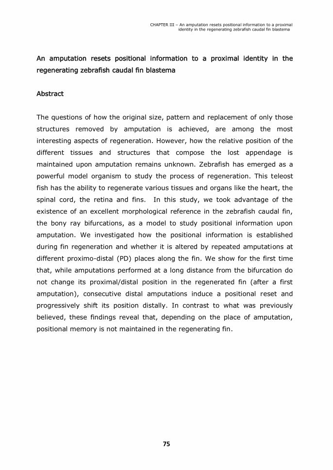
CHAPTER III – An amputation resets positional information to a proximal identity in the regenerating zebrafish caudal fin blastema
75
An amputation resets positional information to a proximal identity in the
regenerating zebrafish caudal fin blastema
Abstract
The questions of how the original size, pattern and replacement of only those
structures removed by amputation is achieved, are among the most
interesting aspects of regeneration. However, how the relative position of the
different tissues and structures that compose the lost appendage is
maintained upon amputation remains unknown. Zebrafish has emerged as a
powerful model organism to study the process of regeneration. This teleost
fish has the ability to regenerate various tissues and organs like the heart, the
spinal cord, the retina and fins. In this study, we took advantage of the
existence of an excellent morphological reference in the zebrafish caudal fin,
the bony ray bifurcations, as a model to study positional information upon
amputation. We investigated how the positional information is established
during fin regeneration and whether it is altered by repeated amputations at
different proximo-distal (PD) places along the fin. We show for the first time
that, while amputations performed at a long distance from the bifurcation do
not change its proximal/distal position in the regenerated fin (after a first
amputation), consecutive distal amputations induce a positional reset and
progressively shift its position distally. In contrast to what was previously
believed, these findings reveal that, depending on the place of amputation,
positional memory is not maintained in the regenerating fin.

CHAPTER III – An amputation resets positional information to a proximal identity in the regenerating zebrafish caudal fin blastema
76
Introduction
Tissue regeneration in humans can occur in a limited extent in structures like
the skin, gut, skeletal muscle, bone, digit tips, liver and blood. However, other
vertebrate species have the extraordinary capacity to regenerate lost tissues
and organs throughout adult life. One of such organisms is the zebrafish, a
well-established model to study general mechanisms of regeneration, since it
is able to regenerate fins, scales, retina, spinal cord and heart among other
internal organs (Iovine, 2007).
Due to its accessibility, caudal fin regeneration is an example of a
powerful and efficient model for regenerative studies. The zebrafish caudal
fin is composed of several segmented bony rays, mesenchymal tissue, blood
vessels and nerve axons. Each bony ray is made of two concave hemirays
and, with the exception of the most lateral rays, is bifurcated in a distal
position within the fin (Poss et al., 2003). These bifurcations are responsible
for generating the characteristic shape of the caudal fin and ultimately for
increasing swimming efficiency.
In the zebrafish caudal fin, an amputation triggers a regenerative
program that occurs in three phases: wound healing, blastema formation
and regenerative outgrowth. Within the first 12 hours-post-amputation
(hpa), the injury is healed through migration of epidermal cells that cover
and close the wound (Poss et al., 2003). In the next 12-48 hpa, the wound
epithelium thickens forming an apical epidermal cap (AEC) and the tissue
proximal to the amputation plane disorganizes, begins to proliferate and
migrates distally to form the blastema, which is a mass of proliferating cells
(Poss et al., 2003). The onset of regenerative outgrowth is at 48 hpa, and at
this stage the blastema becomes subdivided into a distal region comprising
slow proliferative cells and an intensely proliferative proximal region
(Nechiporuk and Keating, 2002). Within 2 weeks after amputation, the
blastema reconstitutes the original architecture of the caudal fin with all its
different tissues and structures (Nechiporuk and Keating, 2002).

CHAPTER III – An amputation resets positional information to a proximal identity in the regenerating zebrafish caudal fin blastema
77
Although we are beginning to understand the molecular mechanisms of
regeneration, it is becoming clear that distinct pathways are activated upon
amputation. Fibroblast growth factor (Fgf) signalling seems to be required for
blastema formation (Whitehead et al., 2005), canonical Wnt/β-catenin
signalling enhances proliferation of progenitors cells while non-canonical
Wnt/Planar cell polarity (PCP) pathway seem to promote the opposite (Stoick-
Cooper et al., 2007b) and Hedgehog (Hh) signalling seems to play a latter
role by controlling bone differentiation (Quint et al., 2002). It is true that a
tight control of cell proliferation and differentiation is critical to regenerate a
fully functional caudal fin. Nonetheless, equally important is to be able to
reconstitute the relative arrangement of the different regenerating tissues and
structures, which means that during fin regeneration there must be ways of
keeping positional memory. This is a fascinating question in the regeneration
field for which we know very little.
In the present study, we took advantage of the zebrafish caudal fin as a
model to study positional information upon amputation, since the stereotypic
PD position of the bony ray bifurcations provides an excellent morphological
reference. We tested how positional information of bony ray bifurcations is
affected by repeated amputations performed at different levels along the PD
axis of the fin. We show that there is a progressive distalization of the position
of the bifurcations in the regenerated fin, when the repeated amputations
were done proximally near the bifurcation (distal amputations). On the other
hand, after a first amputation, its position is maintained in subsequent
amputations done near the base of the fin, therefore away from the
bifurcation (proximal amputations). Thus, we show for the first time that the
positional memory of the bifurcation is maintained in proximal but not in distal
amputations. Furthermore, we analysed the role of Fgf and Hh signalling and
concluded that they do not seem to be the instructive signals that determine
the bifurcation position.

CHAPTER III – An amputation resets positional information to a proximal identity in the regenerating zebrafish caudal fin blastema
78
Results
III.1. Repeated amputations progressively shift the bifurcation position
distally
In Chapter II, I describe an amputation protocol that allowed us to conclude
that the regenerative capacity of the zebrafish caudal fin is not affected by
repeated amputations or ageing. In this protocol, the caudal fin was subjected
to three amputations every month. This protocol was repeated 10 times.
During the first 6 months (corresponding to the first 15 cuts) the third
consecutive amputation (the last before allowing the fin to completely
regenerate) was done three bone segments below the most proximal bony ray
bifurcation (near the bifurcation). In the following 4 months (corresponding to
the next 12 cuts), the third consecutive amputation was done 4 segments
distally from the base of the fin (distant from the bifurcation) (Azevedo et al.,
2011). Although the regenerative capacity was not affected, we detected an
alteration in the original pattern of pigment cells and a distal shift in the
position of the bony ray bifurcations in the regenerated caudal fins (Figure
3.1a,b).
We quantified the number of segments formed between the base of the
fin and the 3rd dorsal ray bifurcation in the regenerated fin in order to
determine the PD position of the bifurcation after each set of consecutive
amputations. We observed that, during the first 6 months, there was an
increase in the number of segments formed between the base of the fin and
the 3rd dorsal ray bifurcation. This reveals that the position of the bifurcations
was progressively shifted distally when compared to its position before
amputation (Figure 3.1c - near bifurcation). In the following 4 months, the
number of segments formed between the base of the fin and the 3rd dorsal
ray bifurcation was maintained, showing that the PD position of the
bifurcations was unaltered (Figure 3.1c – 4 segments). The overall number of
segments within the regenerated caudal fin was unchanged (data not shown)

CHAPTER III – An amputation resets positional information to a proximal identity in the regenerating zebrafish caudal fin blastema
79
Figure 3.1. The bifurcation position is distalized with repeated amputations. (a) The same
caudal fin before amputation and after 15 and 27 amputations. The dashed line in each panel
marks the number of segments from the base of the fin until the bifurcation in the 3rd dorsal
ray. (b) Schematic representation of the bifurcation distalization with the repeated
amputations. (c) Number of segments formed in the 3rd dorsal ray between the base of the
fin and the bifurcation after consecutive amputations. In the first 6 months, the third
consecutive amputation was performed three segments below the bifurcation (near the
bifurcation) and in the following 4 months, the third amputation was done at 4 segments
from the base of the fin (distant from the bifurcation). (d) 3rd dorsal ray segment length
before any amputation and after 24 amputations.

CHAPTER III – An amputation resets positional information to a proximal identity in the regenerating zebrafish caudal fin blastema
80
during the repeated rounds of amputations as well as the segment length
(Figure 3.1d).These results suggest that the bifurcation position is distalized
when the amputations are performed proximally near the bifurcation, but
remains unaltered when the amputations are done near the base of the fin
(distant from the bifurcation).
III.2. The bifurcation position is only shifted distally when the amputations are
performed repeatedly near the bifurcation
One possibility to explain the maintenance of the proximal-distal position of
the bifurcation observed in the last 4 months of our experimental setting
could be that the distalization of the bifurcation reached its maximum limit
after 6 months of consecutive amputations. Another possibility could be that
the increased amputation distance to the bifurcation place, performed in the
last 4 months, would decrease the possible influence of an amputation in the
PD bifurcation position.
To distinguish between these two possibilities, we designed a more
controlled amputation protocol (Figure 3.2a). We performed a first amputation
at 4 segments from the base of the fin (distant from the bifurcation) in 20
adult zebrafish and allowed the fin to completely regenerate. The second,
third and fourth amputations were performed at 4 segments from the base of
the fin in 10 of the animals and, in the remaining 10, the second, third and
fourth amputations were performed at 1 segment below the most proximal
bifurcation (near the bifurcation).
Upon a first amputation at 4 segments from the base of the fin, the
bifurcation was immediately distalized when compared to its position in the
uncut fin (Figure 3.2b). Following the second, third and fourth amputations,
the bifurcation position was maintained in the regenerated fin when the
amputations were done at 4 segments from the base of the fin (Figure 3.2b’)
but it was progressively distalized when the amputations were done 1
segment proximal to the bifurcation (near bifurcation) (Figure 3.2b’’). These
data show that while amputations performed at a long distance from the

CHAPTER III – An amputation resets positional information to a proximal identity in the regenerating zebrafish caudal fin blastema
81
Figure 3.2. The distalization of the bifurcation is dependent on the PD level
of amputation. (a) After a first amputation performed at 4 segments from
the base of the fin, the fish were divided into two groups. One group was
amputated a second, third and fourth time at 4 segments from the base of
the fin and the second group was amputated one segment below the
bifurcation (near the bifurcation). (b) Number of segments formed in the
third dorsal ray between the base of the fin and the bifurcation after
consecutive amputations performed always at 4 segments from the base
of the fin (b’) and after a first amputation performed at 4 segments from
the base of the fin followed by a second, third and fourth amputations near
the bifurcation (b’’).
CHAPTER III – An amputation resets positional information to a proximal identity in the regenerating zebrafish caudal fin blastema
82
bifurcation do not change its PD position in the regenerated fin, consecutive
amputations near the bifurcation induce a positional reset and progressively
shift its position distally.
III.3. shh expression pattern is independent of the place of amputation
We next asked what factors determine the bifurcation position and how they
are influenced by the amputation place.
Sonic hedgehog (Shh) is a strong candidate due to its previously
described dynamic expression, correlated with the formation of a bifurcation
during fin regeneration. It was shown that at 2 days-post-amputation (dpa), a
strong single domain of shh expression is detected at the level of amputation
on the top of each hemiray. By 4 dpa, this shh single domain starts to split
into two groups of cells located laterally in the proximal region of the basal
wound epidermal layer. This change in shh expression from one to two
domains was proposed to be the trigger for bifurcation formation (Laforest et
al., 1998).
Thus, we wanted to determine how this dynamic expression pattern of
shh is modulated by the amputation place and whether Shh would be the
instructive factor to form the bifurcation or a downstream factor in this
process.
To this end we performed two rounds of amputations at different places,
at 1 segment proximal to the bifurcation (near the bifurcation) or at 4
segments from the base of the fin (distant from the bifurcation) and analysed
by in situ hybridization the expression of shh at 3 and 4 dpa. We observed
that, independently of the number or places of amputations, shh was
consistently expressed in two separate cellular domains already at 3 dpa
(Figure 3.3a-h). These results show that shh expression is not modulated by
the amputation place. Moreover, at 4 dpa, in a caudal fin that does not have
any bifurcations due to being subjected to several distal amputations, shh
expression was localized in two groups of cells located laterally in the
proximal region of the basal wound epidermal layer (Figure 3.3i, j). This

CHAPTER III – An amputation resets positional information to a proximal identity in the regenerating zebrafish caudal fin blastema
83
Fig
ure
3.3
. The e
xpre
ssio
n p
att
ern
of
shh d
uring r
egenera
tion d
oes n
ot
change w
ith t
he P
D l
evel or
the n
um
ber
of
am
puta
tions.
(a-d
) Caudal fins w
ere
am
puta
ted a
t 4 s
egm
ents
fro
m t
he b
ase o
f th
e f
in o
nce o
r tw
ice a
nd s
hh e
xpre
ssio
n w
as d
ete
rmin
ed a
t 3
and 4
days
follow
ing t
he a
mputa
tion.
(e-h
) Caudal fins w
ere
am
puta
ted n
ear
the b
ifurc
ation o
nce o
r tw
ice a
nd s
hh e
xpre
ssio
n w
as
dete
rmin
ed a
t 3 a
nd 4
days f
ollow
ing t
he a
mputa
tion.
(i)
Caudal fin w
ith n
o b
ifurc
ations w
as a
mputa
ted a
t 4 s
egm
ents
fro
m t
he
base o
f th
e f
in a
nd s
hh e
xpre
ssio
n w
as e
xam
ined a
t 4 d
ays f
ollow
ing t
he a
mputa
tion.
(J)
Top v
iew
of
the c
audal fin s
how
n in I
. (k
-
v)
Caudal
fins of
2.2
shh:g
fp:A
BC#
15 tr
ansgenic
fish w
ere
am
puta
ted at
1 segm
ent
pro
xim
al
to th
e bifurc
ation (n
ear
the
bifurc
ation)
and a
naly
zed a
t diffe
rent
tim
e-p
oin
ts a
fter
am
puta
tion b
y a
double
im
munosta
inin
g w
ith a
nti
-GFP (
k-o
) a
nd a
nti-Z
ns5
(p-t
) antibodie
s.
(u,v
) m
erg
e o
f 1 d
pa+
18 h
pa (
u)
and 2
dpa (
v).
Arr
ow
s indic
ate
irr
egula
r dom
ain
s o
f expre
ssio
n o
f shh.
Dashed
line r
epre
sents
the a
mputa
tion p
lane.

CHAPTER III – An amputation resets positional information to a proximal identity in the regenerating zebrafish caudal fin blastema
84
strongly suggests that Shh is not sufficient to trigger the formation of
bifurcations.
To analyse with increased cellular resolution the dynamics of shh
expression in the zebrafish regenerating fin, we made use of a transgenic line
expressing GFP under the control of shh promoter (2.2shh:gfp:ABC#15)
(Shkumatava et al., 2004). Using this transgenic zebrafish line, we performed
one amputation, at 1 segment proximal to the bifurcation (near bifurcation)
and analysed the expression of shh, every 6 hours from 1 to 2 dpa. The time
course analysis revealed that the establishment of shh expression pattern
during regeneration is around 1 dpa + 12 hpa. However, in a few cases, shh
expression could be detected at earlier time points, in a small number of cells,
in only one or both sides of the regenerating hemiray (Figure 3.3i). From its
onset of expression (at 1 dpa + 12 hpa) until 2 dpa, shh was always present
with the same pattern of expression, namely in two separate groups of cells
located laterally in the proximal region of the basal epidermal layer (Figure
3.3m-o). Since we have never observed a transition in shh expression from
one to two domains during fin regeneration, these results provide additional
support to the conclusion that Shh may not be the instructor to form the
bifurcation.
In addition, it has also been proposed that Shh plays a role in the
patterning and/or differentiation of osteoblasts within the blastema during fin
regeneration (Quint et al., 2002). In order to determine whether there is a
correlation between the restriction of shh expression in two epidermal
domains and the dynamics of bone formation during regeneration, we
performed a Zns5 (osteoblast marker) immunostaining time-course analysis
(every 6 hours from 1 to 2 dpa) in the 2.2shh:gfp:ABC#15 transgenic fish.
Interestingly, we observed that soon after the onset of shh expression, the
growing bone alters the shape of its tip from a cone to a “V” shape (Figure
3.3s). This suggests that, Zns5+ cells cease to accumulate in the middle of
the blastema and are aligned close to the basal layer of the epidermis where
shh mRNA is produced (Figure 3.3n-t,u,v). Interestingly, we have also
CHAPTER III – An amputation resets positional information to a proximal identity in the regenerating zebrafish caudal fin blastema
85
observed that shh expression domains can be irregular in the form and differs
in the number of shh positive cells in each individual blastema of the same fin
(Figure 3.3n - arrows). Consequently, the visibility of shh separation in two
cellular domains depends on the regenerating ray and blastema shape.
Similarly, irregularities in the shape are also visible in the spatial organization
of Zns5+ cells in the regenerating tip of each ray (Figure 3.3p-t).
Altogether, these results suggest that shh expression in two separate
domains in the basal layer of the epidermis is not determining the PD position
of the bone bifurcation, but could have an important role in bone formation
and growth through osteoblasts alignment by attracting them to the region
where shh is being produced.
III.4. Fgf signalling does not play a role in the determination of the bony ray
bifurcation position
It was already demonstrated that the levels of Fgf signalling activation vary
according to the PD place of amputation. Upon amputation, the expression
levels of Fgf downstream targets such as mkp3, sef and spry4 are higher
following a proximal amputation when compared to a distal amputation (Lee
et al., 2005). This suggests the existence of positional memory that can be
mediated or act through Fgf signalling.
In order to investigate whether Fgf signalling determines the PD position
of the bifurcation in the regenerated fin, we made use of the hsp70:dn-fgfr1
transgenic zebrafish (Lee et al., 2005). This transgenic contains a dominant-
negative fgfr1-egfp fusion gene (dn-fgfr1) driven by a heat-inducible zebrafish
hsp70 promoter. It was previously demonstrated that this construct
attenuates Fgf signalling during fin regeneration in a dose dependent manner.
Upon heat-shock, the regeneration growth rate is affected. This phenotype is
highly sensitive to 1ºC temperature increments (Lee et al., 2005).
The hsp70:dn-fgfr1 transgenic zebrafish were amputated once, at 1
segment proximal to the bifurcation (near the bifurcation) and Fgf signalling
was partially inhibited by heat-shocking at 35°C for 1 hour daily, starting at

CHAPTER III – An amputation resets positional information to a proximal identity in the regenerating zebrafish caudal fin blastema
86
Fig
ure
3.4
. Fgf s
ignallin
g d
oes n
ot s
eem
to p
lay a
role
in th
e d
ete
rmin
atio
n o
f the P
D p
ositio
n
where
the b
ifurc
atio
n is
form
ed (a
) H
eat-s
hock p
roto
col fo
llow
ing a
n a
mputa
tion. T
ransgenic
hsp70:d
n-fg
fr1 fis
h w
ere
am
puta
ted at
1 segm
ent
pro
xim
al
to th
e bifu
rcatio
n and heat-
shocked a
t 35°C fo
r 1 h
our, d
urin
g 6
days, s
tartin
g a
t day 2
post a
mputa
tion. (b
) P
ictu
re o
f a
7 days-p
ost-a
mputa
tion re
genera
ted caudal
fin of
hsp70:d
n-fg
fr1 tra
nsgenic
fis
h w
ith or
with
out th
e h
eat-s
hock p
roto
col. (c
) Num
ber o
f segm
ents
form
ed in
the 3
rd dors
al ra
y b
etw
een
the b
ase o
f the fin
and th
e b
ifurc
atio
n in
heat-s
hocked a
nd n
on-h
eat-s
hocked s
iblin
gs.

CHAPTER III – An amputation resets positional information to a proximal identity in the regenerating zebrafish caudal fin blastema
87
day 2 until day 7 post amputation (Figure 3.4a). The time-window of this
protocol was designed to target the regenerative outgrowth phase (when the
bifurcations are signalled to form) at a temperature that does not block
regeneration. The induction of dn-fgfr1 upon heat-shock was confirmed by the
detection of GFP in the regenerating fins (data not shown). The regenerated
caudal fins after this protocol presented the bifurcation place in the same PD
position as the amputated non heat-shocked siblings, as analysed by counting
the number of segments formed between the base of the fin and the
bifurcation in the 3rd dorsal ray (Figure 3.4b,c). Other protocols of attenuation
of Fgf signalling were tested by heat-shocking at different temperatures,
durations or time-points of regeneration. However, none of the protocols
tested affected the bifurcation position (i.e. the number of segments formed
between the base of the fin and the bone bifurcation in the regenerated
caudal fin) (see Figure S3.5 in the supplementary data). These results
suggest that Fgf signalling is not involved in the determination of the bony ray
bifurcation position during caudal fin regeneration.
Discussion
Our results clearly demonstrate that the amputation place influences the bony
ray bifurcation position and that repeated amputations performed proximally
near the bifurcation will progressively induce a distal shift, changing the
original position of the bifurcation and resetting its positional memory (Figure
3.1 and Figure 3.2). Thus, it is possible that the formation of a blastema after
an amputation proximally near the bifurcation will inhibit the signal
responsible to initiate a bifurcation and consequently delay its formation. This
means that a certain number of segments will need to be formed/
differentiated before a bifurcation is signalled to form.
We wanted to investigate the mechanism controlling the
position/formation of a bifurcation during caudal fin regeneration.

CHAPTER III – An amputation resets positional information to a proximal identity in the regenerating zebrafish caudal fin blastema
88
A previous report indicates that, in caudal fin regeneration, preceding the
formation of a bony ray bifurcation, shh duplicates its single domain of
expression in the basal layer of the epidermis (Laforest et al., 1998). This
indicates that Shh would be a good candidate to signal the formation of a
bifurcation (Laforest et al., 1998; Quint et al., 2002). However, we have
observed that the dynamics of shh expression does not change, being always
expressed in two separate groups of cells in the basal layer of the epidermis,
after two consecutive amputations at different levels relatively to the
bifurcation place: 4 segments distal from the base of the fin (proximal
amputation) or near the bifurcation (distal amputation) (Figure 3.3a-h).
Furthermore, shh expression in two separate domains was clear at 4 dpa even
in a caudal fin that did not have any bifurcations due to being subjected to
several distal amputations (Figure 3.3i,j).
In addition, we have precisely followed shh expression using the
2.2shh:gfp:ABC#15 transgenic zebrafish line. We analyzed shh expression,
every 6 hours from 1 to 2 dpa at 1 segment proximal to the bifurcation (near
the bifurcation) and demonstrated that its expression is initiated at 1 dpa +
12 hpa when it is already detected in two separate domains, maintaining this
expression in all subsequent time-points (Figure 3.3i-o). Altogether, these
results suggest that Shh signalling does not seem to have an instructive role
in settling the position of the bony ray bifurcation, even though it might be
required for the formation of this structure.
To uncover the functional relevance for the expression of shh in two
separate domains in the basal layer of the epidermis, we did a Zns5
expression time-course (osteoblast marker), since it has been proposed that
Shh might play a role in the osteoblasts patterning and/or differentiation
during fin regeneration (Quint et al., 2002). Interestingly, soon after the
detection of shh expression, the bone alters its growing tip, as a cone shape,
and the forming osteoblasts start to be aligned close to the basal layer of the
epidermis in “V” shape, next to shh expressing cells (Figure 3.3s). This
observation suggests that Shh might act has an attractor of bone progenitors

CHAPTER III – An amputation resets positional information to a proximal identity in the regenerating zebrafish caudal fin blastema
89
aligning them, directing the bone growth and possibly controlling the width of
the bony ray in the regenerating fin.
Another possible candidate to control the bifurcation position is Fgf
signalling pathway, which has been proposed as a possible pathway involved
in the regulation of positional memory during regeneration (Lee et al., 2005).
In order to address a potential role of Fgf signalling in the determination of
the bifurcation position, we made use of the heat-shock inducible transgenic
hsp70:dn-fgfr1 to attenuate Fgf signalling in a time controlled manner. All the
different protocols used to transiently attenuate Fgf signalling did not alter the
position of the bony ray bifurcation when compared to the controls, with
unaffected Fgf levels (Figure 3.4 and Figure S3.5). This indicates that Fgf
signalling may not be the factor controlling the formation of a bony ray
bifurcation in the zebrafish regenerating caudal fin.
In the regenerating zebrafish fin it has been reported that retinoic acid
(RA) treatment distalizes the bifurcation point due to the fusion of fin rays
(Geraudie et al., 1995; White et al., 1994). It is not clear though, whether
this is caused by a proximalization of the regenerating tissue, by the
downregulation of shh (Laforest et al., 1998) or even toxicity, perturbing
proper bone formation/patterning (Quint et al., 2002) following RA treatment.
In addition, previous work has demonstrated that the crosstalk between
blastema, distal regenerating epidermis and inter-ray tissue is essential for
signalling the formation of a bifurcation in the zebrafish fin (Murciano et al.,
2002). Local interactions between the different cellular domains present in the
regenerating fin seem to be key regulatory mechanisms in the patterning of a
regenerating appendage. Nevertheless, the signalling(s) that gives positional
information to the regenerating fin tissue remains to be discovered.

CHAPTER III – An amputation resets positional information to a proximal identity in the regenerating zebrafish caudal fin blastema
90
Supplementary data
Figure S3.5. Fgf signalling does not seem to play a role in the determination of the PD
position of the bifurcation. Transgenic hsp70:dn-fgfr1 fish were amputated 1 at segment
proximal to the bifurcation and heat-shocked at: 35ºC for 1 hour, every other day, from day
2 post amputation until day 8 post amputation; 36ºC for 1 hour daily, during 3 days,
starting at day 2 post amputation; 34ºC permanently, from the time of amputation until the
accomplishment of a complete regeneration; once at 38ºC for 1 hour at 2 dpa. The number
of segments formed in the 3rd dorsal ray between the base of the fin and the bifurcation in
the heat shocked zebrafish were counted and compared to the non-heat-shocked siblings
(a) or to the heat-shocked siblings, negative for hsp70:dn-fgfr1 insertion (b).

CHAPTER III – An amputation resets positional information to a proximal identity in the regenerating zebrafish caudal fin blastema
91
Materials and methods
Ethics Statement
All experiments involving animals were approved by the Animal User and
Ethical Committees at Instituto de Medicina Molecular, according with
directives from Direcção Geral Veterinária (PORT 1005/92).
Zebrafish lines, maintenance and surgery
The following zebrafish strains were used in this study: wild-type AB strain
(from ZIRC), Tg(hsp70:dn-fgfr1]pd1 strain (Poss 2005) and
2.2shh:gfp:ABC#15 (Shkumatava et al 2004) . Fish of 6-24 months of age
were anaesthetized in 0.1% tricaine (Sigma- Aldrich), and caudal-fin
amputations were performed with razor blades. Animals were allowed to
regenerate for various times in water kept at 30-33°C, except the
Tg(hsp70:dn-fgfr1]pd1 strain that was keep at 28.5°C.
Adult heat induction experiments
A heated incubator was used to maintain the water of breeding boxes warmed
to the heat-shock temperature of 35°C or 34°C, 36°C and 38°C. To give the
heat-shock, zebrafish were transferred from a temperature of 28,5°C to the
breeding boxes with heated water in the incubator.
In situ Hybridization
The antisense shh RNA probe was synthesized with a digoxigenin labelling kit
(Promega) and as previously described by Henrique et al. (1995). The plasmid
containing shh cDNA was kindly provided by David Wilkinson’s lab.
In situ hybridization of zebrafish fins was perfomed as follows. Fin
regenerates were fixed overnight at 4°C in 4% paraformaldehyde (PFA) in
phosphate-buffered saline (PBS) and transferred to ethanol at room
temperature (RT) and stored at -20°C, at least one overnight. Fins were
rehydrated stepwise through ethanol in PBS-0,1% Triton (PBT) and washed in

CHAPTER III – An amputation resets positional information to a proximal identity in the regenerating zebrafish caudal fin blastema
92
two changes of PBT for 10 minutes. A solution of 6% of H2O2 in PBT was used
during 30 minutes to inactivate endogenous peroxidases, followed by two
washes for 5 minutes in PBT. Proteinase K (10 mg/ml) digestion was
performed for 15 minutes and then stopped by washing with a glycine
solution (2mg/ml in PBT). After two washes for 5 minutes in PBT, fins were
refixed with 3.7% Formaldehyde solution, 0.2% Glutaraldehyde in PBT for 20
minutes followed by another two PBT brief washes. Pre-hybridization was
allowed for ≥1 hour at 70°C, in hybridization solution (Hyb solution)
containing: 60% formamide, 5x SSC (20x ph 6.0), 500mg/ml tRNA, 0,1%
Tween20 (10%), 50mg/ml heparin, in miliQ H2O. Fins were then hybridized in
Hyb solution, containing 5ml/ml digoxigenin-labeled RNA probe, overnight at
70°C. Unhybridized probe was removed using washing solutions I and II
(washing solution I: Formamide 50%, 1x SCC, 0.1% Tween 20; washing
solution II: 50% Wash I, 50 % TBST) at 70°C (wash I: 2 x 15 minutes + 2 x
30 minutes; wash II: 2x 20 minutes). After this fins were washed with TRIS-
buffered saline in 0.1% Tween 20 (TBST), incubated in a blocking solution
(10% sheep serum in TBST) at RT for ≥1 hour and incubated with anti-
digoxigenin antibody coupled to alkaline phosphatase Fab fragment (Roche),
1:2500 in blocking solution (10% goat serum in TBST), overnight at 4°C. The
excess of anti- digoxigenin antibody was removed with at least four TBST
washes for 15 minutes. For the alkaline phosphatase reaction, fins were first
washed in reaction buffer NTMT (5M NaCl, 1M Tris HCl pH 9.5, 1M MgCl2,
Tween20, H2O MQ) for 5 minutes followed by two washes for 10 minutes. The
staining signal was developed with the staining reaction containing 2 µL/mL
NBT and 3.5 µL/mL BCIP (Roche).
Immunohistochemistry
The fins were fixed in a solution of 80% methanol, 20% DMSO (Sigma)
overnight at 4ºC, rehydrated in a methanol-PBS series, permeabilised with
acetone at –20°C for 20 minutes, followed by two washes in PBS. An
additional permeabilisation step was done with a PBST 0.5% solution (PBS

CHAPTER III – An amputation resets positional information to a proximal identity in the regenerating zebrafish caudal fin blastema
93
with 0.5% Triton X-100) for 30 minutes. Fins were then washed several times
with PBS, blocked in PBS with 10% foetal bovine serum (FBS) and incubated
with the primary monoclonal antibody anti-Zns5 antibody (dilution 1:250)
(ZIRC 011604) to mark osteoblasts and anti-GFP antibody (dilution 1:100)
(Abcam) overnight at 4°C. After several washes in PBS fins were incubated
with the secondary antibody overnight at 4°C and then mounted for analysis.
Microscopy
Images of in situ hybridisation were obtained with a Leica Z6APO
stereomicroscope equipped with a Leica DFC490 digital camera.
Immunostaining of the -2.4shh:gfpABC transgenic fish were obtained on a
Zeiss LSM 510 META confocal microscope. Captured Z stacks were analysed
using ImageJ software.


95
CHAPTER IV
Discussion


CHAPTER IV - Discussion
97
IV.1. The potential of the zebrafish caudal fin as regeneration model
“Regeneration is arguably among the most awe inspiring biological
phenomena known to exist” (Gurley and Alvarado, 2008). Discovered
centuries ago, regeneration continues to be a fascinating biological process.
Urodele amphibians are the true champions of regeneration among
vertebrates, being able to regenerate several body parts throughout adult life
including the upper and lower jaw, lens, retina, limb, tail, spinal cord and
intestine (Brockes and Kumar, 2005; Han et al., 2005). Therefore, for many
years amphibians have been the model of choice to study vertebrate
regeneration. However, the lack of a sequenced genome and well-developed
molecular and genetic tools, have been a great limitation for the
understanding of the cellular and molecular mechanisms of vertebrate
regeneration (Poss, 2010; Poss et al., 2003).
In contrast, the teleost zebrafish is amenable for standard molecular and
genetic manipulations and has the genome almost completely sequenced. In
addition, similarly to amphibians, zebrafish has the amazing capacity to
regenerate various tissues and organs like the heart, spinal cord, retina and
fins throughout life. Other advantages of this model organism include a short
generation time, the ability to raise and maintain a large number of animals
and the availability of reagents and technology generated by the zebrafish
community (Poss et al., 2003). For these reasons, zebrafish has recently
emerged as a powerful model organism to study the process of regeneration.
In particular, the zebrafish caudal fin, due to its accessibility, fast and robust
regeneration and simple architecture, is currently one of the most convenient
models for regenerative studies. It is composed of several segmented bony
rays, mesenchymal tissue, blood vessels and nerve axons. The bony rays
consist of 2 concave hemirays, and are bifurcated in the distal part of the fin
(with the exception of the lateral rays) (Poss et al., 2003). These features
combined with a well-established regenerative program composed of
stereotypic successive steps activated upon injury, make the zebrafish caudal
fin regeneration the ideal model to use in the work performed during my PhD
CHAPTER IV - Discussion
98
thesis. The caudal fin regeneration steps include the closure of the wound by
the epidermis to form the regeneration epidermis and the migration of stump
cells distally to form the blastema. In the outgrowth phase, the blastema cells
proliferate, go through morphogenesis, pattern formation, and differentiation
(Poss et al., 2003).
IV.2. Zebrafish caudal fin regeneration does not decline with consecutive
repeated amputations and aging
Repeated amputation experiments are fundamental to uncover the
regenerative capacity limit of lower vertebrates. A few studies have
investigated this in different tissues and model organisms.
Two reports show a progressive accumulation of defects in the
regenerated limb with an increasing number of amputations in both larval and
adult Notophthalmus viridescens newts (Dearlove and Dresden, 1976; Abdel-
Karim and Michael, 1993). In contrast, two other studies demonstrate that
regeneration is successfully accomplished with only minor defects after 16 tail
amputations in adult Trituruscarnifex newts (Margotta et al., 2002; Margotta,
2008). Also in the newt Cynops pyrrhogaster, another recent study, analyzed
the regenerative capacity of the lens. In this study, structural and gene
expression analysis revealed that regeneration efficiency is not compromised
upon 18 amputations spanning 16 years (Eguchi et al., 2011). Whether the
difference in the capacity to regenerate these structures completely without
defects is due to differences between newt species or whether the newt tails
and lens have a higher capacity to regenerate than limbs is unsolved.
Only very recently, the regeneration limit of the zebrafish caudal fin was
investigated. In the first reported study, the gene expression analysis and the
size of regenerated tissue at 7 dpa show that the regenerative capacity of the
zebrafish caudal fin does not decline after 9 amputations (Shao et al., 2011).
In Chapter II, we extended these results by showing that repeated
amputations up to 29 times over a period of 11 months (Figure 2.1A) do not
affect the regenerative capacity of the caudal fin. We show that the size of the
72 hours-post-amputation (hpa) (Figure 2.3A,B) regenerate and 4 weeks-
CHAPTER IV - Discussion
99
post-amputation (wpa) (Figure 2.2) fully regenerated caudal fin did not
significantly change, even though there was a slight decrease in the gene
expression markers analyzed (Figure 2.3C,D). Altogether, these data show
that wound healing, blastema formation and regenerative outgrowth are not
affected when the fin is challenged with 29 consecutive repeated amputations,
demonstrating a virtually unlimited regenerative capacity of the zebrafish
caudal fin.
However, in spite of this amazing capacity to regenerate, we observed
that the bone proximal to the amputation plane (old bone), but not the
regenerated bone (new bone), became progressively thickened with repeated
cycles of amputations (Figure 2.4). Since we could not detect a difference in
the number of osteoblasts (Figure 2.4I, J), the progressive bone thickening
might be a consequence of inappropriate activation of osteoblasts that secrete
matrix far away from the amputation plane. Indeed, there is now strong
evidence that osteoblasts enter the cell cycle following amputation (Johnson
and Bennett, 1999; Knopf et al., 2011; Sousa et al., 2011) and that
differentiated cells can be induced to proliferate even far from the amputation
plane (Knopf et al., 2011; Santos-Ruiz et al., 2002). Thus, while some
dedifferentiated osteoblasts migrate distally to form the blastema, it is
unlikely that newly formed osteoblasts distant from the amputation plane
would participate in blastema formation. Rather, they likely represent a
source of cells replacing those moving into the blastema. Therefore, it is
possible that activation of proliferation causes these cells to re-activate matrix
secretion, which after repeated cycles results in bone thickening.
Alternatively, the increase in bone matrix could be caused by an unbalanced
ratio of bone-forming (osteoblasts) and bone-degrading cells (osteoclasts) or
to a decrease in the production/activation of the enzymes responsible for
collagen degradation. This hypothesis could be further investigated by
determining whether there is a progressive decrease in the number of
osteoclasts with increased number of amputations, using the osteoclasts
markers Calcitonin receptor (Hattersley and Chambers, 1989) and osteoclast-

CHAPTER IV - Discussion
100
associated receptor (OSCAR) (Kim et al., 2002). On the other hand, we could
also analyse if repeated amputations result in a decrease or inactivation of the
enzymes involved in bone resorption, such as the matrix metalloproteinases
(Mmps) or Cathepsin K (Murphy and Lee, 2005). We could also determine if
the overexpression of enzymes implicated in the process of bone resorption
would rescue the thickened bone phenotype.
IV.3. Stem-cell niches maintained by Wnt signaling do not contribute to the
robust regeneration capacity of the zebrafish caudal fin
The almost unlimited regenerative capacity of the zebrafish caudal fin that we
have uncovered could be due to either the presence of stem cells,
dedifferentiation of mature cells or the contribution of both. We hypothesized
that each amputation could activate the pool of putative stem cells that might
be present in different fin tissues, leading to the differentiation of all the
missing structures. Importantly, the decision between self-renewal and the
initiation of differentiation is controlled by signals provided by the tissue
microenvironment, or niche, where stem cells are believed to reside. The Wnt
signalling pathway plays a fundamental role in the control of maintenance and
proliferation initiation of adult stem cells reservoirs (Korinek et al., 1998;
Blanpain and Fuchs, 2006).
In Chapter II, we made use of the heat-shock inducible transgenic
hsp70l:dkk1-gfp to block Wnt/β-catenin signalling (Figure 2.5A). Fin
regeneration was impaired after Wnt signalling inhibition upon heat-shock and
spontaneous regeneration did not occur when fish were relieved from the
heat-shock treatment (Figure 2.5B). However, if the fins were re-amputated
and allowed to have an intact Wnt signalling by keeping them at a non-
inducing temperature, fins regenerated completely and reached the original
length even after several rounds of consecutive Wnt/β-catenin signalling
inhibition and re-amputation (Figure 2.5B,C). These results show that the
ability to regenerate after Wnt signalling inhibition requires a novel
CHAPTER IV - Discussion
101
amputation stimulus and suggest that blastema formation does not depend on
a pool of progenitor cells that requires Wnt for its maintenance. While these
data do not completely rule out a contribution of progenitor cells, it is more
compatible with the alternative model of regeneration based on
dedifferentiation. This is supported by the recent finding that mature
osteoblasts dedifferentiate to form part of the blastema (Knopf et al., 2011;
Sousa et al., 2011) and regenerate bone (Knopf et al., 2011) in the zebrafish
caudal fin. In this model, Wnt signalling could be involved in the mechanisms
of dedifferentiation, migration and/or expansion of the dedifferentiated cells to
form the blastema. To address whether Wnt signalling plays a role in these
early processes of fin regeneration, one could take advantage of hsp70I:dkk1-
gfp transgenic line and, by blocking Wnt signalling in a time-controlled
manner, analyse its contribution to the early regenerative events that will lead
to blastema formation.
IV.4. Dedifferentiation and implications for regenerative medicine
Our data in Chapter II suggests that zebrafish regeneration capacity does not
depend on a stem cell niche controlled by Wnt signalling. This fits with the
model proposed by others in the zebrafish fin (Knopf et al., 2011; Sousa et
al., 2011) and heart regeneration (Jopling et al., 2010; Kikuchi et al., 2010)
in which dedifferentiation might be the major mechanism contributing to the
regeneration process. Thus, vertebrate regeneration does not seem to be
related to a homeostatic event (that relies on a pool of stem cell to replace
cells lost through apoptosis and aging). In contrast, an amputation will trigger
an unknown signal required for cell dedifferentiation, proliferation and
migration to the wound. Importantly, according to several recent studies in
different regeneration models (namely in the zebrafish fin and heart,
salamander limb and mouse digit tip), this dedifferentiation does not lead to a
pluripotent cell state since these studies demonstrate that there is cell-lineage
restriction during the regeneration process (Kikuchi et al., 2011; Knopf et al.,
2011; Kragl et al., 2009; Lehoczky et al., 2011; Rinkevich et al., 2011).

CHAPTER IV - Discussion
102
These discoveries bring significant implications for the regenerative medicine
field since it is now well established that this capacity of differentiated cells to
go through a dedifferentiation mechanism is not specific to lower vertebrates.
Indeed, a major accomplishment in the regenerative medicine field was
achieved when it was reported the possibility to experimentally force
differentiated fibroblasts cells back into a pluripotent stem cell state (in vitro),
from which all cell lineages could be derived (Takahashi and Yamanaka,
2006). This ability to reverse the differentiated state of mammalian cells
opens the possibility to induce in vivo regeneration upon injury or disease in
mammals. To this end, further studies in regenerating model organisms will
be essential.
A notable example of translation from research performed in newts to
mammals is the case of the discovery of the factors required for mammalian
muscle dedifferentiation. The effect of Retinoblastoma protein (RB)
inactivation in mammalian muscle cell cycle re-entry was investigated after
being reported that its inactivation is required for muscle proliferation during
newt limb regeneration (Tanaka et al., 1997). In the case of mammals, it was
found that RB inactivation alone is not sufficient to induce mammalian muscle
cell cycle re-entry (Camarda et al., 2004). This is due to RB inactivation being
compensated by the action of the tumour suppressor alternate reading frame
(ARF), which by itself is sufficient to induce cell cycle arrest (Tago et al.,
2005). However, inactivation of both RB and Arf could successfully induce
mammalian muscle cells to dedifferentiate and proliferate (Pajcini et al.,
2010).
It has been thought that organs which are only composed of
differentiated cells are not able to self renew. Importantly, this old
regenerative medicine concept is now starting to change. An example of this
is the recent and major discovery that mammalian cardiac muscle cells are
able to renew. In this study, elevated carbon 14 was found integrated in the

CHAPTER IV - Discussion
103
heart muscle DNA, from people born before 1955, when nuclear bomb testing
during the Cold War generated high levels of radioactive in the Earth's
atmosphere. This finding demonstrates that cardiac cells divided after birth.
Moreover, with this analysis, it was also possible to estimate that a 20 year
old person renews about 1% of heart muscle cells per year, having about 45
percent of the heart muscle cells renewed by the age of 50 (Bergmann et al.,
2009). This relevant work in the mammalian heart, together with the
identification of cardiomyocyte dedifferentiation as the main mechanism
contributing to the heart regeneration in zebrafish (Jopling et al., 2010;
Kikuchi et al., 2010) are essential starting points to future strategies in the
induction of mammalian cardiac regeneration.
IV.5. Positional memory in regenerating appendages
The questions of how the original size, pattern and replacement of only those
structures removed by amputation is achieved, are among the most
interesting aspects of regeneration. However, how the relative position of the
different tissues and structures that compose the lost appendage is
maintained upon amputation remains unknown. The positional memory
instructors should be present in a gradient or restricted pattern in the intact
adult structure and their ectopic expression or downregulation should affect
the pattern of the regenerated appendage. Studies from the past decades
mainly in amphibian limb regeneration have attempted to identify differences
between proximal and distal regenerates. Relevant work indicates a gradient
of retinoic acid (RA) and of the cell surface protein CD59, with higher levels in
more proximal blastemas when compared to the distal ones (da Silva et al.,
2002; Scadding and Maden, 1994). In addition, treatment with RA
proximalizes the regenerate in a concentration-dependent fashion (Crawford
and Stocum, 1988; Maden, 2002) by increasing the levels of CD59 (da Silva
et al., 2002). These data provide evidence for a model in which cell
interactions take place locally between adjacent cells conferring different
adhesion properties which enable to distinguish proximal from distal
CHAPTER IV - Discussion
104
regenerates (Crawford and Stocum, 1988; Maden, 2002). It was also shown
that proximal amputations have a faster regeneration rate when compared
with distal amputations. This was observed in the salamander limb, as well as
in lower vertebrates and invertebrates suggesting an evolutionary conserved
role that might be important for the setup of positional memory during a
regeneration process (Morgan 1900, Lee et al., 2005).
Relevant work in planarian has identified graded or region-specific
expression of certain signalling molecules which confer positional memory in
the intact animal as well as in regeneration. Shh, Wnt and Bmp signalling
pathways are implicated in the instruction and maintenance of planarian axial
polarity (as described above in the invertebrates section of Chapter I).
Misregulation of these pathways causes severe patterning defects during
regeneration, as well as an abnormal body shape in the intact animal (Poss,
2010).
In the regenerating zebrafish fin it has been proposed that after an
amputation, distal to the bifurcation, RA treatment leads to the fusion of the
bifurcated sister rays and consequently, distalizes the bifurcation point
(Geraudie et al., 1995; White et al., 1994). It is not clear though, whether
this is directly caused by proximalization of the regenerating tissue or
indirectly by the downregulation of shh, caused by the RA treatment (Laforest
et al., 1998), which affects proper bone formation/patterning (Quint et al.,
2002). On the other hand, a recent study hypothesizes that these patterning
defects are a result of toxicity and secondary effects due to the high
concentration of RA used in the earlier studies (Blum and Begemann, 2012).
It was also demonstrated that Fgf targets show higher expression in
proximal regenerates when compared to distal ones. This correlates to an
increased cell proliferation detected in proximal regenerates and to the
possibility of an Fgf gradient in the regenerating fin, suggesting that Fgf
signalling might be implicated in positional memory during fin regeneration
(Lee et al., 2005). In agreement with a supposed role of Shh and Fgf in the
positional memory of the caudal fin is their expression in the intact fin,
CHAPTER IV - Discussion
105
possibly maintaining the positional cues in the adult cells (Poss, 2010).
However, since a gradient was never observed for these (or any) signalling
molecules, their expression in the intact fin could just simply be required to
the continuous growth of the caudal fin observed throughout the life of the
animal. Nevertheless, the signalling(s) that give positional information to the
regenerating fin tissue remains to be discovered.
VI.6. Positional memory of the caudal fin bifurcation is influenced by the
amputation place
In Chapter III, we took advantage of the zebrafish caudal fin as a model to
study positional information upon amputation, since the bony ray bifurcations
provide an excellent morphological reference of the PD axis. We observed how
positional information of the bony ray bifurcation is affected with repeated
amputations at different levels. Our results show that there is a progressive
distalization of the position of this structure in the regenerated fin, when the
repeated amputations are done at 1 segment proximal to the bifurcation (near
the bifurcation) (Figure 3.2). On the other hand, its position is maintained
(after a first amputation) with repeated amputations at a more proximal level
(4 segments distally from the base of the fin) (Figure 3.2). This indicates that
while amputations proximally distant from the bifurcation do not affect its PD
position in the regenerated fin, successive amputations proximally near the
bifurcation induce a positional reset and will progressively shift its place
distally. Thus, it is possible that the formation of a blastema during the
regeneration process, after an amputation proximally near the bifurcation, will
inhibit or delay its formation. This means that a certain number of segments
will need to be formed/differentiated before a bifurcation is signalled to form.

CHAPTER IV - Discussion
106
IV.7. Shh is not the signal for the formation of a bony ray bifurcation
In Chapter III we investigated potential pathways involved in the control of
the position/formation of a bifurcation during caudal fin regeneration. One of
the investigated pathways was Shh, since a previous report indicates that, in
caudal fin regeneration, preceding the formation of a bony ray bifurcation, shh
duplicates its single mesenchymal domain of expression in the basal layer of
the epidermis (Laforest et al., 1998). This would provide a good indication
that Shh could be signalling the formation of a bifurcation during caudal fin
regeneration (Laforest et al., 1998; Quint et al., 2002). According to this idea,
an amputation proximally distant from the bifurcation would induce a delay in
the duplication of the single domain of shh expression when compared to an
amputation proximally near the bifurcation. However, we have observed that
shh expression was not differently expressed after successive amputations at
the two different levels relatively to the bifurcation place, being in both cases
detected in two separate groups of cells in the basal layer of the epidermis
(Figure 3.3a-h). Furthermore, shh expression in two separate domains was
clear at 4 dpa even in a caudal fin that did not have any bifurcations due to
being subjected to several distal amputations (Figure 3.3i,j).
We made use of the shh:gfp reporter transgenic zebrafish line to precisely
follow shh expression every 6 hours from 1 to 2 days post amputation at 1
segment proximal to the bifurcation. We observed that its expression is
initiated at 1 dpa + 12 hpa when it is already detected in two separate
domains in the basal layer of the epidermis. This expression was maintained
in all subsequent time-points (Figure 3.3k-o).
Altogether, these results suggest that Shh signalling does not seem to
have an instructive role in setting the position of the bony ray bifurcation,
even though it might be required for the formation of this structure.
CHAPTER IV - Discussion
107
IV.8. shh expression in two separate epidermal domains might be required for
bone alignment during regeneration
In Chapter III, to uncover the functional relevance of shh expression in two
separate domains in the basal layer of the epidermis, we performed a Zns5
expression time-course (osteoblast marker), since it has been proposed that
Shh might play a role in the osteoblasts patterning and/or differentiation
during fin regeneration (Quint et al., 2002). Interestingly, soon after the
detection of shh expression, the bone alters its growing tip, as a cone shape,
and the forming osteoblasts start to be aligned close to the basal layer of the
epidermis in “V” shape, next to shh expressing cells (Figure 3.3p-t). This
observation suggests that Shh might act has an attractor of bone progenitors
directing the bone growth and width in the regenerating fin. In order to test
this hypothesis, an interesting experiment would be to implant Shh-coated
beads in regenerating and intact fins, and observe if bone cells would migrate
towards the bead.
IV.9. Fgf signalling does not seem to be involved in the determination of the
bifurcation position
Another possible candidate to control the bifurcation position is Fgf signalling,
since it has been implicated as a possible mediator of the positional memory
in the regenerating fin (Lee et al., 2005). In order to address a potential role
of Fgf signalling in instructing positional information and determining the
bifurcation position, we made use of the heat-shock inducible transgenic
hsp70:dn-fgfr1 to attenuate Fgf signalling in a time controlled manner. All the
different protocols used to transiently attenuate Fgf signalling did not alter the
position of the bony ray bifurcation when compared to the controls, with
unaffected Fgf levels (Figure 3.4 and Figure 3.5). This indicates that Fgf
signalling may not be the factor which controls the formation of a bifurcation
in the zebrafish regenerating caudal fin.

CHAPTER IV - Discussion
108
An interesting candidate signalling to investigate next in order to pursue
this work, would be the RA signalling pathway, since it was previously
demonstrated to play a role in the establishment of positional information in
the regenerating amphibian limb (as described above, in this chapter)
(Crawford and Stocum, 1988; da Silva et al., 2002; Maden, 2002; Scadding
and Maden, 1994).
In addition, previous work has demonstrated that the crosstalk between
blastema, distal regenerating epidermis and inter-ray tissue is essential for
signalling the formation of a bifurcation in the zebrafish fin (Murciano et al.,
2002). Therefore, local interactions between the different cellular domains
present in the regenerating fin seem to be key regulatory mechanisms in the
patterning of a regenerating appendage. Therefore, it would be interesting to
further investigate the role of these interactions in the triggering of the
formation of a bony ray bifurcation.
IV.10. Central questions in the field of regeneration
Intriguingly, the classic questions in regeneration research remain much as
they were a long time ago possibly because the powerful genetic and
molecular tools only very recently started to become available. This means
that we are now able to begin to increase the knowledge in the understanding
of the fundamental issues of this fascinating phenomenon that has for long
been in the scientists’ minds.
The question of what defines and controls regenerative potential has
captured the imagination of scientists for centuries. The idea that
regeneration capacity has been progressively lost during evolution is currently
well accepted and several hypotheses have emerged to explain why some
animals regenerate while others fail to do so (Reichman 1984, Bely et al
2009). Nevertheless, the ultimate answer to this question remains to be
addressed.
The origin of the cellular sources of vertebrate regeneration has also
intrigued researchers for a long time. Very recent discoveries in the zebrafish

CHAPTER IV - Discussion
109
heart (Jopling et al., 2010; Kikuchi et al., 2010) and fin (Knopf et al., 2011;
Sousa et al., 2011) have finally shed some light on this subject.
Notwithstanding, the cellular sources of regeneration are still poorly
understood and these findings are only the beginning of the understanding of
this fascinating question.
Another major unresolved issue that has relevant implications in the
regenerative field is to discover the factors necessary to trigger regeneration.
A few developmental genes including Fgf20a (Whitehead et al., 2005), Wnt
ligands (Kawakami et al., 2006; Stoick-Cooper et al., 2007b) and Activin-βA
(Jazwinska et al., 2007) were identified to be expressed early in amphibian
and fish appendage regeneration. However, it remains unknown what is the
mechanism responsible to induce their expression. Possibly cell stress and/or
death are involved, as shown for Wnt3 induction in apoptotic cells in hydra
head regeneration (Chera et al., 2009).
Finally, how the positional memory of the lost body part is maintained
and how the re-growth is controlled are other unresolved mysteries. To date,
a few developmental signalling pathways have been implicated in positional
memory in planarian and hydra regeneration (Bosch, 2007; Chera et al.,
2009; Galliot and Chera, 2010; Reinhardt et al., 2004). Conversely, in
vertebrate appendage regeneration, it remains unknown which are the signals
involved in the maintenance of positional memory, aside from RA and Prod1
in the amphibian limb (Crawford and Stocum, 1988; da Silva et al., 2002;
Maden, 2002; Scadding and Maden, 1994).
IV.11. Future perspectives in the regenerative medicine field
Regenerative medicine aims to find new therapies for patients with severe
injuries or chronic diseases, which do not naturally recover new functional
tissues. Stem cells are the primary source used to repair, regenerate, and
replace tissues and organs. These cells may be derived from embryonic, fetal
or adult tissues. Moreover, they can be allogeneic or autologous, added

CHAPTER IV - Discussion
110
exogenously or recruited from the host, expanded and/or differentiated in
vitro (Atala et al., 2010).
Cell-based therapies have hold promise for a variety of clinical problems
and the goal of a successful treatment ultimately depends upon the ability of
cells to respond to their environment and function in a clinically relevant
manner. This represents one of the most simple, and yet most complex
principles for cell-based therapies. Many factors contribute to decide which
would be the most indicated cell source for the cell-based therapy in a given
patient. The clinical condition and the type of damaged tissue are primary
factors to consider (Atala et al., 2010).
The application of stem/progenitor cell therapy based on the expansion of
adult stem cells is limited to tissues in which these cells are possible to
isolate, culture/expand and re-differentiate in vitro. Moreover, since adult
stem cells are often a very small percentage of the total cells isolated from a
given tissue, generating a pure population is difficult (Koh and Atala, 2004).
Bone marrow and blood-derived stem cells have been the most thoroughly
investigated. However, since the yield of stem cell isolation from these tissues
is low, this motivates efforts to find alternative adult stem cell sources,
namely the umbilical cord and the fat tissue (Atala et al., 2010). The umbilical
cord has been considered an exciting resource for regenerative medicine
applications since it is a widely available source of stem cells with extensive
expansion capabilities in vitro (Chiu et al., 2005). Likewise, the fat tissue,
another abundant adult stem cell source, has also already been shown to
have the potential to differentiate into multiple cell types (Ashjian et al.,
2003; Huang et al., 2004; Zuk et al., 2002).
Overall, the proven differentiation potential of human adult stem cells is
limited. Therefore, this cell replacement strategy will benefit from further
translation from basic discoveries (namely in animal models of regeneration)
regarding the identity and behavior of stem cells into applied therapies.
On the other hand, the high proliferation and pluripotency of embryonic
stem cells are their major advantages and, at the same time, potential

CHAPTER IV - Discussion
111
limitations to the use of such cells for regenerative medicine. Indeed, the
current main challenges for the clinical application of these cells are to
efficiently direct their differentiation to a pure population of given cell type,
without the presence of residual stem cells that can lead to the formation of
tumors upon in vivo implantation (Odorico et al., 2001). These challenges will
likely extend the timeline of usage of these cells in tissue engineering
applications. In contrast, adult tissue-specific stem cells may provide a more
direct route to clinical translation and it is likely that they are a safer cell
source for clinical applications with or without prior differentiation.
Importantly, the understanding of the cellular interaction with
extracellular matrices and biological factors has improved during the past
years allowing significant progress in the in vitro generation of three-
dimensional tissue-engineered skin, cartilage, and blood vessels. It was also
discovered the importance of providing proper physical and biological context
in order to elicit the desired cellular response. Understanding these
interactions will continue to guide the future development of clinically useful
engineered tissues or organs in the practice of regenerative medicine (Atala et
al., 2010).
Stem cell technology shows potential in contributing to regenerative
medicine. Nevertheless, many scientific obstacles will need to be overcome for
each stem cell type before clinical use. Extensive ongoing research indicates
the confidence of researchers in the ability to overcome these obstacles and in
the potential of stem cells to have a positive impact on clinical applications.
Progress in this field will hopefully help to treat many currently incurable
diseases, face the lack of organs available for transplantation and will possibly
allow customization of therapies for each patient.


113
REFERENCES


115
Abdel-Karim A. E. and Michael M. I. (1993). Regenerative abnormalities in hind
limbs of Bufo regularis induced by repeated amputations in early larval stages.
Qatar Univ Sci J 13, 101-108.
Adell, T., Cebria, F. and Salo, E. (2010). Gradients in planarian regeneration and
homeostasis. Cold Spring Harb Perspect Biol 2, a000505.
Akimenko, M. A., Mari-Beffa, M., Becerra, J. and Geraudie, J. (2003). Old
questions, new tools, and some answers to the mystery of fin regeneration. Dev
Dyn 226, 190-201.
Ashjian, P. H., Elbarbary, A. S., Edmonds, B., DeUgarte, D., Zhu, M., Zuk, P. A.,
Lorenz, H. P., Benhaim, P. and Hedrick, M. H. (2003). In vitro differentiation of
human processed lipoaspirate cells into early neural progenitors. Plast Reconstr
Surg 111, 1922-31.
Atala, A., Lanza, R., Thompson, J., Nerem, R. (2010). Foundations of regenerative
medicine.
Azevedo, A. S., Grotek, B., Jacinto, A., Weidinger, G. and Saude, L. (2011). The
regenerative capacity of the zebrafish caudal fin is not affected by repeated
amputations. PLoS One 6, e22820.
Beachy, P. A., Karhadkar, S. S. and Berman, D. M. (2004). Tissue repair and stem
cell renewal in carcinogenesis. Nature 432, 324-31.
Beck, C. W., Izpisua Belmonte, J. C. and Christen, B. (2009). Beyond early
development: Xenopus as an emerging model for the study of regenerative
mechanisms. Dev Dyn 238, 1226-48.
Bely, A. E. and Nyberg, K. G. (2010). Evolution of animal regeneration: re-
emergence of a field. Trends Ecol Evol 25, 161-70.

116
Bergmann, O., Bhardwaj, R. D., Bernard, S., Zdunek, S., Barnabe-Heider, F.,
Walsh, S., Zupicich, J., Alkass, K., Buchholz, B. A., Druid, H. et al. (2009). Evidence
for cardiomyocyte renewal in humans. Science 324, 98-102.
Blanpain, C. and Fuchs, E. (2006). Epidermal stem cells of the skin. Annu Rev Cell
Dev Biol 22, 339-73.
Bleuming, S. A., He, X. C., Kodach, L. L., Hardwick, J. C., Koopman, F. A., Ten
Kate, F. J., van Deventer, S. J., Hommes, D. W., Peppelenbosch, M. P., Offerhaus,
G. J. et al. (2007). Bone morphogenetic protein signaling suppresses tumorigenesis
at gastric epithelial transition zones in mice. Cancer Res 67, 8149-55.
Blum, N. and Begemann, G. (2012). Retinoic acid signaling controls the formation,
proliferation and survival of the blastema during adult zebrafish fin regeneration.
Development 139, 107-16.
Bosch, T. C. (2007). Why polyps regenerate and we don't: towards a cellular and
molecular framework for Hydra regeneration. Dev Biol 303, 421-33.
Bosch, T. C., Anton-Erxleben, F., Hemmrich, G. and Khalturin, K. (2010). The
Hydra polyp: nothing but an active stem cell community. Dev Growth Differ 52, 15-
25.
Brockes, J. P. and Kumar, A. (2005). Appendage regeneration in adult vertebrates
and implications for regenerative medicine. Science 310, 1919-23.
Camarda, G., Siepi, F., Pajalunga, D., Bernardini, C., Rossi, R., Montecucco, A.,
Meccia, E. and Crescenzi, M. (2004). A pRb-independent mechanism preserves the
postmitotic state in terminally differentiated skeletal muscle cells. J Cell Biol 167,
417-23.
Chablais, F. and Jazwinska, A. (2010). IGF signaling between blastema and wound
epidermis is required for fin regeneration. Development 137, 871-9.

117
Chang, B. E., Blader, P., Fischer, N., Ingham, P. W. and Strahle, U. (1997). Axial
(HNF3beta) and retinoic acid receptors are regulators of the zebrafish sonic
hedgehog promoter. EMBO J 16, 3955-64.
Chera, S., Ghila, L., Dobretz, K., Wenger, Y., Bauer, C., Buzgariu, W., Martinou, J.
C. and Galliot, B. (2009). Apoptotic cells provide an unexpected source of Wnt3
signaling to drive hydra head regeneration. Dev Cell 17, 279-89.
Crawford, K. and Stocum, D. L. (1988). Retinoic acid coordinately proximalizes
regenerate pattern and blastema differential affinity in axolotl limbs. Development
102, 687-98.
da Silva, S. M., Gates, P. B. and Brockes, J. P. (2002). The newt ortholog of CD59
is implicated in proximodistal identity during amphibian limb regeneration. Dev Cell
3, 547-55.
Dearlove, G. E. and Dresden, M. H. (1976). Regenerative abnormalities in
Notophthalmus viridescens induced by repeated amputations. J Exp Zool 196, 251-
62.
Dorey, K. and Amaya, E. (2010). Fgf signalling: diverse roles during early
vertebrate embryogenesis. Development 137, 3731-42
Edmondson, S. R., Thumiger, S. P., Werther, G. A. and Wraight, C. J. (2003).
Epidermal homeostasis: the role of the growth hormone and insulin-like growth
factor systems. Endocr Rev 24, 737-64.
Egger, B. (2008). Regeneration: rewarding, but potentially risky. Birth Defects Res
C Embryo Today 84, 257-64.
Eguchi, G., Eguchi, Y., Nakamura, K., Yadav, M. C., Millan, J. L. and Tsonis, P. A.
(2011). Regenerative capacity in newts is not altered by repeated regeneration and
ageing. Nat Commun 2, 384.
Galliot, B. and Chera, S. (2010). The Hydra model: disclosing an apoptosis-driven
generator of Wnt-based regeneration. Trends Cell Biol 20, 514-23.

118
Galliot, B. and Ghila, L. (2010). Cell plasticity in homeostasis and regeneration. Mol
Reprod Dev 77, 837-55.
Gardiner, D. M. (2005). Ontogenetic decline of regenerative ability and the
stimulation of human regeneration. Rejuvenation Res 8, 141-53.
Geraudie, J., Monnot, M. J., Brulfert, A. and Ferretti, P. (1995). Caudal fin
regeneration in wild type and long-fin mutant zebrafish is affected by retinoic acid.
Int J Dev Biol 39, 373-81.
Gurley, K.A. and Sánchez Alvarado, A. (2008). Stem cells in animal models of
regeneration. StemBook
Hall, B. K. (2005). Bones and cartilage : developmental and evolutionary skeletal
biology. Australia ; San Diego, Calif.: Elsevier Academic Press.
Han, M., Yang, X., Taylor, G., Burdsal, C. A., Anderson, R. A. and Muneoka, K.
(2005). Limb regeneration in higher vertebrates: developing a roadmap. Anat Rec B
New Anat 287, 14-24.
Hattersley, G. and Chambers, T. J. (1989). Calcitonin receptors as markers for
osteoclastic differentiation: correlation between generation of bone-resorptive cells
and cells that express calcitonin receptors in mouse bone marrow cultures.
Endocrinology 125, 1606-12.
Huang, J. I., Zuk, P. A., Jones, N. F., Zhu, M., Lorenz, H. P., Hedrick, M. H. and
Benhaim, P. (2004). Chondrogenic potential of multipotential cells from human
adipose tissue. Plast Reconstr Surg 113, 585-94.
Huangfu, D. and Anderson, K. V. (2006). Signaling from Smo to Ci/Gli:
conservation and divergence of Hedgehog pathways from Drosophila to
vertebrates. Development 133, 3-14.
Iovine, M. K. (2007). Conserved mechanisms regulate outgrowth in zebrafish fins.
Nat Chem Biol 3, 613-8.

119
Jazwinska, A., Badakov, R. and Keating, M. T. (2007). Activin-betaA signaling is
required for zebrafish fin regeneration. Curr Biol 17, 1390-5.
Johnson, S. L. and Bennett, P. (1999). Growth control in the ontogenetic and
regenerating zebrafish fin. Methods Cell Biol 59, 301-11.
Jopling, C., Boue, S. and Izpisua Belmonte, J. C. (2011). Dedifferentiation,
transdifferentiation and reprogramming: three routes to regeneration. Nat Rev Mol
Cell Biol 12, 79-89.
Jopling, C., Sleep, E., Raya, M., Marti, M., Raya, A. and Izpisua Belmonte, J. C.
(2010). Zebrafish heart regeneration occurs by cardiomyocyte dedifferentiation and
proliferation. Nature 464, 606-9.
Kawakami, Y., Rodriguez Esteban, C., Raya, M., Kawakami, H., Marti, M., Dubova,
I. and Izpisua Belmonte, J. C. (2006). Wnt/beta-catenin signaling regulates
vertebrate limb regeneration. Genes Dev 20, 3232-7.
Kikuchi, K., Gupta, V., Wang, J., Holdway, J. E., Wills, A. A., Fang, Y. and Poss, K.
D. (2011). tcf21+ epicardial cells adopt non-myocardial fates during zebrafish heart
development and regeneration. Development 138, 2895-902.
Kikuchi, K., Holdway, J. E., Werdich, A. A., Anderson, R. M., Fang, Y., Egnaczyk, G.
F., Evans, T., Macrae, C. A., Stainier, D. Y. and Poss, K. D. (2010). Primary
contribution to zebrafish heart regeneration by gata4(+) cardiomyocytes. Nature
464, 601-5.
Kim, N., Takami, M., Rho, J., Josien, R. and Choi, Y. (2002). A novel member of the
leukocyte receptor complex regulates osteoclast differentiation. J Exp Med 195,
201-9.
Knopf, F., Hammond, C., Chekuru, A., Kurth, T., Hans, S., Weber, C. W., Mahatma,
G., Fisher, S., Brand, M., Schulte-Merker, S. et al. (2011). Bone regenerates via
dedifferentiation of osteoblasts in the zebrafish fin. Dev Cell 20, 713-24.
120
Koh, C. J. and Atala, A. (2004). Tissue engineering, stem cells, and cloning:
opportunities for regenerative medicine. J Am Soc Nephrol 15, 1113-25.
Komiya, Y. and Habas, R. (2008). Wnt signal transduction pathways.
Organogenesis 4, 68-75.
Korinek, V., Barker, N., Moerer, P., van Donselaar, E., Huls, G., Peters, P. J. and
Clevers, H. (1998). Depletion of epithelial stem-cell compartments in the small
intestine of mice lacking Tcf-4. Nat Genet 19, 379-83.
Krafts, K. P. (2010). Tissue repair: The hidden drama. Organogenesis 6, 225-33.
Kragl, M., Knapp, D., Nacu, E., Khattak, S., Maden, M., Epperlein, H. H. and
Tanaka, E. M. (2009). Cells keep a memory of their tissue origin during axolotl limb
regeneration. Nature 460, 60-5.
Laforest, L., Brown, C. W., Poleo, G., Geraudie, J., Tada, M., Ekker, M. and
Akimenko, M. A. (1998). Involvement of the sonic hedgehog, patched 1 and bmp2
genes in patterning of the zebrafish dermal fin rays. Development 125, 4175-84.
Lee, Y., Grill, S., Sanchez, A., Murphy-Ryan, M. and Poss, K. D. (2005). Fgf
signaling instructs position-dependent growth rate during zebrafish fin
regeneration. Development 132, 5173-83.
Lee, Y., Hami, D., De Val, S., Kagermeier-Schenk, B., Wills, A. A., Black, B. L.,
Weidinger, G. and Poss, K. D. (2009). Maintenance of blastemal proliferation by
functionally diverse epidermis in regenerating zebrafish fins. Dev Biol 331, 270-80.
Lehoczky, J. A., Robert, B. and Tabin, C. J. (2011). Mouse digit tip regeneration is
mediated by fate-restricted progenitor cells. Proc Natl Acad Sci U S A 108, 20609-
14.
Maden, M. (2002). Positional information: knowing where you are in a limb. Curr
Biol 12, R773-5.
121
Margotta, V. (2008). Further amputations of the tail in adult Triturus carnifex:
contribution to the study on the nature of regenerated spinal cord. Ital J Anat
Embryol 113, 167-86.
Margotta, V., Filoni, S., Merante, A. and Chimenti, C. (2002). Analysis of
morphogenetic potential of caudal spinal cord in Triturus carnifex adults (Urodele
amphibians) subjected to repeated tail amputations. Ital J Anat Embryol 107, 127-
44.
Meng, X. and Riordan, N. H. (2006). Cancer is a functional repair tissue. Med
Hypotheses 66, 486-90.
Murciano, C., Fernandez, T. D., Duran, I., Maseda, D., Ruiz-Sanchez, J., Becerra,
J., Akimenko, M. A. and Mari-Beffa, M. (2002). Ray-interray interactions during fin
regeneration of Danio rerio. Dev Biol 252, 214-24.
Murphy, G. and Lee, M. H. (2005). What are the roles of metalloproteinases in
cartilage and bone damage? Ann Rheum Dis 64 Suppl 4, iv44-7.
Nechiporuk, A. and Keating, M. T. (2002). A proliferation gradient between
proximal and msxb-expressing distal blastema directs zebrafish fin regeneration.
Development 129, 2607-17.
Odorico, J. S., Kaufman, D. S. and Thomson, J. A. (2001). Multilineage
differentiation from human embryonic stem cell lines. Stem Cells 19, 193-204.
Ornitz, D. M. and Itoh, N. (2001). Fibroblast growth factors. Genome Biol 2,
REVIEWS3005.
Oviedo, N. J. and Beane, W. S. (2009). Regeneration: The origin of cancer or a
possible cure? Semin Cell Dev Biol 20, 557-64.
Pajcini, K. V., Corbel, S. Y., Sage, J., Pomerantz, J. H. and Blau, H. M. (2010).
Transient inactivation of Rb and ARF yields regenerative cells from postmitotic
mammalian muscle. Cell Stem Cell 7, 198-213.
122
Poss, K. D. (2010). Advances in understanding tissue regenerative capacity and
mechanisms in animals. Nat Rev Genet 11, 710-22.
Poss, K. D., Keating, M. T. and Nechiporuk, A. (2003). Tales of regeneration in
zebrafish. Dev Dyn 226, 202-10.
Poss, K. D., Shen, J. and Keating, M. T. (2000a). Induction of lef1 during zebrafish
fin regeneration. Dev Dyn 219, 282-6.
Poss, K. D., Shen, J., Nechiporuk, A., McMahon, G., Thisse, B., Thisse, C. and
Keating, M. T. (2000b). Roles for Fgf signaling during zebrafish fin regeneration.
Dev Biol 222, 347-58.
Quint, E., Smith, A., Avaron, F., Laforest, L., Miles, J., Gaffield, W. and Akimenko,
M. A. (2002). Bone patterning is altered in the regenerating zebrafish caudal fin
after ectopic expression of sonic hedgehog and bmp2b or exposure to cyclopamine.
Proc Natl Acad Sci U S A 99, 8713-8.
Reddien, P. W. (2011). Constitutive gene expression and the specification of tissue
identity in adult planarian biology. Trends Genet 27, 277-85.
Reddien, P. W. and Sanchez Alvarado, A. (2004). Fundamentals of planarian
regeneration. Annu Rev Cell Dev Biol 20, 725-57.
Reichman, O.J. (1984). Evolution of regeneration capabilities. The Am Nat 123,
752-63.
Reinhardt, B., Broun, M., Blitz, I. L. and Bode, H. R. (2004). HyBMP5-8b, a BMP5-8
orthologue, acts during axial patterning and tentacle formation in hydra. Dev Biol
267, 43-59.
Rink, J. C., Gurley, K. A., Elliott, S. A. and Sanchez Alvarado, A. (2009). Planarian
Hh signaling regulates regeneration polarity and links Hh pathway evolution to cilia.
Science 326, 1406-10.

123
Rinkevich, Y., Lindau, P., Ueno, H., Longaker, M. T. and Weissman, I. L. (2011).
Germ-layer and lineage-restricted stem/progenitors regenerate the mouse digit tip.
Nature 476, 409-13.
Sanchez Alvarado, A. (2000). Regeneration in the metazoans: why does it happen?
Bioessays 22, 578-90.
Santos-Ruiz, L., Santamaria, J. A., Ruiz-Sanchez, J. and Becerra, J. (2002). Cell
proliferation during blastema formation in the regenerating teleost fin. Dev Dyn
223, 262-72.
Scadding, S. R. and Maden, M. (1994). Retinoic acid gradients during limb
regeneration. Dev Biol 162, 608-17.
Scartozzi, M., Bianconi, M., Maccaroni, E., Giampieri, R., Del Prete, M., Berardi, R.,
Cascinu, S. (2011). State of the art and future perspectives for the use of insulin-
like growth factor receptor 1 (IGF-1R) targeted treatment strategies in solid
tumors. Discov Med 11, 144-53
Semenova, E., Koegel, H., Hasse, S., Klatte, J. E., Slonimsky, E., Bilbao, D., Paus,
R., Werner, S. and Rosenthal, N. (2008). Overexpression of mIGF-1 in
keratinocytes improves wound healing and accelerates hair follicle formation and
cycling in mice. Am J Pathol 173, 1295-310.
Shao, J., Chen, D., Ye, Q., Cui, J., Li, Y. and Li, L. (2011). Tissue regeneration after
injury in adult zebrafish: the regenerative potential of the caudal fin. Dev Dyn 240,
1271-7.
Shi, Y. and Massague, J. (2003). Mechanisms of TGF-beta signaling from cell
membrane to the nucleus. Cell 113, 685-700.
Shkumatava, A., Fischer, S., Muller, F., Strahle, U. and Neumann, C. J. (2004).
Sonic hedgehog, secreted by amacrine cells, acts as a short-range signal to direct
differentiation and lamination in the zebrafish retina. Development 131, 3849-58.

124
Smith, A., Avaron, F., Guay, D., Padhi, B. K. and Akimenko, M. A. (2006).
Inhibition of BMP signaling during zebrafish fin regeneration disrupts fin growth and
scleroblasts differentiation and function. Dev Biol 299, 438-54.
Sousa, S., Afonso, N., Bensimon-Brito, A., Fonseca, M., Simoes, M., Leon, J.,
Roehl, H., Cancela, M. L. and Jacinto, A. (2011). Differentiated skeletal cells
contribute to blastema formation during zebrafish fin regeneration. Development
138, 3897-905.
Stoick-Cooper, C. L., Moon, R. T. and Weidinger, G. (2007a). Advances in signaling
in vertebrate regeneration as a prelude to regenerative medicine. Genes Dev 21,
1292-315.
Stoick-Cooper, C. L., Weidinger, G., Riehle, K. J., Hubbert, C., Major, M. B., Fausto,
N. and Moon, R. T. (2007b). Distinct Wnt signaling pathways have opposing roles in
appendage regeneration. Development 134, 479-89.
Tago, K., Chiocca, S. and Sherr, C. J. (2005). Sumoylation induced by the Arf
tumor suppressor: a p53-independent function. Proc Natl Acad Sci U S A 102,
7689-94.
Takahashi, K. and Yamanaka, S. (2006). Induction of pluripotent stem cells from
mouse embryonic and adult fibroblast cultures by defined factors. Cell 126, 663-76.
Tanaka, E. M., Gann, A. A., Gates, P. B. and Brockes, J. P. (1997). Newt myotubes
reenter the cell cycle by phosphorylation of the retinoblastoma protein. J Cell Biol
136, 155-65.
Tanaka, E. M. and Reddien, P. W. (2011). The cellular basis for animal
regeneration. Dev Cell 21, 172-85.
Tanaka, E. M. and Weidinger, G. (2008). Micromanaging regeneration. Genes Dev
22, 700-5.
Tsonis, P. A. (2000). Regeneration in vertebrates. Dev Biol 221, 273-84.

125
Tu, S. and Johnson, S. L. (2011). Fate restriction in the growing and regenerating
zebrafish fin. Dev Cell 20, 725-32.
Vilhais-Neto, G. C. and Pourquie, O. (2008). Retinoic acid. Curr Biol 18, R191-2.
Werner, S. and Grose, R. (2003). Regulation of wound healing by growth factors
and cytokines. Physiol Rev 83, 835-70.
White, J. A., Boffa, M. B., Jones, B. and Petkovich, M. (1994). A zebrafish retinoic
acid receptor expressed in the regenerating caudal fin. Development 120, 1861-72.
Whitehead, G. G., Makino, S., Lien, C. L. and Keating, M. T. (2005). fgf20 is
essential for initiating zebrafish fin regeneration. Science 310, 1957-60.
Zuk, P. A., Zhu, M., Ashjian, P., De Ugarte, D. A., Huang, J. I., Mizuno, H., Alfonso,
Z. C., Fraser, J. K., Benhaim, P. and Hedrick, M. H. (2002). Human adipose tissue
is a source of multipotent stem cells. Mol Biol Cell 13, 4279-95.