Embed Size (px)
Citation preview

UNCLASSI FIED
AD299-038
ARM ED SERVICES TECHNICAL INFORMATION AGENCYARLINGTON HALL STATION,-ARLINGTON 12, VIRGINIA
UNCLASSIFHED.
NOTICE: When government or other drawings,, speci-.fications or other data are used for any-purposeother than.in connection with a definitely relatedgovernment procurement operation, the U. S.Government thereby incurs no responsibility, nor any.obligation whatsoever; and the fact that the Govern-ment may have formulated, furnished, or in any waysupplied the said drawings, specifications, or other-data is not :to be regarded by implication or other-wise as in Any.manner licensing the holder or any
..other person or corporation, or conveying any rightsor permission to manufacture, use or sell any"patented invention that may in any way be relatedthereto.
iI
JPRS: 16.105. I-TSO 8NoUG~er 1962
f0 A ItEDtUCED L 'IB N THE BNVRdmt~~o~ p s~ mby
A4.Avorghbinokay~
-USSR.
/6 S. . DEPARTMNTOF CMEC
OFFIC OF TC14NIAL SEVICE
and.nde~ndnceAve,, S, ,_
Prc:$1.60
F 0 R E W OR D
This publication was prepared under contract forthe Joint Publications Research Service, an organisationestablished to -service the tratslation and., foreign-languageresparch needs of the various federal goyernment depart-- merits,.. .- , •-
The.contents, of thla istfater.ial -in nO.way representthe poli.ciest viewsor attitudes of the U. S. Povernment,or of the parties to any dipt-ribution arrangements.
PROCUREMENT OF JPRS REPORTS
All JPRS reports are listed in Monthly Catalga of U. So;Government Publications, available for $4.50 ($6.00 foreign) per.year (including an annual index) from the Superintendent ofDocuments, U.,s. Government Printing Officer. Washington 25, D. C.
"Scientific and technical reports may be obtained from.Sales and Distribution SeRction, Office of Technical Services,Washington 25i D. C. These reports andtheir prices are listedin the Office of Technical Services semimonthly publication,Technical Translations• available at $12.00 per year from theSuperintendent of Documents, U. S. Government Printing Office,Washington 25, D. C.
Photocopies of any JPRS report are available (price uponrequest) from: Photoduplication Service, L~brary of Cpngress,Washington 25, D. C.
JiFRS:1 16,105.
TISsuB URCHAkSNIM 0 ADAPKAI09 OF ANIMALS TO A REDUCEDOXYGEN CONTENT. IN THE ENV]EONMUNT
Following is a translation of an article byNo, A. Verzhbinskaya.jin Xjstia ademiiElLuk SSSR (News of the Aoadamy 5o1enoes3mrSR) ,-No, J, Moscow, 19 62 , pp, 430-442.1
* .So fax& it is far from clear which physlological andbiochemical mechanisms provide for the aoclimatisation ofanimals to hypoxia, in what sequence they..are included in 7
* the total combination of adaptive reactions of the body inV response to the prolonged effect of a hypoxio medium, and
which phyisiologioal and. bioohomioal changes inthe bodyshould be regarded as an index of true :or oomp'lete aoelima-tization .,b hypoxia.
T. e extensive literature'dealing with the effect ofvarious forms.of hypoxia on the functions of animal organismscontains a tremendous numbexr of facts depioting various frag-monte of the aoclimatization process, whioh, however, cannotas. yet be reduced to a single definitive concept of it.
Por the purpose of putting order into the existingextensive material it is essential first of all to select foranalysis only data pertaining to true acclimatization of-ani-mals to hypoxias. Following Ye, X. LKreps (1956), we call thetotality of adaptive physiological and biochemical changesoccurring in response to a prolonged effect of an altered en-vironment and creating a new physiological condition of thebody whioh is better adapted to the altered existential con-ditions "acolimatization to hypozia" Not only good survivalof organisms in the altered medium but, of necessity, alsotheir normal multiplioation, with good survival, normal devel-Opmert, and maturation of the progeny should also be consider-ed an index of complete aoclimatization,
It would be no exaggeration to state that at the pro-sent time the role of tissue adaptation to hypoxia during theao6limatization process has been least clarified,
'Me main cause of variegation of the experimentaldata is the extreme variety of. the experimental conditions.In the groat majority of studies the oxygen content in themedium was lower than that in which true aoolimatization ispnasible. The experienoe of Ye. M. Kreps' laboratory showedthat a slight reduction of the oxygen content in the atmosphereof the chamber, below ten percent (at sea level), oan interferewith the reproductive function of ruts--animals whkoh arevery resistant to hypoxia (ropes and others, 1956a).
__ Li many studies in which ohanges in the activity of
FLssuo oxidative systems have been described only the goodsurvival of animals under hypoxic conditions and the devel-opment of a number of physiological changes in them havebeen noted; no study has been made of the multiplioati'on *Ofanimals and, therefore, there has not been adequate basisfor assuming the development of true acclimatization in thesense in which it is presented here. In a number of studiesa more or less significant increase of' activity of the maincell oxidative systems, determinable under in vitro condi-tions, has been established. Ze I, 3arbasheva (1952, 1953)described an increase in the activity of brain oytochromeoxidaie and. that of other tiss-uesp a slight increase in the.oytocorome C oonteot in the muscles and other changes in th6
* tissues of mice and rats trained for hypoxia for..a month,M Nore marked changes under conditions of more severe but brief-or hypoxia were observed by Z.. K. Sulimo-Sarnuyllo (1932).Delachaux and Tissieres (1956), Delaohaux and Berson (1947),"Harnischfeger and Opitz (19.50) found a considerable increasein the content of oytoohrome 0 in the muscles of rabbits andguinea pigs exposed to the effect of hypoxia in differentforms of experiments, Delachaux and Tissie'res also'found aconsiderable! increase in the myoglobin content of the muscles."In other works these data have not been confi.rmed (Kreps andothers, *1956,a, b, c). Under similar experimental conditionswhich frequently accurately copied those described, theauthors did not find any increase in enzyme activity# essen-ti-al changes in the cytochrome C content of the brain, musclesor other tissues, or in the myoglobin content in the musclesof animals trained for hypoxia.
In the group of works which made a study of. changes intissue metabolism occurring under conditions of a known severehypoxia incompatible with acclimatization, a distinct reduc--ton of activity of oxidative enzymes, a reduction in the rateof turnover of phosphorus-oontaining compounds wei'e alwaysfound (Kreps, Smirnov, Chetvorikov, 1954; Shapot and Gromova,1954; Dornontovich, 1953); preliminary training of the'animalsUnder conditions of moderate hypoxia reduced the magnitudes ofthe changes which followod (Domontovich., 1953).
In previous studies of Kreps' laboratory (Kreps andothers, .1956a, b, c; Kreps, 1956; Voytkevich, 1958) the re-sult:S of a study of the acclimatization process of animals tohypoxia wore presented under conditions of a long (nine yearsnow) Ghronlo experiment. In the first works data were pro-sonted on the condition of oxidative tissue metabolism in the
-f-a- ac.lmnatized -ri an atmiuspiherewith an oxygen content reduced to 10.5 percent at normalpressure. However, at that time, in the fourth generation,tbo multiplication of rats was considerably retarded; the con-ditions of the chamber proved to be too severe,and the hypo ia
s incompatible with the prolonged existence of the animal;
-er a number of generations. After a slight lessening ofthe degree of hypoxia (the oxygen content was raised to 11percent) multiplioation was restored, and at the presenttime we have 12 generations of rats which have been born,bred and have given progeny under conditions-of oxygen de-ficioncy.
In the first works it was shown that the'aotivity ofthe main cellular oxidative enzyme systems did not. uidergoany appreciable changes 'in any of the tiSsues stadied. How-ever, at that time a change was found in the properties ofthe oytoohrome system in the brain and cardiac muscle of."hypoxioll rats.
- The activity of the oolular oxidative enzyme systemswas determined under in vitro oonditions at that times. Inthis form of experiment it is diffioditto select the con-ditions which recreate the physiological level of tissue.metabolism oharaoteristic of it in vivo. Under in vitro oon-ditions the potential activity of.,enzyme systems determiningthe maximum capacity of the system within limits of which thephysiological variations in its activity were carried out wasmeasured. Considerably better suited to investigations of,this kind is the.fOrm of experimenton the intact, uninjuredorganism with its operating regulatory systems.- with a normaloell concentration of all the agents which determine therates of..the enzymio reactions,.
In the presentwork, which in a chronic experimentcontinues the investigation of the acclimatization process ofanimals to hypoxia (Kreps 19.6), the results of a study ofthe rate of turnover of adenosinetriphosphorio acid (ATP) inthe brain in vivo and the permeability of the hematoencephaliobarrier in 11 generations of rats born, bred and reproducingin a hypoxio medium containing 10*5-11 percent oxygen are pro-sented.s The conditions under which the rats were kept havealready been described in detail (Kreps and others, g956a).',hite rats were kept in a gas flow chamber, in which for 12hours a day a mixture of air and nitrogen was administered;the carbon dioxide and moisture in the chamber were absorbedby soda lime and silica gels In the present work, for tech-nical reasons, it was impossible to study all 11 generationsof rats; the first and setond generations were investigatedand then, from the eighth through the 11th. The generationsbetween the second and eighth were not studied,
Method
In this work the method of determining the turnoverof ATP in the brains of warm-blooded animals which we wotkedout (Verzhbinskaya, 1957, a, b, 1958) was used. The methodis based on the utilization of small doses of radioaotivephosphorus injected into the animals for very short times,
-3-
•to, ten and 15 minutes, with the obligatory determlna- Ition of the blood content of the brain and the introductionof a ciorroctive ftotor for the radioactivity of cerebralbleed-. In every experiment four rats were investigated(two t1hypotio" and two control rats). The animals were in-jeotedL intraperitoneally with NaIR04 labeled for P in aquantity of 0.2 mioroourie of P-"÷.10 miorograms of P3 1 pergram of body weight in a volume of 0.3 0o: per rat. After
fivet ten grid 15 minutes in various experiments and 60 min-utes .X.ter the injection of the labeled phosphate the ani-MaIj w•re frozen in liquid oxygen. At the last moment be-fore immersing the animalls head in the liquid oxygen anincision was made in the neck through the carotid artery,and several drops of blood were collected in liquid 'oxygen.By th:ls method simultaneous fixation of the blood -and brainwas achlieved, which is very Important in experiments of shortduration. The stony-hard brain 4as cut out of the skull arndgroutndL up by means of liqu~id oxyken into a fine powder,, thebuilt: of whioh was transterred to previously weighed flasksWith half-frozen five-percent triohloracetic acid, In thesmall flaSk.containing ten-peroent triohloraoetio acid acerta:Lrn quantity of frozen blood was taken up,and, finally,a smali quantity of brain powder (23-60 milligrams) was putin a, weighed flask containing 0,2 percent Na0l solution@ The.portion of brain powder put in the five peroent trichloraoe-tic, ac.d was used for the determination of the content andradiouotivity of inorganic phosphate of the brain (IP), ofbrain adenosinetriphosphate (ATP) and brain creatine phos-phate (0P); the portion.of brain powder put into the hypo-tonic: NaCl solution was used for a determination of the con-tent of 61ood in the brain and for introducing a correctivefactor fo0• the radioactivity -of blood phosphates;and, finally,in blood put into the tan-percent triohlorao.tic acid a deter-mine tion was made of the content and radioactivity of IP andATP o:r 1;he blood,
The oontent of P in fractions of IF and ATP of thebrain anzd blood was determined in isobutanol extracts of anIP preiolpitate by the Delore method and in isobutapol ex-traetti!i of a merourial procipitate of ATP-ADF after a ton-minute hydrolysis of it in 1 N HUl at 1000. Phosphomolyb-dato van reduced with SnCl according to the At V, Kotellniko|va m:ethod (1937)o Co~trol tests were also performed inwhicih the phosphomolybdate was extracted with isobutanol in
a redttcod form by the Fiske and Subbarow method,
on at ''-:5-23-3FL end-type counter and a type B scaler.The figures obtained in the experiments contained all
the) naoiessary data for calculation of the speolfio radio-a~cti•vLity (SR), ocrrooted for the radioactivity of bloode eift I th brain and in th F and ik F fr aot ions o h
Firain T h en, the RSR[relative. speofio radioaotivity of--the br•,%',- ATP was calculated, which-in percentages express-ed the ratio S-AlTP a The radioaotivity of tho CrHR-brain IP
fraction of the brain was used for a more complete recordof radioactivity passing from the inoraanic phosphate .frac-tion of the brain into the organic phosphate fraotion of thebrain, Consictring that only ADP is the primary acceptor ofthe labeled P3 during the course of Oxidative phosphoryla-tion and that the radioactivity in OP can occur only as theresult of interestorification of OP and ATP, we found itmore acourate to refer the entire radioactivity measured infractions of ATP and CP of the brain to the P oohntent inATP alone;, in this way the SR of ATP was calculated, whichincludes the entire. radioactivity of the labile ma•roergioadonylio nuoleotides of the brain. In a corresponding manner,the RSR of ATP was caldulated,which, according to our concept,more fully refleots the rate of the oxidative phosphorylationprooess in the brain.
The ratio SR-IP of brain gave the RSR of IP of theSR-IP of blood
brain and characterized the permeability of the hematoenoeph-alic barrier to P.
* The admixture of blood in the brain was determined bythe benzidine method, worked out for quantitative determirna-tion of blood hemoglobin by Sing and Baker (1931, 1932) andadapted by us for a determination of the bloodoontent in thetissues. Small portions of the brain powder with 0.& percentNaQl and portions of blood were weighed on an analytical bal-ance and dilut.ed with two percent NaCi to 1i400, shaken vig-orously, and. left in the refrigerator until the next day. Inthe morning the tests were centrifu ed (ten minutes at 3000-4+000 rpm)i and the centrifugates were used for further deter-muinat ion.
A color oalibration scale of blood was made for thebrain extracts, For this purpose the blood was first diluted.with NaQ1 to it2000, ltO000 and 1%8000, and then each dilu-tion mentioned was again diluted by five times with one of thebrain extracts. By this method a dilution series of blood wasobtained of 1:10,000, 1:20,000 and 1:i0,0000 in which four-fifths of the volume was constituted by the brain extract andone-fifth of the volume by a salt solution of blood hemoglob-
Into a number of test-tubes, eaoh oontaining 0.5 00nftebniie m-u-----------of -- -- blo... diuto.A tw
parallel tests) was poured; two blank tests were performed,each containing 0.5 cc of benzidine reagent, 0.4 cc of brainextract and 0,1 cc of 0,2 percent NaCl, The test-tubes werecarefully shaken, and 0,73 percent Hz0 2 was added in a quan-
[ti ty of 0.25 cc to each. Color doveloped in two j
F hours, changing from a transient blue to a persistent reae.Aifter two hours, 20 percent-acetic acid was added to allthe test-tubes . to a final volume of 3 ct, mixedp and ex-amined oolorimetrically after eight minutes on a photo-eleotrio .PK-M oolorimeter against a blank test which oon-tained a mixture of all the omponents with the exception ofH202. A calibration curve was constructed as follows: onthe absoi~ssa axis the extinction of decreasing blood dilu-tions was plotted; -on the ordinate axis# the percentage ofblood in the brain. The brain samples were treated simul-taneously with the blood-samples and'were examined oolori-metrically against a corresponding blank test. Oorrespon-dence of the brain color in a dilution of 1 : 400 with thecolor of blood in dilutions., of ltlO,000, 1;20,000. or 14D0,000corresponds to a 1-9 2-, or 4-% blood content in the brain*The intermediate blood concentrations were found from the,ourve.
The benzidine reagent was prepared from recrystal-lized benzidine according to the method of the authors.
Results of the Bxperiments
In Table 1 the arithmetic means and the mean errorsof the values for the SR and RSA of ATP and IP of the brainand the values of the SR of IF of blood in control rats andin rats acclimatized to hypoxia are shown five and 15 minutesafter they had been injeoted with radioactive phosphate.
-By analyzing the first two horizontal columns in theTable,whioh contain the data of five-minute experiments forcontrol rats and those acclimatized to hypoxiap we can finda distinos statistically significant increase in the renew-al rate of brain ATP in the latter.
The turnover rate of ATP in brain metabolism, .oalou-lated according to the data of five-minute experiments(Verzhbinskaya, 1957b, 1958), is equal to the following incontrol rats: 60t5 mioromoles of P of ATP per gram of brainper hour and, for the acclimatized rats, 85±9'micromoles ofP of ATP.
'The results of 15-minute experiments also show theconsiderably greater renewal rate of ATP* in the.brains ofacclimatized rats by comparison with the controls. From thedata of 15-minute experiments it is impossible to calculatethe renewal rate of ATP in the brain metabolism, because dur-ing this period the labile groups of AT? are recreated twicein control rats and vhree times in "niypoxio racs during Ie
metabolio prooess. These conditions are unsuitable for oal-culating the renewal rate of ATP in the prooess of oxidativemetabolism. However, the fact of the higher renewal, rate ofL ATP of the brains of acclimatized rats is also confirmed byJthe 13-minute exposure periods.
-6-
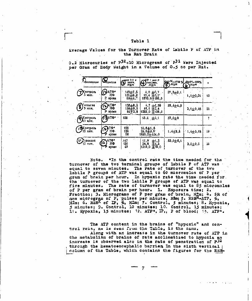
Table 1
Average Values for the Turnover Rate of Labile P of ATF inthe Rat Brain
0.2 Mioroouries of p32410 Micrograms ofP03 Were Injectedper Gram of Body Wei&t in a Volume of 065 cc per Rate
S% .. .... .... ...
0 ATOO~ 145*7,5 4,0 +0,7 27, 8:2, i, M. HO M 31.8,2 15 4±,i v+±.o t I ,oo21 f 9
""1 TO 1382 4,7 ±0.,50 35.4±-L4,0.123 ,9",8 -1 6,1 4:•,4 2,1±0,46" 2
80 I _24~,8 18426,0___ " " Xp . O IR 51±2,8 1255,0 ±ss,o ._
•HTPoJb TOO 1U 15,4.±3,1 j 37,009 " 7
i . 1. •, & 0 1,4±3,8• 1,4±0,18 1
I117I~H T' .4 11,9' 1,2 8..3,0±6,1
15 HO 13~ 24~ ~2,8±0,5 :1
Note. *In the control rats the time needed for theturnover of the two terminal groups of labile P of ATP wasequal to seven minutes. The rate of turnover of the twrolabile P groups of ATP was equal to 60 mioromoles of.P pergram of brain per hour. In hypoxic rate the time needed forthe turnover of the two labile P groups of ATP was equal tofLive minutes. The rate of turnover was equal to 85 micromolesof P per gram of brain per hour, I& Exposure time; 2,Fraction; 3. Micrograms of P per geam of brain, M.m; 4*. SR ofane microgr m of P, pulses per minute, Ntm; 5. RSiK"ATP, ý,M±m; 6. RSRK of IF, 7, M ,m; 7. Control, 5 minutes; 8. Hypoxia,5 minutes; 9, Control$ 10 minutes; 10. Control, 13 minutes;11. Hypoxia, 15 minutes; 12, ATP*, IP,, P of blood; 13, ATP*.
The ATP content in the brains of "hypoxia" and eon-troi rats, aJ 6 Sw Wil 'ilSJ
Alone with an increase in the turnover rate of ATP inthe metabolism of brains of rats acclimatized to hypoxia aninorease is observed also in the rate of penetration of p32through the hematoenoephalio barrien In the sixth vertioal
olumn of the Table, which contains the figures for the R_4-
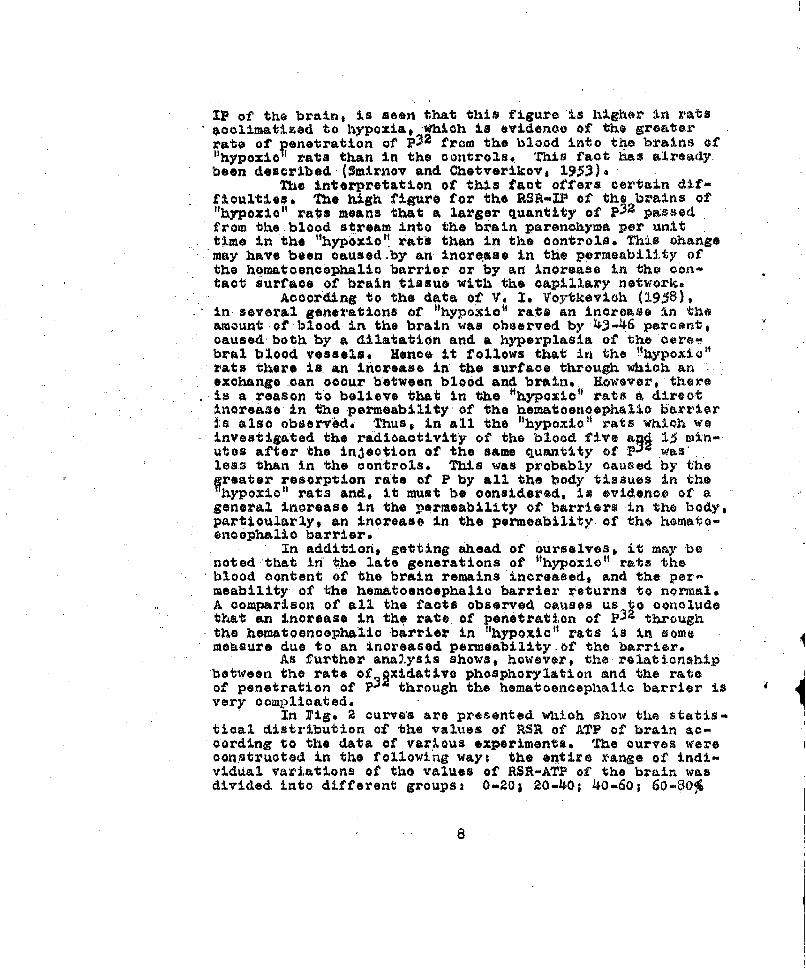
IP of the brain, is seen that this figure is higher in ratsacclimatized to hypoxia,#Which is evidenoe of the greaterrate of Penetration of P32 from the blood into the brains of"hypoxiO 1 rats than in the controls* This fact has already.been desoribed,(Smirnov and Chetverikovs 1933)a
The interpretation of this fact offers certain dif-fioulties. The high figure for the RSR-IP of the brains of"hypoxio" rats means that a larger quantity of p32 passedfrom the.blood stream into the brain parenchyma per unittime in the "hyp'oioi rats than in the controls. This changemay have been caused.by an inorease in the permeability ofthe homatooncoephalic barrier or by an increase in the con-tact surface of brain tissue with the capillary network.
According to the data of V. Is Voytkevich (1938),i in several generations of "hypoioc" rats an increase In theamount of blood in the brain was observed by 43-46 percent,oaused both by a dilatation and a hyperplasia of the cere-bral blood vessels. Hence it follows that in the "•hypoxic"rats there is an increase in the surfaoe through which anexchange can occur between blood and brain. However, thereis a reason to believe that in the tthypOziOt rats a direotIncrease in the permeability-of the hematoenoephalic barrieris also observad. Thus, in all the "hypoxic" rats which weinvestigated the radioactivity of the blood five a 15 min-utes after the injection of the same quantity of P2' was"less than In the controls. This was probably caused by theWreater resorption rate of P by all the body tissues in thehypoxio" rats and, it must be considered, is evidence of a
general increase in the permeability of barriers in the body,partioularly, an inorease in the permeability of the hemato-encephalic barrier.
In addition, getting ahead of ourselves, it may benoted that in 'he late generations of "hypoxio" rats theblood content of the brain remains increased, and the per-meability of the hematoenoephalio barrier returns to normal.A comparison of all the facts observed causes us to concludethat an increase in the rate of penetration of P32 throughthe hematoenoephalio barrier in "hypoxia" rats is in somemeasure due to an increased permeability.of the barrier.
As further analysis shows, however, the relationshipbetween the rate of _xidative phosphorylation and the rateof penetration of P through the hematoencephalic barrier isvery complicated.
In Pig* 2 curve's are presented which show the statis-tical distribution of the values of RSR of ATh of brain ac-cording to the data of various experiments. 'the curves wereconutruoted in the following ways the entire range of indi-vidual variations of the values of RSR-ATP of the brain wasdivided into different groups: 0-20; 20-40; 40-60; 60-800
8
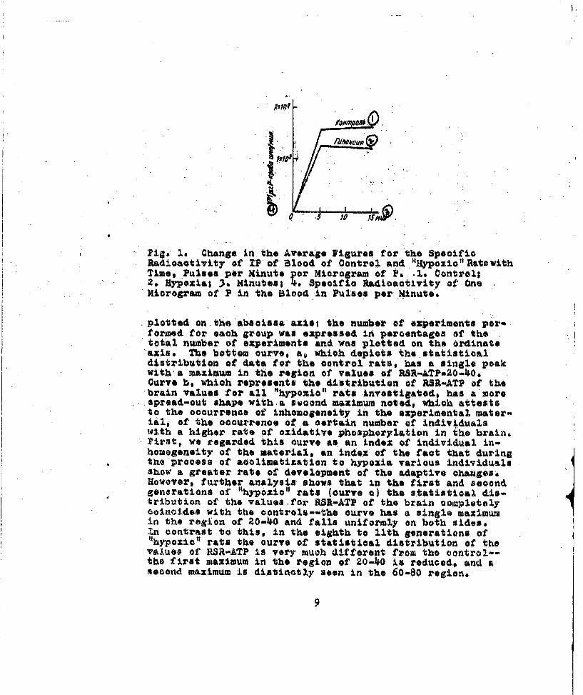
Pig# Is Ch~angs, in the Average Fi.gures for the SpecificRadioactivity of 1P of Blood of Control and. "Hypoxio'l Rats withTime, Pul1ses per Hinute 10 r Microgram of Ps -I* Control;.2o Hypoxial Is, Minutes, to. Specific Radioactivity ot One
Xiorogram fPi the Ood nPle o.iu
thotal gneater of aprienofsdandwpseptoftdo the odatdvinatge.distibuion f dta or te cntro rasbras ingcmletpeak
withs, axium n te reionof alus o6RS-80 region.
;z %_, 0
, 40
t20
.0077 40 /A 7WT
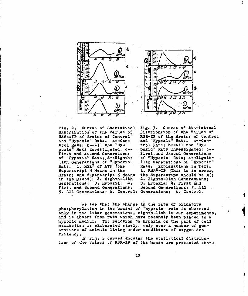
Figs. 2, COurves of Statistioal Fig. 3. Curves of StatisticalDistribution of the Values of Distribution of the Values ofRSR-ATP of Brains of Control RSR-IP of the Brains of Controland "Hypoxib" Rats, a--Con- and "Hypoxio" Rats. a--Con-trol Rats; b--All the "Hy- trol Rats; b*-All the "Hy-poxilo" Rats Investigated; 0-- poxic" Rats Investigated; c--First and Second Generations First.and Second Generationsof "Hypoxio" Rats; d--Eighth- of "Hypoxic" Rats; d--Bighth-llth Generations of "Hypoxio" l1th Generations of "Hypoxie"Rats. Is RSRM of ATP Ithe Rats. fxplanations in Text.Superscript M Means in the 1 RSRK-IP 'E•ma is In error,arain; the Superscript K Heans the Superscript should be M]jin the Blood]' 2. Eighth-llth 2. Eighth-llth Generations;Generations; 3* Hypoxia; 4- 3. Hypoxia; 4, First andFirst and Second Generations; Second Generations; 3o All5. All Generations; 6. Control. Generations; 6. Control,
We see that the change in the rate of oxidativephosphorylation in the brains of "hypoxioal rats is observedonly in the later generations, eighth-llth in our experiments,and is absent from rats which have recently been placed in ahypoxio medium. The reaction to hypoxia on the part of cellmetabolism is elaborated slowly, only over a number of gen-orations of animals living under conditions of oxygen de-fioiency.
In Fig, 3 curves showing the statistical distribu-tion of the values of RSR-IP of the brain are presented ohar-
10
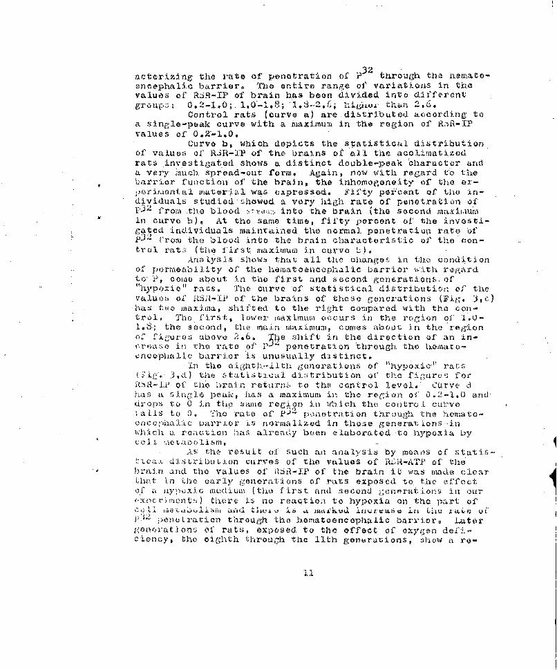
acterizing the rate of penotration of }P32 through the htemato-encephalic barrier0 The entire range of' variations in thevalues of RSR-IP of brain has been divided into differentgroups: 0.2-1.0; 1.0-1.;•-; lb._; hits tn ... "
Control rats (curve a) are distributed according toa single-peak curve with a maximum in the region of RAR-IPvalues of 0.2-1.0.
Curve b, which depicts the statistical distributionof values of RiR-]IP of the brains of all the acolimatized.rats investigated shows a distinct double-peak character anda very Rq.uch spread-out form. Again, now with reCard t:o the
arri~er function' of' the brain, the inhomogeneity of the ex-per•airontal mater:ial was expressed. Fifty pt'cent of the in-dividuals studied ý.showod -a very high rate of' penetration of02 4 from the blood r'w,. into the brain (the second maxiiaurmin curve b), At the same timiie, fifty percent of the investi-gated individuals maintained the normal. penetration rate ofP32, from the blood into the brain characteristic of the con-trol rats: (the first mnaximum in curve L•)
Anna.45 isshows t:hat all the ohanges in thu condition
of pormeability of the hematoenecophalic ba.rrier wiLt regardto i1, come about in the first and second gonerations. of"hypoxio" rats. The curve of statistical dlistribution of thevaluoesi of I--P of the brains of those g,ýeunerations (Fig. 3,e)has two maxima, shifted to the right compared with the con-trol, The first, -lover maximm- occurs in the rogion of i,0-1.6; the seoncd1 tfhe main mkaximum, comes about in the regionof figures a"bove 2.6, The shift in the direction of an in-cr,.,ase in rhe rate of' P-I'; penetrat-ion through the hemato-enceplialic barrier is unusually diistinct.
.in thle eighth.--ilth g.nerations of '"hypoxico' rats('i 3,d) tire ,,tatis;t.ical di:.i;ribution of the fi.(gures forf.gR-ih of tLh ?rain reotrnsE to the control level. Curve d
a single peak, has a maximum in the re,-ion of 03.2-1.0 andU"Or's 'to 0 in the, simie reg; on in wb.i.oh the controal cut'veaeLls to 0! 0 Tho rate of' p) Y.n.et'tration throug.i tfte houeato-
enCr~OuAI;lic ;arri-er is normalized in tho:;e generations <inwhich a. roaiction hIas already boan elaborated-tO hypoxia by
-i 'y 1.3,o e n e 1ior ,e. to
A.s the result oi such ai anlalysis by moanls of stat is-tiLa.. distri hution cuirves of the values of" RL.ai-ATI-' of' thebrain and. the values of Ll6A1-IP of the brain it was made clear(Amt in the early generations of rats exposed to. the effectof.,1 It iypoxi; a LI. urt C the, first and second generations iin our"i.c~inon t.) tl.Lere is no reaction to hy.poxia on the part of'
CCII '- -- - - -I~~ti ..L'tJ X->lVAi~~M' Lb 'A IWktLKUQ.1 IlII:IUtIt O C J i ll- t 4h1 VELO
pAZ :r)CLrLUrtion through the hboratoencophalic barrio. r a tergente).yat ion:s of rats, exposed to the effect of oxygen 'defijciency, the eighth thirough the l1th generations, show a re-
iii
encepha~lic barrier, observedl during the first period, of' ac-olimatization, is of biologic value, A positive result inrtile series of' erperiments undertaken would make it possible
ic barrier in the first period of acclimatization an activeadaptive• reaction to hypoxia.
In the literature it has been firmly esta.blishedthat the nor•mal aocompliskment of the barrier function in thebrain is maintained, by the energy of oxidative metabolism.Auy nmore or less marked disturbance of lt--anaorobiosis,hypoxia, disorder of cytoobrowe oxidase, restriction, of thesupply o~f substratea--hypoglyoemia--all those effsets ar'e at-companied by an increase in the permeability of the hewato-encephalic barrier to vital dyes, to p• and Other substances(Tschirgi, 1952; H~ess, 1955; $Aakay, 3.957; Verzhbins'kaya, 1957,
.. 195$),In a number of experiments control and '"hypoxic" rats
were in~eot•ed with su~bstances which increase the permeabilityof' the hematoenoephalic barrier to p32 in the sateS dosesipituitrin (0.1 Oc intraporitoneally), histamine (0,.5 miliigram. intravenously) and, finally,, simply a large vo~lume ofhypotanic saline solution (5 cc of a 0,2. percent NaCI solu-tion intraperitonealty). All these agents produced a• consid-.orable increase, by two or three times, in the JR$R-I? of theob5 •in, which signifies an increased rate Of penetrat•ion of
P hough the barrier (Table 2). Tn all experiments oft h~s series & considerable, 50 percent, reduction was observ-oct in the rate of oxidative phosphorylation in the brain (see'fableel and 2),
* i~Because of' the inadequacy of our knowledge about therhnysiolog4ai mechanism of penetration of substances,s parti'-oularly P' ,througb the barrier between 1t he bloodi and thebrain, it is impossible to put 'trust in the faot that thereis a reduction in the rate of oxidative phosphorylationunder these conditions. There is reason to believe that thisis only an apparent phenomenon. In connection with the prob-1cm being analyzed, it is important to note• only that theinitial increase in the permeability of' the hematoencephalicbarrier is not associated with an increased rate of oxidativephosphorylation, permeability ch1anges of the barri.er probablyoccur secoondarily, as the result of c~hanges in the rate. ofoxtdative metabolism. This given us the right to ex~press theidea that the incoreased rate of penetration of P5 2 throughIthe hematoencephalic barrier, observed during• the first periodof acclimatization of rats• to hypoxia, should be consider.ed an
tier function occurring as the r'esult of' a deficienc;y of th~eenergy of" oxidative metabol.ism of the brain during- this petted.=
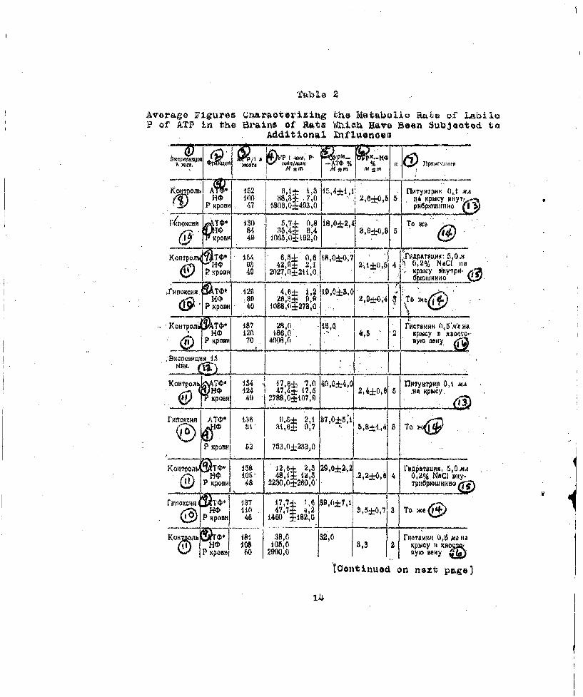
Ta~ble 2
Avera~ge Figures Qnraoterizizng the Metboliq R"LVt O~f !!ýP of ATP in the Brain$ Of Rats Wbhiokh Have Been~ Subjeoted to
Additional Infliaenoes
'0 ', 1 1 99. P, p 1 j,-
Kpo~ oA'r 152 0i,t+ 1, ""* 1 MlyNTRKM 01 AMHO0 100.J 3 70I .2605s ti*& KPUCY NNiYT,
r130 0, 68 i8,0±4,4 HTa we1, x )m 41) IN150 15f 92,0 [
8'UP04 TV ~ , 15A 9.5+ d0,&8
9O 93 42,n± 2,1 1 -)7 jt±,14 0 2% NACI nBDPK(B1 all 49 207,0±2t1,O. ýWC WHIW-
.rMo.M~120 4,6;L 1,2 i10,013,0 $* R.Sf r 26 -iS 15,9 2,104 C1'BM TbiA~ *
.F H41 120 1800.", 2 Kpb~cy 0 XIBO(.?Qo(j f~Kposm 70 400 0 ylo Belly,
KOTO T&' I 1.64 47,e b,-7,0 40,0±4,0 1 fl tTPHywpn 0,1 UA
VHV 124 I47 41 17,6 2 ,4±0,6 5 .94 Hpb4cy..
!Dtomm RATO1* 136 DZ 2,1 B7,0±5:158±,
P753,6±18330,O
Kowijj.Q'4 158 1 2,b 2,3 I29,0±2,2 S'~aruu ,OWAH0 i05) 48,1 12,5 .2,2±0,6 4 0,20A NaCI say.
CD p orn 46 22,W,0±280:0' T F 116pbowhum
r'mmem 137 17,7~ :, 39,(J±7,1I1, . 47,7 41, S5:E, 3 T AenI6 A60 ±182,0 ~ u<
Kcn~J~~ZT ' 1B~38,0 182,0 r~crmm~ o,5 ma tiai'(~IHO 108 1 ~03,3 2 XPley Ht ,xno=sp lXpOM 801 29000~ty~e
[Continuad on next psg83
[Table 2, continued from previous pagel
lo ~~1L~ Tbio,.5 Ariues 2 Frcti.u~ . 4crocuriesof P per Gram of Brain; 4. SR of One Microgram of P Pulsesper Minute, Mtm; .3. RSRM--ATP Percent, M±m; 6. RSRk--IPPercont, M.+n; 7. Notes; 8. Control; 9. ATP*, IP, P ofBlood; 10. fypoxia; 11. Control; 12. &xposure Time 15Minutes; 13,3 ituitrin, 0.1 oc per Rat Intraporitoneally;1.4° Same; 15. Hydration: 5.0 cc of 0.2 Percent NaCI perRat Intraperitoneally; 16, Histamine, 0.5 Milligram perRat tnto the Caudal Vein.
Conclusions
The process of acclimatization of mammals to an oxy-ien deficiency takes place over a long time, over a numberof generations of animals,
In the first period of acclimatization, which, ac-cording to the data of this work, lasted at least for. thelifetimes of two genarations of rats, the survival of theanimals under conditions of oxygen deficiency is assured bythe mobilization of different physiological reserves of thebody. All the reactions of the organism observed duringthis period are dlirected at maintenance of a normal oxida-tion in the brain, the organ which de]ends most on the enor-gy of oxidative metabolism. During this period the condi-tion of the organism cannot be called good. It is at theboundary of irreversible changes. Maintenance of tho normaloxidation level in the brain and in other vital organs isachieved by an increased consumption of oxidative energy bythe body as a whole during this period. A vicious cycle iscreated, capable of leading to a disorder. of the energy meta-boiism of the body.
The increased permeability of the homatoencophalicbarrier and the absonce of a reaction to hypoxia on the partof the call metabolism in the brain can servo as one of tl)eindications of the unfavorable, unbalanced condition of the;•ody durinj the first period of acclimatization.
The period when the tissue reaction to hypoxia.�'�.�~cand the permeability of the hematoonceplia3.io barrier
roturnsý to normial should be called the "seceond period of ac-climat!izati.on; thte period of true acclimatizationo Throughbho •iaterial of tho present %ýor. t:..is poriod. was found be-
iyp,'.•xLc ::LUh1t i3y this 1)oe'riod the capacity oif more offici-a,it cýiid oconomicial titilizatl.on of the energy of oxidativo
mcuotaboLii,;m i"s olaio3rtedo The increased rate of oxidativopho.'i,•orylatio; in) the brain tissues probably occurs because
15
of a greater conjugation of oxidation With phosphorylatlon6The increased efficiency of oxidative metabolism contributesto the normalization of the barrier fun.otion of the brain,
The rosults of tho proenaL .zivoutigation were ob-tained through a study of the oxidative phosphorylation inthe brains of intact, uninjured animals existing normallyand capable of multiplication. There were no disordersintroduced ýy the in vitro experiment, no destruction of thetissue structure, no disorder of nervous or hunoral regula-tion, ate. This increases the reliability of the resultsobtainod.
Finally, it seems important to us to note thatunder experimental conditions throughout the lives of 11generations of higher vertebrates a cumulative effect wasfound from generation to Ceneration, that is, a hereditarilyreinforced effect of an altered environment on the conditionof'the most important.process of the energy balance--theprocess of oxidative phosphorylation in the brain tissues,
i3±b il ograplhy.
Is, 1aarbashova.Z. I., 1952. New Data on the Mechanism ofAcclimatization to Hypoxia. Collection.Kislorod-naya Terapiya i Kislorodnaya Nedostatochnost', 85,Kiev, Publisbint Ho.use of the Academy of SciencesUkSSR. 1938. Specific and Non-Specific F'eaturesof Acclimatization to Hypoxia, Colleotion. Pro blemyEvolyutsii Fiziol . Funktsiy, Posy. 75-Letiyu AkadeL. A. Orbelia Moscow--Leningrad.
2o Verzhbinskaya No A', 1957,.a. Material on the.Evolutionbf the Energy Balance of the 3rains of VertebrateseAuthor's Abstraot Of Dissertation, Leningrad,-�.1957be The Process of Oxidative Phosphorylationin the Brain and the Formation of the Hemato-encephalioaarrier i.n the Order of Vertebrates.Collection.Voprosy Biokhimii Nervnoy Sisteaiv,'(Problems of Nervous Syst-em B.iochemistry). 187-Kiev,--1938a The. Svolution of the Process of Oxi-dative Phosphorklation in the t•rains of Vertebrates.Collectiono Narodnokhazyaystvennoye .ISpol'zovaniyeEnergii Radioaktivnykh Izluoheniy v Mirnykh Tselyakh(National Economic Utilization of Radioactive Energyfor Peaceful Purposes), 24, Moscow,.
3. Voytkevich V. Il, 1958. The Effect of Chronic Hlrpoxia onthe Blood Sunply of the .B.aine. Cuiiectioni 'iziol.
-i Patol, Dykhaniya. Gipoksiya. i Oksi noterapi.a(Physiology and Pathology of Respiration, iHypoxiatand Oxygen Therapy), 56, Kiev, Publishing House of
16
the Academy of Sciences Uki~Re4~ Domonovikch.Ye. X. 19!ý8. Some Physiao:Loioal Meohnisnis
of Adap taton of an OgmniS- +e Hiywn ýV~iniiPizioia i Pat~lo D khanii a, Gi. ksiya, 3. Qlsigenoterl-a 3. &, b7, Kiev,_Publishing. House of the Academy of
~t~oesUkSSR**5o Kcotellnikova A. V., 1957. Method of Determining the
'Specoifio Aotivity "of Inorganic Fhosphi~at by theIsobtyl'b o4thodw. 8iokhimiXW (Biochemistry), 22, 4
6e Kropq Ye. Me, 1936. Tissue Adaptation to Chronicd Hypoxia,*Ove; a Number of Generations* Collection,. DokliadyAl~a 20-m Mozhdunarodnont Kongresse 2tixi6l. v Bryussele(Reports at the Twentieth World Congress of phy-sia-lody.. in. Brussoik),#"1292, Moscov.'-1938. a e Respirm-tion o f Srain Tissue in Hypoxia,., Collesotiori. IFiiolw
qiokhiimiya Nervnoy Sistemy.. NeW1 DAt& on the Compara-tiv ~tdyof ahshou Metabolism of the B~rain in
Different F'undtionml States. Causeda by kHypoxia of theBrain&. Colleoti.4n, fiokhimix No Ssemv. 125.,
K). KOps. Ye, 1q, sVerazhbinskaya --g As, Cherlykayeva Yoe' YU .,Chirkovskaya Ye. V and Gavurina Ti. K., 3.956a.-Ad~ptation of Animals to ýChro'nic Hypoxia. Communi-cation I4 Fiziol, Zh*.SSS (journal. of Physiology of'the USýR), TTL - 9--195b. Qormruniot~ion 114,
9. suJlimo-samuyl Z., K 9326 The Eff $at of .Reducead Par-tial Pressure 6f Oxygen on Tissue Respiration.Author's. Abstraot 'of Dissertatiorl, Leningrad.
10o Sm3.rfov A. A. and Chetverikov D. A,, i953)t .tud~y of IPhos-phior~l~s Ntabolism in- the Br~in Diiring Hypoxia withthe Aid. of Radioactive Phosphorus, Doki AAN ý.iSR(Reports of the. Academy of Scienoes 9017 37d3
111. shapot V,0 $4, Gromova X& GoI.1 934,i Energy 3alance-of the.lBrain-and the P.Froblem of Hypoxio States. Collootion'aBlokhirniya Nervhoy SistemX, 1939, Kiev.
12. 3nSakay Le, 1937* Dyna~mic Aspects of the 5lood.-Urain ..Barriera..Metabolism of the' Nervo~us System, Ed, 2Richter, Porgamon iPrass.
13. BiLng ta C. and Baker 1R..4s, l931* The Determination of -
iib in.Minute inolant.q of Liiinni hy thl ts Method. J..siole.' Cheme, 9,2, 3*--1932. Purification of Ben'Zidinea nd an I poved Reagent for Estimating Hemaglobin inal1ooci.- Js. _iols. Chemn, 95,' 21 3837-
17
14+. Dolaoliaux At and Tissi~res A.0 19kk6, L'Adaptation.Tisaulaire.& 1'HypoXYdOse- av MoL~dAct'&, 3/4+,
15. flelaclhaux A. and 13orson Jo, 194+7, Hoy o*Atx 4'4+63.
16. karnischfagegr S~. and.0pitz £., 193C). ft~erdon Cyto-chromgebhalt V1erso1hiedenen Kaninch~eilorgane rnach1iollenan~pas sung. Arcoh. f do ptos#.?hirs ol, 252, 627.,
17. lle s A..,. 19,33. 3161od- ran'arior and.G;round Substanceo~f tho Contral Nervous System, Aro r1l. Psy-
183 'll s s i iTr esL 19ý4 3. iLe T4Ux do Cy-toohrome C-d~ars le'1Thlsci1o Squ~at Iique du Rat aprbs Traitement auAlnri~tpophonol, au Dosoxycorticos.terq~on et-auTos~tostorbne.. Nrohs. Intern, Ph~ysio. 5,25
19. Ta3chirgi R, D, 19.52., 131ood-J3rain t34rrior1 C, Thei3iol.ogy -off MUertaJ Health and Disease 34 Paul £3.Inc.. Mod Book JDepartmo of Hiarper~ and iBrotb..'
:n s t I t it 9' 6f PhyslologY iniani2..P.kavlov of-:tho Aoademy Raecivbd 8 May 19.59.
of -Scionces
1-83