Embed Size (px)
Citation preview
ULTRASTRUCTURAL MALFORMATIONS OF THE CEREBRAL
MICROVESSELS IN PATHOLOGICAL CONDITIONS AN ELECTRON
MICROSCOPIC STUDY
Zoltán Süle, M.Sc.
Ph.D. thesis
Experimental and Clinical Neuroscience Doctoral Programme
Doctoral School of Medicine
Supervisors:
Eszter Farkas, M.Sc., Ph.D.
Prof. András Mihály, M.D., Ph.D., D.Sc.
Department of Anatomy, Histology and Embryology
Faculty of Medicine, University of Szeged
Szeged
2010

Ph.D. thesis Publications 2
LIST OF PUBLICATIONS, RELATED TO THIS THESIS
I. Fabene PF, Weiczner R, Marzola P, Nicolato E, Calderan L, Andrioli A, Farkas E, Süle
Z, Mihály A, Sbarbati A: Structural and functional MRI following 4-aminopyridine-
induced seizures: a comparative imaging and anatomical study.
Neurobiol Dis. 2006 Jan; 21(1):80-9 IF: 4.128
II. Farkas E, Süle Z, Tóth-Sz ki V, Mátyás A, Antal P, Farkas IG, Mihály A, Bari F: Tumor
necrosis factor-alpha increases cerebral blood flow and ultrastructural capillary damage
through the release of nitric oxide in the rat brain.
Microvasc Res. 2006 Nov; 72(3):113-9 IF: 2.477
III. Süle Z, Mracskó É, Bereczki E, Sántha M, Csont T, Ferdinándy P, Bari F, Farkas E:
Capillary injury in the ischemic brain of hyperlipidemic, apolipoprotein B-100 transgenic
mice.
Life Sci. 2009 Jun 19; 84(25-26):935-9 IF: 2.560
Cumulative Impact Factor = 10.153 (ISI JCR 2009)
LIST OF ABSTRACTS PUBLISHED IN CITED JOURNALS, RELATED TO THIS
THESIS
i. Süle Z, Tóth-Sz ki V, Antal P, Mátyás A, Mihály A, Bari F, Farkas E: TNF- -induced
microvascular damage is mediated by nitrogen monoxide in the rat brain.
Clin.Neurosci./Ideggyógy. Szle. 2006, 59 (1 klsz.) IF: -
ii. Süle Z, Bari F, Sántha M, Bereczki E, Farkas E: Capillary injury in the ischemic brain of
hyperlipidemic, apolipoprotein b-100 transgenic mice.
J. Vasc. Res., 2008, 45, (Suppl. 2), 122 IF: 2.792
iii. Süle Z, Kovács GG, Mihály A, Farkas E: Microvascular aberrations in the white matter
in Alzheimer's disease.
J. Neurol. Sci., 2009, 283, (1-2), 286 IF: 2.324
Cumulative Impact Factor = 5.116 (ISI JCR 2009)
Ph.D. thesis Abbreviations 3
LIST OF ABBREVIATIONS
1VO one-vessel occlusion 2VO two-vessel occlusion a astrocytic endfoot A
beta-amyloid AD Alzheimer s disease ANOVA analysis of variance apoB-100 apolipoprotein B-100 ASC atherosclerosis BBB blood-brain barrier BM basement membrane CBF cerebral blood flow CNS central nervous system coll collagen deposition Cpu caudate putamen CT computer tomography e endothelial cell em endothelial mitochondria en endothelial nucleus ep endothelial cytoplasm er erythrocyte EAAT excitatory amino acid transporter FWM frontal white matter F female FM focal mild -GTP gamma-glutamyl transpeptidase
GLUT-1 glucose transporter isoform-1 Gm gray matter HSPG heparin sulfate proteoglycan i.c. intracarotid i. m. intramuscular i.p intraperitoneal i.v. intravenous l capillary lumen p pericyte pn nucleus of pericyte LDL low density lipoprotein L-NAME N(G)-nitro-L-arginine-methyl-ester LS lateral septum LV lateral ventricle M male MRI magnetic resonance imaging NO nitric-oxide NOS nitrogen-monoxide synthase O occipital P parietal PD Parkinson s disease PWM parietal white matter rCBF regional cerebral blood flow
Ph.D. thesis Abbreviations 4
LIST OF ABBREVIATIONS (contd.)
ROS reactive oxygen species
SPECT single photon emission computed tomography
Tg transgenic tj tight junction TNF
tumor necrosis factor-alpha VDB ventral diagonal band VEGF vascular endothelial growth factor VLDL very low density lipoprotein Wt wild-type WM white matter y years

Ph.D. thesis Contents 5
CONTENTS Page
1. INTRODUCTION 6 1.1. General architecture of the brain capillaries in healthy conditions 6 1.2. Morfological units of the BBB 7 1.2.1. Endothelial cells 7 1.2.2. Pericytes 8 1.2.3. Astrocytes 9 1.2.4. Basement membrane 9 1.3. Ultrastructure of the blood-brain barrier in pathological conditions 10 1.4 Cerebrovascular and other risk factors for Alzheimer s disease 10 1.4.1. Cerebrovascular risk factors for AD 11 1.4.2. Effects of the neuroinflammation on AD 11 1.4.3. Effects of the hyperlipidemia on AD 12 2. OBJECTIVES 14 3. MATERIALS AND METHODS 15 3.1. Experimental models / study population 15 3.1.1. Effects of normal aging and Alzheimer s disease on cerebral white matter
microvessels a human study 15 3.1.2. Effects of circulating TNF on blood-brain barrier ultrastructure a rat study 16 3.1.3. Effects of hyperlipidemia and/or ischemia on blood-brain barrier
ultrastructure a mouse study 18 3.2. Electron microscopy 18 3.2.1. Sample preparation, and examination 19 3.2.2. Determination of microvascular damage and quantitative analysis 20 3.2.3. Statistical analysis 21 4. RESULTS 22 4.1. The ultrastructure of cerebral white matter microvessels in normal aging and
Alzheimer s disease 22 4.2. Effects of circulating TNF on blood-brain barrier ultrastructure 25 4.3. Effects of hyperlipidemia and/or ischemia on blood-brain barrier
ultrastructure 27
5. DISCUSSION 30 5.1. Alterations of cerebral white matter microvessels in normal aging and
Alzheimer s disease 31 5.1.1. Cerebral blood flow and microvascular alterations in normal aging 32 5.1.2 Cerebrovascular pathology in AD 33 5.2. BBB ultrastructure in inflammatory processes 34 5.3. The ultrastructure of the BBB in hyperlipidemia and/or ischemia 36 6. CONCLUSIONS 39 7. ACKNOWLEDGEMENTS 40 8. REFERENCES 41 9. APPENDIX 50
Ph.D. thesis Introduction 6
1. INTRODUCTION
1.1. General architecture of the brain capillaries in healthy conditions
Cerebral capillaries represent the finest branches of the vascular tree in the human brain.
They form anastomoses, and create a three dimensional vascular network. The surface area
of the brain microvasculature is approximately 100 cm2/g tissue; the mean intercapillary
distance in the human brain is about 40 µm (Duvernoy et al., 1983). It is a well known
phenomenon, that the density and distribution of the capillaries vary in different brain areas.
For example, the microvascular density in the gray matter is approximately three times
higher than that of the white matter. Moreover, the functional activity and nutrient claim of the
given brain region determine the distribution of its capillary network. This concept is
supported by the observation, that local cerebral blood flow and capillary length per brain
volume show a remarkable correlation (Gjedde and Diemer, 1985). Also, synapse-rich layers
are highly vascularized, cell body populations contain less dense microvascular networks,
and neural fiber bundles receive a relatively moderate blood supply. Finally, vascular
distribution may be defined by the specific tasks of a brain area: the motor centers receive
lower blood supply than the sensory and association centers.
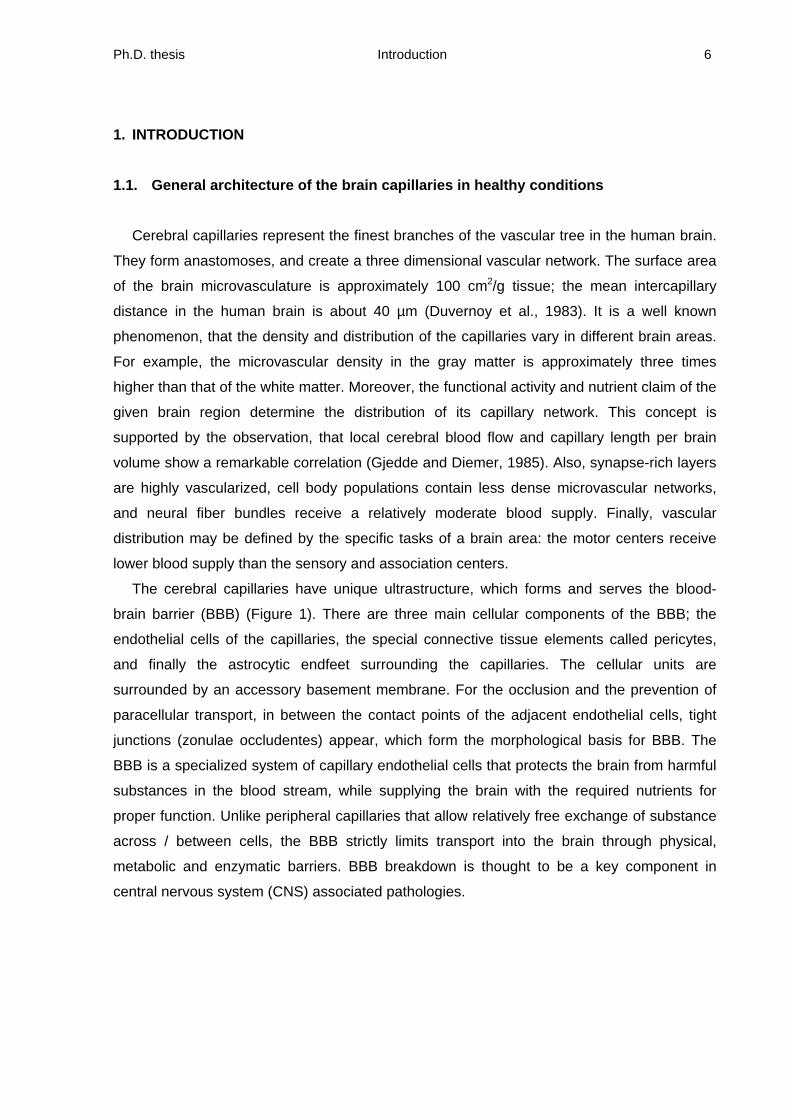
The cerebral capillaries have unique ultrastructure, which forms and serves the blood-
brain barrier (BBB) (Figure 1). There are three main cellular components of the BBB; the
endothelial cells of the capillaries, the special connective tissue elements called pericytes,
and finally the astrocytic endfeet surrounding the capillaries. The cellular units are
surrounded by an accessory basement membrane. For the occlusion and the prevention of
paracellular transport, in between the contact points of the adjacent endothelial cells, tight
junctions (zonulae occludentes) appear, which form the morphological basis for BBB. The
BBB is a specialized system of capillary endothelial cells that protects the brain from harmful
substances in the blood stream, while supplying the brain with the required nutrients for
proper function. Unlike peripheral capillaries that allow relatively free exchange of substance
across / between cells, the BBB strictly limits transport into the brain through physical,
metabolic and enzymatic barriers. BBB breakdown is thought to be a key component in
central nervous system (CNS) associated pathologies.
Ph.D. thesis Introduction 7
Figure 1. Ultrastructure of a cerebral capillary s cross section. A: an electron microscopic image of a typical cortical capillary from the frontoparietal cortex of a Wistar rat. B: graphic redrawn of the vessel. Abbreviations: a: astrocytic endfeet; bm: basement membrane; em: endothelial mitochondria; en: endothelial nucleus; ep: endothelial cytoplasm; l: capillary lumen; p: pericyte; tj: tight junction. (With kind permission of Eszter Farkas.)
1.2. Morphological units of the BBB
1.2.1. Endothelial cells
The endothelial cells of the BBB show some special features which are unique and set
them apart from peripheral endothelial cells. Comparative morphometric analysis of the wall
of a cerebral versus a muscular capillary revealed that the wall thickness of brain capillaries
is almost 40% lower than that of capillaries in muscles (Coomber and Stewart, 1985). The
narrower wall of the cerebral capillaries could possibly be complementary to the restrictive
permeability of the BBB, allowing nutrients a shortened transport time to cross the BBB and
enter the brain parenchyma. The cerebral endothelial cytoplasm contains only a few
pinocytotic vesicles and the capillary wall is not fenestrated (Abbott, 2005).
There are various transport mechanisms through the BBB. Paracellular diffusion (in
between the neighboring cells) is not frequent because of the tight junctions; only the water is
able to traffic in this way in non-pathological conditions. The transcellular transport
mechanisms make use of various energy-dependent and non energy-dependent pathways.
Quantitative biochemical studies provided evidence for a functional polarity of the BBB. The
luminal and abluminal membrane surfaces of the capillary endothelial cells are different
Ph.D. thesis Introduction 8
according to their specific functions (Betz and Goldstein, 1978). An electron microscopic
immunogold study showed, that the concentration of the glucose transporter isoform 1
(GLUT-1), the principal glucose transporter of the BBB, is approximately 4-fold higher in the
abluminal than in the luminal endothelial membrane (Farrell and Pardridge, 1991).
The number of mitochondria and their volume in BBB endothelial cells is higher as
compared with the peripheral endothelial layer (Oldendorf et al., 1977), which is thought to
be required for the active transport of nutrients and waste products through the BBB. The
barrier function is further supported by an enzymatic barrier at the cerebral endothelial layer,
capable of metabolizing drugs and nutrients (Minn et al., 1991; Brownlees and Williams
1993; Brownson et al., 1994). These enzymes are principally directed at metabolizing
neuroactive blood-borne peptides. Enzymes such as -glutamyl transpeptidase ( -GTP),
alkaline phosphatase, and aromatic acid decarboxylase are in elevated concentration in
cerebral microvessels, yet often in low concentration or absent in non-neuronal capillaries.
1.2.2. Pericytes
Pericytes in the periphery are flat, undifferentiated, contractile connective tissue cells,
which surround capillary walls. The association of pericytes to blood vessels has been
suggested to regulate endothelial cell proliferation, survival, migration, differentiation, and
vascular branching (Hellström et al., 2001). Pericytes send out cellular projections, which
penetrate the basal lamina and cover approximately 20-30% of the microvascular
circumference (Frank et al., 1987). Pericytes are thought to contribute to endothelial cell
proliferation via selective inhibition of endothelial cell growth (Antonelli-Orlidge et al., 1989).
In support of this notion, the lack of pericytes has lead to endothelial hyperplasia and
abnormal vascular morphogenesis in the brain (Hellström et al., 2001). Electrophysiological
measurements have showed, that pericytes in the cerebellum and in retina are able to
control the capillary diameter according to the extracellular Ca2+ level or presence of various
vasoactive neurotransmitters, such as noradrenaline or adenosine triphosphate (Peppiatt et
al., 2006).
Pericytes of the BBB might be derived from or convert to microglia, since the cerebral
pericytes demonstrate the capacity to phagocytize exogenous proteins form the CNS, and
display macrophage surface markers (Coomber and Stewart, 1985).
Ph.D. thesis Introduction 9
1.2.3. Astrocytes
Astrocytes are star-shaped glial cells of the CNS, which envelop more than 99% of the
BBB endothelium (Hawkins and Davis, 2005). Astrocytes serve as a guiding mesh and play
an active role in the induction of the BBB. Their processes terminate as endfeet at the basal
lamina of the blood vessels, and form a limiting membrane (membrana limitant gliae
perivascularis) around them.
There is significant body of evidence to indicate that astrocyte interaction with the cerebral
endothelium helps determine BBB function, morphology (i.e. tightness), and protein
expression (Beck et al., 1984; Arthur et al., 1987; Cancilla et al., 1983). In vitro cell culture
models helped to clarify the astrocytic roles in this process. Cerebral microvascular
endothelial cells in themselves were able to form a BBB-like phenotype but with loss of some
important functions (leakier tight junctions, downregulated transport and enzyme systems)
(Krämer et al., 2001). Some of those properties can be reconditioned in experimental
models, which are based on cerebral endothelial cells co-cultured with astrocytic cell lines.
Rubin et al. (1991) clearly demonstrated that the tight junctional proteins are upregulated by
astrocytes. Even more interesting is that astrocytes have the ability for the induction of BBB
markers on peripheral endothelial cells (Kuchler-Bopp et al., 1999).
In addition to the constitution of the BBB, another most important function of the mature
astrocytes is the regulation of cerebral blood flow (CBF) and vascular tone, which is based
on the expression and function of serotonergic and cholinergic receptors on the perivascular
endfeet (Luiten et al., 1996; Elhusseiny et al., 1999; Cohen et al., 1997).
1.2.4. Basement membrane
A well-defined basement membrane (BM) covering the endothelial cells and embracing
pericytes supports the cerebral capillary walls. The BM is composed of three different
sublayers: a lamina rara interna along the abluminal endothelial surface, a middle layer
called lamina densa, and an outmost lamina rara externa medial to the astrocytic endfeet.
The average thickness of the cerebral capillary BM is about 30-40 nm. The BM constitutes of
extracellular matrix components such as type IV collagen, heparan sulfate proteoglycan
(HPSG), tenascin, laminin and extrinsic fibronectin. The major structural element of the BM is
type IV collagen, which is preferentially located in the lamina densa, while the proteins
laminin and HSPG are more closely associated with the two lamina rarae. Cell adhesion to

Ph.D. thesis Introduction 10
the basal lamina relies on integrins, which are transmembrane receptors that bridge the
cytoskeletal elements of a cell to the extracellular matrix. The BM provides mechanical
support for cell attachment, serves as a substratum for cell migration, separates adjacent
tissue, and can act as a barrier to the passage of macromolecules.
1.3. Ultrastructure of the blood-brain barrier in pathological conditions
The morphological features of brain capillaries can be best studied with electron
microscopy. Under various pathological conditions, the cerebral capillaries display typical
malformations. In aging, or cerebral ischemia, the apical surface of the endothelial cells
appear irregular, display microvillus-like processes into the lumen, and empty vacuoles may
form occasionally (Oomen et al., 2009; Süle et al., 2009). Pericytes often show type IV
collagen and dense body (lysosomes) accumulation in their cytoplasm (Farkas et al., 2000).
Damage or disruption of the BBB causes swelling of the astrocytic endfeet, which typically
appears as hypodense pericapillary areas in electron microscopic images. The most
common ultrastructural deviations of the basement membrane concern the accumulation of
extracellular matrix proteins, which results in BM thickening and/or fibrous collagen
deposition.
1.4. Cerebrovascular and other risk factors for Alzheimer s disease
Sporadic (i.e. not genetically inherited) AD is considered as a multifactorial, progressive
neurodegenerative disorder. In addition to amyloid toxicity and defect in tau-phosphorilation,
various risk factors, such as cerebral hypoperfusion, neuroinflammation and
hypercholesterolemia contribute to the development of AD (Fig. 2).
1.4.1. Cerebrovascular risk factors for AD
Vascular risk factors are considered as major contributors to disease evolution and
progression in AD. As such, moderate but persistent reduction in regional CBF compromises
memory processes and contributes to the development and progression of dementia (Farkas
et al., 2007). The association of decreased CBF (particularly in the temporal and parietal
cortices) with AD has been firmly established (de la Torre, 2002; Farkas and Luiten, 2001;
Matsuda, 2001), and the degree or pattern of cerebral hypoperfusion in mild cognitive
Ph.D. thesis Introduction 11
impairment has been proposed as a predictive marker for the progression to AD (Borroni et
al., 2006; Hirao et al., 2005).
Persistently low baseline CBF has been proposed to induce ultrastructural pathology of
the cerebral microvessels (Farkas and Luiten, 2001) (Fig. 2). The BBB displays
hypoperfusion-related ultrastructural abnormalities in the form of BM thickening and fibrous
collagen deposits, which develop chronically during aging and dementia (Farkas and Luiten,
2001). The accumulation of collagen fibers in the microvascular BM may hinder specific BBB
transport for important nutrients such as glucose and essential amino acids (Farkas and
Luiten, 2001). In the aging brain, a significant correlation has been established between
collagen deposits in the microvascular wall and advancing age in the frontal and occipital
white matter (Farkas et al., 2006). Further, in AD, the proportion of capillaries displaying
collagen accumulation in the microvascular BM in the cingulate cortex was considerably
higher than in age-matched controls (Farkas et al., 2000). Whether such BM pathology is
related to cerebral hypoperfusion has been tested in rats, of which the common carotid
arteries were permanently occluded (2-vessel occlusion, 2VO). Electron microscopic
examination revealed microvascular BM thickening and collagen deposits 14 months after
2VO onset, which were comparable to those seen in the human post mortem studies. The
proportion of affected capillaries in the hippocampus in 2VO rats was almost twice that in the
controls (De Jong et al., 1999). Based on the above findings, chronic cerebral hypoperfusion
is suggested to be a causative, accelerating condition for ultrastructural BBB damage, which
is also the focus of our current investigation.
The cause for cerebral hypoperfusion in AD has not been unequivocally defined; a
number of competing and complementary hypotheses exist. The development of
atherosclerotic plaques in the carotid arteries or any other cerebral resistance vessels
narrows the vascular lumen and renders the vessel wall too rigid for the fine regulation of
vascular diameter, thereby compromising optimal blood supply to the brain. Indeed, clinical
studies have demonstrated a link between carotid intima-media thickness and cognitive
decline (Silvestrini et al., 2009), and the association between atherosclerosis in the carotid
arteries and a higher risk for AD (van Oijen et al., 2007).
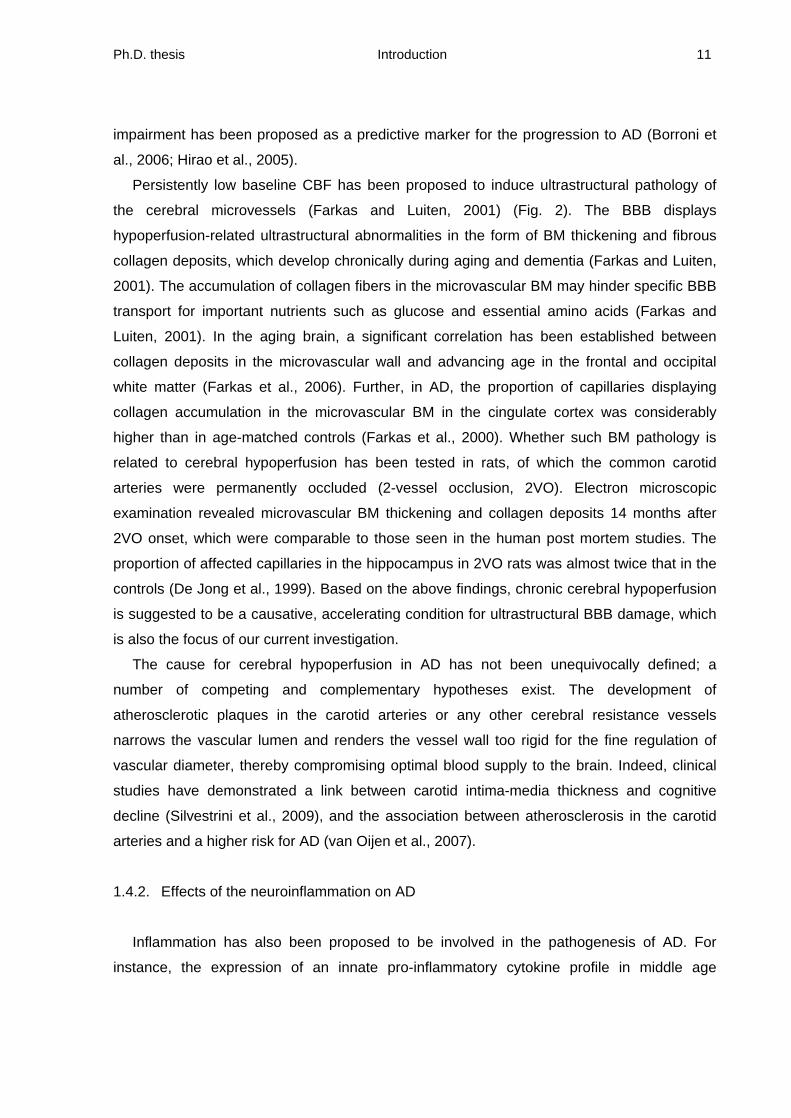
1.4.2. Effects of the neuroinflammation on AD
Inflammation has also been proposed to be involved in the pathogenesis of AD. For
instance, the expression of an innate pro-inflammatory cytokine profile in middle age
Ph.D. thesis Introduction 12
appeared as an early risk factor of AD in old age (van Exel et al., 2009). According to a
widely held view, beta-amyloid (A ) deposits in the brain parenchyma activate microglia,
which, in turn release pro-inflammatory cytokines and reactive oxygen species (ROS). This
chronic cascade may eventually lead to neuronal damage (Neuroinflammation Working
Group, 2000). While inflammation within the brain is thus thought to be a potentially major
neurodegenerative process, links between AD and inflammation in the periphery have also
been suggested (McNaull et al., 2010). As such, peripheral blood mononuclear cells produce
higher levels of pro-inflammatory cytokines upon stimulation in mild cognitive impairment and
early AD (Magaki et al., 2007). However, no association between circulating inflammatory
mediators and cerebromicrovascular injury that occurs in AD has been investigated. Here we
set out to characterize ultrastructural BBB damage after intracarotid infusion of the pro-
inflammatory cytokine, tumor necrosis factor-alpha (TNF ) in the rat, and to define whether
nitric oxide (NO) is a mediator of TNF in this regard.
1.4.3. Effects of the hyperlipidemia on AD
High plasma lipid content is known to favor plaque formation. Incidentally, high dietary
cholesterol intake increases, while the use of statins reduces the risk for AD (Sparks, 2008;
Sparks et al., 2008). Hypercholesterolemia caused by elevated plasma concentration or
abnormal metabolism of low density lipoprotein (LDL, an important carrier of cholesterol),
accelerates atherosclerosis (Grundy et al., 1985; Rudel et al., 1986). Furthermore, recent
evidence suggests that the triglyceride-rich very low density lipoprotein (VLDL) also
contributes to atherogenesis possibly through the inflammatory activation of vascular or foam
cells (Libby, 2007; Persson et al., 2006). Apolipoprotein B-100 (apoB-100) is a constant
surface component of both the cholesterol carrier LDL, and the triglyceride-rich VLDL in
circulating blood plasma. It plays a pivotal role in VLDL assembly in the liver, and binds to
specific receptors on the cell membranes to direct the lipoproteins to their proper metabolic
sites (Blasiole et al., 2007; Olofsson and Boren, 2005). In atherogenesis, apoB-100
assembles atherogenic lipoproteins and acts as a mediator in the interaction between LDL
and proteoglycans in the vascular wall, thereby promoting lipoprotein retention in the
vascular intima. The elevated production or decreased removal of apoB-100-containing LDL
from plasma has been associated with an increased susceptibility for atherosclerosis
(Grundy et al., 1985). In AD, apoB-100 was found up-regulated in the serum as shown by a
proteomics study surveying potential plasma biomarkers for AD (Song et al., 2009).

Ph.D. thesis Introduction 13
Genetically engineered mice expressing human apoB-100 (Tg(apoB-100)) were
generated in order to model hyperlipidemia with increased serum cholesterol or triglyceride
concentrations similar to human conditions (Chiesa et al., 1993; Csont et al., 2007; Linton et
al., 1993), and to investigate related cardiovascular pathologies in experimental models
(Csont et al., 2007; Purcell-Huynh et al., 1995). The established cardiovascular pathology of
Tg(apoB-100) mice prompted us to explore potential, ultrastructural microvascular
abnormalities in their brains, which may be relevant for the evolution of cerebromicrovascular
injury AD.
Figure 2. Various risk factors, considered in this study, which are expected to cause blood-brain barrier (BBB) damage that may contribute to Alzheimer s disease. The focus of our investigations was the ultrastructural malformations of the BBB. Conditions that were investigated in various experimental paradigms are highlighted in italics.
Ph.D. thesis Objectives 14
2. OBJECTIVES
The general aim of our study was to describe the possible ultrastructural aberrations of the
cerebral microvasculature in various pathological conditions. The disease processes
specified below all occur alone or in combination - in AD with ischemic components.
First, we sought to determine whether:
a) the microvasculature of the cerebral periventricular white matter is injured in
Alzheimer s disease;
b) the potential microvascular damage affects all white matter areas (i.e. frontal, parietal,
occipital) equally.
Second, we set out to:
a) investigate the effect of systemic inflammation (e.g. high level of the circulating
proinflammatory cytokine TNF ) on the ultrastructure of the blood-brain barrier;
b) determine whether NO is a mediator of the expected TNF -induced alterations in
blood-brain barrier ultrastructure.
Third, we aimed to investigate whether hyperlipidemia:
a) causes cerebral microvascular lesions in itself;
b) augments ischemia-related capillary damage.
Ph.D. thesis Materials and methods 15
3. MATERIALS AND METHODS
3.1. Experimental models / study population
All animal experiments were approved by the Ethical Committee of the University of
Szeged. In the human study, samples were collected based on informed consent, approved
by the Regional Ethics Committee for Human Medical Biology Research of the University of
Szeged.
3.1.1. Effects of normal aging and Alzheimer s disease on cerebral white matter
microvessels a human study
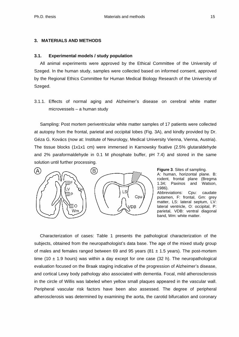
Sampling: Post mortem periventricular white matter samples of 17 patients were collected
at autopsy from the frontal, parietal and occipital lobes (Fig. 3A), and kindly provided by Dr.
Géza G. Kovács (now at: Institute of Neurology, Medical University Vienna, Vienna, Austria).
The tissue blocks (1x1x1 cm) were immersed in Karnowsky fixative (2.5% glutaraldehyde
and 2% paraformaldehyde in 0.1 M phosphate buffer, pH 7.4) and stored in the same
solution until further processing.
Figure 3. Sites of sampling. A: human, horizontal plane. B: rodent, frontal plane (Bregma 1.34; Paxinos and Watson, 1986). Abbreviations: Cpu: caudate putamen, F: frontal, Gm: grey matter, LS: lateral septum, LV: lateral ventricle, O: occipital, P: parietal, VDB: ventral diagonal band, Wm: white matter.
Characterization of cases: Table 1 presents the pathological characterization of the
subjects, obtained from the neuropathologist s data base. The age of the mixed study group
of males and females ranged between 69 and 95 years (81 ± 1.5 years). The post-mortem
time (10 ± 1.9 hours) was within a day except for one case (32 h). The neuropathological
evaluation focused on the Braak staging indicative of the progression of Alzheimer s disease,
and cortical Lewy body pathology also associated with dementia. Focal, mild atherosclerosis
in the circle of Willis was labeled when yellow small plaques appeared in the vascular wall.
Peripheral vascular risk factors have been also assessed. The degree of peripheral
atherosclerosis was determined by examining the aorta, the carotid bifurcation and coronary
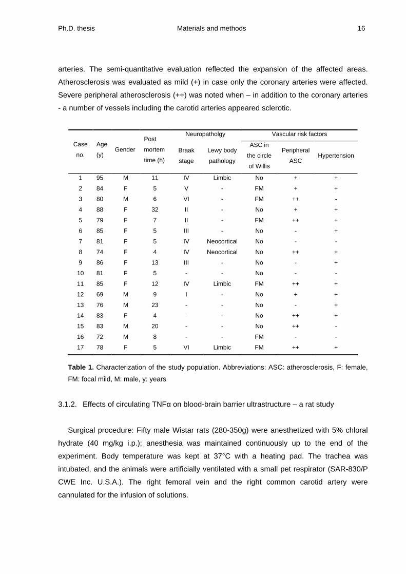
Ph.D. thesis Materials and methods 16
arteries. The semi-quantitative evaluation reflected the expansion of the affected areas.
Atherosclerosis was evaluated as mild (+) in case only the coronary arteries were affected.
Severe peripheral atherosclerosis (++) was noted when
in addition to the coronary arteries
- a number of vessels including the carotid arteries appeared sclerotic.
Neuropatholgy Vascular risk factors
Case
no.
Age
(y) Gender
Post
mortem
time (h)
Braak
stage
Lewy body
pathology
ASC in
the circle
of Willis
Peripheral
ASC Hypertension
1 95 M 11 IV Limbic No + +
2 84 F 5 V - FM + +
3 80 M 6 VI - FM ++ -
4 88 F 32 II - No + +
5 79 F 7 II - FM ++ +
6 85 F 5 III - No - +
7 81 F 5 IV Neocortical No - -
8 74 F 4 IV Neocortical No ++ +
9 86 F 13 III - No - +
10 81 F 5 - - No - -
11 85 F 12 IV Limbic FM ++ +
12 69 M 9 I - No + +
13 76 M 23 - - No - +
14 83 F 4 - - No ++ +
15 83 M 20 - - No ++ -
16 72 M 8 - - FM - -
17 78 F 5 VI Limbic FM ++ +
Table 1. Characterization of the study population. Abbreviations: ASC: atherosclerosis, F: female,
FM: focal mild, M: male, y: years
3.1.2. Effects of circulating TNF on blood-brain barrier ultrastructure a rat study
Surgical procedure: Fifty male Wistar rats (280-350g) were anesthetized with 5% chloral
hydrate (40 mg/kg i.p.); anesthesia was maintained continuously up to the end of the
experiment. Body temperature was kept at 37°C with a heating pad. The trachea was
intubated, and the animals were artificially ventilated with a small pet respirator (SAR-830/P
CWE Inc. U.S.A.). The right femoral vein and the right common carotid artery were
cannulated for the infusion of solutions.
Ph.D. thesis Materials and methods 17
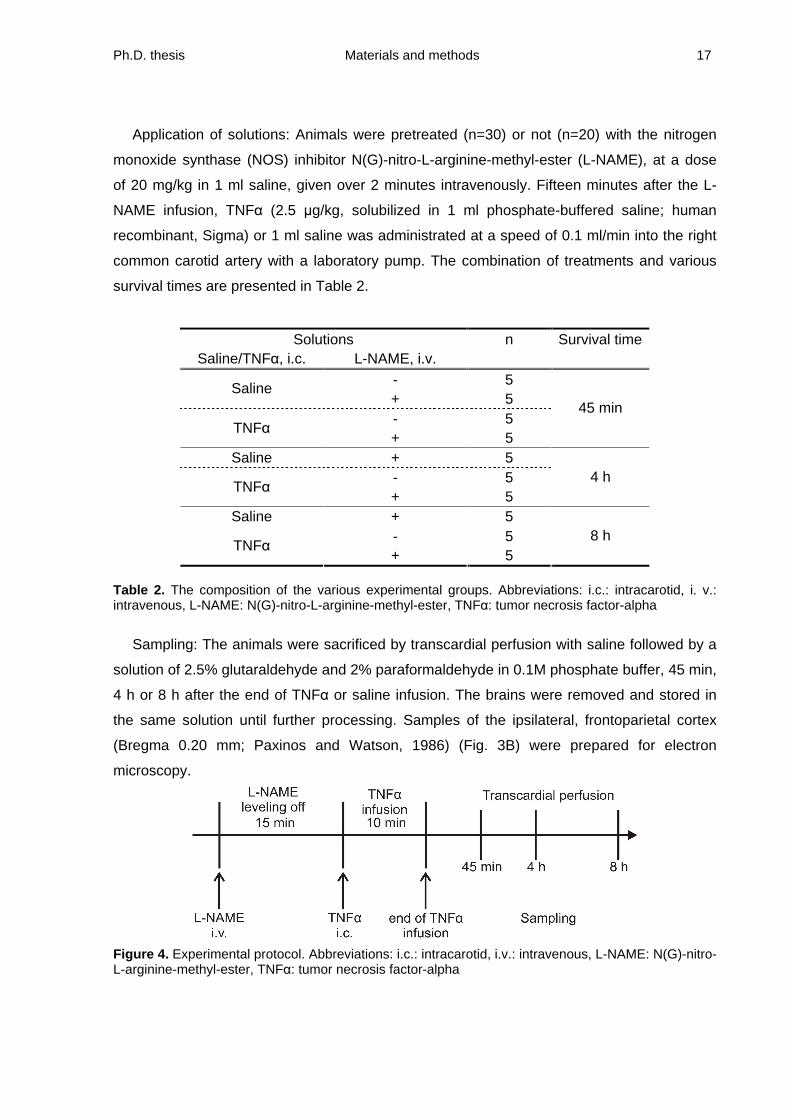
Application of solutions: Animals were pretreated (n=30) or not (n=20) with the nitrogen
monoxide synthase (NOS) inhibitor N(G)-nitro-L-arginine-methyl-ester (L-NAME), at a dose
of 20 mg/kg in 1 ml saline, given over 2 minutes intravenously. Fifteen minutes after the L-
NAME infusion, TNF (2.5 g/kg, solubilized in 1 ml phosphate-buffered saline; human
recombinant, Sigma) or 1 ml saline was administrated at a speed of 0.1 ml/min into the right
common carotid artery with a laboratory pump. The combination of treatments and various
survival times are presented in Table 2.
Solutions n Survival time
Saline/TNF , i.c.
L-NAME, i.v.
- 5 Saline
+ 5 - 5
TNF
+ 5
45 min
Saline + 5 - 5
TNF
+ 5 4 h
Saline + 5 - 5
TNF
+ 5 8 h
Table 2. The composition of the various experimental groups. Abbreviations: i.c.: intracarotid, i. v.: intravenous, L-NAME: N(G)-nitro-L-arginine-methyl-ester, TNF : tumor necrosis factor-alpha
Sampling: The animals were sacrificed by transcardial perfusion with saline followed by a
solution of 2.5% glutaraldehyde and 2% paraformaldehyde in 0.1M phosphate buffer, 45 min,
4 h or 8 h after the end of TNF
or saline infusion. The brains were removed and stored in
the same solution until further processing. Samples of the ipsilateral, frontoparietal cortex
(Bregma 0.20 mm; Paxinos and Watson, 1986) (Fig. 3B) were prepared for electron
microscopy.
Figure 4. Experimental protocol. Abbreviations: i.c.: intracarotid, i.v.: intravenous, L-NAME: N(G)-nitro-L-arginine-methyl-ester, TNF : tumor necrosis factor-alpha

Ph.D. thesis Materials and methods 18
3.1.3. Effects of hyperlipidemia and/or ischemia on blood-brain barrier ultrastructure
a
mouse study
Transgenic model and dietary regime: Transgenic and wild-type mice were generated,
raised, characterized (i.e. serum lipid profile), and kindly supplied by the research group of
Miklós Sántha (Laboratory of Animal Genetics and Molecular Neurobiology, Institute of
Biochemistry, Biological Research Center, Szeged) and Tamás Csont (Cardiovascular
Research Group, Department of Biochemistry, Faculty of Medicine, University of Szeged).
Transgenic mice expressing human apolipoprotein B-100 (Tg(apoB-100) were generated as
previously described (Bjelik et al., 2007; Csont et al., 2007). Six-week-old Tg(apo-b 100)
(n=23) and wild-type (Wt) mice (C5/B6, n=26) were fed for 17-19 weeks with standard
laboratory rodent chow (Szinbad Ltd., Hungary), or 2% cholesterol-enriched diet prepared by
supplementing the standard diet with cholesterol. The animals received food ad libitum
throughout the study; the body weight at the end of the dietary regime was similar for all
experimental groups (Table 3). Serum lipid levels were determined in blood samples to justify
the development of hyperlipidemia (Csont et al., 2007). Table 3 gives a summary of the
composition, body weight and serum lipid concentration for the different experimental groups.
Serum lipid levels Experimental groups
Genotype Diet
n
male/female
Body weight
g
mean±SEM
Total
cholesterol
(mmol/L)
LDL
cholesterol
(mmol/L)
Triglycerides
(mmol/L)
Wild-type 13 (7/6) 28±1.3 2.54±0.17 1.14±0.10 0.99±0.13
Tg(apoB-
100)
Standard 11 (4/7) 27±1.5 2.68±0.11 1.09±0.12 1.63±0.20*
Wild-type 13 (6/7) 28±1.9 2.70±0.24 1.37±0.13 0.78±0.11
Tg(apoB-
100)
Cholesterol-
enriched 12 (6/6) 27±1.0 3.51±0.33*
1.87±0.19*
0.79±0.19
Table 3. Composition of experimental groups, body weight, and serum lipid concentration in wild-type or apoB-100 transgenic mice, on standard or cholesterol-enriched diet. Data are given as mean±SEM., n=6, p*<0.05 vs. all other groups. Abbreviation: Tg(apoB-100): human apolipoprotein B-100 expressing transgenic.
Ph.D. thesis Materials and methods 19
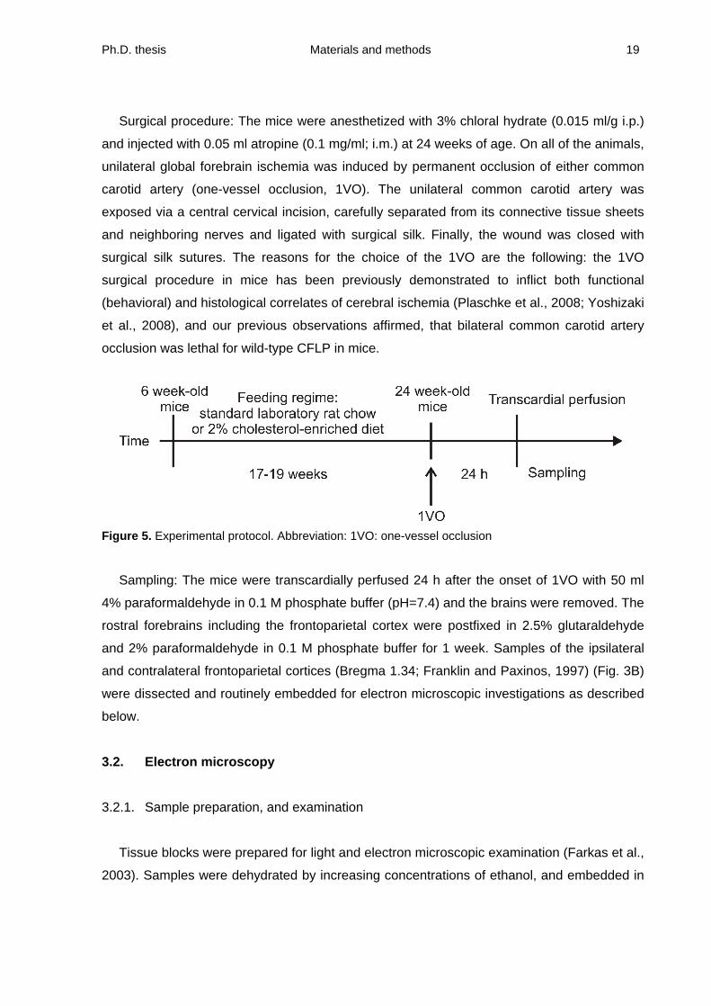
Surgical procedure: The mice were anesthetized with 3% chloral hydrate (0.015 ml/g i.p.)
and injected with 0.05 ml atropine (0.1 mg/ml; i.m.) at 24 weeks of age. On all of the animals,
unilateral global forebrain ischemia was induced by permanent occlusion of either common
carotid artery (one-vessel occlusion, 1VO). The unilateral common carotid artery was
exposed via a central cervical incision, carefully separated from its connective tissue sheets
and neighboring nerves and ligated with surgical silk. Finally, the wound was closed with
surgical silk sutures. The reasons for the choice of the 1VO are the following: the 1VO
surgical procedure in mice has been previously demonstrated to inflict both functional
(behavioral) and histological correlates of cerebral ischemia (Plaschke et al., 2008; Yoshizaki
et al., 2008), and our previous observations affirmed, that bilateral common carotid artery
occlusion was lethal for wild-type CFLP in mice.
Figure 5. Experimental protocol. Abbreviation: 1VO: one-vessel occlusion
Sampling: The mice were transcardially perfused 24 h after the onset of 1VO with 50 ml
4% paraformaldehyde in 0.1 M phosphate buffer (pH=7.4) and the brains were removed. The
rostral forebrains including the frontoparietal cortex were postfixed in 2.5% glutaraldehyde
and 2% paraformaldehyde in 0.1 M phosphate buffer for 1 week. Samples of the ipsilateral
and contralateral frontoparietal cortices (Bregma 1.34; Franklin and Paxinos, 1997) (Fig. 3B)
were dissected and routinely embedded for electron microscopic investigations as described
below.
3.2. Electron microscopy
3.2.1. Sample preparation, and examination
Tissue blocks were prepared for light and electron microscopic examination (Farkas et al.,
2003). Samples were dehydrated by increasing concentrations of ethanol, and embedded in

Ph.D. thesis Materials and methods 20
Durcupan epoxy resin (Fluka). Semi-thin sections were cut on an ultramicrotome (Ultracut E,
Reichert-Jung) and stained on object glasses with a 1:1 mixture of 1% methylene blue and
1% Azure II blue. The samples were then coverslipped with DPX and analyzed under a light
microscope (Nikon E600). Ultrathin sections were cut from the same blocks and collected in
200-mesh copper grids. The preparations were then contrasted with 5% uranyl acetate and
Reynolds lead citrate solution. Finally, the samples were analyzed with a Philips TM10
transmission electron microscope. Photographs were taken with a computer-assisted digital
camera (MegaView II, Soft Imaging Systems, Germany).
3.2.2. Determination of microvascular damage and quantitative analysis
All cortical layers were systematically scanned in the rodent experiments (rat:
approximately 0.13 mm2 tissue surface ~25±7 capillary cross sections; mice: about 0.14 mm2
cortical area ~ 28±8 capillaries), and an entire section in the human study (approximately
0.49 mm2 of sample surface ~8±4 microvessels) on a randomly selected sample grid.
First, the lumen diameter of microvessels was measured. Microvessels only of a defined
lumen diameter were included in the analysis (i.e. rodent: d<7mm, human d<12 mm). In case
the vessel profile was not an exact cross section (circle) but slightly oval, the shortest
diameter was taken. The diameters of all the investigated vessels in each sample were
averaged, and this mean was used as a single value for further statistical analysis.
Vascular density was calculated for a standard surface area with the help of the sample grid
as follows. The number of encountered microvascular profiles was divided by the examined
surface area, which was determined by counting the number of grid squares of a standard
size provided by the distributor. Vascular density was then expressed as the number of
microvascular profiles on 1mm2 surface.
Analysis of the changed cellular elements and the BM:
In the rodent studies, capillaries displaying finger-like endothelial processes protruding the
vessel lumen (microvilli) or swollen astrocytic endfeet were counted and the number was
expressed as percentage of the total number of capillaries examined. The ratio of intact
capillaries (devoid of all the above pathology) was also calculated and expressed as
percentage.
In the human study, the analysis focused on BM pathology, which was noted when fibrous
collagen deposition in the BM occurred. This was recognized as fiber bundles in lateral view,
Ph.D. thesis Materials and methods 21
displaying a 64 nm periodicity typical of collagen type I, or as tightly packed circles within the
BM in cross sectional view. Another investigated pathological feature was the accumulation
of fibrous collagen in the pericytes surrounding the blood vessels. The number of small
vessels with either of the above BM pathology was counted and expressed as percentage of
the total number of microvessels examined.
The mean lumen diameters and the ratio of microvessels displaying deposited fibrous
collagen in their pericytes were correlated with the progression of Alzheimer s
neuropathology defined with Braak stages, and were compared with the age of the subjects .
3.2.3. Statistical analysis
In the rat study, a two-way ANOVA paradigm of the software SPSS 12.0 was used
(variables: treatment and survival time). In the mouse study, data were analyzed with a three-
way ANOVA for genotype (Tg(apoB-100) vs. Wt), diet (cholesterol-enriched vs. standard)
and ischemia (ipsilateral vs. contralateral cortex). In both rodent experiments, ANOVA was
followed by a Fisher least significant difference (LSD) post-hoc test for group comparisons. In
the human study, statistical analysis was performed with the non-parametric Mann-Whitney
U-test, and correlation analysis was performed with a Pearson s one-tailed correlation test.
Results were considered to be significantly different at a probability level of p<0.05* and
p<0.01**.
Ph.D. thesis Results 22
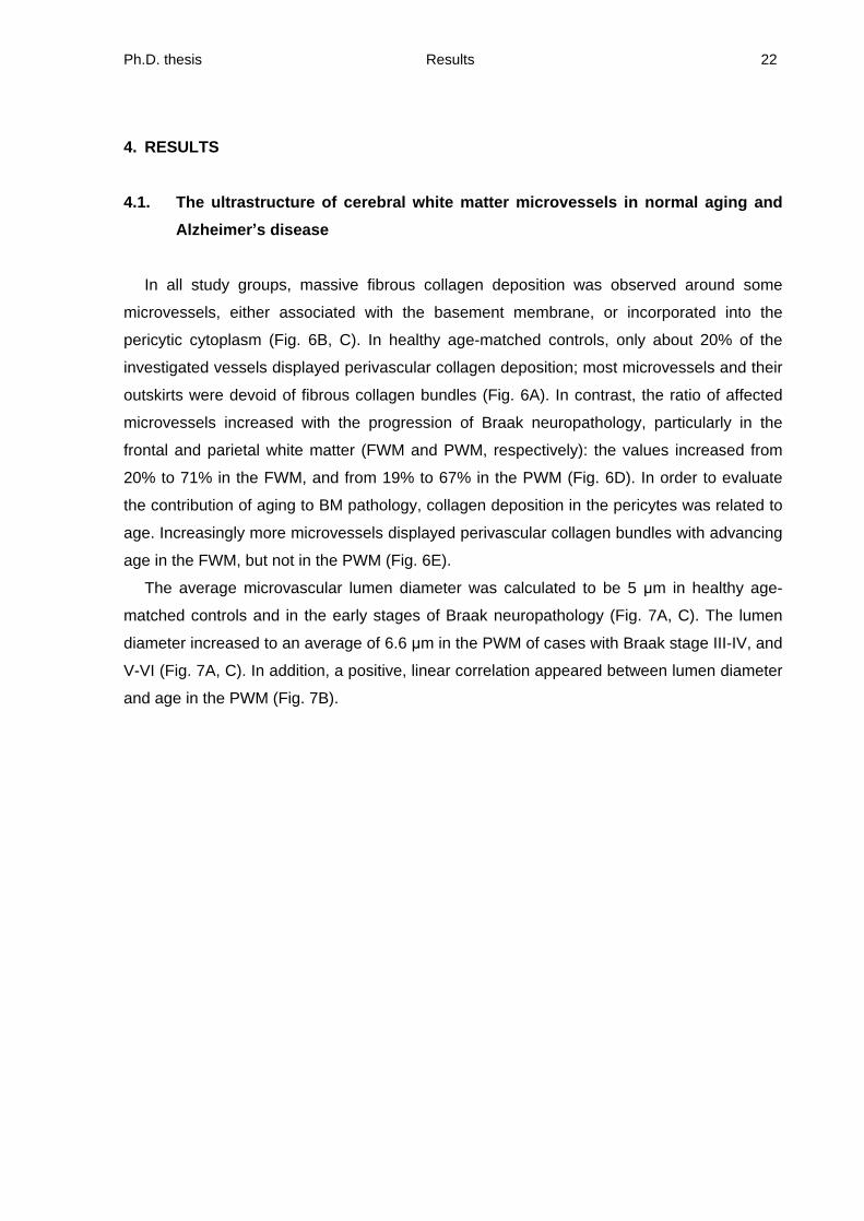
4. RESULTS
4.1. The ultrastructure of cerebral white matter microvessels in normal aging and
Alzheimer s disease
In all study groups, massive fibrous collagen deposition was observed around some
microvessels, either associated with the basement membrane, or incorporated into the
pericytic cytoplasm (Fig. 6B, C). In healthy age-matched controls, only about 20% of the
investigated vessels displayed perivascular collagen deposition; most microvessels and their
outskirts were devoid of fibrous collagen bundles (Fig. 6A). In contrast, the ratio of affected
microvessels increased with the progression of Braak neuropathology, particularly in the
frontal and parietal white matter (FWM and PWM, respectively): the values increased from
20% to 71% in the FWM, and from 19% to 67% in the PWM (Fig. 6D). In order to evaluate
the contribution of aging to BM pathology, collagen deposition in the pericytes was related to
age. Increasingly more microvessels displayed perivascular collagen bundles with advancing
age in the FWM, but not in the PWM (Fig. 6E).
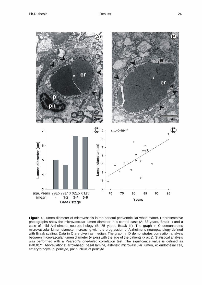
The average microvascular lumen diameter was calculated to be 5 m in healthy age-
matched controls and in the early stages of Braak neuropathology (Fig. 7A, C). The lumen
diameter increased to an average of 6.6 m in the PWM of cases with Braak stage III-IV, and
V-VI (Fig. 7A, C). In addition, a positive, linear correlation appeared between lumen diameter
and age in the PWM (Fig. 7B).

Ph.D. thesis Results 23
Figure 6. Ultrastructural analysis of microvessels in the frontal and parietal periventricular white matter. The photographs are representative electron microscopic images of an intact vessel profile (A; 81 years, Braak -), deposition of collagen in the vascular wall (B; 80 years, Braak IV) and fibrous collagen in the pericyte (C; 95 years, Braak IV). The bar chart in D demonstrates the increasing ratio of fibrous collagen deposited in pericytes with the progression of Alzheimer s neuropathology defined with the Braak scaling. Data in D are given as median. The graph in E demonstrates correlation analysis between the ratio of fibrous collagen deposited in pericytes (y axis) with the age of the patients (x axis). Statistical analysis was performed with a Pearson s one-tailed correlation test. The significance value is defined as P<0.05*. Abbreviations: arrowhead: basal lamina, asterisk: microvascular lumen, coll: collagen deposition, e: endothelial cell, en: nucleus of endothelial cell, er: erythrocyte, FWM: frontal periventricular white matter, p: pericyte PWM: parietal periventricular white matter.
Ph.D. thesis Results 24
Figure 7. Lumen diameter of microvessels in the parietal periventricular white matter. Representative photographs show the microvascular lumen diameter in a control case (A; 88 years, Braak -) and a case of mild Alzheimer s neuropathology (B; 85 years, Braak III). The graph in C demonstrates microvascular lumen diameter increasing with the progression of Alzheimer s neuropathology defined with Braak scaling. Data in C are given as median. The graph in D demonstrates correlation analysis between microvascular lumen diameter (y axis) with the age of the patients (x axis). Statistical analysis was performed with a Pearson s one-tailed correlation test. The significance value is defined as P<0.01**. Abbreviations: arrowhead: basal lamina, asterisk: microvascular lumen, e: endothelial cell, er: erythrocyte, p: pericyte, pn: nucleus of pericyte
Ph.D. thesis Results 25
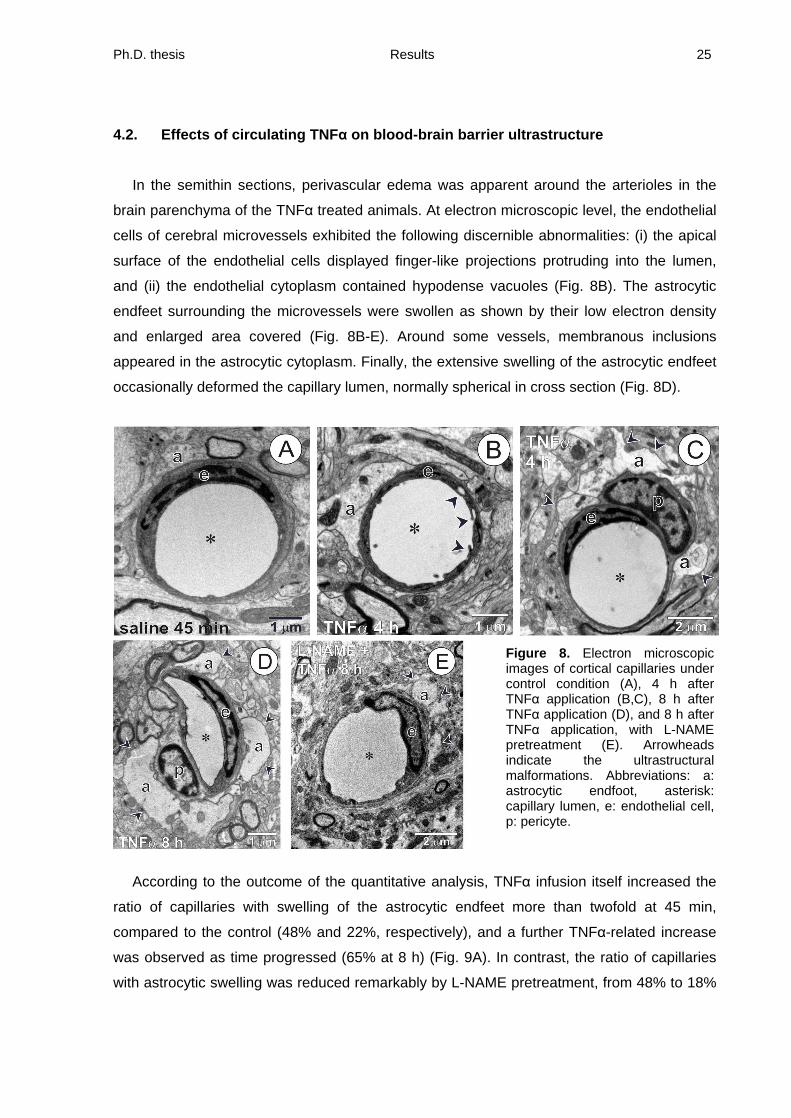
4.2. Effects of circulating TNF on blood-brain barrier ultrastructure
In the semithin sections, perivascular edema was apparent around the arterioles in the
brain parenchyma of the TNF treated animals. At electron microscopic level, the endothelial
cells of cerebral microvessels exhibited the following discernible abnormalities: (i) the apical
surface of the endothelial cells displayed finger-like projections protruding into the lumen,
and (ii) the endothelial cytoplasm contained hypodense vacuoles (Fig. 8B). The astrocytic
endfeet surrounding the microvessels were swollen as shown by their low electron density
and enlarged area covered (Fig. 8B-E). Around some vessels, membranous inclusions
appeared in the astrocytic cytoplasm. Finally, the extensive swelling of the astrocytic endfeet
occasionally deformed the capillary lumen, normally spherical in cross section (Fig. 8D).
According to the outcome of the quantitative analysis, TNF infusion itself increased the
ratio of capillaries with swelling of the astrocytic endfeet more than twofold at 45 min,
compared to the control (48% and 22%, respectively), and a further TNF -related increase
was observed as time progressed (65% at 8 h) (Fig. 9A). In contrast, the ratio of capillaries
with astrocytic swelling was reduced remarkably by L-NAME pretreatment, from 48% to 18%
Figure 8. Electron microscopic images of cortical capillaries under control condition (A), 4 h after TNF application (B,C), 8 h after TNF application (D), and 8 h after TNF application, with L-NAME pretreatment (E). Arrowheads indicate the ultrastructural malformations. Abbreviations: a: astrocytic endfoot, asterisk: capillary lumen, e: endothelial cell, p: pericyte.

Ph.D. thesis Results 26
at 45 min, from 61% to 34% at 4 h, and from 65% to 51% at 8 h after TNF infusion (Fig.
9A).
Figure 9. Quantitative data of pericapillary astrocytic swelling (A), and lumen diameter of capillaries (B). Statistical analysis was performed with an ANOVA model for two variables (treatment, survival time). Data are given as mean±SEM. Significance values are defined as P<0.05*.
Although the statistical analysis has not revealed any significant changes in the lumen
diameter of capillaries between experimental groups, both TNF and L-NAME reduced
microvascular diameter noticeably (Fig. 9B). In particular, TNF alone reduced the lumen
diameter from the control value of 4.56 m to 4.08 m at 45 min, which progressively
decreased to 3.86 m at 4 h, and to 3.61 m at 8 h (Fig. 9B, white dashed bars). After the
treatment with L-NAME alone or in combination with TNF , the capillary lumen diameter
varied in the interval 3.51-3.99 m irrespective of survival time, in contrast with the 4.56 m
for the saline control group (Fig. 9B, black dashed bars).
Ph.D. thesis Results 27
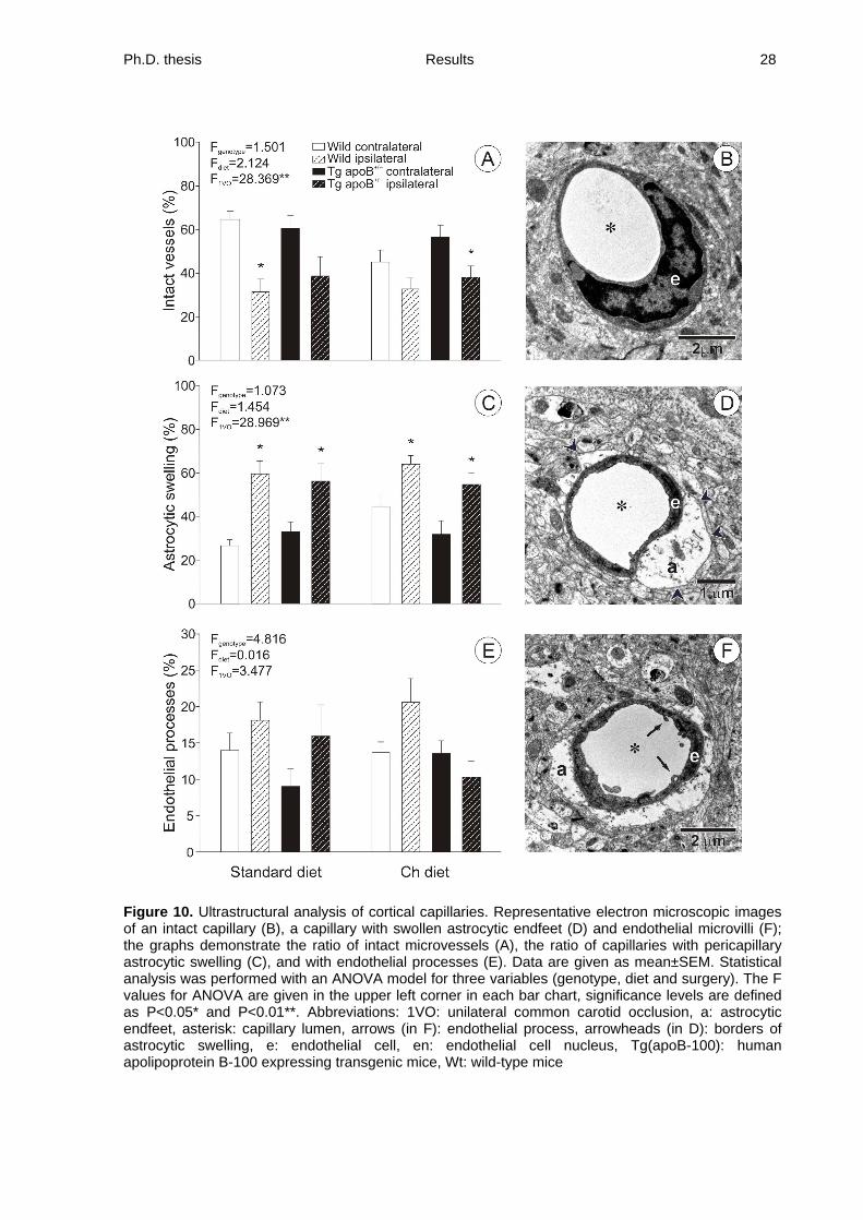
4.3. Effects of hyperlipidemia and/or ischemia on blood-brain barrier ultrastructure
Forebrain ischemia induced in the ipsilateral frontoparietal cortex with respect to 1VO
markedly increased the ratio of cortical capillaries with pericapillary astrocytic swelling from
36.5±4.0% to 62.1±4.2% in Wt mice (dietary groups merged for values, contralateral vs.
ipsilateral, respectively; P<0.001) and from 33.2±4.6% to 56.0±5.4% in Tg(apoB-100) mice
(dietary groups merged for values, contralateral vs. ipsilateral, respectively; P<0.003; Fig.
10C).
The luminal endothelial surface appeared to be irregular, displaying microvilli,
indentations, and occasional vacuoles (Fig. 10F). The ratio of microvessels displaying
endothelial microvilli demonstrated no definite change, though a slight increase appeared in
the ipsilateral cortex after 1VO when compared to the contralateral side (dietary groups
merged for values: 20.1±2.3 vs. 14.3±1.6% in the Wt mice; 13.6±2.7 vs. 11.7±1.7% in
Tg(apoB-100) mice, ipsilateral vs. contralateral, respectively; Fig. 10E). The ratio of
capillaries devoid of any of the above pathology (intact microvessels) decreased from an
average of 65.2±3.7% to 32.1±5.6% in Wt mice (dietary groups merged for values,
contralateral vs. ipsilateral, respectively; P<0.001) and from 56.78±5.60% to 38.64±5.06% in
Tg(apoB-100) mice (dietary groups merged for values, contralateral vs. ipsilateral,
respectively; P<0.004) due to 1VO (Fig. 10A and B). The microvascular integrity was not
altered by either the transgenic genotype or the experimental diet.
Ph.D. thesis Results 28
Figure 10. Ultrastructural analysis of cortical capillaries. Representative electron microscopic images of an intact capillary (B), a capillary with swollen astrocytic endfeet (D) and endothelial microvilli (F); the graphs demonstrate the ratio of intact microvessels (A), the ratio of capillaries with pericapillary astrocytic swelling (C), and with endothelial processes (E). Data are given as mean±SEM. Statistical analysis was performed with an ANOVA model for three variables (genotype, diet and surgery). The F values for ANOVA are given in the upper left corner in each bar chart, significance levels are defined as P<0.05* and P<0.01**. Abbreviations: 1VO: unilateral common carotid occlusion, a: astrocytic endfeet, asterisk: capillary lumen, arrows (in F): endothelial process, arrowheads (in D): borders of astrocytic swelling, e: endothelial cell, en: endothelial cell nucleus, Tg(apoB-100): human apolipoprotein B-100 expressing transgenic mice, Wt: wild-type mice
Ph.D. thesis Results 29
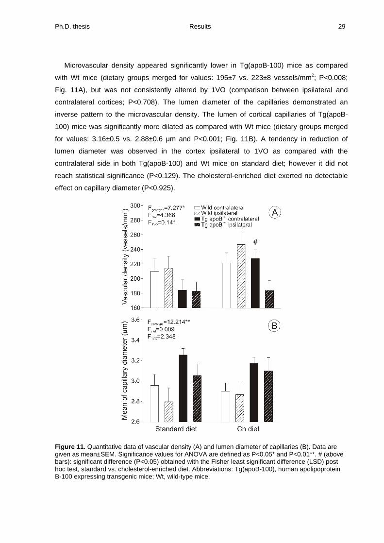
Microvascular density appeared significantly lower in Tg(apoB-100) mice as compared
with Wt mice (dietary groups merged for values: 195±7 vs. 223±8 vessels/mm2; P<0.008;
Fig. 11A), but was not consistently altered by 1VO (comparison between ipsilateral and
contralateral cortices; P<0.708). The lumen diameter of the capillaries demonstrated an
inverse pattern to the microvascular density. The lumen of cortical capillaries of Tg(apoB-
100) mice was significantly more dilated as compared with Wt mice (dietary groups merged
for values: 3.16±0.5 vs. 2.88±0.6 m and P<0.001; Fig. 11B). A tendency in reduction of
lumen diameter was observed in the cortex ipsilateral to 1VO as compared with the
contralateral side in both Tg(apoB-100) and Wt mice on standard diet; however it did not
reach statistical significance (P<0.129). The cholesterol-enriched diet exerted no detectable
effect on capillary diameter (P<0.925).
Figure 11. Quantitative data of vascular density (A) and lumen diameter of capillaries (B). Data are given as mean±SEM. Significance values for ANOVA are defined as P<0.05* and P<0.01**. # (above bars): significant difference (P<0.05) obtained with the Fisher least significant difference (LSD) post hoc test, standard vs. cholesterol-enriched diet. Abbreviations: Tg(apoB-100), human apolipoprotein B-100 expressing transgenic mice; Wt, wild-type mice.
Ph.D. thesis Discussion 30
5. DISCUSSION
Injuries of the cerebral microcirculation are hypothesized as either the main or contributing
risk factors for the progression of pathophysiologic mechanisms leading to various disease
manifestations, such as vascular dementia, AD, neuroinflammation, or hyperlipidemia. The
most typical forms of microvascular malformations, which affect the cellular and non-cellular
components of the blood-brain barrier (BBB) are basement membrane thickening,
perivascular collagen deposition, astrocyte endfoot swelling and the formation of microvillus-
like endothelial processes extending into the microvascular lumen. Therefore capillary
degeneration in the above pathological states leads to serious functional consequences. First
of all, the transport mechanisms through the BBB are hampered and the local regulatory
mechanisms are less effective. The partial or complete breakdown of the BBB promotes the
non-specific permeability of the BBB, while the facilitated transport of indispensable materials
(e.g. glucose, amino acids) is damaged. The opening of the BBB favors different pathologic
processes. From the circulatory system, toxic agents are able to traffic into the brain
parenchyma, and nutrient supply becomes suboptimal. These processes contribute to
cognitive dysfunction, and have been described to be part of the etiology of AD (Abbott et al.,
2010; Popescu et al., 2009).
In order to maintain constant and optimal blood supply to the brain through a
morphologically compromised cerebrovascular network, compensatory mechanisms must
turn on. One possible way to manage normal flow is the adjustment of microvascular density
in order for nutrient supply to meet the requirements of the nervous tissue. Vascular
remodeling mediated by angiogenesis through vascular endothelial growth factor (VEGF) is
considered as a potential mechanism to save brain areas jeopardized by suboptimal nutrient
supply through damaged capillary walls. In support of this view, the expression of various
angiogenic proteins has been identified in the microvessels of AD brains, as compared with
non-demented controls (Thirumangalakudi et al., 2006). In contrast, in the periphery
(especially in the myocardium) of hypercholesterolemic rats, Western blot analysis revealed
the downregulation of VEGF, which was reversed by resveratrol (Penumathsa 2008).
Angiogenesis and vascular remodeling are important elements in several chronic
inflammatory diseases; for instance, increased VEGF expression has been found in the
spinal cord in a guinea pig model of multiple sclerosis (Kirk and Karlik, 2003).
In addition to the extension of the microvascular network, other mechanism may also
compensate for the hindered nutrient supply through a damaged microvascular system.
Ph.D. thesis Discussion 31
Increased microvascular caliber may be the maintenance of optimal basal blood flow.
Interestingly, arteriolar lumen diameter was found dilated in AD brains (Stopa et al, 2008).
Since arterioles are involved in flow regulation rather than nutrient trafficking, the dilated
arterioles may promote better blood supply to areas in need in the AD brain.
5.1. Alterations of cerebral white matter microvessels in normal aging and
Alzheimer s disease
The main goal of the study was to determine, whether the microvasculature of cerebral
periventricular white matter (WM) is injured in AD, and whether the possible microvascular
damage appears in the frontal, parietal and occipital white matter areas evenly. The present
study on the human post-mortem samples has provided electron microscopic evidence of the
microvascular wall pathology in the periventricular WM in AD. Observations of our human
study presented the following results. Perivascular fibrous collagen deposition almost linearly
increased with advancing age in the FWM and microvascular lumen diameter displayed a
linear, positive correlation with age in the PWM. On the other hand, perivascular fibrous
collagen accumulation more than tripled in AD patients with neuropathology of Braak V-VI in
the PWM.
Numerous experimental observations described WM alterations in cerebral diseases.
Between the different subparts of the WM, damage to the periventricular WM is thought to
play a role in cognitive failures (Challa et al., 2004), and malformations in the subcortical
area are hypothesized to be responsible for depressive symptoms. Various non-invasive
imaging techniques helped to identify WM changes in AD and Parkinson s disease (PD). In
AD, WM injuries have been described as hypodense areas in computer tomographic (CT)
images, while in T2-weighted magnetic resonance imaging (MRI), the same structures
appeared as hyperintensive regions (Pantoni and Garcia, 1997). The histopathologic
correlates of the WM lesions have been described as myelin rarefaction at light microscopic
level. More detailed studies with specific markers confirmed that in the injured WM region,
demyelination, apoptotic process of oligodendrocytes and gliosis occur (Brun and Englund,
1986). Such WM malformations are thought to have vascular or ischemic origin. In a number
of cases, chronic cerebral hypoperfusion has been shown to play a key role in the evolution
of WM rarefaction and dementia (Miki et al, 2009; Farkas et al., 2004). On the other hand,
chronic cerebral hypoperfusion is a known process not only in the state of dementia, but also
in normal aging.
Ph.D. thesis Discussion 32
5.1.1. Cerebral blood flow and microvascular alterations in normal aging
Our data have demonstrated increasing collagen deposition in the microvascular wall with
aging, particularly in the FWM. In addition, microvascular lumen diameter was increasingly
more dilated with advancing age in the PWM. Chronic cerebral hypoperfusion is proposed to
initiate the fine pathology of the microvascular wall, as was shown experimentally in rats with
permanent, bilateral common carotid artery occlusion (Farkas and Luiten, 2001). Changes in
cerebral perfusion are a well studied phenomenon in various neurodegenerative diseases.
Cross-sectional studies on healthy volunteers helped to clarify CBF changes in normal aging
(Schultz et al., 1999). When regional, cortical CBF of healthy middle-aged (65-69 years) and
old (80-84 years) subjects was compared with the help of SPECT, a significant reduction of
about 5% in baseline flow was shown in the parietal and temporal cortex of the old group as
compared with the middle-aged group (Claus et al., 1998). The aged had significantly lower
CBF, than the younger individuals, and correlation analysis demonstrated that the CBF
negatively correlated to life time (Scheel et al., 2000). Most of the data reports the reduction
of total CBF in aging and/or dementias, but a CT analysis demonstrated the reduction of the
rCBF in WM areas, as well. (Siennicki-Lantz et al., 1998).
Chronically decreased CBF results in lower metabolic rates of oxygen and glucose in the
progress of normal aging. Due to insufficient nutrition, all cell types of the brain may undergo
degenerative processes. Both the parenchyma, and the fine cerebrovascular network may
display structural malformations. The ultrastructure of the microvessels in the aging brain has
been investigated with electron microscopy in aging animals; the loss and elongation of the
capillary endothelial cells (Mooradian, 1988) and an increased size of pericytic mitochondria
was observed, the latter of which was attributed to an active state of the pericytes (Hicks et
al, 1983). Besides the cellular elements of the BBB, the basement membrane is also
involved in the ultrastructural malformations in aging (Farkas and Luiten, 2001). BM
pathologies manifest in the form of BM thickening and fibrous deposition of extracellular
matrix components such as collagen, which is suggested to form due to the increased
production or the decreased breakdown of the connective tissue elements. In the human
periventricular WM, a previous study of ours has identified BM aberrations to correlate
markedly with increasing age, especially in the frontal and occipital WM (Farkas et al, 2006).
In addition, BM pathology was found to be related to the degree of atherosclerosis that
occurred in peripheral vessels such as the aorta (Farkas et al., 2006). Since atherosclerosis
Ph.D. thesis Discussion 33
is strongly related to hyperlipidemia, our finding points to investigating the role of
hyperlipidemia in cerebral microvascular malformations, which is presented below.
In addition to the fine ultrastructural aberrations, the microvascular architecture may also
change with increasing age, but the results in this respect appear to be somewhat
controversial. In human neocortex, increased capillary density attributed to tissue shrinkage
was observed, but other research groups reported decreased microvascular density in aged
humans (Abernethy et al., 1993). We ourselves could not demonstrate any noteworthy
association between age and microvascular density in the human periventricular WM (Farkas
et al., 2006). However, in animal experiments, a comprehensive comparison of the cortical
and hippocampal capillaries of adult and old rats showed that microvascular density was
reduced with age (Sonntag et al., 1997). The data available so far on microvascular density
in the aging brain, therefore, remain to be inconclusive.
5.1.2. Cerebrovascular pathology in AD
The current analysis of the human samples showed notable perivascular, fibrous collagen
deposition in the WM microvascular walls with Braak neuropathology stages V-VI, especially
in the PWM region. These results are in agreement with MRI observations in leukoaraiosis
(i.e. WM lesions) by Targosz-Gajniak et al. (2009). These authors have demonstrated that
WM lesions appeared in all AD patients examined, the amount and size of the WM lesions
correlated with age, and that the severity of the lesions in the periventricular WM increased
with the progression of AD.
The structural changes of microvessels show here share similar characteristics with those
found in capillaries in the cerebral cortex and, more interestingly, in the WM after chronic,
experimental, cerebral hypoperfusion in rats (De Jong, 1999; Farkas and Luiten, 2001). In
the experimental model of chronic cerebral hypoperfusion, the typical capillary abnormalities
included 25% higher occurrence of degenerative pericytes around the blood vessels and
25% higher incidence of basement membrane thickening, with or without fibrosis, in
hypoperfused animals. These data demonstrate that decreased cerebral blood flow induces
ultrastructural damage of cerebral microvessels (Farkas and Luiten, 2001).
Experimental cerebral hypoperfusion that evokes ischemia has been found to have a
deleterious impact on the medullary tissue. The injury appears in the form of glial activation
and rarefaction of the WM, as already mentioned under point 5.1. (Farkas et al., 2004; Ueno
et al., 2002).
Ph.D. thesis Discussion 34
We have additionally shown that the average size of microvascular lumen increases with
the severity of AD, especially in the PWM. The increased lumen diameter is suggested to be
a compensatory mechanism to maintain normal cerebral perfusion and transport in spite of
the increased rigidity and thickness of the microvascular wall as demonstrated by massive
collagen deposition.
Our data have demonstrated that the enhanced perivascular accumulation of collagen and
the dilation of micorvascular lumen in the periventricular WM are region-specific. The most
affected site was the parietal region, where the collagen deposition increased most intensely,
and the microvascular lumen dilated most noticeably. The microvascular alterations
presented here suggest a potentially increased vulnerability of the frontal and parietal areas.
Indeed, in normal aging, the reduction of WM integrity as assessed with diffusion tensor
imaging was most pronounced in the FWM (Head et al., 2004).
The susceptibility of the frontal and parietal WM to injury may be rooted in the
angioarchitecture and perfusion of the region. Accumulating evidence demonstrates that the
vascular pattern of WM areas defines perfusion and is related to the metabolic vulnerability
of the region (de Reuck, 1971; Farkas et al., 2004; Moody et al., 1990).
5.2. BBB ultrastructure in inflammatory processes
Tumor necrosis factor-alpha (TNF ) is a proinflammatory cytokine, which is produced by
various cell types, e.g. macrophages and lymphocytes. TNF has been demonstrated to
cause expression of proadhesive molecules on the endothelium, which results in leukocyte
accumulation, adherence, and migration from capillaries into the brain. TNF promotes
inflammation by stimulation of capillary endothelial cell proinflammatory responses and
thereby provides leukocyte adhesion and infiltration into the ischemic brain.
TNF is a well-known and potent vasodilatator in various experimental in vivo models.
The suggested pathway for the vasodilatory influence of TNF involves nitric-oxide. NOS
inhibitors successfully block the vasoactive property of the TNF (Brian and Faraci, 1998;
Shibata et al., 1996).
In addition to the functional role of TNF in cerebral blood flow regulation, TNF can
impose structural damage to the cerebral microvasculature. In our present study, the
ultrastructural analysis of the cerebrocortical capillaries was performed after TNF treatment
in rats. The most obvious aberration concerned the pericapillary astrocytic endfeet, which
appeared considerably swollen, seen around the microvessels as distorted, dilated areas
Ph.D. thesis Discussion 35
with low electron density in electron microscopic images. Astrocytic swelling is a marked
feature not only of inflammatory reactions, but also of seizure-related states (Fabene et al.,
2006).
In line with our findings, intracarotid administration of TNF in newborn piglets resulted in
the opening of the BBB. The opening of the BBB in pathologic states can be clearly
demonstrated macroscopically after intravenous Evans blue administration (Jancso et al.,
1998), or at light microscopic level by sodium fluorescein (Abraham et al., 1996). Evans blue
is a vital dye with very high affinity for serum albumin or large molecular weight peptides.
Because serum albumin normally cannot cross the BBB, and Evans blue is bound to
albumin, albumin-bound Evans blue enters the brain tissue when the BBB has been
compromised. Sodium fluorescein labeling is based on the same principle for small
molecular weight substances. Besides the different macromolecules labeled by histologic
tracers, whole cellular components of the circulating blood are also able to leave the
circulation via the impaired BBB. For example, lymphocytes have been shown to
transmigrate into the brain parenchyma from the blood after the opening of the BBB by TNF
(Farkas et al., 2003).
An electron microscopic 3-D reconstruction analysis demonstrated that, in normal
conditions, the astrocytes cover almost the whole surface of the cerebral vasculature, only
small hypothesized microglial processes extend across the perivascular glial sheet to make
direct connection with the endothelial cells (Mathiisen et al., 2010). The healthy glial sheet,
which is formed by the astrocytes is not just a structural component of the BBB, but also the
site of important physiologic processes. The main functions of the mature astrocytes are the
maintenance of the extracellular milieu in the brain parenchyma, and the regulation of
neurotransmitter level. Normally, the astrocytes are responsible for the re-uptake of
glutamate from the synaptic cleft by their specific plasma membrane proteins, which is an
activity-dependent mechanism. The excitatory amino acid transporter (EAAT) protein family
contribute to this process. The damage of astrocytes influences the level of excitatory
neurotransmitters via the alteration of the amount of EAAT sites. Comparative
immunhistochemical analysis proved that with the increasing Braak stage a decreased
expression of EAAT2 and increased expression of GFAP (i.e. gliosis) occured (Simpson et
al., 2010).
The astrocytes have a main role in the induction of normal formation of brain capillaries.
Astrocytes are necessary in endothelial cell cultures to induce BBB phenotype, and
experimental evidence have shown that astrocytes induce several BBB properties also in

Ph.D. thesis Discussion 36
peripheral endothelial cells (Kuchler-Bopp et. al, 1999). Using in vitro cell cultures, several
specific transport systems are up-regulated in BBB models exposed to astrocytes; however
the chemical nature of the inductive signal(s) produced by the astrocytes is currently unclear.
The most highly suspected candidates are the glial cell-line derived neurotrophic factors,
transforming growth factors- 1, interleukin-6, and basic fibroblast growth factor (Correale and
Villa, 2009), which are involved in the formation of normal BBB phenotype. In addition to the
experimental findings described above, the astrocytes play an important role in the clearance
and degradation of beta-amyloid plaques in AD (Wyss-Coray et al., 2003), so functionally
intact astrocytes may postpone the development of dementia.
Taken together, if the astrocytic endfeet are swollen as shown here, the cross-talk with the
endothelium becomes disturbed or discontinues, which can lead to BBB dysfunction. In
support of this view, the BBB disruption indicated by the extravasation of Evans blue was
associated with astrocytic swelling that involved aquaporin-4, a major water channel
implicated in the formation of injury-related brain edema (Venero et al., 2001; Vizuete et al.,
1999).
5.3. The ultrastructure of the BBB in hyperlipidemia and/or ischemia
The first aim of the study was to investigate whether hyperlipidemia (i.e. the expression of
human apoB-100 in transgenic mice, and/or cholesterin-rich diet) causes cerebral
microvascular lesions. Comparison of the cerebral capillary network proved that the
Tg(apoB-100) mice exhibited decreased density and increased capillary lumen diameter as
compared with Wt mice. The density of the cerebral capillary network is established during
early development, and dynamically reacts to environmental challenges and
pathophysiological conditions (e.g. chronic hypoxia leads to increased cerebral capillary
density; stroke induces angiogenesis) (Dore-Duffy and LaManna, 2007). The expression of
vascular endothelial growth factor is hypothetised as an important determinant of
angiogenesis during the development in the brain (Plate, 1999). Genetic hyperlipidemia in
rabbits or hypercholesterolemia in mice impaired both basal and stimulated angiogenesis in
the ischemic rabbit hindlimb, and in an artificial disk implanted into the thorax of mice (Jang
et al., 2000; Van Belle et al., 1997). In these studies the hyperlipidemia-related hindrance of
angiogenesis is thought to be an important risk factor for the changed vascular architecture
mediated by the VEGF signaling pathway. Alteration of the VEGF nitric oxide signaling
amend the impaired angiogenesis in hypercholesterolemic mice, and the injection of VEGF
Ph.D. thesis Discussion 37
re-established angiogenesis in the hyperlipidemic, ischemic rabbit hindlimb (Jang et al.,
2000; Van Belle et al., 1997). Therefore, in hyperlipidemic Tg(apoB-100) mice (Chiesa et al.,
1993; Csont et al., 2007; Linton et al., 1993; Purcell-Huynh et al., 1995), the capillary density
in the cerebral cortex is proposed to be lower due to their high plasma cholesterol or
triglyceride profile, thus inhibiting angiogenesis possibly through hampered VEGF signaling.
Parallel with the alteration of the capillary density, the dilation of the capillary lumen was
observed. The increase of the lumen diameter is proposed as a complementary and
compensatory mechanism in order to maintain standard cerebral perfusion rate (i.e. the
normal value of the cerebral blood flow) despite a less dense capillary network in Tg(apoB-
100) as compared with Wt mice. With the rearrangement of the microvascular architecture in
Tg(apoB-100) mice, altered cerebrovascular reactivity may occur in different
pathophysiological conditions, such as ischemia. In addition, a recent study has indicated,
that the reduced capillary density may contribute to apoptosis and progressive neuronal cell
death in Tg(apoB-100) mice by chronically depriving the brain of its vital nutrients (Bereczki
et al., 2008).
The second aim was to determine whether the expression of human apoB-100 in
transgenic mice and/or a high-cholesterol diet augment ischemia-related cerebral capillary
damage. Unilateral forebrain ischemia was induced by 1VO in Tg(apoB-100) and in Wt mice
to answer this question. Previously, 1VO carried out in Mongolian gerbils led to perivascular
glial swelling, and irregularities on the luminal vascular surface with endothelial projections
(Naganuma, 1990). Similar microvascular pathology has been described in senescence-
accelerated mice (Lee et al., 2000; Ueno et al., 1998). Therefore our investigation focused on
the ultrastructure of perivascular astrocytic endfeet and the endothelial surface. In the
present study, the swelling of astrocytic endfeet and the presence of endothelial microvilli
became more prominent in the ischemic hemisphere. These data agree with previous
observations made in models of cerebral ischemia: (i) electron microscopic analysis of
cerebral microvessels revealed compressed capillaries consistently surrounded by swollen
astrocytic endfeet; and (ii) the widespread appearance of cerebral endothelial microvilli
(Dietrich et al., 1986). These morphological features imply some harmful functional changes:
astrocytic pathology compromises the integrity of the blood brain barrier (Haseloff et al.,
2005), while endothelial microvilli might increase microvascular resistance, leading to
moderate hemodynamic impediments (Dietrich et al., 1986).
The data presented here showed that hyperlipidemia in human apoB-100 transgenic mice
and chronically elevated dietary cholesterol alone or in combination did not have any impact
Ph.D. thesis Discussion 38
on capillary ultrastructure in the non-ischemic hemisphere, and did not exacerbate ischemia-
induced microvascular lesions in the mouse brain. Morphological description for
hyperlipidemia-related vascular pathology has been experimentally demonstrated on larger
caliber vessels. For instance, Tg(apoB-100) mice kept on high-fat diet, or
hypercholesterolemic rabbits displayed marked atherosclerosis in the aorta (Purcell-Huynh et
al., 1995; Sasaki et al., 1988). A comparison between the aorta and cerebral microvessels of
hypercholesterolemic rabbits showed that the aorta became atherosclerotic, while the brain
microvessels remained free of lipid depositions (Sasaki et al., 1988). Based on these and our
data, high serum lipid level-related, morphological, vascular lesions in experimental models
are assumed to be confined to large-caliber arteries (e.g. aorta, carotid arteries), rather than
cerebral capillaries.
Still, hyperlipidemia may induce biochemical reactions in brain microvessels (Mooradian
et al., 1995; Robert et al., 1982). The cerebral capillaries of cholesterol-fed rabbits displayed
some biochemical modifications in the capillary walls such as decreased collagen and
hexose content (Robert et al., 1982). Also, increased dietary cholesterol enhanced
malondialdehyde production in brain microvascular walls in rabbits (Mooradian et al., 1995).
Even though high dietary cholesterol thus induces biochemical modifications in the cerebral
microvascular endothelial cells, the impact of hyperlipidemia on capillary ultrastructure may
not become clearly evident. There has been an ongoing debate whether hyperlipidemia is a
direct risk factor for ischemic stroke (Demchuk et al., 1999; Landau, 1999). The data
presented here indicate that apoB-100 expression-related hyperlipidemia (both
hypercholesterolemia and hypertriglyceridemia) lowers the density of cerebral capillaries, but
imposes no obvious ultrastructural microvascular malformations. Cerebral capillary density
reduced by high plasma lipid levels as presented here may affect cerebrovascular reactivity
during pathophysiological challenges ischemia being one of the most prominent types.

Ph.D. thesis Conclusions 39
6. CONCLUSIONS
The studies presented here all aimed to investigate the fine ultrastructure of cerebral
capillaries in various pathological conditions that appear as risk factors for AD. First, the
morphology of the microvessels of the human cerebral WM in AD was assessed. Second,
cerebral microvascular damage as a result of inflammatory processes was characterized.
Third, the condition of brain capillaries in severe ischemia and hyperlipidemia was
investigated. We have found that, with the exception of hyperlipidemia, the ultrastructure of
the BBB becomes compromised in all these conditions, which is suggested to have
functional implications. Since the BBB is the site of selective nutrient and waste product
trafficking between blood and the nervous tissue, its structural integrity is crucial to fulfill its
function and meet the metabolic demands of the central nervous system.
AD is a progressive mental disorder with undefined origin. It has been firmly established
that cerebrovascular pathology and chronically reduced cerebral blood flow are hallmarks of
AD, cerebral hypoperfusion is a predictive marker of the disease, and that cerebrovascular
risk factors contribute to the disintegration of cognitive function over the course of the
disease. Our present study confirms the occurrence of microvascular injury in AD brains, and
reveals conditions (i.e. ischemia, inflammatory processes), which are causative for the
development of cerebral microvascular damage. Our results shed light on the complexity of
causative elements that may add up to damage the BBB, and ultimately lead to memory
dysfunction.
Ph.D. thesis Acknowledgements 40
7. ACKNOWLEDGEMENTS
First of all, I am indebted to my excellent supervisor, Eszter Farkas, Ph.D. (Senior Research
Associate, Department of Medical Physics and Informatics, Faculty of Medicine, University of
Szeged) for her admirable patience and assistance from the initial steps to the final touches
of my Ph.D. work throughout.
I would like to express special thanks to Prof. András Mihály, M.D., Ph.D., D.Sc. (Professor
and Chairman of the Department of Anatomy, Histology and Embryology, Faculty of
Medicine, University of Szeged), for his continuous support during my experimental work.
I was honored to be a member of the Cerebrovascular Research Group of Prof. Ferenc
Bari, Ph.D., D.Sc. (Professor and Chairman of the Department of Medical Physics and
Informatics, Faculty of Medicine, University of Szeged). Working in his team has extended
my knowledge in the field of cerebrovascular research.
I am grateful to Zsolt Rázga, Ph.D. (Research Associate, Department of Pathology, Faculty
of Medicine, University of Szeged) and his previous and current assistants, to Mária Bakacsi
and Erika Németh for providing the facilities for EM work, and for the accurate technical help
with the use of the EM apparatus.
I am greatly appreciate the support of Géza G. Kovács, M.D., Ph.D. (Associate Professor,
Institute of Neurology, Medical University of Vienna, Vienna, Austria) and thank him for
collecting and offering the post-mortem human samples for our study.
I am thankful to the following assistants of our Department: Adrienne Mátyás and Gabriella
Pap for their caring technical help.
Finally, I would like to give my special thanks to my sister Erika and her family for their
everyday care and support, and to the prominent members of my circle of friends, namely
Levente Csaba, István Dobó, Antal Radvánszky, Dániel Tari, and László Tímár.
Ph.D. thesis References 41
8. REFERENCES
1. Abbott NJ.: Dynamics of CNS barriers: evolution, differentiation, and modulation. Cell Mol
Neurobiol. 2005; 25(1):5-23.
2. Abbott NJ, Patabendige AA, Dolman DE, Yusof SR, Begley DJ.: Structure and function of
the blood-brain barrier. Neurobiol Dis. 2010; 37(1):13-25.
3. Abernethy WB, Bell MA, Morris M, Moody DM.: Microvascular density of the human
paraventricular nucleus decreases with aging but not hypertension. Exp Neurol. 1993;
121(2):270-4.
4. Abraham CS, Deli MA, Joo F, Megyeri P, Torpier G.: Intracarotid tumor necrosis factor-
alpha administration increases the blood-brain barrier permeability in cerebral cortex of
the newborn pig: quantitative aspects of double-labelling studies and confocal laser
scanning analysis. Neurosci Lett. 1996;208(2):85-8.
5. Antonelli-Orlidge A, Smith SR, D'Amore PA.: Influence of pericytes on capillary
endothelial cell growth. Am Rev Respir Dis. 1989; 140(4):1129-31.
6. Arthur FE, Shivers RR, Bowman PD.: Astrocyte-mediated induction of tight junctions in
brain capillary endothelium: an efficient in vitro model. Brain Res. 1987; 433(1):155-9.
7. Beck DW, Vinters HV, Hart MN, Cancilla PA.: Glial cells influence polarity of the blood-
brain barrier. J Neuropathol Exp Neurol. 1984 ;43(3):219-24.
8. Bereczki E, Bernát G, Csont T, Ferdinandy P, Scheich H, Sántha M.: Overexpression of
human apolipoprotein B-100 induces severe neurodegeneration in transgenic mice.
J Proteome Res. 2008;7(6):2246-52.
9. Betz AL, Goldstein GW.: Polarity of the blood-brain barrier: neutral amino acid transport
into isolated brain capillaries. Science. 1978; 202(4364):225-7.
10. Bjelik A, Pákáski M, Bereczki E, Gonda S, Juhász A, Rimanóczy A, Zana M, Janka Z,
Sántha M, Kálmán J.: APP mRNA splicing is upregulated in the brain of biglycan
transgenic mice. Neurochem Int. 2007; 50(1):1-4.
11. Blasiole DA, Davis RA, Attie AD.: The physiological and molecular regulation of
lipoprotein assembly and secretion. Mol Biosyst. 2007; 3(9):608-19.
12. Borroni B, Grassi M, Costanzi C, Archetti S, Caimi L, Padovani A.: APOE genotype and
cholesterol levels in lewy body dementia and Alzheimer disease: investigating genotype-
phenotype effect on disease risk. Am J Geriatr Psychiatry. 2006; 14(12):1022-31.
13. Brian JE Jr, Faraci FM.: Tumor necrosis factor-alpha-induced dilatation of cerebral
arterioles. Stroke. 1998; 29(2):509-15.
Ph.D. thesis References 42
14. Brownlees J, Williams CH.: Peptidases, peptides, and the mammalian blood-brain
barrier. J Neurochem. 1993; 60(3):793-803.
15. Brownson EA, Abbruscato TJ, Gillespie TJ, Hruby VJ, Davis TP.: Effect of peptidases at
the blood brain barrier on the permeability of enkephalin. J Pharmacol Exp Ther. 1994;
270(2):675-80.
16. Brun A, Englund E.: A white matter disorder in dementia of the Alzheimer type: a
pathoanatomical study. Ann Neurol. 1986;19(3):253-62.
17. Cancilla PA, DeBault LE.: Neutral amino acid transport properties of cerebral endothelial
cells in vitro. J Neuropathol Exp Neurol. 1983; 42(2):191-99.
18. Challa VR, Thore CR, Moody DM, Anstrom JA, Brown WR.: Increase of white matter
string vessels in Alzheimer's disease. J Alzheimers Dis. 2004; 6(4):379-8.
19. Chiesa G, Johnson DF, Yao Z, Innerarity TL, Mahley RW, Young SG, Hammer RH,
Hobbs HH.: Expression of human apolipoprotein B100 in transgenic mice. Editing of
human apolipoprotein B100 mRNA. J Biol Chem. 1993; 268(32):23747-50.
20. Claus JJ, Breteler MM, Hasan D, Krenning EP, Bots ML, Grobbee DE, Van Swieten JC,
Van Harskamp F, Hofman A.: Regional cerebral blood flow and cerebrovascular risk
factors in the elderly population. Neurobiol Aging. 1998;19(1):57-64.
21. Cohen Z, Molinatti G, Hamel E.: Astroglial and vascular interactions of noradrenaline
terminals in the rat cerebral cortex. J Cereb Blood Flow Metab. 1997; 17(8):894-904.
22. Coomber BL, Stewart PA.: Morphometric analysis of CNS microvascular endothelium.
Microvasc Res. 1985; 30(1):99-115.
23. Correale J, Villa A.: Cellular elements of the blood-brain barrier. Neurochem Res. 2009;
34(12):2067-77.
24. Csont T, Bereczki E, Bencsik P, Fodor G, Görbe A, Zvara A, Csonka C, Puskás LG,
Sántha M, Ferdinandy P.: Hypercholesterolemia increases myocardial oxidative and
nitrosative stress thereby leading to cardiac dysfunction in apoB-100 transgenic mice.
Cardiovasc Res. 2007; 76(1):100-9.
25. De Jong GI, Farkas E, Stienstra CM, Plass JR, Keijser JN, de la Torre JC, Luiten PG.:
Cerebral hypoperfusion yields capillary damage in the hippocampal CA1 area that
correlates with spatial memory impairment. Neuroscience. 1999; 91(1):203-10.
26. de la Torre JC.: Vascular basis of Alzheimer's pathogenesis. Ann N Y Acad Sci. 2002;
977:196-215.
27. De Reuck J.: The human periventricular arterial blood supply and the anatomy of cerebral
infarctions. Eur Neurol. 1971;5(6):321-34.
Ph.D. thesis References 43
28. Demchuk AM, Hess DC, Brass LM, Yatsu FM.: Is cholesterol a risk factor for stroke?:
Yes. Arch Neurol. 1999; 56(12):1518-20.
29. Dietrich WD, Ginsberg MD, Busto R, Watson BD, Yoshida S.: Vascular aspects and
hemodynamic consequences of central nervous system injury. Cent Nerv Syst Trauma.
1986; 3(4):265-80.
30. Dore-Duffy P, LaManna JC.: Physiologic angiodynamics in the brain. Antioxid Redox
Signal. 2007; 9(9):1363-71.
31. Duvernoy H, Delon S, Vannson JL.: The vascularization of the human cerebellar cortex.
Brain Res Bull. 1983; 11(4):419-80.
32. Elhusseiny A, Cohen Z, Olivier A, Stanimirovi DB, Hamel E.: Functional acetylcholine
muscarinic receptor subtypes in human brain microcirculation: identification and cellular
localization. J Cereb Blood Flow Metab. 1999; 19(7):794-802.
33. Fabene PF, Weiczner R, Marzola P, Nicolato E, Calderan L, Andrioli A, Farkas E, Süle Z,
Mihaly A, Sbarbati A.: Structural and functional MRI following 4-aminopyridine-induced
seizures: a comparative imaging and anatomical study. Neurobiol Dis. 2006; 21(1):80-9.
34. Farkas E, De Jong GI, Apró E, de Vos RA, Steur EN, Luiten PG.: Similar ultrastructural
breakdown of cerebrocortical capillaries in Alzheimer's disease, Parkinson's disease, and
experimental hypertension. What is the functional link? Ann N Y Acad Sci. 2000; 903:72-
82.
35. Farkas E, De Jong GI, de Vos RA, Jansen Steur EN, Luiten PG.: Pathological features of
cerebral cortical capillaries are doubled in Alzheimer's disease and Parkinson's disease.
Acta Neuropathol. 2000; 100(4):395-402.
36. Farkas E, de Vos RA, Donka G, Jansen Steur EN, Mihály A, Luiten PG.: Age-related
microvascular degeneration in the human cerebral periventricular white matter. Acta
Neuropathol. 2006; 111(2):150-7.
37. Farkas E, De Vos RA, Jansen Steur EN, Luiten PG.: Are Alzheimer's disease,
hypertension, and cerebrocapillary damage related? Neurobiol Aging. 2000; 21(2):235-
43.
38. Farkas E, Donka G, de Vos RA, Mihály A, Bari F, Luiten PG.: Experimental cerebral
hypoperfusion induces white matter injury and microglial activation in the rat brain. Acta
Neuropathol. 2004; 108(1):57-64.
39. Farkas E, Luiten PG.: Cerebral microvascular pathology in aging and Alzheimer's
disease. Prog Neurobiol. 2001; 64(6):575-611.
Ph.D. thesis References 44
40. Farkas E, Luiten PG, Bari F.: Permanent, bilateral common carotid artery occlusion in the
rat: a model for chronic cerebral hypoperfusion-related neurodegenerative diseases.
Brain Res Rev. 2007; 54(1):162-80.
41. Farkas IG, Czigner A, Farkas E, Dobó E, Soós K, Penke B, Endrész V, Mihály A.: Beta-
amyloid peptide-induced blood-brain barrier disruption facilitates T-cell entry into the rat
brain. Acta Histochem. 2003; 105(2):115-25.
42. Farrell CL, Pardridge WM.: Blood-brain barrier glucose transporter is asymmetrically
distributed on brain capillary endothelial lumenal and ablumenal membranes: an electron
microscopic immunogold study. Proc Natl Acad Sci U S A. 1991; 88(13):5779-83.
43. Frank RN, Dutta S, Mancini MA.: Pericyte coverage is greater in the retinal than in the
cerebral capillaries of the rat. Invest Ophthalmol Vis Sci. 1987; 28(7):1086-91.
44. Franklin KBJ, Paxinos G: The mouse brain in stereotaxic coordinates. Academic Press,
San Diego, 1997.
45. Gjedde A, Diemer NH.: Double-tracer study of the fine regional blood-brain glucose
transfer in the rat by computer-assisted autoradiography. J Cereb Blood Flow Metab.
1985; 5(2):282-9.
46. Grundy SM, Vega GL, Kesäniemi YA.: Abnormalities in metabolism of low density
lipoproteins associated with coronary heart disease. Acta Med Scand Suppl. 1985;
701:23-37.
47. Haseloff RF, Blasig IE, Bauer HC, Bauer H.: In search of the astrocytic factor(s)
modulating blood-brain barrier functions in brain capillary endothelial cells in vitro. Cell
Mol Neurobiol. 2005; 25(1):25-39.
48. Hawkins BT, Davis TP.: The blood-brain barrier/neurovascular unit in health and disease.
Pharmacol Rev. 2005; 57(2):173-85.
49. Head D, Buckner RL, Shimony JS, Williams LE, Akbudak E, Conturo TE, McAvoy M,
Morris JC, Snyder AZ.: Differential vulnerability of anterior white matter in nondemented
aging with minimal acceleration in dementia of the Alzheimer type: evidence from
diffusion tensor imaging. Cereb Cortex. 2004; 14(4):410-23.
50. Hellström M, Gerhardt H, Kalén M, Li X, Eriksson U, Wolburg H, Betsholtz C.: Lack of
pericytes leads to endothelial hyperplasia and abnormal vascular morphogenesis. J Cell
Biol. 2001; 153(3):543-53.
51. Hicks P, Rolsten C, Brizzee D, Samorajski T.: Age-related changes in rat brain
capillaries. Neurobiol Aging. 1983;4(1):69-75.

Ph.D. thesis References 45
52. Hirao K, Ohnishi T, Hirata Y, Yamashita F, Mori T, Moriguchi Y, Matsuda H, Nemoto K,
Imabayashi E, Yamada M, Iwamoto T, Arima K, Asada T.: The prediction of rapid
conversion to Alzheimer's disease in mild cognitive impairment using regional cerebral
blood flow SPECT. Neuroimage. 2005; 28(4):1014-21.
53. Jancsó G, Domoki F, Sántha P, Varga J, Fischer J, Orosz K, Penke B, Becskei A, Dux M,
Tóth L.: Beta-amyloid (1-42) peptide impairs blood-brain barrier function after intracarotid
infusion in rats. Neurosci Lett. 1998; 253(2):139-41.
54. Jang JJ, Ho HK, Kwan HH, Fajardo LF, Cooke JP.: Angiogenesis is impaired by
hypercholesterolemia: role of asymmetric dimethylarginine. Circulation. 2000;
102(12):1414-9.
55. Kirk SL, Karlik SJ.: VEGF and vascular changes in chronic neuroinflammation.
J Autoimmun. 2003;21(4):353-63.
56. Krämer SD, Abbott NJ, Begley DJ: Biological models to study blood brain barrier
permeation. In: Pharmacokinetic Optimization in Drug Research: Biological,
Physicochemical and Computational Strategies. 2001; 127-153
57. Kuchler-Bopp S, Delaunoy JP, Artault JC, Zaepfel M, Dietrich JB.: Astrocytes induce
several blood-brain barrier properties in non-neural endothelial cells. Neuroreport. 1999;
10(6):1347-53.
58. Landau WM.: Is cholesterol a risk factor for stroke?: No. Arch Neurol. 1999; 56(12):1521-
4.
59. Lee EY, Lee SY, Lee TS, Chi JG, Choi W, Suh YH.: Ultrastructural changes in
microvessel with age in the hippocampus of senescence-accelerated mouse (SAM)-P/10.
Exp Aging Res. 2000; 26(1):3-14.
60. Libby P.: Fat fuels the flame: triglyceride-rich lipoproteins and arterial inflammation. Circ
Res. 2007; 100(3):299-301.
61. Linton MF, Farese RV Jr, Chiesa G, Grass DS, Chin P, Hammer RE, Hobbs HH, Young
SG.: Transgenic mice expressing high plasma concentrations of human apolipoprotein
B100 and lipoprotein(a). J Clin Invest. 1993; 92(6):3029-37.
62. Luiten PG, de Jong GI, Van der Zee EA, van Dijken H.: Ultrastructural localization of
cholinergic muscarinic receptors in rat brain cortical capillaries. Brain Res. 1996; 720(1-
2):225-9.
63. Magaki S, Mueller C, Dickson C, Kirsch W.: Increased production of inflammatory
cytokines in mild cognitive impairment. Exp Gerontol. 2007; 42(3):233-40.

Ph.D. thesis References 46
64. Mathiisen TM, Lehre KP, Danbolt NC, Ottersen OP.: The perivascular astroglial sheath
provides a complete covering of the brain microvessels: an electron microscopic 3D
reconstruction. Glia. 2010; 58(9):1094-1103.
65. Matsuda H.: Cerebral blood flow and metabolic abnormalities in Alzheimer's disease. Ann
Nucl Med. 2001; 15(2):85-92.
66. McNaull BB, Todd S, McGuinness B, Passmore AP.: Inflammation and anti-inflammatory
strategies for Alzheimer's disease--a mini-review. Gerontology. 2010; 56(1):3-14.
67. Miki K, Ishibashi S, Sun L, Xu H, Ohashi W, Kuroiwa T, Mizusawa H.: Intensity of chronic
cerebral hypoperfusion determines white/gray matter injury and cognitive/motor
dysfunction in mice. J Neurosci Res. 2009;87(5):1270-81.
68. Minn A, Ghersi-Egea JF, Perrin R, Leininger B, Siest G.: Drug metabolizing enzymes in
the brain and cerebral microvessels. Brain Res Brain Res Rev. 1991; 16(1):65-82.
69. Moody DM, Bell MA, Challa VR.: Features of the cerebral vascular pattern that predict
vulnerability to perfusion or oxygenation deficiency: an anatomic study. AJNR Am J
Neuroradiol. 1990;11(3):431-9.
70. Mooradian AD.: Effect of aging on the blood-brain barrier. Neurobiol Aging. 1988;
9(1):31-9.
71. Mooradian AD, Lung CC, Pinnas JL.: Cholesterol enriched diet enhances
malondialdehyde modification of proteins in cerebral microvessels of rabbits. Neurosci
Lett. 1995; 185(3):211-3.
72. Naganuma Y.: Changes of the cerebral microvascular structure and endothelium during
the course of permanent ischemia. Keio J Med. 1990; 39(1):26-31.
73. Oldendorf WH, Cornford ME, Brown WJ.: The large apparent work capability of the
blood-brain barrier: a study of the mitochondrial content of capillary endothelial cells in
brain and other tissues of the rat. Ann Neurol. 1977; 1(5):409-17.
74. Olofsson SO, Boren J.: Apolipoprotein B: a clinically important apolipoprotein which
assembles atherogenic lipoproteins and promotes the development of atherosclerosis.
J Intern Med. 2005; 258(5):395-410.
75. Oomen CA, Farkas E, Roman V, van der Beek EM, Luiten PG, Meerlo P.: Resveratrol
preserves cerebrovascular density and cognitive function in aging mice. Front Aging
Neurosci. 2009; 1:4.
76. Pantoni L, Garcia JH.: Cognitive impairment and cellular/vascular changes in the cerebral
white matter. Ann N Y Acad Sci. 1997; 826:92-102.
Ph.D. thesis References 47
77. Paxinos G, Watson C: The Rat Brain in Stereotactic Coordinates, 2nd ed., Academic
Press, New York, 1986.
78. Penumathsa SV, Koneru S, Samuel SM, Maulik G, Bagchi D, Yet SF, Menon VP, Maulik
N.: Strategic targets to induce neovascularization by resveratrol in hypercholesterolemic
rat myocardium: role of caveolin-1, endothelial nitric oxide synthase, hemeoxygenase-1,
and vascular endothelial growth factor. Free Radic Biol Med. 2008;45(7):1027-34.
79. Peppiatt CM, Howarth C, Mobbs P, Attwell D.: Bidirectional control of CNS capillary
diameter by pericytes. Nature. 2006; 443(7112):700-4.
80. Persson J, Nilsson J, Lindholm MW.: Cytokine response to lipoprotein lipid loading in
human monocyte-derived macrophages. Lipids Health Dis. 2006; 5:17.
81. Plaschke K, Staub J, Ernst E, Marti HH.: VEGF overexpression improves mice cognitive
abilities after unilateral common carotid artery occlusion. Exp Neurol. 2008; 214(2):285-
92.
82. Plate KH.: Mechanisms of angiogenesis in the brain. J Neuropathol Exp Neurol. 1999;
58(4):313-20.
83. Popescu BO, Toescu EC, Popescu LM, Bajenaru O, Muresanu DF, Schultzberg M,
Bogdanovic N.: Blood-brain barrier alterations in ageing and dementia. J Neurol Sci.
2009; 283(1-2):99-106
84. Purcell-Huynh DA, Farese RV Jr, Johnson DF, Flynn LM, Pierotti V, Newland DL, Linton
MF, Sanan DA, Young SG.: Transgenic mice expressing high levels of human
apolipoprotein B develop severe atherosclerotic lesions in response to a high-fat diet.
J Clin Invest. 1995; 95(5):2246-57.
85. Robert AM, Miskulin M, Godeau G, Tixier JM, Milhaud G.: Action of calcitonin on the
atherosclerotic modifications of brain microvessels induced in rabbits by cholesterol
feeding. Exp Mol Pathol. 1982; 37(1):67-73.
86. Rubin LL, Hall DE, Porter S, Barbu K, Cannon C, Horner HC, Janatpour M, Liaw CW,
Manning K, Morales J, Tanner LI, Tomaselli KJ, Bard F.: A cell culture model of the
blood-brain barrier. J Cell Biol. 1991; 115(6):1725-35.
87. Rudel LL, Parks JS, Johnson FL, Babiak J.: Low density lipoproteins in atherosclerosis.
J Lipid Res. 1986; 27(5):465-74.
88. Sasaki N, Saito Y, Yosida S.: Effect of hypercholesterolemia on
cholinephosphotransferase activity in rabbit and rat vessel walls. Atherosclerosis. 1988;
74(3):241-6.
Ph.D. thesis References 48
89. Scheel P, Ruge C, Petruch UR, Schöning M.: Color duplex measurement of cerebral
blood flow volume in healthy adults. Stroke. 2000;31(1):147-50.
90. Schultz SK, O'Leary DS, Boles Ponto LL, Watkins GL, Hichwa RD, Andreasen NC.: Age-
related changes in regional cerebral blood flow among young to mid-life adults.
Neuroreport. 1999; 10(12):2493-6.
91. Shibata M, Parfenova H, Zuckerman SL, Leffler CW.: Tumor necrosis factor-alpha
induces pial arteriolar dilation in newborn pigs. Brain Res Bull. 1996; 39(4):241-7.
92. Siennicki-Lantz A, Lilja B, Rosén I, Elmstahl S.: Cerebral blood flow in white matter is
correlated with systolic blood pressure and EEG in senile dementia of the Alzheimer type.
Dement Geriatr Cogn Disord. 1998; 9(1):29-38.
93. Silvestrini M, Paolino I, Vernieri F, Pedone C, Baruffaldi R, Gobbi B, Cagnetti C,
Provinciali L, Bartolini M.: Cerebral hemodynamics and cognitive performance in patients
with asymptomatic carotid stenosis. Neurology. 2009; 72(12):1062-8.
94. Simpson JE, Ince PG, Lace G, Forster G, Shaw PJ, Matthews F, Savva G, Brayne C,
Wharton SB: Astrocyte phenotype in relation to Alzheimer-type pathology in the ageing
brain. Neurobiol Aging. 2010; 31(4):578-90.
95. Song F, Poljak A, Smythe GA, Sachdev P.: Plasma biomarkers for mild cognitive
impairment and Alzheimer's disease. Brain Res Rev. 2009; 61(2):69-80.
96. Sonntag WE, Lynch CD, Cooney PT, Hutchins PM.: Decreases in cerebral
microvasculature with age are associated with the decline in growth hormone and insulin-
like growth factor 1. Endocrinology. 1997;138(8):3515-20.
97. Sparks DL.: The early and ongoing experience with the cholesterol-fed rabbit as a model
of Alzheimer's disease: the old, the new and the pilot. J Alzheimers Dis. 2008; 15(4):641-
56.
98. Sparks DL, Kryscio RJ, Sabbagh MN, Connor DJ, Sparks LM, Liebsack C.: Reduced risk
of incident AD with elective statin use in a clinical trial cohort. Curr Alzheimer Res. 2008;
5(4):416-21.
99. Stopa EG, Butala P, Salloway S, Johanson CE, Gonzalez L, Tavares R, Hovanesian V,
Hulette CM, Vitek MP, Cohen RA.: Cerebral cortical arteriolar angiopathy, vascular beta-
amyloid, smooth muscle actin, Braak stage, and APOE genotype. Stroke. 2008
Mar;39(3):814-21.
100. Süle Z, Mracskó E, Bereczki E, Sántha M, Csont T, Ferdinandy P, Bari F, Farkas E.:
Capillary injury in the ischemic brain of hyperlipidemic, apolipoprotein B-100 transgenic
mice. Life Sci. 2009; 84(25-26):935-9.

Ph.D. thesis References 49
101. Targosz-Gajniak M, Siuda J, Ochud o S, Opala G.: Cerebral white matter lesions in
patients with dementia - from MCI to severe Alzheimer's disease. J Neurol Sci. 2009;
283(1-2):79-82.
102. Thirumangalakudi L, Samany PG, Owoso A, Wiskar B, Grammas P.: Angiogenic
proteins are expressed by brain blood vessels in Alzheimer's disease. J Alzheimers Dis.
2006;10(1):111-8.
103. Ueno M, Akiguchi I, Hosokawa M, Shinnou M, Sakamoto H, Takemura M, Higuchi K.:
Ultrastructural and permeability features of microvessels in the olfactory bulbs of SAM
mice. Acta Neuropathol. 1998; 96(3):261-70.
104. Ueno M, Tomimoto H, Akiguchi I, Wakita H, Sakamoto H.: Blood-brain barrier
disruption in white matter lesions in a rat model of chronic cerebral hypoperfusion.
J Cereb Blood Flow Metab. 2002; 22(1):97-104.
105. Van Belle E, Rivard A, Chen D, Silver M, Bunting S, Ferrara N, Symes JF, Bauters C,
Isner JM.: Hypercholesterolemia attenuates angiogenesis but does not preclude
augmentation by angiogenic cytokines. Circulation. 1997; 96(8):2667-74.
106. van Exel E, Eikelenboom P, Comijs H, Frölich M, Smit JH, Stek ML, Scheltens P,
Eefsting JE, Westendorp RG.: Vascular factors and markers of inflammation in offspring
with a parental history of late-onset Alzheimer disease. Arch Gen Psychiatry. 2009;
66(11):1263-70.
107. van Oijen M, de Jong FJ, Witteman JC, Hofman A, Koudstaal PJ, Breteler MM.:
Atherosclerosis and risk for dementia. Ann Neurol. 2007; 61(5):403-10.
108. Venero JL, Vizuete ML, Machado A, Cano J.: Aquaporins in the central nervous
system. Prog Neurobiol. 2001;63(3):321-36.
109. Vizuete ML, Venero JL, Vargas C, Ilundáin AA, Echevarría M, Machado A, Cano J.:
Differential upregulation of aquaporin-4 mRNA expression in reactive astrocytes after
brain injury: potential role in brain edema. Neurobiol Dis. 1999;6(4):245-58.
110. Wyss-Coray T, Loike JD, Brionne TC, Lu E, Anankov R, Yan F, Silverstein SC,
Husemann J.: Adult mouse astrocytes degrade amyloid-beta in vitro and in situ. Nat Med.
2003; 9(4):453-7.
111. Yoshizaki K, Adachi K, Kataoka S, Watanabe A, Tabira T, Takahashi K, Wakita H.:
Chronic cerebral hypoperfusion induced by right unilateral common carotid artery
occlusion causes delayed white matter lesions and cognitive impairment in adult mice.
Exp Neurol. 2008; 210(2):585-91.
Ph.D. thesis Appendix 50
9. APPENDIX
COPIES OF PUBLICATIONS, RELATED TO THIS THESIS
I. Fabene PF, Weiczner R, Marzola P, Nicolato E, Calderan L, Andrioli A, Farkas E, Süle
Z, Mihály A, Sbarbati A: Structural and functional MRI following 4-aminopyridine-
induced seizures: a comparative imaging and anatomical study.
Neurobiol Dis. 2006 Jan; 21(1):80-9
II. Farkas E, Süle Z, Tóth-Sz ki V, Mátyás A, Antal P, Farkas IG, Mihály A, Bari F: Tumor
necrosis factor-alpha increases cerebral blood flow and ultrastructural capillary damage
through the release of nitric oxide in the rat brain.
Microvasc Res. 2006 Nov; 72(3):113-9
III. Süle Z, Mracskó É, Bereczki E, Sántha M, Csont T, Ferdinándy P, Bari F, Farkas E:
Capillary injury in the ischemic brain of hyperlipidemic, apolipoprotein B-100 transgenic
mice.
Life Sci. 2009 Jun 19; 84(25-26):935-9
COPIES OF ABSTRACTS PUBLISHED IN CITED JOURNALS, RELATED TO THIS
THESIS
i. Süle Z, Tóth-Sz ki V, Antal P, Mátyás A, Mihály A, Bari F, Farkas E: TNF- -induced
microvascular damage is mediated by nitrogen monoxide in the rat brain.
Clin.Neurosci./Ideggyógy. Szle. 2006, 59 (1 klsz.)
ii. Süle Z, Bari F, Sántha M, Bereczki E, Farkas E: Capillary injury in the ischemic brain of
hyperlipidemic, apolipoprotein b-100 transgenic mice.
J. Vasc. Res., 2008, 45, (Suppl. 2), 122
iii. Süle Z, Kovács GG, Mihály A, Farkas E: Microvascular aberrations in the white matter
in Alzheimer's disease.
J. Neurol. Sci., 2009, 283, (1-2), 286