Embed Size (px)
Citation preview

Two motor units
Rat tibialis anterior muscle after single axon stimulation. PAS

Motor Unit Potential (MUP)Motor Unit Potential (MUP)
7 Turns3 Phases6 Segments
Amplitude Phase
Duration
Rising Time
Satellite
Turns
Baseline Crossing
Segment
Single Potential, Phases < 4 Polyphasic Potential, Phases > 4
Uptake area of concentric electrode
EMG TestsEMG Tests
� Insertion activity
� Activity at rest
� MUP analysis
� Interference pattern
� Turn/ Amplitude analysis
Muscular ContractionMuscular ContractionStriated MusclePeripheral Nerve
No
Contraction
Discrete
Contraction
Moderate
Contraction
Maximum
Contraction
Motor Neuron
Motor Unit
0 MUP
1 MUP
3 MUPs
++ MUPs
Motor Unit Potentials
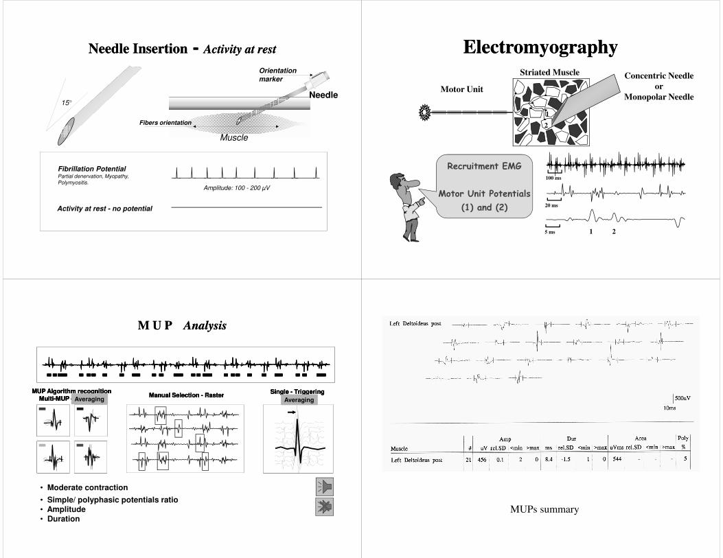
Needle Insertion - Activity at restNeedle Insertion - Activity at rest
Fibrillation PotentialPartial denervation, Myopathy,
Polymyositis.Amplitude: 100 - 200 µV
Activity at rest - no potential
Muscle
Orientation
marker
Needle
Fibers orientation
15°
ElectromyographyElectromyography
Concentric Needle
or
Monopolar Needle
Striated Muscle
Motor Unit
1
2
Recruitment EMG
Motor Unit Potentials
(1) and (2)
100 ms
20 ms
5 ms 1 2
• Moderate contraction
Single - TriggeringManual Selection - Raster
M U P AnalysisM U P Analysis
MUP Algorithm recognitionMulti-MUP - Averaging
Manual Selection - RasterManual Selection - RasterMUP Algorithm recognition
Multi-MUP - AveragingMUP Algorithm recognition
Multi-MUP - Averaging
• Simple/ polyphasic potentials ratio• Amplitude• Duration
Single - TriggeringSingle - Triggering
AveragingAveraging
�
AveragingAveraging
MUPs summary

Muscular dystrophy
Type grouping

DCN EMG
MAXIMUM EFFORT
EMG Needles:
- Disposable concentric
- Disposable monopolar
Auditory control of:
- Resting silence
- Sound of MUPs
- Frequency firing
EMG - Recruitment Interference
Pattern
EMG - Recruitment Interference
Pattern
< 50 ms ( 20 Hz )
Amplitude depends on Muscle, 4-2 mV
�
Turns/ AmplitudeTurns/ Amplitude
• 1 second EMG signal
• Concentric or Monopolar Needle
• Few sites in same muscle
• 20 measurements at diff. force
• Number of Turns > 100 µV
• Mean Turns Amplitude
T1
T2
T3
T4
T5
S1S2
S3S4
A4
A2
A1A3
Change in signal direction = Turn Segment Amplitude > 100 µV
A = AmplitudeS = Segment
Myopathy
Neuropathy
Polymyositis
tib ant
Polio Sequele
tib ant
Amplitude
Turns
Amplitude
Turns
Short Segments / Amplitude / ActivityShort Segments / Amplitude / Activity
Myopathy
Neuropathy
Sum of activity periodsin % (or ms) of full IP
NSS Amplitude Duration
Yes < 0.5 mV < 1.5 msYes 0.5 > < 2mV < 3 ms No > 2 mV < 5 ms
• 500 ms EMG signal then x 2
• NSS Number of Small Segments
• UCA Upper Centile Amplitude
Small Segment
Activity
Normal turn/amplitude analysis Normal activity/envelope

Turn-amp and
activity-envelope
analyses in
myopathy
Turn-amp and activity-
envelope analyses in
neurogenic conditions

Pathologies in Striated MusclePathologies in Striated Muscle
Normal
Neuropathy
Myopathy
Myasthenia
CNEMG
Same effort, same muscle,
different pathologies
Ground
�Neuromuscular junction
disease. See DecrementUnstable MUPs