Embed Size (px)
Citation preview
(CANCER RESEARCH 52, 1553-1560, March 15, 1992]
Tumor Types Derived from Epithelial and Myoepithelial Cell Lines of R3230ACRat Mammary Carcinoma1
Anna Sapino, Mauro Papotti, Brunella Sanfìlippo,Patrizia Gugliotta, and Gianni Bussola!iDepartment of Biomedicai Sciences and Human Oncology, University of Turin, Via Santena 7, Turin, 1-10126, Italy
ABSTRACT
Epithelial and myoepithelial cells coexist in the rat R3230AC mammary tumor. To test the hypothesis that these two cell types constituteinteractive but independent neoplastic populations, we obtained in vitrocell lines with epithelial or myoepithelial patterns and transplanted themin syngeneic animals. One stabilized line (EPI) and four cloned lines (A,C, D, E) with epithelial characteristics, confirmed by positive reactionsfor keratins in immunocytochemical and immunoblot tests, constantlygave rise in vivo to carcinomas, which, however, lacked structural andfunctional patterns typical of the original tumor. A fusiform shape andimmunocytochemical characteristics of myoepithelial cells were observedin three clones (H, I, L), which in vivo gave rise to sarcomatous andmixed carcinosarcomatous neoplasms. These data are consistent with theabove hypothesis and indicate that breast carcinomas derive from epithelial cells, while sarcomatous and carcinosarcomatous neoplasms canoriginate from myoepithelial cell proliferation. This study provides datasuggesting myoepithelial cell involvement in the development of pathological entities occurring in the human breast and displaying mixedepithelial and stromal neoplastic components, i.e.. cystosarcoma phyl-loides and sarcomatous metaplasia in carcinomas.
INTRODUCTION
The R3230AC mammary carcinoma, transplantable inFischer rats, was originally described by Hilf et al. (1) as a milk-producing adenocarcinoma. Under estrogen stimulation, it displays marked increase of milk protein production and cellularvacuolation. The biochemical properties and hormone dependency of this tumor have been investigated by several authors(2-12).
We observed that the peculiar functional secretory similarityof this carcinoma to the normal mammary gland is matched bya parallel similarity in structure; both epithelial and basallylocated myoepithelial cells are present, outlining neoplasticpseudoglandular structures and showing the typical immunocytochemical and ultrastructural characteristics of these cells inthe normal breast (13).
The R3230AC mammary carcinoma therefore represents anexperimental model with unique morphological and functionalproperties. We have established and cloned in vitro cell linesfrom this tumor and studied their morphology, immunocyto-chemistry, and function. These cell lines were further investigated by transplanting them back into syngeneic rats, wherethey gave rise to carcinomas, sarcomas, and mixed carcinosar-comas. This experimental model provides data that help tounderstand the significance and genesis of peculiar and stillunexplained pathological entities occurring in the humanbreast, i.e., cystosarcoma phylloides and sarcomatous metaplasia in carcinomas.
Received 7/22/91; accepted 1/8/92.The costs of publication of this article were defrayed in part by the payment
of page charges. This article must therefore be hereby marked advertisement inaccordance with 18 U.S.C. Section 1734 solely to indicate this fact.
1This work was supported by grants from the CNR (Rome), AIRC (Milan),
and the Ministry of the University.2To whom requests for reprints should be addressed.
MATERIALS AND METHODS
Tumor. R3230AC transplantable mammary tumor was obtainedfrom Biomeasure (Hopkinton, MA) and maintained by serial s.c. transplantations into the dorsal region of female Fischer 344 inbred rats(100-120 g body weight; Nossan, Correzzana, Milan, Italy).
Tumor fragments were obtained from nonnecrotic areas of the donorrat, mechanically dispersed in a Potter homogenizer and suspended inRPMI. Approximately 1 mm3 of tumor (or 1-3 x IO6neoplastic cells)
was used for monthly transplantations. The tumor consistently grew toa 2-em mass within 20 days. Animals were killed by decapitation every25-35 days; tumor tissue was in part inoculated in recipient rats asdescribed above, in part fixed in absolute ethanol or methacarn forhistology and immunohistochemistry (antibodies listed in Table 1), andin part processed for cell culture lines (see below).
In order to evaluate the response to estrogen stimulation, tumor-bearing animals were given for 1 month weekly s.c. injections ofestradici valerate (20 mg/kg body weight) according to the method ofHilf (14).
Establishment of Primary Stabilized and Cloned Cultures. The tumortissue, removed aseptically, was minced into small pieces (~1 mm3)
with scalpels and digested with 125 units/mg collagenase (type II)(Worthington Biochemical Corp., Freehold, NJ) in RPMI (GIBCO-Island Biological Co., Grand Island, NY), 10% PCS3 (GIBCO), 200
units/ml penicillin, 200 Mg/ml streptomycin (EUROBIO), and 2.5 mg/ml Fungizone (GIBCO) for 2 h at 37*C.
Single cell suspensions and several small tumor fragments werewashed twice in RPMI supplemented with 10% FCS without collagenase and plated in T25 flasks (Falcon Plastics, Los Angeles, CA) at aconcentration of about 3 x 10s cells/flask.
Incubation of the flasks for 24 h at 37'C in a 5% CO2 humidified
atmosphere allowed most of the fragments and free cells to attach. Inorder to obtain an epithelial cell line (EPI) from the primary culture,elongated cells were removed following trypsin-EDTA treatment (15)with a minor modification proposed by Kuzumaki et al. (16). Brieflythe mixed cell sheet was rinsed once with Ca2+-and Mg2+-free PBS andincubated for 3 min at 37*C in the presence of fresh trypsin-EDTA.
The less rapidly detaching cells, mostly islands of epithelium-like cells,were rinsed and supplied with fresh medium. This procedure wasrepeated every 3 days until epithelial like cells covered more than 90%of the culture surface.
The stabilized EPI cells are presently at the 100th passage. Subcultures were routinely carried out in RPMI, 10% FCS, and antibiotic.
Cell Cloning. The mixed population obtained from primary cultures(of R3230AC tumor) as well as the stabilized (EPI) epithelial cells werecloned following the dilution plating technique proposed by Sato et al.(17). A single cell suspension was appropriately diluted and 96-wellmicrotest plates (Falcon) were seeded; each well was inoculated with acell suspension yielding an average of 1 cell/well. Those wells containing only one cell as ascertained by microscope inspection were considered available for clone establishment; the others were discarded.
Conditioned media were prepared by adding to the routine medium10% of the supernatant obtained from confluent cultures of epithelialcells filtered through 0.22-/im Millipore filters and stored at 4*C before
use. We obtained 3 clones (I, H, L) from the mixed population ofprimary culture and 4 clones (A, C, D, E) from the EPI cell line.
The cell lines, at early passages, were reinjected into syngeneicanimals and subcultured until passage 80.
3The abbreviations used are: FCS, fetal calf serum; PBS, phosphate-bufferedsaline; NTEN, 50 miviTris, pH 7.4-0.15 M NaCI-2 mM EDTA-0.1% Nonidet P-40; sm, smooth muscle.
1553
on May 18, 2018. © 1992 American Association for Cancer Research. cancerres.aacrjournals.org Downloaded from
MAMMARY CARCINOMA CELL LINES
In Vitro Hormonal Stimulation. Primary cultures, stabilized EPI cells,and clones D and E were cultured in Petri dishes and on glass coverslipsfor 9 days in a basal medium containing 10 * M 17/3-estradiol (Merck,
Darmstadt, Germany) and 10% of charcoal-adsorbed PCS (18).The medium was changed every 3 days.a-Lactalbumin production was estimated immunocytochemically.Light Microscopy. The morphological appearance of living cultures
in flasks was observed on a Leitz Labovert inverted microscope fittedwith phase contrast. Mixed cells from primary cultures, EPI cells, andclones were also grown on glass coverslips; fixed in methanol; andstained by the Papanicolaou method for cytological examination.
For immunohistochemical tests, cells were grown on glass coverslips,fixed in methanol for 5 min at -20'C, permeabilized in acetone for 5 sat —¿�20*C,and réhydratée!with pig normal serum (diluted 1:5(1in PBSfor 20 min). Cells were incubated at 37'C for 60 min with the primary
antibodies listed in Table 1 and then with the appropriate fluorescein-labeled secondary antisera (Sera-Lab, Ltd., Sussex, England) diluted1:10 in PBS for 30 min at room temperature, following a standardindirect immunofluorescence procedure.
Electron Microscopy. EPI cells and cells from clone I were fixed ina solution of 1-2% glutaraldehyde in 0.2 M cacodylate buffer (pH 7.3)and 2% aqueous osmium tetroxide for l h at 4*C, dehydrated through
graded alcohols, cleared in propylene oxide, and embedded in Epon-Araldite. Ultrathin sections were counterstained with uranyl acetateand lead citrate and examined with a Siemens Elmiskop 1 or a PhilipsEM 401 electron microscope.
Cell Extracts. The established cell line EPI and the clones (C, D, H,I, L) were homogenized in a buffer 20 mM Tris (pH 7.4), 0. l M NaCl,5 mM MgCl2, 1% Nonidet P-40, 0.5% sodium deoxycholate, 0.1 mM2-mercaptoethanol, and 2000 lU/ml Trasylol. Extracts were stored at-80*C until used. Protein concentration was determined by the method
of Lowry as modified by Markwell et al. (19).Immunoblotting. Total proteins (100 ng) were separated by sodium
dodecyl sulfate polyacrylamide gel electrophoresis on 8% acrylamidegel slabs and transferred to nitrocellulose filters in a Ployblot apparatus(ABN, Emeryville, CA) according to the manufacturer's instructions.
Nitrocellulose was then saturated in 3% bovine serum albumin inNTEN for 3 h at 37'C, incubated in the same buffer containing the
anticytokeratin 19 monoclonal antibody 1165 (Amersham International, England) diluted '/iooor an anti-a-sm-actin monoclonal antibodydiluted 1:800 (Sigma Chemical Co., St. Louis, MO) for 16-18 h at4"C, washed twice for 10 min each in NTEN, incubated with the
secondary rabbit anti-mouse antibody (Dako) diluted 1:500 for 3 h at4"C, washed twice for 10 min each in NTEN, incubated with '"!-
protein A (0.1 /¿Ci/ml)(Amersham), and then washed as before, airdried, and exposed for 2 days at —¿�80°Cto Hyperfilm MP autoradi-
ographic films (Amersham).Transplantation of Cell Lines into Animals. Cultured cells (approxi
mately 0.8-4 x IO6, suspended in 0.5-0.8 ml RPMI), obtained fromearly passages (3rd-8th) were injected s.c. into female Fischer rats. Theoccurrence and time of growth of tumors were recorded. Tumorsgrowing in syngeneic rats from cultured lines were retransplanted intoother rats, following the procedure outlined above. Tissue blocks werefixed in methacarn (20) and embedded in paraffin. Sections were
routinely stained with hematoxylin and eosin and processed for im-munocytochemical characterization using the antibodies listed in Table1. To detect immunological binding, the avidin-biotinylated peroxidasecomplex procedure (21) was used, after treatment of the sections withthe appropriate biotinylated secondary antisemiti (Vector, Burlingame,CA).
In Vivo Hormonal Stimulation. Animals bearing palpable tumorsobtained from EPI, D, E, H, and I cells were treated with estrogenfollowing the protocol used in R3230AC-bearing animals (see above).
RESULTS
Serially Transplanted R3230AC Tumors. The gross and microscopic appearances of the R3230AC mammary tumor, described in detail elsewhere (13), were examined at eachtransplantation.
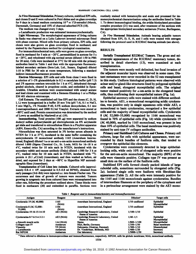
The tumors were mostly well delimited, but focal invasion ofthe adjacent muscular layers was observed in some cases. Distant métastaseswere never recorded in the 52 rats transplantedin this study. Uniform histological patterns were observed withacinar and/or gland-like structures lined by polygonal epithelialcells and basal, elongated myoepithelial cells. The originaltumor stained positively for «-sm-aetin in the elongated basalcells, thus confirming their myoepithelial nature (Fig. la).
Epithelial secretory cells were stained with different antibodies to keratin. AE1, a monoclonal recognizing acidic cytokera-tins, was positive only in single squamous cells while AE3, amonoclonal to basic cytokeratins, recognized a few epithelialcells and the majority of basal myoepithelial cells. Cytokeratin8 (M, 52,000-55,000) recognized by 1166 monoclonal wasfound in 70% of epithelial cells (Fig. \b) while cytokeratin 19(M, 40,000), marked by 1165 monoclonal, was present in lessthan 10% of epithelial cells. The basal membrane was positivelystained by anti-type IV collagen antibodies.
Primary and Stabilized Cell Cultures and Clones. Primary cellcultures, large flat cells with epithelial appearance, were surrounded by thick bundles of fusiform cells, that tended toovergrow the epithelial-like elements.
Cytokeratins were consistently detected in large epithelial-looking cells, while only 10% of elongated cells were positivefor the anti-sm-actin monoclonal. The majority (80%) of thecells were vimentin positive. Collagen type IV was present assmall dots on the surface of the fusiform cells.
Stabilized EPI cells formed closely packed islands of largecuboidal cells, sometimes surrounded by elongated cells (Fig.la). Isolated single cells were fusiform with fibroblast-likeappearance (Table 2). All the cells were intensely positive forthe 1165 and 1166 monocle muÃsagainst cytokeratins. Bundlesof intermediate filaments at the periphery of the cytoplasm andin a perinuclear arrangement were stained by the AE3 mono-
Table 1 Reagents used in immunohistochemistry and immunofluorescence
AntigenCytokeratin
19 (M,40,000)Cytokeratin
8 (M,52,000-55,000)Cytokeratins
19-16-15-14-10Cytokeratins
8-7-6-5-4-3-2-1..-Smooth
muscleactinCollagentypeIVVimentina-LactalbuminRat
MFGMReagent1165(MAb)1166(MAb)AE1
(MAb)AE3
(MAb)1
A4(MAb)AntiserumV9
(MAb)AntiserumAntiserumSourceAmersham
International,EnglandAmersham
International,EnglandCambridge
Research Laboratory,UnitedStatesofAmericaCambridge
Research Laboratory,UnitedStatesofAmericaSigmaHeyl,
Berlin,GermanyDakopatts,Glostrup,DenmarkDr.
Vonderhaar. Bethesda, MDOurlaboratoryDilution"1.
'1(1undilutedI/
IO-undiluted1/100-1/101/40-1/11/800-1/1001/350-1/501/300-1/101/400-1/201/5000-1/50"
Data referred to dilutions in immunoperoxidase and immunofluorescence, respectively. MFGM, milk fat globule membrane; MAli,1554Target
cellsEpithelialMyoepithelialEpithelialEpithelialEpithelialMyoepithelialMyoepithelialBasal
membraneMesenchymalSecretorySecretorymonoclonal
antibody.
on May 18, 2018. © 1992 American Association for Cancer Research. cancerres.aacrjournals.org Downloaded from
MAMMARY CARCINOMA CELL LINES
%«&3K& <•>i. ms*!f»"•$4
&vyK&?r «r.->iw?., •¿�":'-b>;,*,v.*. v^..^:.
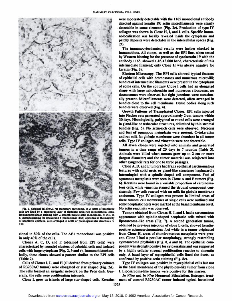
Fig. 1. Original R3230AC rat mammary carcinoma. In a, nests of neoplasticcells are lined by a peripheral layer of flattened actin-rich myoepithelial cells.Immunoperoxidase staining with a-smooth muscle actin monoclonal, x 250. Inb, immunostaining for cytokeratin 8 (monoclonal 1166) is positive in the majorityof neoplastic epithelial cells arranged in nests or pseudoglandular structures, xISO.
clonal in 80% of the cells. The AE1 monoclonal was positivein only 40% of the cells.
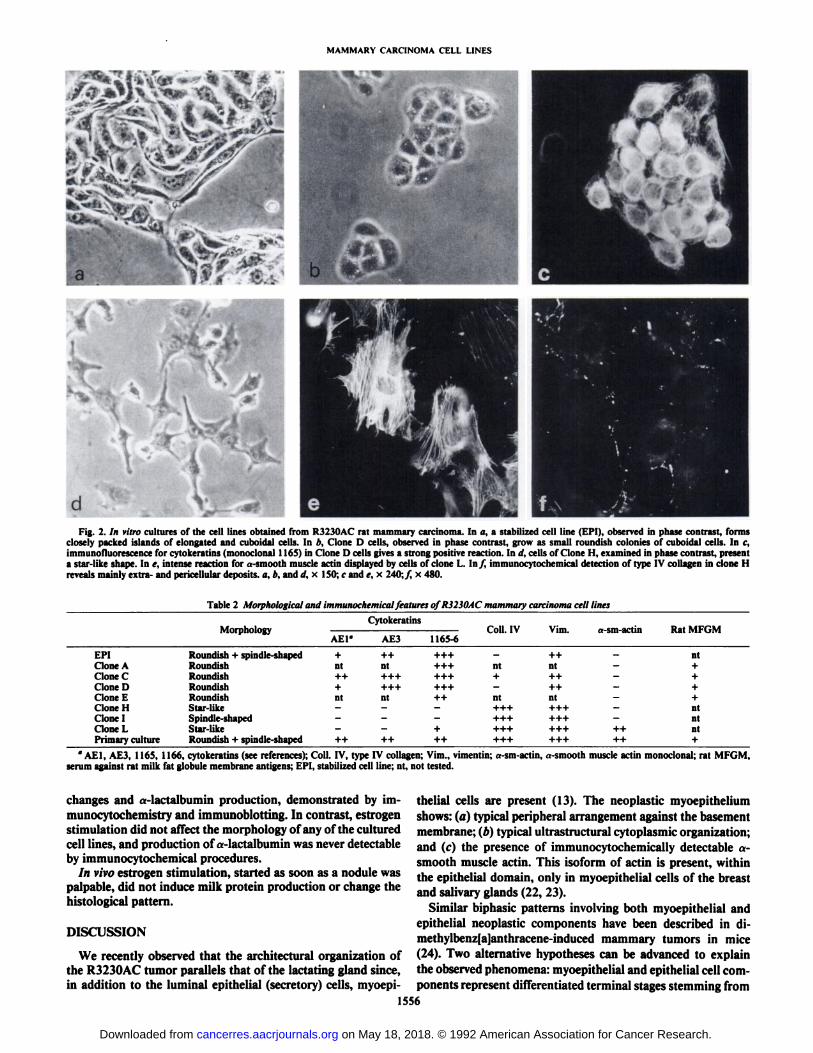
Clones A, C, D, and E (obtained from EPI cells) werecharacterized by rounded clusters of cuboidal cells and isolatedcells with large cytoplasm (Fig. 2, b and r). Immunohistochem-ically, these clones showed a pattern similar to the EPI cells(Table 2).
Cells of Clones I, L, and H (all derived from primary culturesof R3230AC tumor) were elongated or star shaped (Fig. 2d).The cells formed an irregular network on the Petri dish. Generally, the cells were proliferating intensely.
Clone L grew as islands of large star-shaped cells. Keratins
were moderately detectable with the 1165 monoclonal antibodydirected against keratin 19; actin micro lìlaments were clearlydetectable in some elements (Fig. 2e). Production of type IVcollagen was shown in Clone H, I, and L cells. Specific immu-nolocalization was locally revealed inside the cytoplasm andpatchy deposits were detectable in the intercellular spaces (Fig.2f>. '
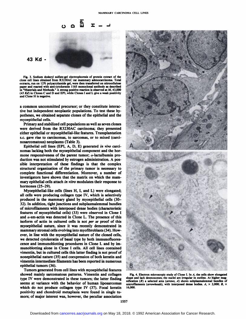
The immunocytochemical results were further checked inimmunoblots. All clones, as well as the EPI line, when testedby Western blotting for the presence of cytokeratin 19 with theantibody 1165, showed a M, 43,000 band, characteristic of thisintermediate filament; only Clone H was always negative forkeratin (Fig. 3).
Electron Microscopy. The EPI cells showed typical featuresof epithelial cells with desmosomes and numerous microvilli;bundles of intermediate filaments were present in the cytoplasmof some cells. On the contrary Clone I cells had an elongatedshape with large mitochondria and numerous ribosomes; nodesmosomes were observed but tight junctions were occasionally present. Microfilaments were detected, often arranged inbundles close to the cell membrane. Dense bodies along suchbundles were observed (Fig. 4).
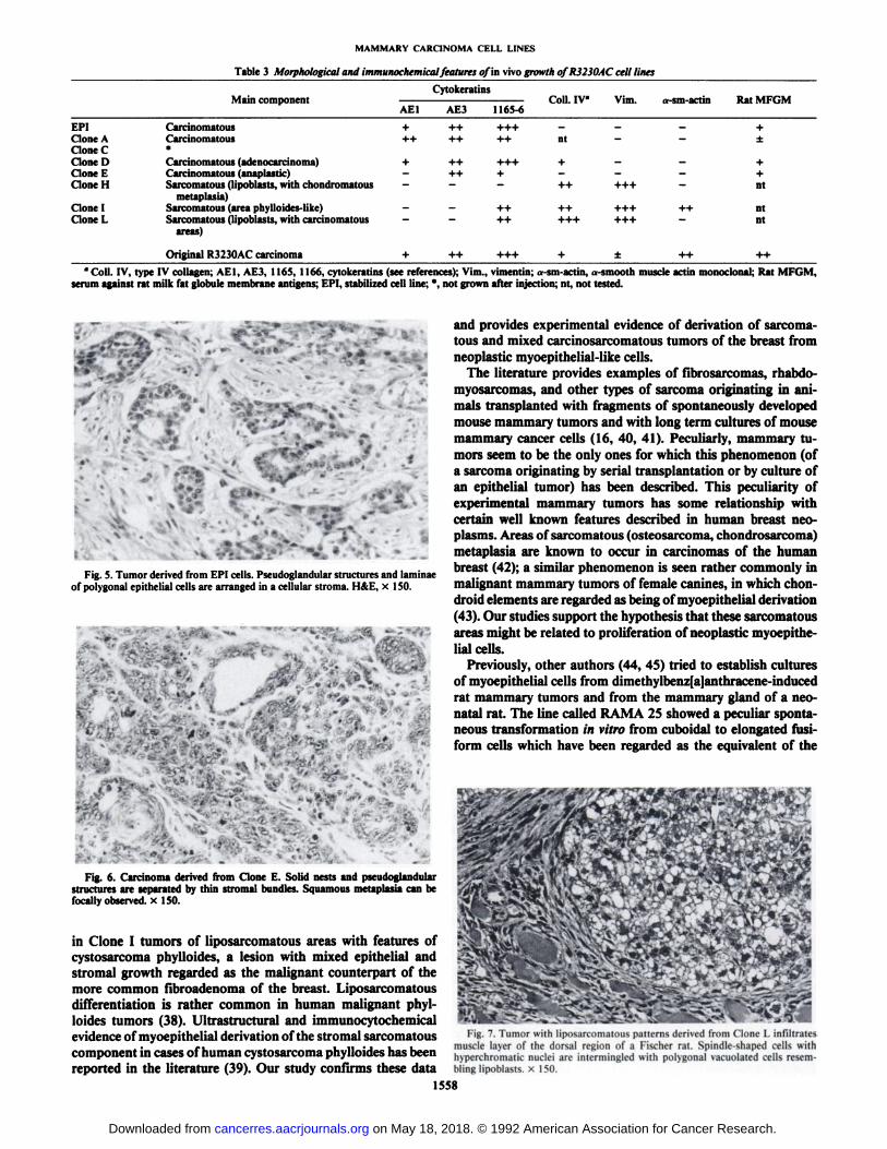
Growth Patterns of Transplanted Clones. EPI cells injectedinto Fischer rats generated approximately 2 cm tumors within30 days. Histologically, polygonal or round cells were arrangedin gland-like or trabecular structures, delimited by thin stromalbundles (Fig. 5). No actin-rich cells were observed. Necrosisand foci of squamous metaplasia were present. Cytokeratinsand rat milk fat globule membrane were abundant in all tumorcells. Type IV collagen and vimentin were not detectable.
All seven clones were injected into animals and generatedtumors in a time range of 20 days to 7 months (Table 3).Animals were killed when tumors grew up to 2 cm or more(largest diameter) and the tumor material was reinjected intoother syngeneic rats for one to three passages.
Clone A, D, and E tumors had frank epithelial carcinomatousfeatures with solid nests or gland-like structures haphazardlyintermingled with a spindle-shaped cell component. Foci ofsquamous metaplasia were seen in Clone A and E tumors (Fig.6). Keratins were found in a variable proportion of carcinomatous cells, while vimentin stained the stromal component consistently. Few cells reacted with rat milk fat globule membraneantiserum. Type IV collagen was present in limited areas ofthese tumors; cell membranes of single cells were outlined andsome neoplastic nests were marked at the basal membrane level.No actin reactivity was observed.
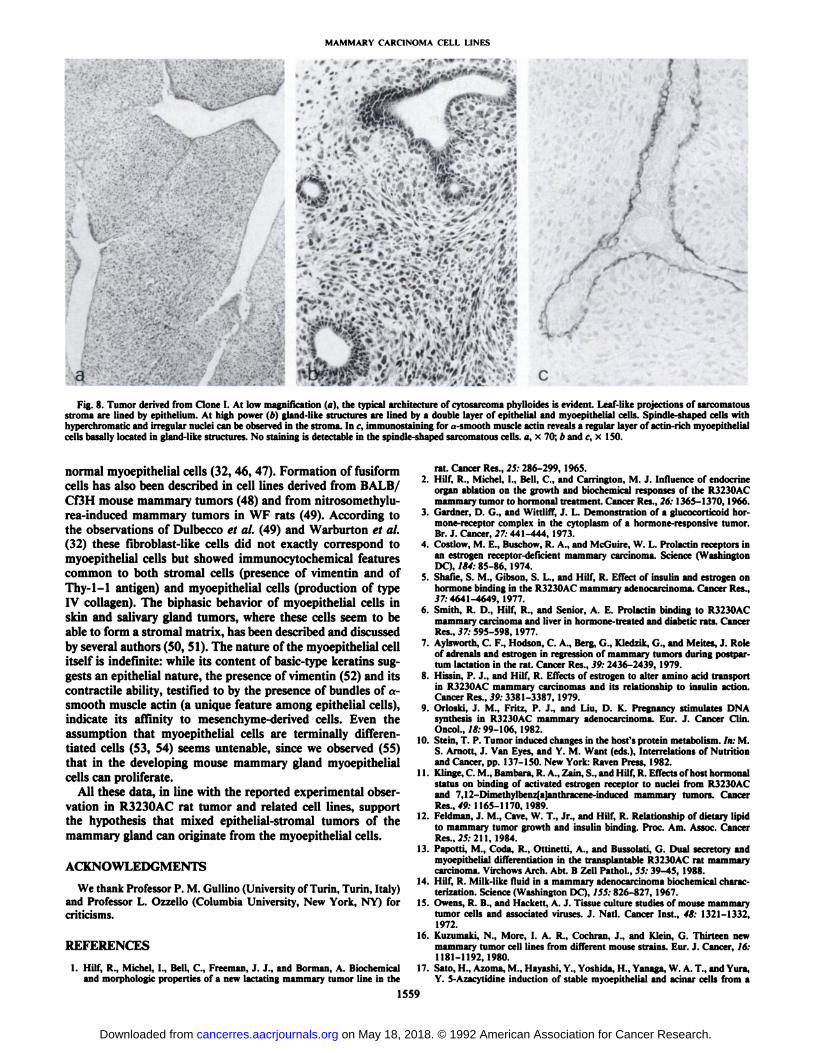
Tumors obtained from Clones H, I, and L had a sarcomatousappearance with spindle-shaped neoplastic cells mixed withliposarcoma-like areas (Fig. 7). A second generation tumor,obtained by previous injection of clone L cells, showed keratin-positive adenocarcinomatous foci while in a tumor originatedfrom Clone H, areas of chondromatous metaplasia were present. Clone I had a peculiar morphology, strongly resemblingcystosarcoma phylloides (Fig. 8, a and b). The epithelial component was strongly positive for cytokeratins and was supportedby a highly cellular stromal proliferation reactive to vimentinonly. A basal layer of myoepithelial cells lined the ducts, asconfirmed by positive actin staining (Fig. 8e).
Type IV collagen was positive in myoepithelial cells but notin the basal membrane of the phylloides-like tumor from cloneI. Liposarcoma-like tumors were positive for this marker.
In Vitro and in Vivo Hormonal Stimulation. Estrogen treatment of control R3230AC tumor induced typical lactational
1555
on May 18, 2018. © 1992 American Association for Cancer Research. cancerres.aacrjournals.org Downloaded from
MAMMARY CARCINOMA CELL LINES
\psr^£âA ; \à » ~ * J^* *;
» . •¿�,.
Fig. 2. /n vitro cultures of the cell lines obtained from R3230AC rat mammary carcinoma. In a, a stabilized cell line (EPI), observed in phase contrast, formsclosely packed islands of elongated and cuboidal cells. In b. Clone D cells, observed in phase contrast, grow as small roundish colonies of cuboidal cells. In <•,
immunofluorescence for cytokeratins (monoclonal 1165) in Clone D cells gives a strong positive reaction. In d, cells of Clone H, examined in phase contrast, presenta star-like shape. In e, intense reaction for «-smoothmuscle actin displayed by cells of clone I.. In/, immunocytochemical detection of type IV collagen in clone Hreveals mainly extra- and pericellular deposits, a, e, and d, x ISO; c and e, x 240: / x 480.
Table 2 Morphological and immunochemical features 0/R3230AC mammary carcinoma cell lines
EPICloneACloneCCloneDCloneECloneHCloneICloneLPrimary
cultureMorphologyRoundish
+spindle-shapedRoundishRoundishRoundishRoundishStar-likeSpindle-shapedStar-likeRoundish
+ spindle-shapedAE1"+nt+
j-+nt—-—++CytokeratinsAE3
1165-6++
+++nt+++++++++++++++nt++——¿�--—+++
++Coll.
IV Vim.a-sm-actin++nt
nt-+++—¿�—++—¿�ntnt-++++++—¿�++++++-++++++++++++++ ++Rat
MFGMnt++++ntntnt+
°AE1, AE3, 1165, 1166, cytokeratins (see references); Coll. IV, type IV collagen; Vim., \ ¡mentiti;«-sm-actin,«-smoothmuscle actin monoclonal; rat MFGM,
serum against rat milk fat globule membrane antigens; EPI, stabilized cell line; nt, not tested.
changes and a-lactalbumin production, demonstrated by im-
munocytochemistry and immunoblotting. In contrast, estrogenstimulation did not affect the morphology of any of the culturedcell lines, and production of a-lactalbumin was never detectableby immunocytochemical procedures.
In vivo estrogen stimulation, started as soon as a nodule waspalpable, did not induce milk protein production or change thehistológica! pattern.
DISCUSSION
We recently observed that the architectural organization ofthe R3230AC tumor parallels that of the lactating gland since,in addition to the luminal epithelial (secretory) cells, myoepi-
thelial cells are present (13). The neoplastic myoepitheliumshows: (a) typical peripheral arrangement against the basementmembrane; (b) typical ultrastructural cytoplasmic organization;and (c) the presence of immunocytochemically detectable a-smooth muscle actin. This isoform of actin is present, withinthe epithelial domain, only in myoepithelial cells of the breastand salivary glands (22, 23).
Similar biphasic patterns involving both myoepithelial andepithelial neoplastic components have been described in di-methylbenz[a]anthracene-induced mammary tumors in mice(24). Two alternative hypotheses can be advanced to explainthe observed phenomena: myoepithelial and epithelial cell components represent differentiated terminal stages stemming from
1556
on May 18, 2018. © 1992 American Association for Cancer Research. cancerres.aacrjournals.org Downloaded from
MAMMARY CARCINOMA CELL LINES
o o I - -I
43 Kd -
Fig. 3. Sodium dodecyl sulfate-gel electrophoresis of protein extract of theclone cell lines obtained from R3230AC rat mammary adenocarcinoma. Totalextracts, run on 12% polyacrylamide gel, were then transferred on nitrocellulosepaper and reacted with anti-cytokeratin 1165 monoclonal antibody as describedin "Materials and Methods." A strong positive reaction is observed at M, 43,000
(43 Kd) in Clones C and D and EPI, while Clones I and L give a weak positivityand Clone H is negative.
a common uncommitted precursor; or they constitute interactive but independent neoplastic populations. To test these hypotheses, we obtained separate clones of the epithelial and themyoepithelial cells.
Primary and stabilized cell populations as well as seven cloneswere derived from the R3230AC carcinoma; they presentedeither epithelial or myoepithelial-like features. Transplantations.c. gave rise to carcinomas, to sarcomas, or to mixed (carci-nosarcomatous) neoplasms (Table 3).
Epithelial cell lines (EPI, A, D, E) generated in vivo carcinomas lacking both the myoepithelial component and the hormone responsiveness of the parent tumor; «-lactalbumin production was not stimulated by estrogen administration. A possible interpretation of these findings is that the complexstructural organization of the primary tumor is necessary tocomplete functional differentiation. Moreover, a number ofinvestigators have shown that the matrix on which the mammary epithelial cells attach in vitro modulates their response tohormones (25-29).
Myoepithelial-like cells (lines H, I, and L) were elongated;all cells were producing collagen type IV, which is selectivelyproduced in the mammary gland by myoepithelial cells (30-32). In addition, tight junctions and subplasmalemmal bundlesof microfilaments with interposed dense bodies (characteristicfeatures of myoepithelial cells) (33) were observed in Clone Iand a-sm-actin was detected in Clone L. The presence of thisisoform of actin in cultured cells is not per se proof of thismyoepithelial nature, since it was recently demonstrated inmammary stromal cells evolving into myofibroblasts (34). However, in line with the myoepithelial nature of the cloned cells,we detected cytokeratin of basal type by both immunofluores-cence and immunoblotting procedures in Clone L and by im-munoblotting alone in Clone I cells. All cell lines containedvimentin, but in cultured cells this latter finding is not proof ofnonepithelial nature (35) and coexpression of both keratin andvimentin intermediate filaments has been reported in numerousepithelial tumors (36).
Tumors generated from cell lines with myoepithelial featuresshowed mainly sarcomatous patterns. Vimentin and collagentype IV were demonstrated in these tumors; the latter findingseems at variance with the behavior of human liposarcomaswhich do not produce collagen type IV (37). Focal keratinpositivity and chondroid metaplasia were found in single tumors; of major interest was, however, the peculiar association
1557
A •¿�1
BFig. 4. Electron microscopic study of Clone I. In 1. the cells show elongated
shape and lack desmosomes; the nuclei are irregular in outline. At higher magnification (B) a selected area (arrows. A) shows subplasmalemmal bundles ofmicrofilaments (arrowheads), with interposed dense bodies. A, x 2,000; B, x16,000.
on May 18, 2018. © 1992 American Association for Cancer Research. cancerres.aacrjournals.org Downloaded from
MAMMARY CARCINOMA CELL LINES
Table 3 Morphological and immunochemical features of in vivo growth ofR3230AC cell lines
Cytokeratins.t '.'"'111 VI MU ('l'Ut. Il 1 V^VHli Õ T ' lili. U-3Ill-a\.lIII IVtll ITI! VIITIAEl
AE31165-6EPIClone
ACloneCCloneDCloneECloneHClone
ICloneLCarcinomatous
+ ++ +++ - --Carcinomatous++ ++ ++ nt —¿�—¿�•Carcinomatous
(adenocarcinoma) + ++ +++ + --Carcinomatous(anaplastic) —¿� ++ + —¿� —¿�—¿�Sarcomatous
(lipoblasts, with chondromatous - - - ++ +++-metaplasia)Sarcomatous
(area phylloides-like) —¿� —¿� ++ ++ +++++Sarcomatous(lipoblasts, with Carcinomatous —¿� —¿� ++ +++ +++—¿�areas)+±++ntntnt
Original R3230AC carcinoma" Coll. IV, type IV collagen; AEl, AE3, 1165, 1166, cytokeratins (see references); Vim., vimentin; a-sm-actin, «smooth muscle actin monoclonal; Rat MFGM,
serum against rat milk fat globule membrane antigens; EPI, stabilized cell line; *. not grown after injection; nt, not tested.
>•
..•.. v.. A---'- *
%„¿�-•—¿�. vi,-:'
^
Fig. 5. Tumor derived from EPI cells. Pseudoglandular structures and laminaeof polygonal epithelial cells are arranged in a cellular stroma. H&E, x 150.
* t!'^v
. JK* -*»-.* .-.
Fig. 6. Carcinoma derived from Clone E. Solid nests and pseudoglandularstructures are separated by thin stromal bundles. Squamous metaplasia can befocally observed, x 150.
in Clone I tumors of liposarcomatous areas with features ofcystosarcoma phylloides, a lesion with mixed epithelial andstromal growth regarded as the malignant counterpart of themore common fibroadenoma of the breast. Liposarcomatousdifferentiation is rather common in human malignant phylloides tumors (38). Ultrastructural and immunocytochemicalevidence of myoepithelial derivation of the stromal sarcomatouscomponent in cases of human cystosarcoma phylloides has beenreported in the literature (39). Our study confirms these data
and provides experimental evidence of derivation of sarcomatous and mixed carcinosarcomatous tumors of the breast fromneoplastic myoepithelial-like cells.
The literature provides examples of fibrosarcomas, rhabdo-myosarcomas, and other types of sarcoma originating in animals transplanted with fragments of spontaneously developedmouse mammary tumors and with long term cultures of mousemammary cancer cells (16, 40, 41). Peculiarly, mammary tumors seem to be the only ones for which this phenomenon (ofa sarcoma originating by serial transplantation or by culture ofan epithelial tumor) has been described. This peculiarity ofexperimental mammary tumors has some relationship withcertain well known features described in human breast neoplasms. Areas of sarcomatous (osteosarcoma, chondrosarcoma)metaplasia are known to occur in carcinomas of the humanbreast (42); a similar phenomenon is seen rather commonly inmalignant mammary tumors of female canines, in which ohmiciro id elements are regarded as being of myoepithelial derivation(43). Our studies support the hypothesis that these sarcomatousareas might be related to proliferation of neoplastic myoepithelial cells.
Previously, other authors (44, 45) tried to establish culturesof myoepithelial cells from dimethylbenz[a]anthracene-inducedrat mammary tumors and from the mammary gland of a neonatal rat. The line called RAMA 25 showed a peculiar spontaneous transformation in vitro from cuboidal to elongated fusiform cells which have been regarded as the equivalent of the
Fig. 7. Tumor with liposarcomatous patterns derived from Clone L infÃltralesmuscle layer of the dorsal region of a Fischer rat. Spindle-shaped cells withhyperchromatic nuclei are intermingled with polygonal vacuolated cells resembling lipoblasts. x 150.
1558
on May 18, 2018. © 1992 American Association for Cancer Research. cancerres.aacrjournals.org Downloaded from
MAMMARY CARCINOMA CELL LINES.v."''Ã.;!;î•¿�•••¿�•m**
Fig. 8. Tumor derived from Clone I. At low magnification (a), the typical architecture of cytosarcoma phylloides is evident. Leaf-like projections of sarcomatousstromu are lined by epithelium. At high power (b) gland-like structures are lined by a double layer of epithelial and myoepithelial cells. Spindle-shaped cells withhyperchromatic and irregular nuclei can be observed in the strenna. In c, immunostaining for a-smoolh muscle actin reveals a regular layer of actin-rich myoepithelialcells basally located in gland-like structures. No staining is detectable in the spindle-shaped sarcomatous cells, a, x 70; b and c,x 150.
normal myoepithelial cells (32, 46, 47). Formation of fusiformcells has also been described in cell lines derived from BALB/CDH mouse mammary tumors (48) and from nitrosomethylu-rea-induced mammary tumors in WF rats (49). According tothe observations of Dulbecco et al. (49) and Warburton et al.(32) these fÃbroblast-like cells did not exactly correspond tomyoepithelial cells but showed immunocytochemical featurescommon to both stromal cells (presence of vimentin and ofThy-1-1 antigen) and myoepithelial cells (production of typeIV collagen). The biphasic behavior of myoepithelial cells inskin and salivary gland tumors, where these cells seem to beable to form a stromal matrix, has been described and discussedby several authors (50, 51). The nature of the myoepithelial cellitself is indefinite: while its content of basic-type keratins suggests an epithelial nature, the presence of vimentin (52) and itscontractile ability, testified to by the presence of bundles of «-smooth muscle actin (a unique feature among epithelial cells),indicate its affinity to mesenchyme-derived cells. Even theassumption that myoepithelial cells are terminally differentiated cells (53, 54) seems untenable, since we observed (55)that in the developing mouse mammary gland myoepithelialcells can proliferate.
All these data, in line with the reported experimental observation in R3230AC rat tumor and related cell lines, supportthe hypothesis that mixed epithelial-stromal tumors of themammary gland can originate from the myoepithelial cells.
ACKNOWLEDGMENTS
We thank Professor P. M. Cullino (University of Turin, Turin, Italy)and Professor L. Ozzello (Columbia University, New York, NY) forcriticisms.
REFERENCES
1. Hilf, R., Michel, I., Bell, C., Freeman, J. J.. and Borman, A. Biochemicaland morphologic properties of a new lactating mammary tumor line in the
10.
11.
12.
13.
14.
IS,
16
17,
rat. Cancer Res., 25: 286-299, 1965.Hilf, R., Michel, I., Bell, C., and Carrington, M. J. Influence of endocrineorgan ablation on the growth and biochemical responses of the R3230ACmammary tumor to hormonal treatment. Cancer Res., 26:136S-1370, 1966.Gardner, D. G., and Wittliff, J. L. Demonstration of a glucocorticoid hormone-receptor complex in the cytoplasm of a hormone-responsive tumor.Br. J. Cancer, 27:441-444, 1973.Costlow, M. E., Buschow, R. A., and McGuire, W. L. Prolactin receptors inan estrogen receptor-deficient mammary carcinoma. Science (WashingtonDC), 184: 85-86, 1974.ShaFie, S. M., Gibson, S. L., and Hilf, R. Effect of insulin and estrogen onhormone binding in the R3230AC mammary adenocarcinoma. Cancer Res.,37; 4641-4649, 1977.Smith, R. D., Hilf, R., and Senior, A. E. Prolactin binding to R3230ACmammary carcinoma and liver in hormone-treated and diabetic rats. CancerRes., 3 7: 595-598, 1977.Aylsworth, C. F., Hodson, C. A., Berg, G., Kledzik, G., and Meites, J. Roleof adrenals and estrogen in regression of mammary tumors during postpar-tum lactation in the rat. Cancer Res., 39: 2436-2439, 1979.Hissin, P. J., and Hilf, R. Effects of estrogen to alter amino acid transportin R3230AC mammary carcinomas and its relationship to insulin action.Cancer Res., 39: 3381-3387, 1979.Orloski, J. M., Fritz, P. J., and Liu, D. K. Pregnancy stimulates DNAsynthesis in R3230AC mammary adenocarcinoma. Eur. J. Cancer Clin.Oncol., /*: 99-106, 1982.Stein, T. P. Tumor induced changes in the host's protein metabolism. In: M.
S. Arnott, J. Van Eyes, and Y. M. Want (eds.). Interrelations of Nutritionand Cancer, pp. 137-150. New York: Raven Press, 1982.Klinge, C. M., Bambara, R. A., Zain, S., and Hilf, R. Effects of host hormonalstatus on binding of activated estrogen receptor to nuclei from R3230ACand 7,12-Dimethylbenz[a]anthracene-induced mammary tumors. CancerRes.,«: 1165-1170, 1989.Feldman, J. M., Cave, W. T., Jr., and Hilf, R. Relationship of dietary lipidto mammary tumor growth and insulin binding. Proc. Am. Assoc. CancerRes., 25:211, 1984.Papotti, M., Coda, R., Ottinetti, A., and Bussolati, G. Dual secretory andmyoepithelial differentiation in the transplantable R3230AC rat mammarycarcinoma. Virchows Arch. Abt. B Zeli Pathol., 55: 39-45, 1988.Hilf, R. Milk-like fluid in a mammary adenocarcinoma biochemical characterization. Science (Washington DC), 155:826-827, 1967.Owens, R. B., and Hackett, A. J. Tissue culture studies of mouse mammarytumor cells and associated viruses. J. Nati. Cancer Inst., 48: 1321-1332,1972.Kuzumaki, N., More, 1. A. R., Cochran, J., and Klein, G. Thirteen newmammary tumor cell lines from different mouse strains. Eur. J. Cancer, 16:1181-1192, 1980.Sato, H., Azoma, M., Hayashi, Y., Yoshida, H., Yanaga, W. A. T., and Yura,Y. 5-Azacytidine induction of stable myoepithelial and acinar cells from a
1559
on May 18, 2018. © 1992 American Association for Cancer Research. cancerres.aacrjournals.org Downloaded from
MAMMARY CARCINOMA CELL LINES
human salivary intercalated duct cell clone. Cancer Res., 47: 4453-4459,1987.
18. Sapino, A., Pietribiasi, F., Bussolati, G., and Marchisio, P. C. Estrogen- andtamox¡fun-induced rearrangement of cytoskeletal structures in breast cancerMCF-7 cells. Cancer Res., 46: 2526-2531,1986.
19. Markwell, M. A. K., Haas, S. M., Bieber, L. C, and Tolbert, N. E. Amodification of Lowry procedure to simplify protein determination in membrane and lipoprotein samples. Anal. Biochem., 87: 206-210, 1978.
20. Mitchell, D., Ibrahim, S., and Gusterson, B. A. Improved immunohistochem-ical localization of tissue antigens using modified methacarn fixation. J.Histochem. Cytochem., 33:491-495, 1985.
21. Hsu, S. M., Raine, L., and Fanger, H. Use of avidin-biotin-peroxidasecomplex (ABC) technique. A comparison between ABC and unlabeled antibody (PAP) procedures. J. Histochem. Cytochem., 29: 577-580, 1981.
22. Skalli, O., Ropraz, P., Trzeciak, A., Benzonana, G., Gillessen, D., andGabbiani, G. A monoclonal antibody against »-smoothmuscle actin. A newprobe for smooth muscle differentiation. J. Cell Biol., 103:2787-2796,1986.
23. Gugliotta, P., Sapino, A., Mucri, L., Skalli, O., Gabbiani, G., and Bussolati,G. Specific demonstration of myoepithelial cells by anti-a smooth muscleactin antibody. J. Histochem. Cytochem., 36:659-663, 1988.
24. Rehm, S. Chemically induced mammary gland adenomyoepitheliomas andmyoepithelial carcinomas of mice. Immunohistochemical and ultrastructuralfeatures. Am. J. Pathol., 136: 575-584, 1990.
25. Kratochw ill, K. Organ specificity in mesenchymal induction demonstrated inembryonic development of the mammary gland in the mouse. Dev. Biol., 20:46-71, 1969.
26. Emmerman, J. T., Enami, J., Pitelka, D. R., and Nandi, S. Hormonal effectson intracellular and secreted casein in cultures of mouse mammary epithelialcells in floating collagen membranes. Proc. Nati. Acad. Sci. USA, 74:4466-4470, 1977.
27. Ormerod, J. E., and Rudland, P. S. Mammary gland morphogenesis in vitro:formation of branched tubules in collagen gels by a cloned rat mammary cellline. Dev. Biol., 91: 360-375, 1982.
28. Lee, E. Y-H., Parry, G., and Bissel, M. J. Modulation of secreted proteinsof mouse mammary epithelial cells by the collagenous substrata. J. Cell.Biol., 98:146-155, 1984.
29. Stockdale, F. E., Wiens, D. J., and Levine, J. F. Cell-cell interactions in themediation of hormone dependent differentiation of mammary epithelium.In: Progress in Developmental Biology, Part B, pp. 417-424. New York:Alan R. Liss, Inc., 1986.
30. Warburton, M. J., Ferns, S. A., and Rudland, P. S. Enhanced synthesis ofbasement membrane proteins during the differentiation of rat mammarytumor epithelial cells into myoepithelial-like cells in vitro. Exp. Cell Res.,137: 373-380, 1982.
31. Warburton, M. J., Mitchell, D., Ormerod, E. J., and Rudland, P. S. Distribution of myoepithelial and basement membrane proteins in the resting,pregnant, lactating and involuting rat mammary gland. J. Histochem. Cytochem., 30: 667-676, 1982.
32. Warburton, M. J., Ferns, S. A., Hughes, C. M., Sear, C. H. J., and Rudland,P. S. Generation of cell types with myoepithelial and mesenchymal pheno-types during the conversion of rat mammary tumor epithelial stem cells intoelongated cells. J. Nati. Cancer Inst., 78: 1191-1201, 1987.
33. Ozzello, L. Ultrastructure of the human mammary gland. Pathol. Annu., 6:1-59, 1951.
34. Rennov-Jessen, L., van Deurs, B., Cells, J. E., and Petersen, O. W. Smoothmuscle differentiation in cultured human breast gland stromal cells. Lab.Invest., 63: 532-543, 1990.
35. Lazarides, E. Intermediate filaments: a chemically heterogeneous, develop-mentally regulated class of proteins. Annu. Rev. Biochem., 51: 219-250,1982.
36. McGuire, L. J., Ng, J. P. V., and Lee, J. C. K. Coexpression of cytokeratin
and vimentin. Appi. Pathol., 7: 73-84, 1989.37. Ogawa, K., Oguchi, M., Yamabe, H., Nakashima, \.. and Hamashima, Y.
Distribution of collagen type IV in soft tissue tumors. An immunohistochem-ical study. Cancer (Phila.), 5«:269-277, 1986.
38. Qizilbash, A. H. Cystosarcoma phyllodes with liposarcomatous stroma. Am.J. Clin. Pathol., 65: 321-327, 1976.
39. Mechtersheiner, G., Krüger,K. H., Born, I. A., and Möller,P. Antigenicprofile of mammary fibroadenoma and cystosarcoma phillodes. A study usingantibodies to estrogen and progesterone receptors and to a panel of cellsurface molecules. Pathol. Res. Pract., 186:427-438, 1990.
40. Sanford, K. K., Dunn, T. B., Westfall, B. B., Covalesky, A. B., Dupree, L.T., and luirle. W. R. Sarco matous change and maintenance of differentiationin long term cultures of mouse mammary carcinoma. J. Nati. Cancer Inst.,26:1139-1159, 1961.
41. Fasske, E., Fetting, R., Morgenroth, K., Jr., and Themann, H. The sarcom-atous transformation of mammary carcinoma of mice in isologous transplantâtes,in cell cultures, and réimplantâtes.Oncology (Basel), 21: 189-213,1967.
42. Kahn, L. B., Uys, C. J., Dale, J., and Rutherfoord, F. Carcinoma of thebreast with metaplasia to chondrosarcoma: a light and electron microscopicstudy. Histopathology, Z-93-106, 1978.
43. von Bombard, D., and von Sandersleben, J. Überdie Feinstruktur conMammamischtumoren der Hündin.II. Das Vorkommen von Myoepithelzel-len in chondroiden Arealen. Virchows Arch. Abt. A Pathol. Anat. Histol.,362:157-167, 1974.
44. Warburton, J., Ormerod, E. J., Monaghan, P., Ferns, S., and Rudland, P. S.Characterization of a myoepithelial cell line derived from a neonatal ratmammary gland. J. Cell. Biol., 91: 827-836, 1981.
45. Dunnington, D. J., Hughes, C. M., Monaghan, P., and Rudland, P. S.Phenotypic instability of rat mammary tumor epithelial cells. J. Nati. CancerInst., 71:1227-1240,1983.
46. Bennett, D. C., Peachey, L. A., Durbin, H., and Rudland, P. S. A possiblemammary stem cell line. Cell, 15: 283-298, 1978.
47. Rudland, P. S., Paterson, F. C., Monaghan, P., Twiston-Davies, A. C., andWarburton, M. J. Isolation and properties of rat cell lines morphologicallyintermediate between cultured mammary epithelial and myoepithelial-likecells. Dev. Biol., 113: 388-405, 1986.
48. Hager, J. C., Fligiel, S., Stanley, W., Richardson, A. M., and Heppner, G.H. Characterization of a variant-producing tumor cell line from a heterogeneous strain BALB/cfC3H mouse mammary tumor. Cancer Res., 41:1293-1300, 1981.
49. Dulbecco, R., Henahan, M., Bowman, M., Okada, S., Battifora, H., andUnger, M. Generation of fibroblast-like cells in vitro: a possible new celltype. Proc. Nati. Acad. Sci. USA, 78: 2345-2349, 1981.
50. Azzopardi, J. G., and Smith, O. D. Salivary gland tumours and their mucins.J. Pathol. Bacterio!., 77:131-140, 1959.
51. Varela-Duran, J., Diaz-Flores, L., and Varela-Nunez, R. Ultrastructure ofchondroid syringoma. Role of the myoepithelial cell in the development ofthe mixed tumor of the skin, and soft tissues. Cancer (Phila.), 44: 148-156,1979.
52. Warburton, M. J., Hughes, C. M., Ferns, S. A., and Rudland, P. S. Localization of vimentin in myoepithelial cells of the rat mammary gland. Histochem.J.,2/: 679-685, 1989.
53. Ferguson, D. J. P. Ultrastructural characterization of the proliferative (stem?)cells within the parenchyma of the normal "resting" breast. Virchows Arch.Abt. A Pathol. Anat., 407: 379-385, 1985.
54. Joshi, K., Smith, J. A., Perusinghe, N., and Monoghan, P. Cell proliferationif the human mammary epithelium. Differential contribution by epithelialand myoepithelial cells. Am. J. Pathol., 124: 199-206, 1986.
55. Sapino, A., Macri, L., Gugliotta, P., and Bussolati, G. Immunohistochemicalidentification of proliferating cell types in mouse mammary gland. J. Histochem. Cytochem., 38: 1541-1547, 1990.
1560
on May 18, 2018. © 1992 American Association for Cancer Research. cancerres.aacrjournals.org Downloaded from

1992;52:1553-1560. Cancer Res Anna Sapino, Mauro Papotti, Brunella Sanfilippo, et al. Lines of R3230AC Rat Mammary CarcinomaTumor Types Derived from Epithelial and Myoepithelial Cell
Updated version
http://cancerres.aacrjournals.org/content/52/6/1553
Access the most recent version of this article at:
E-mail alerts related to this article or journal.Sign up to receive free email-alerts
Subscriptions
Reprints and
To order reprints of this article or to subscribe to the journal, contact the AACR Publications
Permissions
Rightslink site. Click on "Request Permissions" which will take you to the Copyright Clearance Center's (CCC)
.http://cancerres.aacrjournals.org/content/52/6/1553To request permission to re-use all or part of this article, use this link
on May 18, 2018. © 1992 American Association for Cancer Research. cancerres.aacrjournals.org Downloaded from