Embed Size (px)
Citation preview
Central Annals of Clinical Pathology
Cite this article: Abrahão-Machado LF, Abrahao-Machado EF, Abrahao-Machado ECF, Guimarães FP, Alvarenga M, et al. (2015) Tumor-To-Tumor Metas-tasis: Intracranial Meningioma Harboring Metastatic Breast Carcinoma. Ann Clin Pathol 3(2): 1049.
*Corresponding authorLucas F Abrahão-Machado, Department of Pathology, Barretos Cancer Hospital – Pio XII Foundation, RuaAntenor Duarte Villela, 1331, BairroDr Paulo Prata, Postal Code: 14784-400, Barretos, SP, Brazil; Tel: 551733216600 ext. 6987; Fax: 551733216600 ext.7117; Email:
Submitted: 09 February 2015
Accepted: 29 April 2015
Published: 30 April 2015
ISSN: 2373-9282
Copyright© 2015 Abrahão-Machado et al.
OPEN ACCESS
Keywords•Tumor-to-tumor metastasis•Breast carcinoma•Meningioma•E-cadherin
Case Report
Tumor-To-Tumor Metastasis: Intracranial Meningioma Harboring Metastatic Breast CarcinomaLucas F Abrahão-Machado¹*, Eduarda F Abrahao-Machado², Elaine C F Abrahao-Machado3, Fernanda Possas Guimarães3, Marcelo Alvarenga3, Ana Maria Adami3, César Augusto Alvarenga3, JoãoFlávio de Mattos Araújo3 and Gustavo Zucca-Matthes4
1Department of Pathology, Barretos Cancer Hospital – Pio XII Foundation,Barretos, Brazil2Jundiai Medical College, Jundiai, Brazil3Catholic University of Campinas (PUCCAMP), Campinas, Brazil4Department of Mastology and Reconstructive Surgery, Barretos Cancer Hospital – Pio XII Foundation,Barretos, SP, Brazil
Abstract
Tumor-to-tumor metastasis is an uncommon event and meningioma has been found as the most common intracranial tumor hosting metastasis, with the majority arising from breast cancer. In the current report we present an interesting case of a meningioma containing metastatic breast carcinoma. We also discuss the radiologic and morphologic features and the pathologic mechanisms of this phenomenon.A 60-year-old woman with history of previous breast cancerdeveloped headache, confusion and aphasia. Magnetic resonance imaging showed a left frontal extraaxial mass, focally hyperintense on T2-weighted images and heterogeneously enhancing.A left frontal craniotomy was performed and the tumor completely removed. Pathological and immumohistochemical examination revealed metastatic breast carcinoma within meningothelial meningioma with E-cadherin expression in both tumors.There are not specific radiologic features of metastasis inside meningiomas but some findings might be suggestive and, although it is a remote possibility, should always be cogitated. Our findings suggest that the role of E-cadherin seems to be significant in promoting intertumoral cellular adhesion.
INTRODUCTIONMetastasis from a tumor to another different tumor is an
uncommon occurrence but not as rare as it was believed. There are several published cases demonstrating renal cell carcinoma as the most frequent recipient of metastases from other tumor, followed by meningioma, whereas lung cancer as the main donor tumor, followed by breast cancer [1-13].
Fried in 1930 was the first author to describe a case of tumor-to-tumor metastasis to a meningioma [14]. Since then many other cases have been reported and meningioma seemed to be the main brain tumor recipient of metastases. Most of metastases to meningioma are originated from breast cancer [15,16]. Some authors described a close link between these two tumors, explained by the risk factors that they share and hormones effects, particularly growth induced by progesterone [17-19].
While meningiomas support growth of metastases in general [11] because its slow growth rate and rich vascularity [9], the cellular adhesion molecule E-cadherin, expressed in both tumors, suggests a specific tendency for breast cancer to metastasize to meningiomas [1,13].
In this article, we report a case of a tumor-to-tumor metastasis, which consists of a metastatic breast carcinoma in a meningioma. Besides the pathologic mechanisms and aspects of this occurrence, the radiologic features are also discussed.
CASE PRESENTATION
Clinical and surgical course
A 60-year-old woman was admitted to the hospital with headache, confusion, aphasia and right hemiparesis. Medical history revealed that she had undergone a left-sided mastectomy
Central
Abrahão-Machado et al. (2015)Email:
Ann Clin Pathol 3(2): 1049 (2015) 2/5
for breast cancer three years ago, due a pathologic diagnosis of invasive ductal carcinoma histological grade II and nuclear grade 2 with metastasis to three axillary lymph nodes. The neoplastic cells were positive to estrogen and progesterone receptors and negative to HER2 (C-erb-B2) on immunohistochemistry. After surgery, adjuvant radiotherapy plus chemotherapy and Tamoxifen were administered and the oncology team followed up the patient.
Preoperative neurological examinations revealed that the patient was somnolent, with no verbal response (aphasia), right hemiparesis and hyporeflexia. She opened the eyes in response to voice and did movements towards painful stimuli, what conferred Glasgow Coma Scale 9.
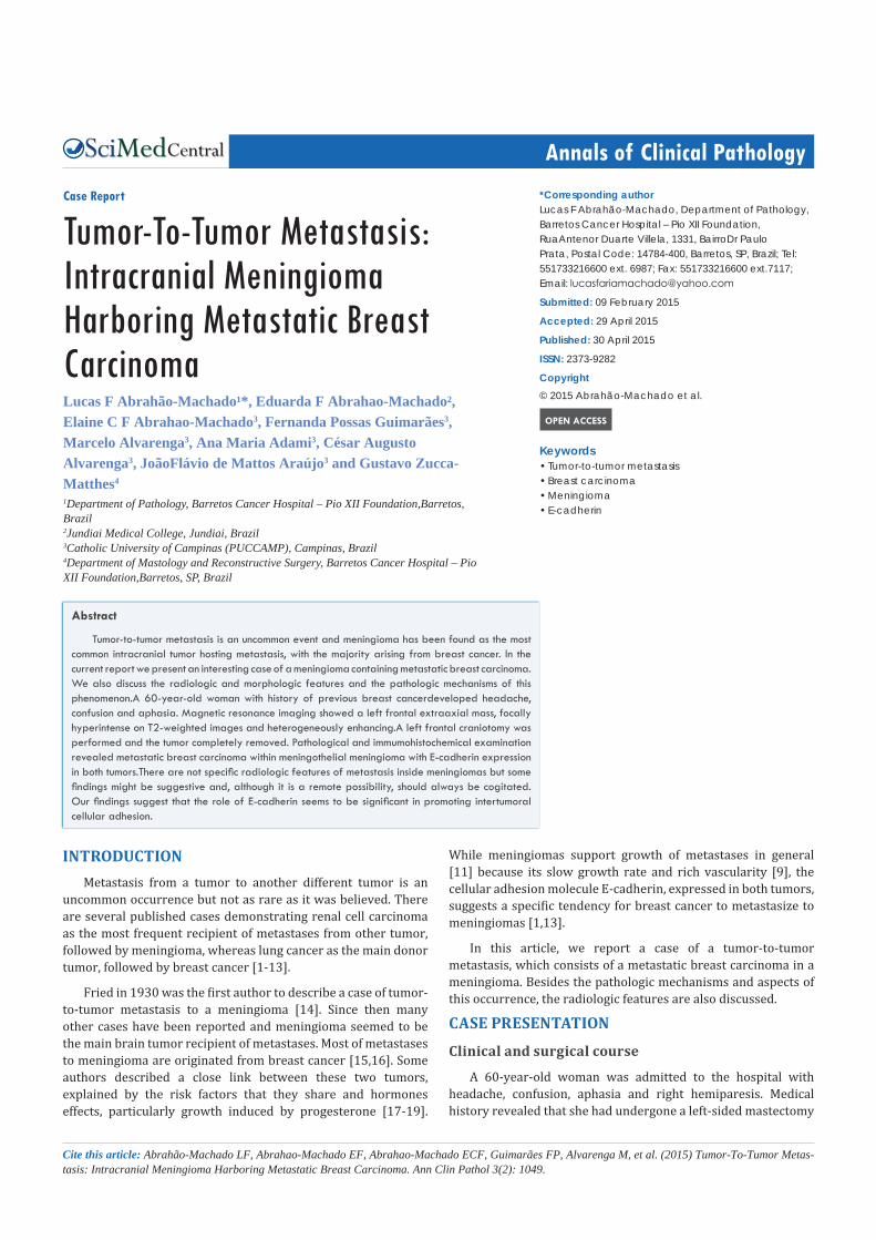
A magnetic resonance imaging (MRI) was performed and showed an extraaxialmass in the left frontal convexity, with a wide dural base, occupying the olfactory groove and causing midline shift (Figure 1A and B). An important perifocal edema was also observed (Figure 1C). The mass was predominantly isointense but focally hyperintense on T2-weighted images, showing an intense heterogeneous enhancement after contrast administration (Figure 1B).
The patient underwent a left frontal craniotomy and the tumor was totally excised. The surgical specimen consisted of a greyish irregular mass with aspect of a meningioma. Brain invasion was not found during the operation. She had an uneventful and favorable postoperative course and was transferred back to oncology division afterwards.
Histologic examination
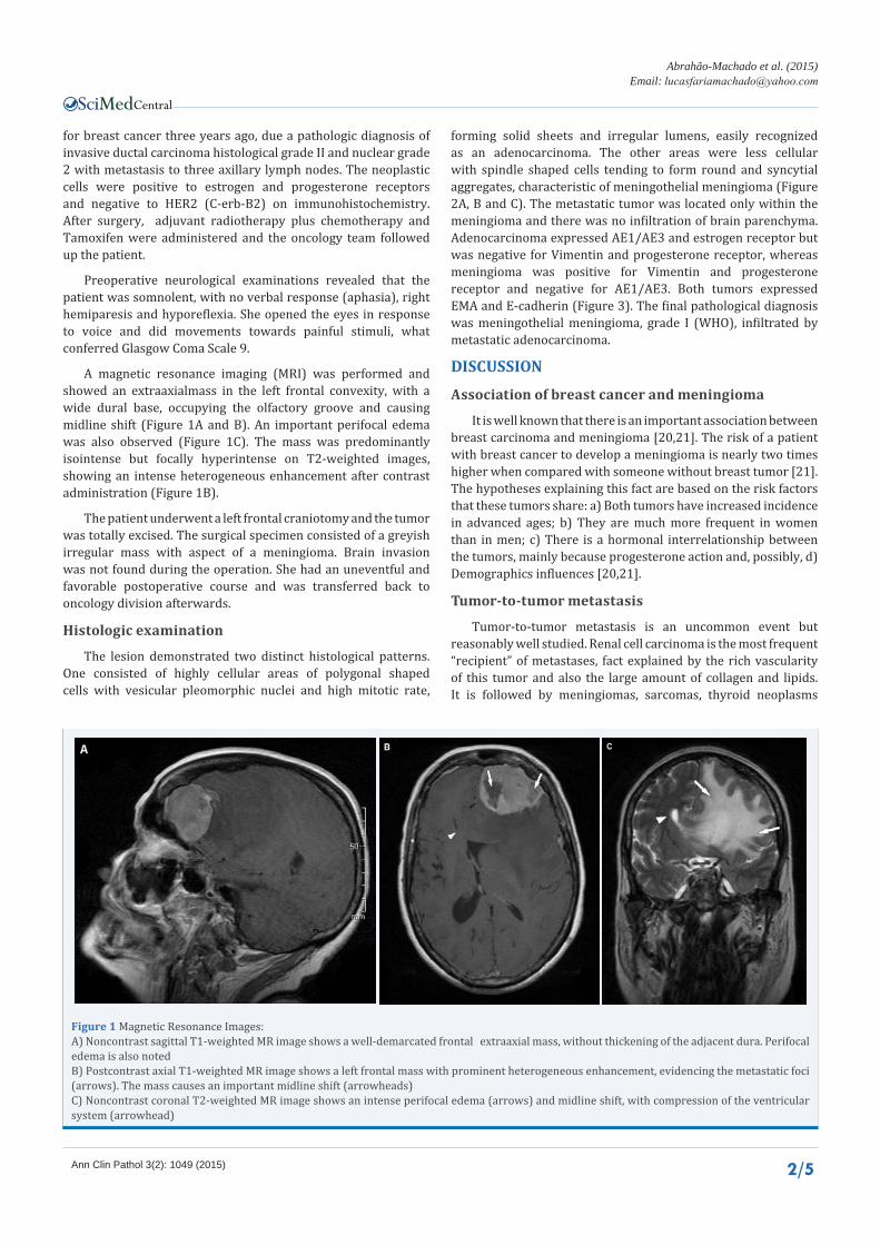
The lesion demonstrated two distinct histological patterns. One consisted of highly cellular areas of polygonal shaped cells with vesicular pleomorphic nuclei and high mitotic rate,
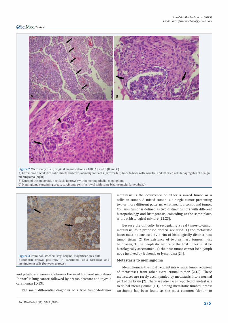
forming solid sheets and irregular lumens, easily recognized as an adenocarcinoma. The other areas were less cellular with spindle shaped cells tending to form round and syncytial aggregates, characteristic of meningothelial meningioma (Figure 2A, B and C). The metastatic tumor was located only within the meningioma and there was no infiltration of brain parenchyma. Adenocarcinoma expressed AE1/AE3 and estrogen receptor but was negative for Vimentin and progesterone receptor, whereas meningioma was positive for Vimentin and progesterone receptor and negative for AE1/AE3. Both tumors expressed EMA and E-cadherin (Figure 3). The final pathological diagnosis was meningothelial meningioma, grade I (WHO), infiltrated by metastatic adenocarcinoma.
DISCUSSION
Association of breast cancer and meningioma
It is well known that there is an important association between breast carcinoma and meningioma [20,21]. The risk of a patient with breast cancer to develop a meningioma is nearly two times higher when compared with someone without breast tumor [21]. The hypotheses explaining this fact are based on the risk factors that these tumors share: a) Both tumors have increased incidence in advanced ages; b) They are much more frequent in women than in men; c) There is a hormonal interrelationship between the tumors, mainly because progesterone action and, possibly, d) Demographics influences [20,21].
Tumor-to-tumor metastasis
Tumor-to-tumor metastasis is an uncommon event but reasonably well studied. Renal cell carcinoma is the most frequent “recipient” of metastases, fact explained by the rich vascularity of this tumor and also the large amount of collagen and lipids. It is followed by meningiomas, sarcomas, thyroid neoplasms
Figure 1 Magnetic Resonance Images:A) Noncontrast sagittal T1-weighted MR image shows a well-demarcated frontal extraaxial mass, without thickening of the adjacent dura. Perifocal edema is also notedB) Postcontrast axial T1-weighted MR image shows a left frontal mass with prominent heterogeneous enhancement, evidencing the metastatic foci (arrows). The mass causes an important midline shift (arrowheads)C) Noncontrast coronal T2-weighted MR image shows an intense perifocal edema (arrows) and midline shift, with compression of the ventricular system (arrowhead)
Central
Abrahão-Machado et al. (2015)Email:
Ann Clin Pathol 3(2): 1049 (2015) 3/5
Figure 2 Microscopy; H&E, original magnifications x 100 (A), x 400 (B and C):A) Carcinoma ductal with solid sheets and cords of malignant cells (arrows, left) back to back with syncitial and whorled cellular agregates of benign meningioma (right)B) Ducts of the metastatic neoplasia (arrows) within meningothelial meningiomaC) Meningioma containing breast carcinoma cells (arrows) with some bizarre nuclei (arrowhead).
Figure 3 Immunohistochemistry; original magnification x 400:E-cadherin shows positivity in carcinoma cells (arrows) and meningioma cells (between arrows)
metastasis is the occurrence of either a mixed tumor or a collision tumor. A mixed tumor is a single tumor presenting two or more different patterns, what means a compound tumor. Collision tumor is defined as two distinct tumors with different histopathology and histogenesis, coinciding at the same place, without histological mixture [22,23].
Because the difficulty in recognizing a real tumor-to-tumor metastasis, four proposed criteria are used: 1) the metastatic focus must be enclosed by a rim of histologically distinct host tumor tissue; 2) the existence of two primary tumors must be proven; 3) the neoplastic nature of the host tumor must be histologically ascertained; 4) the host tumor cannot be a lymph node involved by leukemia or lymphoma [24].
Metastasis to meningioma
Meningioma is the most frequent intracranial tumor recipient of metastases from other extra cranial tumor [2,15]. These metastases are rarely accompanied by metastasis into a normal part of the brain [2]. There are also cases reported of metastasis to spinal meningiomas [1,4]. Among metastatic tumors, breast carcinoma has been found as the most common “donor” to
and pituitary adenomas, whereas the most frequent metastases “donor” is lung cancer, followed by breast, prostate and thyroid carcinomas [1-13].
The main differential diagnosis of a true tumor-to-tumor
Central
Abrahão-Machado et al. (2015)Email:
Ann Clin Pathol 3(2): 1049 (2015) 4/5
meningiomas, followed by lung cancer [15]. Caroli et al, in 2006, reviewed 63 published cases of metastases to an intracranial tumor from any extracranial primary tumor and demonstrated that 53 cases (84%) had meningioma as the recipient tumor. In 31 cases (49%) the metastatic tumor was breast carcinoma and in 19 cases (28%) lung cancer [15]. These data suggest a possible affinity between these tumors, since when compared with brain metastases without meningioma, the percentage is inverted (nearly 20% are metastases from breast cancer and 50% from the respiratory tract) [25]. The most acceptable explanations for this phenomenon are the following: a) meningioma is a slow-growing tumor, what provides a long period of time for the metastasis to develop; b) meningioma is a richly vascularized tumor, what increases the chances of receiving hematogenic metastasis; c) there is an important association between these two tumors, showed by epidemiologic studies and also explained by hormones influences; d) the lack of immune response by meningioma favors metastasis development and installation; e) the amount of collagen and lipids within meningioma also benefits breast cancer metastatic cells [9,15]. Therefore, meningiomas would be a favorable environment for breast cancer metastasis, supporting the old but contemporary “soil and seed” Paget’s hypothesis [26]. Meningioma would be, in this case, a perfect fertile soil for the seeding of the carcinoma cells.
The cellular adhesion molecule, E-cadherin, has been considered an important factor of the increased affinity between these tumors, explained by the intercellular interaction among the cells [1,13]. This is considered to be in keeping with other speculationsof a specific tendency to breast carcinoma metastasizes to meningioma. Watanabe et al [13] and Aghi et al [1] showed that psammomatousmeningiomas, which are only 7% among all meningiomas and generally positive for E-cadherin, constitute 33% of the meningiomas recipients of metastases. Fibrous meningiomas, which are negative for E-cadherin, represent only 2% of the metastatic recipients. Furthermore, there is no case reported in the literature of metastasis to a malignant meningioma, which is E-cadherin negative [13]. Thus this molecule seems to be one of the factors that favor breast cancer metastasis to meningioma.
Radiologic features
Meningiomas can be diagnosed by computed tomography (CT) without contrast in 63% of the cases and, when contrast is administered, in 90% of the cases [27]. CT generally shows a juxtadural rounded and well circumscribed tumor with wide base and isodensity or hyperdensity. Non-enhancing areas may be viewed in benign meningiomas if they contain cystic or necrotic changes, but these areas also appear when malignant rather than benign changes are present [2]. Peritumoral edema and osseous changes such as hyperostosis and bone destruction can be present. MRI usually demonstrates isointensity on T1 and T2-weighted images, contributing to characterize the extraaxial location of this tumor.
There are few cases reported in the literature on MRI of metastatic carcinoma in meningioma and it appears that, at this time, there are no specific radiologic signs which would accurately differentiate benign meningiomas from meningiomas harboring metastatic cells [2,7,8,13]. Nevertheless foci of intense
enhancement in a background of moderate enhancement on CT or MRI studies after contrast administration have been reported in some patients with histologically proved metastasis within the meningioma [7,8,13, 28].
Proton spectroscopic MRI (sMRI) has been related as a useful diagnostic method for determining the malignant potential of meningiomas and for suggesting metastatic foci. It evaluates the aggressiveness of the tumor or even the presence of another malignant tumor by measuring intra-tumor metabolite levels [13]. Perfusion MRI (pMRI) assess the degree of tumor microvasculature and can show regional haemodynamic differences within a meningioma containing metastatic carcinoma, which suggests the presence of two different tumor tissues not apparent on a conventional MRI study [28]. Notwithstanding the precise diagnosis is still obtained only with microscopic examination.
CONCLUSION It is very difficult to identify a true tumor-to-tumor metastasis
before histological examination. Moreover only few publications describe MRI and CT features of metastasis in tumor and they are not conclusive. However foci of more intense enhancement on MRI, after contrast administration, inside the tumoral mass, which sometimes are simply foci of hemorrhage or necrosis, might be suspicious for metastasis inside a meningioma.
In the present case, MRI did not show any specific finding of tumor-to-tumor metastasis and, although the images showed an intense and heterogeneous enhancement after contrast administration, which could be suspect, the correct diagnosis was only obtained with histologic examination and immunohistochemistry.
Metastasis from a tumor to another different tumor is a phenomenon that must be always considered when a tumor shows two or more striking different histological patterns, especially when these patterns are mixed. Thus is necessary to recognize the criteria for tumor-to-tumor metastasis in order to exclude a collision tumor and a mixed tumor as well as search for the medical history of the patient. Our case fulfils the criteria and both tumors demonstrated immunohistochemical expression for E-cadherin, the cellular adhesion molecule, what suggest that meningioma cells and breast carcinoma cells have an intense affinity, explained by the intercellular interaction promoted by this molecule. Hence these tumors seem to be more intimately connected than merely associated by the risk factors in common.
REFERENCES1. Aghi M, Kiehl TR, Brisman JL. Breast adenocarcinoma metastatic to
epidural cervical spine meningioma: case report and review of the literature. J Neurooncol. 2005; 75: 149-155.
2. Baratelli GM, Ciccaglioni B, Dainese E, Arnaboldi L. Metastasis of breast carcinoma to intracranial meningioma. J Neurosurg Sci. 2004; 48: 71-73.
3. Chambers PW, Davis RL, Blanding JD, Buck FS. Metastases to primary intracranial meningiomas and neurilemomas.Archives of pathology & laboratory medicine. 1980; 104: 350-354.
4. Hockley AD. Metastatic carcinoma in a spinal meningioma. J Neurol Neurosurg Psychiatry. 1975; 38: 695-697.
Central
Abrahão-Machado et al. (2015)Email:
Ann Clin Pathol 3(2): 1049 (2015) 5/5
Abrahão-Machado LF, Abrahao-Machado EF, Abrahao-Machado ECF, Guimarães FP, Alvarenga M, et al. (2015) Tumor-To-Tumor Metastasis: Intracranial Menin-gioma Harboring Metastatic Breast Carcinoma. Ann Clin Pathol 3(2): 1049.
Cite this article
5. Inatomi H, Yamada Y, Okamura T. A case of prostate carcinoma metastasizing to renal cell carcinoma. International journal of urology: official journal of the Japanese Urological Association. 1996; 3: 155-157.
6. Kwak TI, Kim DS, Kim JJ, Yoon DK, Cho JH, Koh SK. Lung cancer metastasizing to ipsilateral renal cell carcinoma and the contralateral perirenal space. BJU Int. 1999; 83: 512-513.
7. Lee A, Wallace C, Rewcastle B, Sutherland G. Metastases to meningioma. AJNR Am J Neuroradiol. 1998; 19: 1120-1122.
8. Pamphlett R. Carcinoma metastasis to meningioma. J Neurol Neurosurg Psychiatry. 1984; 47: 561-563.
9. Petraki C, Vaslamatzis M, Argyrakos T, Petraki K, Strataki M, Alexopoulos C. Tumor to tumor metastasis: report of two cases and review of the literature. Int J Surg Pathol. 2003; 11: 127-135.
10. Ricketts R, Tamboli P, Czerniak B, Guo CC. Tumor-to-tumor metastasis: report of 2 cases of metastatic carcinoma to angiomyolipoma of the kidney. Arch Pathol Lab Med. 2008; 132: 1016-1020.
11. Schmitt HP. Metastases of malignant neoplasms to intracranial tumours: the “tumour-in-a-tumour” phenomenon. Virchows Arch A Pathol Anat Histopathol. 1984; 405: 155-160.
12. Sella A, Ro JY. Renal cell cancer: best recipient of tumor-to-tumor metastasis. Urology. 1987; 30: 35-38.
13. Watanabe T, Fujisawa H, Hasegawa M, Arakawa Y, Yamashita J, Ueda F. Metastasis of breast cancer to intracranial meningioma: case report. Am J Clin Oncol. 2002; 25: 414-417.
14. Fried BM. Metastatic Inoculation of a Meningioma by Cancer Cells from a Bronchiogenic Carcinoma. Am J Pathol. 1930; 6: 47-52.
15. Caroli E, Salvati M, Giangaspero F, Ferrante L, Santoro A. Intrameningioma metastasis as first clinical manifestation of occult primary breast carcinoma. Neurosurgical review. 2006; 29: 49-54.
16. Lodrini S, Savoiardo M. Metastases of carcinoma to intracranial meningioma: report of two cases and review of the literature. Cancer. 1981; 48: 2668-2673.
17. BICKERSTAFF ER, SMALL JM, GUEST IA. The relapsing course of
certain meningiomas in relation to pregnancy and menstruation. J Neurol Neurosurg Psychiatry. 1958; 21: 89-91.
18. Higashi H, Fukutomi T, Watanabe T, Adachi I, Narabayashi M, Shibui S, et al. Seven cases of breast cancer recurrence limited to the central nervous system without other visceral metastases. Breast cancer. 2000; 7:153-156.
19. Rubinstein AB, Schein M, Reichenthal E. The association of carcinoma of the breast with meningioma. Surg Gynecol Obstet. 1989; 169: 334-336.
20. Custer BS, Koepsell TD, Mueller BA. The association between breast carcinoma and meningioma in women. Cancer. 2002; 94: 1626-1635.
21. Kubo M, Fukutomi T, Akashi-Tanaka S, Hasegawa T. Association of breast cancer with meningioma: report of a case and review of the literature. Jpn J Clin Oncol. 2001; 31: 510-513.
22. Anlyan FH, Heinzen BR, Carras R. Metastasis of tumor to second different tumor: collision tumors. JAMA. 1970; 212: 2124.
23. Inoshita T, Laurain AR, Youngberg GA, Musil G. Metastasis of bronchogenic carcinoma to the skin involved by melanoma. Arch Pathol Lab Med. 1984; 108: 595-598.
24. Campbell LV Jr, Gilbert E, Chamberlain CR Jr, Watne AL. Metastases of cancer to cancer. Cancer. 1968; 22: 635-643.
25. Suki D. The epidemiology of brain metastases. In: Intracranial metastases; current management strategies. Sawaya R, editor. Malden, Mass., USA: Blackwell Futura; 2004.
26. Paget S. The distribution of secondary growths in cancer of the breast. 1889. Cancer Metastasis Rev. 1989; 8: 98-101.
27. Perry A. Meningiomas. In: Practical surgical neuropathology: a diagnostic approach Perry A, Brat DJ, editors. Philadelphia, PA: Churchill Livingstone/Elsevier; 2010.
28. Benedetto N, Perrini P, Scollato A, Buccoliero AM, Di Lorenzo N. Intracranial meningioma containing metastatic colon carcinoma. Acta Neurochir (Wien). 2007; 149: 799-803.