Embed Size (px)
Citation preview

Published online 4 June 2008 Nucleic Acids Research, 2008, Vol. 36, No. 12 4067–4078doi:10.1093/nar/gkn356
TSA downregulates Wilms tumor gene 1 (Wt1)expression at multiple levelsMohammad Shahidul Makki1, Thorsten Heinzel2 and Christoph Englert1,3,*
1Leibniz Institute for Age Research – Fritz Lipmann Institute, Beutenbergstrasse 11, 07745,2Institute of Biochemistry and Biophysics, Friedrich Schiller University of Jena, Philosophenweg 12, 07743 and3Friedrich Schiller University of Jena, Furstengraben 1, 07743 Jena, Germany
Received January 29, 2008; Revised May 19, 2008; Accepted May 20, 2008
ABSTRACT
The Wilms tumor gene WT1 encodes a zinc-fingertranscription factor that is inactivated in a subsetof pediatric kidney cancers. During embryogenesis,WT1 is expressed in a time- and tissue-specificmanner in various organs including gonads andkidney but also in the hematopoietic system.Although widely regarded as a tumor suppressorgene, wild-type WT1 is overexpressed in a varietyof hematologic malignancies, most notably in acutelymphoblastic leukemia as well as myelodysplasticsyndromes. Reduction of WT1 expression levelsleads to decrease of proliferation and apoptosis ofleukemic cells, suggesting that in certain contextsWT1 might act as an oncogene. We show here thathistone deacetylase inhibitors like Trichostatin A(TSA) can promptly and dramatically downregulateWt1 expression levels in different cell lines. Thiseffect was mostly due to the cessation of transcrip-tion and was mediated by sequences located inintron 3 of Wt1. In addition, TSA also causedenhanced degradation of the Wt1 protein by the pro-teasome. This was at least in part due to inductionof the ubiquitin-conjugating enzyme UBCH8. Thus,downregulation of Wt1 expression might contributeto the beneficial effects of histone deacetylase inhi-bitors that are currently used in clinical trials ascancer therapeutics.
INTRODUCTION
The Wilms tumor gene WT1 was originally identified as atumor suppressor gene lost in 10–15% of Wilms tumors(1,2) and is a member of the GC- rich TATA-less andCCAAT-less class of RNA pol II genes (3). The WT1transcript encompasses 3.5 kb and encodes a four zinc-finger containing protein with an essential role in the
development of several organs, most notably the kidney(4–6). More than 20 different WT1 gene products withmolecular masses of 52–65 kDa are generated by a com-bination of alternative RNA splicing, the usage of differ-ent start codons and RNA editing (7).Expression of the wild-type WT1 gene has been found
in most cases of acute myelocytic leukemia (AML), acutelymphocytic leukemia (ALL), chronic myelocytic leuke-mia (CML) and myelodysplastic syndrome (MDS) athigher levels than those in normal bone marrow or per-ipheral blood (8–10). WT1 is used as a prognostic factorand marker for minimal residual disease in cases of acuteleukemia (9,11). Furthermore, various types of solidtumors, including lung, breast, thyroid, esophageal andcolorectal cancers express wild-type WT1 at higher levelscompared to those in corresponding normal tissues (12).In several studies, the role of Wt1 in cell proliferation,
differentiation and leukemogenesis has been analyzed.In the chronic myeloid leukemia cell line K562 as well asin primary leukemic cells from human patients, WT1antisense oligomers inhibited growth via reduction ofWT1 protein levels (13). In the same cell line, ribozyme-mediated downregulation of WT1 led to inhibition ofcell proliferation and apoptosis (14). Similarly, siRNA-mediated reduction of WT1 mRNA levels in various leu-kemic cell lines including those from AML and CMLpatients inhibited proliferation and induced apoptosis(15). Taken together, all these studies indicate that Wt1may be necessary for leukemic or solid tumor growth sur-vival and that under certain circumstances Wt1 could actas an oncogene (12). This is corroborated by the recentobservation in mice that the chimeric oncoprotein AML1-ETO exerts its leukemogenic function in cooperation withWt1 expression (16).Conversely, removal of WT1 may have an anticancer
effect. That this is indeed the case was recently demon-strated by vaccination of patients with AML, MDS aswell as lung or breast cancer with a WT1 peptide. WT1vaccination led to an increase in WT1-specific cytotoxic Tlymphocytes and subsequent cancer regression without
*To whom correspondence should be addressed. Tel: +49 3641 656042; Fax: +49 3641 656040; Email: [email protected]
� 2008 The Author(s)
This is an Open Access article distributed under the terms of the Creative Commons Attribution Non-Commercial License (http://creativecommons.org/licenses/
by-nc/2.0/uk/) which permits unrestricted non-commercial use, distribution, and reproduction in any medium, provided the original work is properly cited.
Downloaded from https://academic.oup.com/nar/article-abstract/36/12/4067/1139248by gueston 13 February 2018
damage to other normal tissues (17). Thus, in partic-ular cancers WT1 could be a therapeutic target and down-regulation of WT1 might be a promising anticancerstrategy.For transcription to begin in eukaryotes, concerted
actions of multiple protein factors are required. Themajor hurdle in activating transcription in vivo is thehighly compacted nature of chromatin, which preventsaccess of the transcription machinery to the DNA tem-plate. Posttranslational modifications of histones such asacetylation, phosphorylation, methylation, ubiquitination,ADP-ribosylation, sumoylation and biotinylation areassumed to be important factors that control chromatinaccessibility and subsequent gene transcription. It is thisassociation between histone modification and the activitystate of the chromatin for which the expression ‘histonecode’ has been coined (18).The best understood modification is acetylation of
core histones, which is carried out by histone acetyl trans-ferases (HATs); the steady state levels of acetylationare maintained by the opposing activities of HATs andhistone deacetylases (HDACs) (19). So far at least 18HDACs have been identified in humans and have beengrouped into four different classes (20). Class I members(HDACs 1–3 and 8) are most closely related to theSaccharomyces cerevisiae transcriptional regulator RPD3.Class II HDACs (4–7, 9 and 10) display similarity to yeastHDA1; class III comprises the NAD+-dependent sirtuindeacetylases SIRT 1–7 and class IV consists of proteinsrelated to the recently identified human HDAC11, whichshares features of both class I and II. HDACs associatewith a number of oncoproteins and tumor suppressorsand in case of their aberrant activation or inactivationthe concomitant HDAC activity can lead to undue changesin gene expression and in turn to diseases, e.g. cancer (21).This observation has stimulated the identification andcharacterization of HDAC inhibitors as means to counter-act disease-associated aberrant gene expression.Trichostatin A (TSA), a Streptomyces product was origi-
nally identified as a fungicidic antibiotic. It inhibits allclass I and II HDACs and has potent antiproliferativeproperties in cancer cells (22,23). Differential display anal-ysis revealed that expression of only 2–5% of genes inTSA-treated cells is significantly altered (24). The basisfor this gene selectivity is not yet understood, but it sug-gests that only a highly restricted set of genes is sensitive tochanges in histone acetylation (25,26). Another HDACinhibitor, suberoylanilide hydroxamic acid (SAHA) inhi-bits the growth of prostate cancer cells in culture as well asin a xenograft model (27). Moreover, TSA also inhibitshypoxia-induced angiogenesis in the Lewis lung carci-noma model (28). Thus, histone deacetylases are promis-ing drug targets and clinical trials using first generationHDAC inhibitors as anticancer reagents are currentlyunder way (29).Here, we report the effect of HDAC inhibitors like TSA
on Wt1 expression. TSA led to a drastic downregulationof Wt1 mRNA levels in different human and mouse celllines. This effect was mostly due to a reduction of Wt1transcription. In addition, TSA induced degradation ofWt1 protein via the proteasome. This effect was at least
in part mediated by the ubiquitin-conjugating enzymeUBCH8.
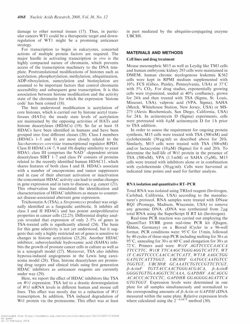
MATERIALS AND METHODS
Cell lines and drug treatment
Mouse mesonephric M15 as well as Leydig like TM3 cellsand human embryonic kidney 293 cells were maintained inDMEM, human chronic myelogenous leukemia K562cells were kept in RPMI medium supplemented with10% FCS (Gibco, Paisley, Pennsylvania, USA) at 378Cwith 5% CO2. For drug studies, exponentially growingcells were trypsinized, seeded at 40% confluency, grownfor 24 h and then treated with TSA (Sigma, St. Louis,Missouri, USA), valproic acid (VPA, Sigma), SAHA(Merck, Whitehouse Station, New Jersey, USA) or MS-275 (Alexis Biochemicals, San Diego, California, USA)for 24 h. In actinomycin D (Sigma) experiments, cellswere pretreated with 4 mM actinomycin D for 1 h priorto TSA addition.
In order to assess the requirement for ongoing proteinsynthesis, M15 cells were treated with TSA (500 nM) andcycloheximide (50mg/ml) as described above for 20 h.Similarly, M15 cells were treated with TSA (500 nM)and/or lactacystine (10 mM) (Sigma) for 6 and 20 h. Todetermine the half-life of Wt1 protein in the presence ofTSA (500 nM), VPA (1.5mM) or SAHA (5mM), M15cells were treated with inhibitors alone or in combinationwith cycloheximide (50mg/ml). Cells were harvested atindicated time points and used for further analysis.
RNA isolation and quantitative RT–PCR
Total RNA was isolated using TRIzol reagent (Invitrogen,Carlsbad, California, USA) according to the manufac-turer’s protocol. RNA samples were treated with DNaseRQ1 (Promega, Madison, Wisconsin, USA) to removeany genomic DNA. cDNA was synthesized from 2 mgtotal RNA using the SuperScript II RT kit (Invitrogen).
Real-time PCR reaction was carried out employing theQuantiTect SYBR green real-time PCR kit (Qiagen,Hilden, Germany) on a Biorad iCycler in a 96-wellformat. PCR conditions were: 958C for 15min, followedby 40 cycles of three-step PCR including melting for 30 s at958C, annealing for 30 s at 608C and elongation for 30 s at728C. Primers used were: Wt1F AGTTCCCCAACCATTCCTTC, Wt1R TTCAAGCTGGGAGGTCATTT, WT1F CAGTTCCCCAACCACTCATT, WT1R AAGCTGGGATGTCATTTGGT, UBCH8F GATGCCAATGTCCTGGTGT, UBCH8R GCAAATCTGTCCGTTCTCGT,�-ActinF TGTTACCAACTGGGACGACA, �-ActinRGGGGTGTTGAAGGTCTCAAA, GAPDHF AACAGCGACACCCACTCCTC, GAPDHR GGAGGGGAGATTCAGTGTGGT. Expression levels were determined in oneplate for all samples simultaneously and normalized tothe corresponding amounts of �-Actin or GAPDH cDNAmeasured within the same plate. Relative expression levelswhere calculated using the 2���CT method (30).
4068 Nucleic Acids Research, 2008, Vol. 36, No. 12
Downloaded from https://academic.oup.com/nar/article-abstract/36/12/4067/1139248by gueston 13 February 2018
Chromatin immunoprecipitation (ChIP) assay
A total of 2� 106 M15 cells (treated with 500 nM TSA orethanol for 24 h) were cross-linked with 1% formaldehydefor 10min at 378C. Cells were resuspended in 250 mlSDA lysis buffer (1% SDS, 50mM Tris, pH 8.1, 10mMEDTA, supplemented with protease inhibitors) followedby sonication to an average DNA length of 200–1000 bp.The sample was cleared by centrifugation and diluted10-fold in ChIP dilution buffer (0.01% SDS, 1.1%Triton X-100, 1.2mM EDTA, 16.7mM Tris, pH 8.1 and167mM NaCl, supplemented with protease inhibitors).Aliquots of the diluted samples were kept as input controls.Anti-acetylated histone H4 (AcH4) antibody (Upstate,Charlottesville, Virginia, USA) was added to each tube,which were rotated at 48C overnight. A total of 30 ml ofprotein A beads was then added to each of the tubes, whichwere further rotated for 2 h at 48C. The beads were washedonce with 1ml of the following buffers: (i) low salt immunecomplex wash buffer (0.1% SDS, 1% Triton X-100, 2mMEDTA, 20mM Tris, pH 8.1 and 150mM NaCl); (ii) highsalt immune complex wash buffer (0.1% SDS, 1% TritonX-100, 2mM EDTA, 20mM Tris, pH 8.1 and 500mMNaCl); (iii) LiCl immune complex wash buffer (0.25MLiCl, 1% NP-40, 1% deoxycholic acid, 1mM EDTA and10mM Tris, pH 8.1) and (iv) TE buffer (10mM Tris,pH 8.1, 1mM EDTA). The beads were eluted with 250 mlof elution buffer (1% SDS, 0.1MNaHCO3) and incubatedovernight at 658C to reverse cross-links. The eluates weretreated with proteinase K and extracted with phenol/chloroform followed by ethanol precipitation. The DNAwas dissolved in TE buffer and analyzed by PCR.The following PCR primers were used; promoter:CCTCCTGGCTCCTCCTCTT (sense) and CGCTGCCTTGAACTCCTTAC (antisense); intron (688–847): AGATTGGGTGGGGGAATG (sense) and CCAAGGATGGGAGAGAAAGA (antisense); intron (2015–2212): CACTTGCATCTTTGGTGCTT (sense) and TTGCTCCCATTTTCTCTGCT (antisense); intron (5478–5614): TAGCTTCGGAGTCCATTTCC (sense) and GTGTCTGCGTCCCTCACC (antisense); intron (8658–8758): TGTAATGACCCTGTCACGAA (sense) and GTTACACGGGCTGGACAACT (antisense). PCR products were amplified for 32cycles.
Immunoblot analysis
Cells were harvested, washed with PBS and lyzed in ice-cold protein lysis buffer (50mM Tris pH 7.6, 400mMNaCl, 0.5% NP-40, 1mM PMSF and 1�protease inhibi-tor cocktail (Roche, Mannheim, Germany)). Lysates wereclarified by centrifugation (15000g for 15min at 48C) andthe supernatant was analyzed immediately or stored at�808C. Equivalent amounts of protein (25–40 mg) wereresolved by 10–12% SDS–PAGE and transferred onto aPVDF membrane (Hybond P, Amersham Biosciences,Piscataway, New Jersey, USA). Membranes were incu-bated with anti-Wt1 (1:1000, Santa Cruz, California,USA) anti-b-Actin (1:5000, Sigma), anti-acetylated his-tone H4 (1:5000, Upstate) anti-UBE2L6 (1:1000,Abgent, San Diego, California, USA), anti-HA (1:1000,Cell Signaling, Danvers, Massachusetts, USA) or anti-V5
(1:10000, Invitrogen) primary antibodies overnight at 48C.Blots were then incubated with horseradish peroxidase-conjugated secondary antibody. Bands were visualizedby enhanced chemiluminescence (Amersham Pharmacia,Uppsala, Sweden) followed by exposure to autoradiogra-phy film (Amersham Pharmacia). To detect acetylated his-tone H4, cells were lyzed in protein loading buffer(0.313M Tris pH 6.8, 10% SDS, 40% glycerol, 0.05%bromophenol blue and 2% b-ME) and sonicated threetimes for 20 s each and separated on 15% SDS–PAGE.Quantification of immunoreactive bands on films was per-formed using the Bio-Rad Quantity One Software pro-gram. Values for Wt1 were divided by those for b-Actinand are depicted as percent relative to the control set as100%.In vivo ubiquitination assay was performed as described
(31). Ubiquitinated Wt1 was detected by western blottingusing an anti-HA antibody.
Nuclear run-off assay
Nuclear run-off assay was performed as described (32).Radiolabeled RNA was hybridized with a Hybond-Nnylon membrane (Amersham) containing immobilizedfragments of Gapdh (1 mg of a 558 bp fragment) and Wt1(2mg of a EcoRI-ApaI fragment). Hybridization was per-formed overnight at 658C with 1� 106 c.p.m. labeled RNAper sample using 2ml of the Rapid-hyb buffer(Amersham) according to the manufacturer’s recommen-dations. To quantitate the signals, radioactive spots wereexcised from the membranes and measured in a scintilla-tion counter.
Constructs, transfection and reporter gene assay
For the Wt1 promoter construct, a 3318 bp Wt1 upstreamfragment was amplified by PCR with primersmWt1F2KpnI aCGTGGTACCGCCAGTGTCTCTTTTCTTCCA and P1XhoI ACGTCTCGAGCGCTG CCTTGAACTCCTTACC containing a KpnI and an XhoI restric-tion site, respectively, using a PAC clone harboring theentire Wt1 genomic locus (33). PCR was performedas mentioned above except elongation was carried outfor 3min at 728C using Triple-Master enzyme mix(Eppendorf, Hamburg, Germany). PCR products weregel purified and ligated into the pGEMT-Easy vector(Promega). The promoter fragment was removed fromthe vector by XhoI/KpnI digestion and cloned intopGL3-Basic. Cloning of Wt1 intron 1 and intron 3 intothe pGEMT-Easy vector has been described (33). Afterexcision with NotI and subsequent fill-in reaction byKlenow enzyme, introns 1 and 3 were cloned into aSmaI-linearized and dephosphorylated pGL3-Promotervector to create pGL3Int1 and pGL3Int3, respectively.For short-term stable transfections, 2� 106 M15 cells
were transfected with 10 mg of reporter plasmid or therespective empty vector (pGL3-Basic or pGL3-Promoter)as control, along with 1 mg of pBABE-puro usingSuperFect (Qiagen) according to the manufacturer’srecommendations. Selection was started after 24 h using1 mg puromycin/ml. At day 5 after transfection, 1� 105
cells of the selected mass populations were seeded into
Nucleic Acids Research, 2008, Vol. 36, No. 12 4069
Downloaded from https://academic.oup.com/nar/article-abstract/36/12/4067/1139248by gueston 13 February 2018

24-well culture dishes and grown overnight followed bytreatment with the indicated concentrations of TSA forfurther 24 h. Subsequently, reporter gene activity wasassessed using the Luciferase Assay System (Promega).Values were normalized to protein content in each sampleand divided by those obtained for the control vector con-taining samples. All transfection results are presented as theaverage from at least three independent experiments.For in vivo ubiquitination assay, 293 cells were trans-
fected with 5 mg Wt1 expression construct together with anequal amount of a His-ubiquitin expression plasmid. 20 hafter transfection cells were treated with lactacystine foradditional 20 h.For UBCH8 overexpression studies, 293 cells were
transfected in 6 cm plates with 0.5, 1 and 2 mg of a plasmidencoding UBCH8 harboring a V5 epitope tag in absenceor presence of 1 mg Wt1 expression construct. ThepcDNA3.1 plasmid transfected cells were used as a con-trol. Cells were harvested 24 h posttransfection and Wt1levels were determined by western blot.
siRNA transfection
For UBCH8 knockdown experiments 293 cells wereseeded at 1� 105 cells per well in a 6-well plate in 2mlmedium 1 day prior to transfection. siRNA (ON-TARGET plus SMARTpool duplex, Dharmacon,Lafayette, Colorado, USA) directed against UBCH8 wasdiluted to 100 ml in serum-free media to achieve a finalconcentration of 50 nM, and 10 ml siFect TransfectionReagent (Qiagen) was added. Samples were vortexed,incubated at room temperature for 10min and thenadded drop-wise to the cells. Twenty-four hours post-transfection media was replaced and cells were grownadditionally for 36 h, followed by incubation in presenceof 500 nM TSA for 20 h. Transfection without siRNA andTSA was used as a control.
RESULTS
Differential effects of HDAC inhibitors onWt1 expression
While we were analyzing epigenetic changes at the murineWt1 locus in Wt1 expressing and Wt1 nonexpressing cells,we observed a dramatic downregulation of Wt1 mRNAlevels following treatment of M15 cells with TSA. Thiseffect was dose- and time-dependent (Figure 1A). TSAhad no effect on �-Actin expression suggesting that theeffect on Wt1 expression was specific and not due to celldeath. We then wanted to quantify the effect and usedreal-time PCR. Within 24 h TSA at 100 nM concentrationcaused Wt1 downregulation to about 40% and at 500 nMto < 5% of the original levels (Figure 1A, left). With500 nM TSA maximal reduction was reached after 12 h(Figure 1A, right).We further analyzed whether this effect was specific for
M15 cells, which express high levels of endogenous Wt1(Figure 1E) or could be observed in other cell types. Wetherefore treated the two human cell lines 293 and K562 aswell as murine TM3 cells with TSA and could observesignificant reduction of Wt1 mRNA levels in all celllines tested (Figure 1B and data not shown). However,
TSA concentrations required as well as the magnitude ofthe effect varied between the different cell lines. In order toassess a possible effect of TSA on cell cycle progression,we have analyzed cell cycle distribution of TSA-treatedcells by flow cytometric analysis. While M15 cells accumu-lated in G2 phase, K562 cells showed a G1 arrest(Supplementary Figure 1A). Thus, the effect of TSA oncell cycle did not correlate with its effect on Wt1 expres-sion. Of note, analysis of cell viability indicated a toxiceffect of TSA only at a concentration of 1 mM(Supplementary Figure 1B) and confirmed that the effectson Wt1 expression were not due to cell death.
To test whether TSA was unique among HDAC inhibi-tors in that it downregulated Wt1, we included SAHA,MS-275 and VPA in our experiments. M15 cells weretreated for 24 h with various concentrations of the threerespective substances and Wt1 mRNA levels were mea-sured by quantitative real-time PCR. In SAHA-treatedcells, Wt1 mRNA levels were strongly reduced withincreasing drug concentrations (Figure 1C, left), albeitmuch higher doses than with TSA were required. In con-trast, VPA had no significant effect on Wt1 expression(Figure 1C, right). Acetylation of histone H4 was usedas a control for VPA activity. When we used the HDACinhibitor MS-275, significant reduction in Wt1 expressionlevels could be observed (Figure 1D, left); this effect waseven more dramatic in K562 cells (Figure 1D, right).These data suggest that the loss of Wt1 mRNA amountis independent of the levels of Wt1 expression (Figure 1E);moreover, it is a phenomenon shared by many but not allHDAC inhibitors. TSA and SAHA inhibit both class Iand class II HDACs, whereas VPA and MS-275 havebeen reported to be class I-selective (34,35). Thus, thedifferential effect of TSA/SAHA/MS-275 and VPA onWt1 gene regulation cannot be attributed to a specificclass of HDACs.
TSA downregulatesWt1 at the transcriptional level
Downregulation of Wt1 mRNA upon TSA treatmentprompted us to investigate the responsible mechanism.One possibility could be that TSA may affect the stabilityof Wt1 mRNA. Alternatively, TSA could directly influ-ence Wt1 transcription. To investigate the first possibility,M15 cells were treated with the transcriptional inhibitoractinomycin D 1h before the addition of TSA. Subse-quently, steady-state Wt1 mRNA was measured by real-time PCR. Most of the repressive effect of TSA on Wt1mRNA levels could be blocked by pretreatment with acti-nomycin D (Figure 2A). This suggested that TSA does notsignificantly alter Wt1 mRNA stability. In order to exam-ine whether TSA would act at the level of transcription,we performed a nuclear run-off assay using untreated andTSA-treated M15 cell nuclei. Already, after 3 h, TSAtreatment profoundly suppressed synthesis of new Wt1transcripts (Figure 2B and C). Longer TSA treatments(12 and 24 h) resulted in further loss in Wt1 mRNA synth-esis. These results suggest that in response to TSA treat-ment, Wt1 is downregulated at the transcriptional leveland that TSA has no significant effect on Wt1 mRNAstability.
4070 Nucleic Acids Research, 2008, Vol. 36, No. 12
Downloaded from https://academic.oup.com/nar/article-abstract/36/12/4067/1139248by gueston 13 February 2018

A
C
0
0.5
1
1.5
2
2.5
Control 0.5 mM 1.5 mM 5 mM
Rel
ativ
e W
t1 e
xpre
ssio
n
β-Actin
C 0.5 5.0 mM
Ac-H4VPA (M15)
0
0.2
0.4
0.6
0.8
1
1.2
Control 100 nM 500 nM 1 µM Control 3 h 6 h 12 h 24 h
Rel
ativ
e W
t1
TSA (M15)
* *
C 0.1 1.00.5 mM
b-Actin
Wt1
Control 100 nM 500 nM 1 µM 5 µM
*
**
0
0.2
0.4
0.6
0.8
1
1.2
Rel
ativ
e W
t1 e
xpre
ssio
n
TSA (M15)
*
* ** **
C 3 6 12 24 h
ß-Actin
Wt1
0
0.2
0.4
0.6
0.8
1
1.2
Control 0.5 µM 2.5 µM 5 µM 10 µM
**
*
*
0
0.2
0.4
0.6
0.8
1
1.2
Control 0.5 µM 2.5 µM 5 µM 10 µM
Rel
ativ
e W
T1
expr
essi
on
*
** **
E
MS-275 (M15) MS-275 (K562)
0
0.2
0.4
0.6
0.8
1
1.2
Control 100 nM 500 nM 1 µM
Rel
ativ
e W
T1
expr
essi
on
0
0.2
0.4
0.6
0.8
1
1.2
Rel
ativ
e W
T1
expr
essi
on
****
TSA (293)
0
1
2
3
4
5
6
7
293 K562 M15
β-Actin
M15 K562 293
WT1
TSA (K562)B
0
0.2
0.4
0.6
0.8
1
1.2
Control 1 µM 2.5 µM 5 µM 10 µM
Rel
ativ
e W
t1 e
xpre
ssio
nR
elat
ive
Wt1
exp
ress
ion
Rel
ativ
e W
t1 e
xpre
ssio
n
**** **
SAHA (M15)
D
Figure 1. HDAC inhibitors affect Wt1 expression. (A) Quantitative real-time PCR of Wt1 expression in M15 cells that had been treated for 24 h withTSA at different concentrations (left) or with 500 nM TSA for different time points (right). (B) Analysis as in (A) employing the human cell lines 293(left) and K562 (right). (C) Analysis as in (A) using the HDAC inhibitors suberoylanilide hydroxamic acid (SAHA, left) and valproic acid (VPA,right) and M15 cells. (D) Analysis as in (A) using the HDAC inhibitor MS-275 in M15 (left) and K562 cells (right). (E) Relative Wt1 expression indifferent cell lines was measured by quantitative real-time PCR. HDAC inhibitor treatment was done for 24 h when not specified otherwise. Insets in(A) show gel pictures of the respective RT–PCR products; the inset in (C) shows a western blot performed with an anti-acetylated histone H4antibody to confirm VPA activity. The inset in (E) shows WT1 protein levels. Bars represent �SD from three independent experiments (�P< 0.05;��P< 0.005; paired Student’s t-test).
Nucleic Acids Research, 2008, Vol. 36, No. 12 4071
Downloaded from https://academic.oup.com/nar/article-abstract/36/12/4067/1139248by gueston 13 February 2018
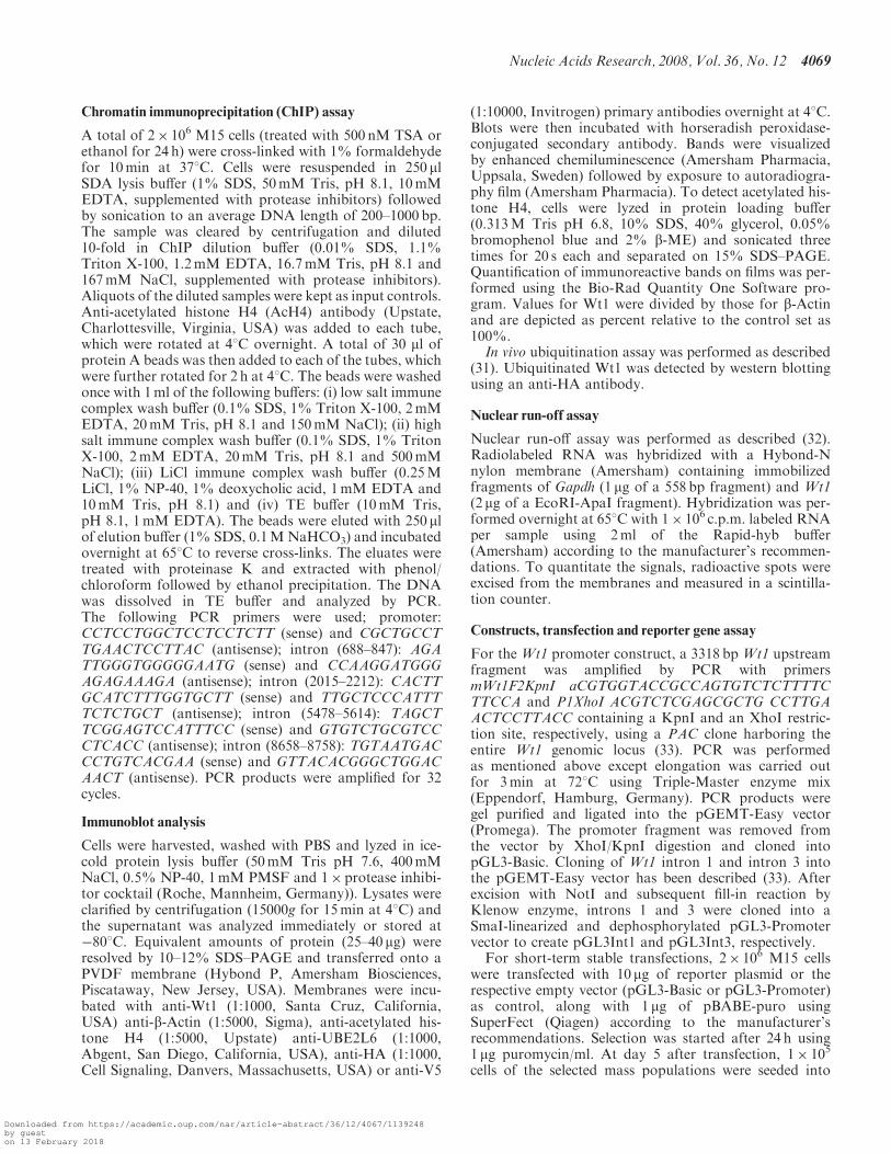
We then asked, whether downregulation of Wt1 expres-sion by TSA required new protein synthesis. We thereforepretreated M15 cells with cycloheximide to inhibit newprotein synthesis and then challenged the cells with TSAfor 20 h. As shown in Figure 2D, such treatment could notprevent TSA-dependent Wt1 mRNA downregulation.This result supports the notion that new protein synthesisis not needed for TSA-dependent downregulation of Wt1expression.To identify the TSA responsive region in theWt1 gene, a
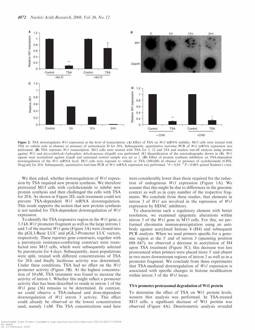
3.3 kbWt1 promoter fragment as well as the large introns 1and 3 of the murineWt1 gene (Figure 3A) were cloned intothe pGL3-Basic LUC and pGL3-Promoter LUC vectors,respectively. These reporter gene constructs, together witha puromycin resistance-conferring construct were trans-fected into M15 cells, which were subsequently selectedby puromycin for 4 days. Subsequently, mass populationswere split, treated with different concentrations of TSAfor 20 h and finally luciferase activity was determined.Under these conditions, TSA had no effect on the Wt1promoter activity (Figure 3B). At the highest concentra-tion of 10 nM, TSA treatment was found to increase theactivity of intron 1. Whether this might reflect a promoteractivity that has been described to reside in intron 1 of theWt1 gene (36) remains to be determined. In contrast,we could observe a TSA-induced and dose-dependentdownregulation of Wt1 intron 3 activity. This effectcould already be observed at the lowest concentrationused, namely 1 nM. The TSA concentrations used here
were considerably lower than those required for the reduc-tion of endogenous Wt1 expression (Figure 1A). Weassume that this might be due to differences in the genomiccontext as well as in copy number of the respective frag-ments. We conclude from these studies, that elements inintron 3 of Wt1 are involved in the repression of Wt1expression by HDAC inhibitors.
To characterize such a regulatory element with betterresolution, we examined epigenetic alterations withinintron 3 of the Wt1 gene in M15 cells. For this, we per-formed chromatin immunoprecipitation using an anti-body against acetylated histone 4 (H4) and subsequentPCR analysis. When we used primers specific for a geno-mic region at the 50 end of intron 3 (spanning position688–847), we observed a decrease in acetylation of H4upon TSA treatment (Figure 3C); this decrease was lesspronounced when primers were placed more 30 and absentin two more downstream regions of intron 3 as well as in apromoter fragment. We conclude from these experimentsthat TSA-mediated downregulation of Wt1 expression isassociated with specific changes in histone modificationwithin intron 3 of the Wt1 locus.
TSA promotes proteasomal degradation ofWt1 protein
To determine the effect of TSA on Wt1 protein levels,western blot analysis was performed. In TSA-treatedM15 cells, a significant decrease of Wt1 protein wasobserved (Figure 4A). Densitometric analysis revealed
Gapdh
Wt1
C 3 h 12 h 24 h
0
0.2
0.4
0.6
0.8
1
1.2
Control TSA Control TSA
Rel
ativ
e W
t1 e
xpre
ssio
n
**
C
A
D
B
–Act D +Act D
0
0.2
0.4
0.6
0.8
1
1.2
Control TSA Control TSAR
elat
ive
Wt1
exp
ress
ion
** *
– CHX +CHX
0
0.2
0.4
0.6
0.8
1
1.2
Control 3 h 12 h 24 h
Rel
ativ
e W
t1
Figure 2. TSA downregulates Wt1 expression at the level of transcription. (A) Effect of TSA on Wt1 mRNA stability. M15 cells were treated withTSA or vehicle only in absence or presence of actinomycin D for 20 h. Subsequently, quantitative real-time PCR of Wt1 mRNA expression wasperformed. (B) TSA represses Wt1 transcription. M15 cells were treated with TSA for 3, 12 and 24 h and nuclear run-off analysis using probesagainst Wt1 and glyceraldehyde-3-phosphate dehydrogenase (Gapdh) was performed. (C) Quantification of the autoradiographs shown in (B). Wt1signals were normalized against Gapdh and untreated control sample was set as 1. (D) Effect of protein synthesis inhibition on TSA-dependentdownregulation of the Wt1 mRNA level. M15 cells were exposed to vehicle or TSA (500 nM) in absence or presence of cycloheximide (CHX,50 mg/ml) for 20 h. Subsequently, quantitative real-time PCR of Wt1 mRNA expression was performed. �P< 0.05; ��P< 0.005; paired Student’s t-test.
4072 Nucleic Acids Research, 2008, Vol. 36, No. 12
Downloaded from https://academic.oup.com/nar/article-abstract/36/12/4067/1139248by gueston 13 February 2018
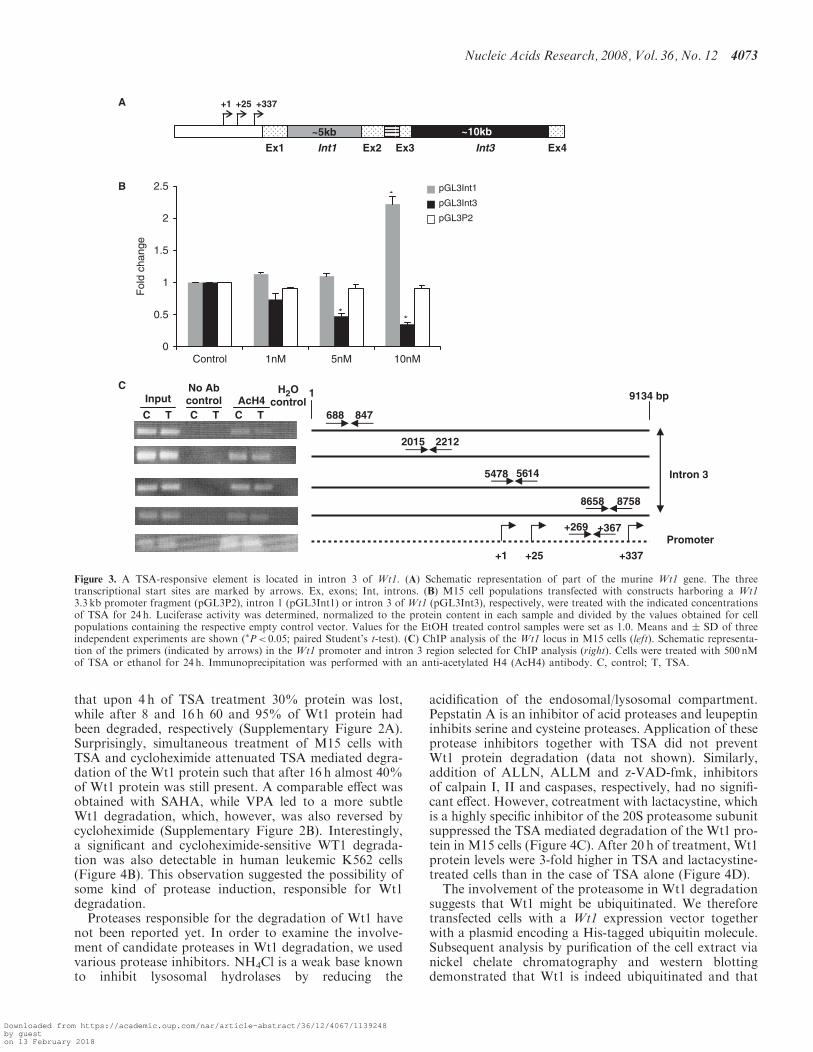
that upon 4 h of TSA treatment 30% protein was lost,while after 8 and 16 h 60 and 95% of Wt1 protein hadbeen degraded, respectively (Supplementary Figure 2A).Surprisingly, simultaneous treatment of M15 cells withTSA and cycloheximide attenuated TSA mediated degra-dation of the Wt1 protein such that after 16 h almost 40%of Wt1 protein was still present. A comparable effect wasobtained with SAHA, while VPA led to a more subtleWt1 degradation, which, however, was also reversed bycycloheximide (Supplementary Figure 2B). Interestingly,a significant and cycloheximide-sensitive WT1 degrada-tion was also detectable in human leukemic K562 cells(Figure 4B). This observation suggested the possibility ofsome kind of protease induction, responsible for Wt1degradation.
Proteases responsible for the degradation of Wt1 havenot been reported yet. In order to examine the involve-ment of candidate proteases in Wt1 degradation, we usedvarious protease inhibitors. NH4Cl is a weak base knownto inhibit lysosomal hydrolases by reducing the
acidification of the endosomal/lysosomal compartment.Pepstatin A is an inhibitor of acid proteases and leupeptininhibits serine and cysteine proteases. Application of theseprotease inhibitors together with TSA did not preventWt1 protein degradation (data not shown). Similarly,addition of ALLN, ALLM and z-VAD-fmk, inhibitorsof calpain I, II and caspases, respectively, had no signifi-cant effect. However, cotreatment with lactacystine, whichis a highly specific inhibitor of the 20S proteasome subunitsuppressed the TSA mediated degradation of the Wt1 pro-tein in M15 cells (Figure 4C). After 20 h of treatment, Wt1protein levels were 3-fold higher in TSA and lactacystine-treated cells than in the case of TSA alone (Figure 4D).The involvement of the proteasome in Wt1 degradation
suggests that Wt1 might be ubiquitinated. We thereforetransfected cells with a Wt1 expression vector togetherwith a plasmid encoding a His-tagged ubiquitin molecule.Subsequent analysis by purification of the cell extract vianickel chelate chromatography and western blottingdemonstrated that Wt1 is indeed ubiquitinated and that
0
0.5
1
1.5
2
2.5
Control 1nM 5nM 10nM
Fol
d ch
ange
pGL3Int1
pGL3Int3
pGL3P2
**
*
A
B
+1 +25 +337
Int1 Int3
~5kb
Ex1 Ex2 Ex3 Ex4
~10kb
C T C T C T
Input No Abcontrol AcH4
H2Ocontrol
1 9134 bp
688 847
2015 2212
5478 5614
8658 8758
+1 +25 +337
+269 +367
Intron 3
Promoter
C
Figure 3. A TSA-responsive element is located in intron 3 of Wt1. (A) Schematic representation of part of the murine Wt1 gene. The threetranscriptional start sites are marked by arrows. Ex, exons; Int, introns. (B) M15 cell populations transfected with constructs harboring a Wt13.3 kb promoter fragment (pGL3P2), intron 1 (pGL3Int1) or intron 3 of Wt1 (pGL3Int3), respectively, were treated with the indicated concentrationsof TSA for 24 h. Luciferase activity was determined, normalized to the protein content in each sample and divided by the values obtained for cellpopulations containing the respective empty control vector. Values for the EtOH treated control samples were set as 1.0. Means and � SD of threeindependent experiments are shown (�P< 0.05; paired Student’s t-test). (C) ChIP analysis of the Wt1 locus in M15 cells (left). Schematic representa-tion of the primers (indicated by arrows) in the Wt1 promoter and intron 3 region selected for ChIP analysis (right). Cells were treated with 500 nMof TSA or ethanol for 24 h. Immunoprecipitation was performed with an anti-acetylated H4 (AcH4) antibody. C, control; T, TSA.
Nucleic Acids Research, 2008, Vol. 36, No. 12 4073
Downloaded from https://academic.oup.com/nar/article-abstract/36/12/4067/1139248by gueston 13 February 2018
this modification can be enhanced by inhibition of theproteasome (Figure 4E).
Ubiquitin-conjugating enzyme UBCH8 is involved inWT1degradation
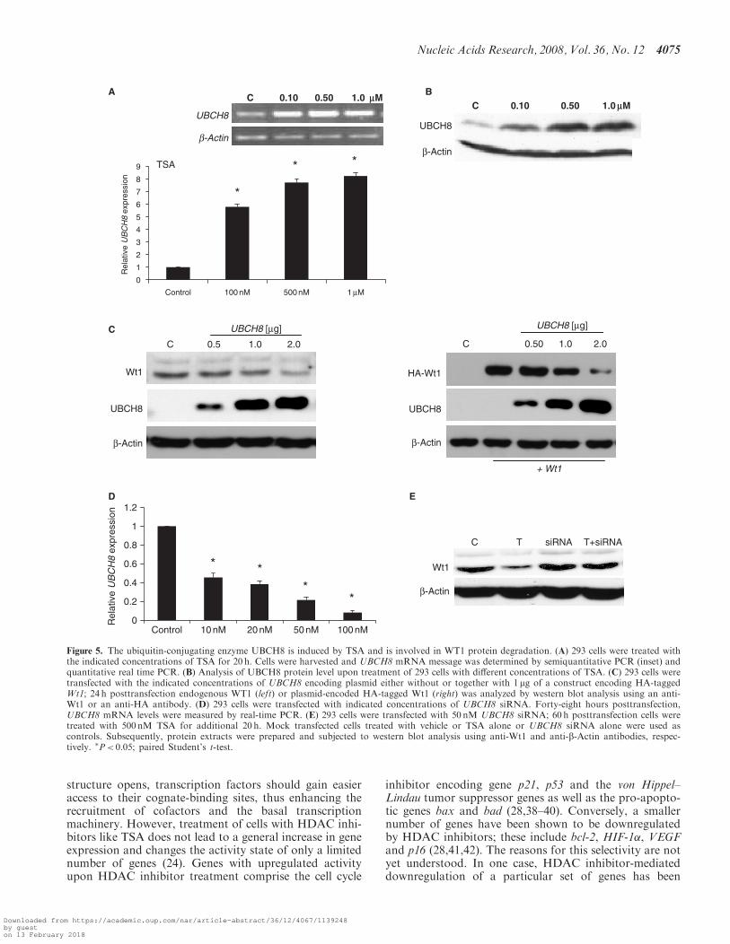
Ubc8 (named Ubce8 in mouse and UBCH8 in humans) isan ubiquitin-conjugating enzyme, which is 4–5-fold upreg-ulated in F9 cells upon TSA treatment (37). In order toanalyze whether this enzyme might also be involved inTSA-induced WT1 degradation, we investigated theexpression of UBCH8 in TSA-treated 293 cells by semi-quantitative PCR and further quantification by real-timePCR in a dose-dependent manner. At a concentration of100 nM TSA, 6-fold UBCH8 induction was observed thatwas further increased with 500 nM and 1 mM TSA after20 h (Figure 5A). A similar effect was observed in M15cells (data not shown). The increase in UBCH8 expressionwas also reflected at the protein level (Figure 5B). Ubc8/UBCH8 induction was not only exerted by TSA but alsoby VPA as well as SAHA in M15 as well as K562 cells(Supplementary Figure 3). To analyze whether WT1 is asubstrate of UBCH8, overexpression studies were per-formed. The 293 cells were transiently transfected withincreasing concentrations of an UBCH8 encoding plasmidin absence or presence of a construct encoding HA-taggedWt1. The levels of endogenous as well as transfected WT1were significantly decreased upon increasing concentra-tions of UBCH8 (Figure 5C and D). Constant levels ofb-Actin indicated that the effect on WT1 is specific andnot due to general protein degradation.
The functional significance of UBCH8 expression levelsfor WT1 degradation was further substantiated by siRNAexperiments in 293 cells. Application of a siRNA poolagainst UBCH8 mRNA resulted in a dose-dependentand significant reduction in UBCH8 expression level(Figure 5D). Transfection of 293 cells with UBCH8siRNA prevented degradation of the endogenous WT1protein upon TSA treatment (Figure 5E). These experi-ments support the involvement of UBCH8 in the degrada-tion of WT1 upon TSA treatment.
Thus, we conclude that in addition to an effect of theHDAC inhibitor TSA on transcription of the Wt1 gene,TSA also leads to the induction of proteasomal degrada-tion of WT1 at the protein level. This is at least in partmediated by induction of UBCH8 expression, whichencodes an ubiquitin-conjugating enzyme.
DISCUSSION
Conceptually, hyperacetylation of histones following inhi-bition of HDAC activity would be predicted to promotea general increase in gene expression. As chromatin
TSA
C 0.5 1 2 4 8 16 h
Wt1
β-Actin
TSA +CHX
β-Actin
Wt1
20 h6 h
C T Lac T+Lac C T Lac T+Lac
Wt1
β-Actin
D
C
A
SAHA
SAHA+CHX
WT1
β-Actin
WT1
β-Actin
C 4 8 16 hB
0
20
40
60
80
100
120
Control TSA Lac TSA/Lac
Wt1
pro
tein
(%
) (*P< 0.05)
E Vector Wt1+Ub
+ Lac
Wt1-Ubconjugates
Figure 4. TSA induces Wt1 protein degradation. (A) M15 or (B) K562cells were treated with TSA (500 nM) or SAHA (5 mM) alone or incombination with cycloheximide (50 mg/ml); protein extracts were pre-pared after various time points and subjected to western blot analysisusing anti-Wt1 and anti-b-Actin antibodies. (C) M15 cells were treatedwith TSA (500 nM) alone, lactacystine (10 mM) alone or in combinationwith TSA; protein extracts were prepared after 6 and 20 h and subjected
to western blot analysis using anti-Wt1 and anti-b-Actin antibodies.(D) Quantification of the western blot analysis shown in (C). Wt1 signalswere normalized against b-Actin and the untreated control sample wasset as 100%. For the analysis, only the data from the 20h time-pointwere used. (E) Wt1 protein is ubiquitinated. Wt1 was coexpressed withHis-ubiquitin in 293 cells by transient transfection. His-ubiquitinatedproteins were purified and Wt1 was detected by western blotting withanti-HA antibody. C, control; T, TSA; Lac, lactacystine.
4074 Nucleic Acids Research, 2008, Vol. 36, No. 12
Downloaded from https://academic.oup.com/nar/article-abstract/36/12/4067/1139248by gueston 13 February 2018
structure opens, transcription factors should gain easieraccess to their cognate-binding sites, thus enhancing therecruitment of cofactors and the basal transcriptionmachinery. However, treatment of cells with HDAC inhi-bitors like TSA does not lead to a general increase in geneexpression and changes the activity state of only a limitednumber of genes (24). Genes with upregulated activityupon HDAC inhibitor treatment comprise the cell cycle
inhibitor encoding gene p21, p53 and the von Hippel–Lindau tumor suppressor genes as well as the pro-apopto-tic genes bax and bad (28,38–40). Conversely, a smallernumber of genes have been shown to be downregulatedby HDAC inhibitors; these include bcl-2, HIF-1�, VEGFand p16 (28,41,42). The reasons for this selectivity are notyet understood. In one case, HDAC inhibitor-mediateddownregulation of a particular set of genes has been
C 0.10 0.50 1.0 mM
β-Actin UBCH8
+ Wt1
0
1
2
3
4
5
6
7
8
9
Control 100 nM 500 nM 1 µM
Rel
ativ
e U
BC
H8
expr
essi
on
TSA
*
* *
BC 0.10 0.50 1.0 mM
b-Actin
UBCH8
C
Wt1
β-Actin
UBCH8
C 0.5 1.0 2.0
UBCH8 [µg]
C 0.50 1.0 2.0
UBCH8 [µg]
HA-Wt1
β-Actin
UBCH8
E
0
0.2
0.4
0.6
0.8
1
1.2
Control 10 nM 20 nM 50 nM 100 nM
Rel
ativ
e U
BC
H8
expr
essi
on
* * *
*
D
Wt1
β-Actin
C T siRNA T+siRNA
A
Figure 5. The ubiquitin-conjugating enzyme UBCH8 is induced by TSA and is involved in WT1 protein degradation. (A) 293 cells were treated withthe indicated concentrations of TSA for 20 h. Cells were harvested and UBCH8 mRNA message was determined by semiquantitative PCR (inset) andquantitative real time PCR. (B) Analysis of UBCH8 protein level upon treatment of 293 cells with different concentrations of TSA. (C) 293 cells weretransfected with the indicated concentrations of UBCH8 encoding plasmid either without or together with 1 mg of a construct encoding HA-taggedWt1; 24 h posttransfection endogenous WT1 (left) or plasmid-encoded HA-tagged Wt1 (right) was analyzed by western blot analysis using an anti-Wt1 or an anti-HA antibody. (D) 293 cells were transfected with indicated concentrations of UBCH8 siRNA. Forty-eight hours posttransfection,UBCH8 mRNA levels were measured by real-time PCR. (E) 293 cells were transfected with 50 nM UBCH8 siRNA; 60 h posttransfection cells weretreated with 500 nM TSA for additional 20 h. Mock transfected cells treated with vehicle or TSA alone or UBCH8 siRNA alone were used ascontrols. Subsequently, protein extracts were prepared and subjected to western blot analysis using anti-Wt1 and anti-b-Actin antibodies, respec-tively. �P< 0.05; paired Student’s t-test.
Nucleic Acids Research, 2008, Vol. 36, No. 12 4075
Downloaded from https://academic.oup.com/nar/article-abstract/36/12/4067/1139248by gueston 13 February 2018
shown to result from physical interaction ofa transcription factor, namely NF-kB p65 with the acety-lated form of a signaling molecule, Stat1, and subsequentreduction of p65 DNA binding (43).We report here that the Wilms tumor suppressor gene
Wt1 also belongs to the class of genes that are downregu-lated by HDAC inhibitors. Downregulation was observedwith TSA as well as with SAHA, both inhibitors of class Iand II HDACs. VPA, an inhibitor of class I HDACsonly (34) did not cause a reduction in Wt1 expression,while MS-275, another class I-selective HDAC inhibitoralso downregulated Wt1 expression. The different beha-vior of MS-275 and VPA on Wt1 expression might beexplained by their different biological potency (the respec-tive EC50 values depend on the specific substrates anddiffer by a factor of 103–104) or by their different targetprofile. It has been shown that while VPA can be classifiedas a specific class I HDAC inhibitor, MS-275 is only classI-selective and can, e.g. also efficiently inhibit the class IIHDAC9 (35). Thus, from these data, we cannot concludewhich specific HDACs mediate downregulation ofWt1 mRNA. The latter was observed in the murine celllines M15 and TM3 as well as in human K562 and 293cells. This indicates that reduction of Wt1 expression byHDAC inhibitors is a general and not a cell-line-specificphenomenon.Strikingly, downregulation of Wt1 expression was very
rapid; within 3 h of TSA treatment Wt1 mRNA wasreduced to almost 50%. This suggests that repression ofWt1 mRNA expression is an early event and that the half-life of Wt1 mRNA must be � 3 h. Mechanistically, loss ofWt1 mRNA could be mediated by two possible mechan-isms. First, TSA could affect the rate of Wt1 transcriptionor second,Wt1mRNA stability. Our experiments employ-ing the transcriptional (RNA polymerase) inhibitoractinomycin D indicate that TSA has no effect on Wt1mRNA stability and downregulation of Wt1 mRNAupon TSA treatment requires ongoing RNA synthesis.Moreover, we were able to show by nuclear run-offassay that TSA strongly affected Wt1 transcription.In order to explore the possible mechanisms, we inves-
tigated whether ongoing protein synthesis was requiredfor TSA-mediated downregulation of Wt1. For this, weused the protein translation inhibitor cycloheximideand observed that this inhibitor did not prevent downre-gulation of Wt1 mRNA. Thus, Wt1 mRNA loss does notrequire de novo protein synthesis. Conceivably, loss ofWt1expression by blocking HDAC activity could be mediatedby histones or by nonhistone proteins. Increasing acetyla-tion of histones associated with Wt1 regulatory regionscould cause the movement of nucleosomes on the Wt1locus and thus mask critical cis-acting sites. Nucleosomeshave been shown to move and be remodeled on specificloci, including the lysozyme gene and the IL-12p40 (44,45).Alternatively, as a consequence of HDAC inhibition atranscriptional activator of Wt1 expression could looseits function or a potential repressor could be activatedas a consequence of hyperacetylation. Acetylation/deace-tylation of several transcription factors including p53,NF-kB, E2f1 and Stat1 have been described (43,46–48).
Further studies will be required to identify the trans-actingfactors mediating the TSA effect on Wt1 expression.
We also wanted to identify the cis-element that isresponsible for the TSA effect on Wt1 expression. Con-siderable work has been done regarding the tissue- andtemporal-specific expression of Wt1; however, the Wt1locus is remarkably complex and our knowledge of itsregulation is still quite limited. In terms of elementsregulating Wt1 expression in particular tissues, a 258 bphematopoietic cell-specific enhancer that is located inintron 3 and bound by GATA-1 and c-Myb has beendescribed (49). The same intron harbors a 460 bp silencerthat represses WT1 transcription in nonrenal cells suchas leukemic (K562 and HL60) and cervical carcinomacells (HeLa) (50). Moreover, a construct including thepromoter, two enhancer elements located in intron 3 andat the 30 end of the WT1 gene could induce robust expres-sion of a reporter gene in respective transgenic leukemiacells (51).
We reasoned that transient transfections would not bewell suited for the identification of regulatory elementsmediating the TSA effects, since the chromatin configura-tion might not be physiological. We have thereforeemployed short-term stably transfected cells for theseexperiments. When we used a luciferase construct linkedto a 3.3 kb Wt1 promoter fragment, TSA did not have anyeffect. Conversely, a reporter construct harboring intron 3of Wt1 led to more than 60% downregulation in reporteractivity upon TSA treatment. Unexpectedly, we did seea TSA-mediated enhancement in intron 1 stimulatedreporter gene activity. While no enhancers have been iden-tified in intron 1 of Wt1, a promoter that controls expres-sion of an antisense Wt1 transcript has been reported toreside in the first intron (36). It might be possible thatTSA exerts an effect on the activity of this promoter. ByChIP, we could also show that histones H4 in a region inthe 50 part of intron 3 are differentially acetylated in thepresence and absence of TSA, respectively. Other regionswithin intron 3 did not seem to be affected. This is inagreement with the existence of a regulatory region inintron 3 of the humanWT1 locus that has been mentionedabove. Whether the sequences that respond to TSA over-lap with the enhancer and silencer sequences in the humanWT1 gene remains to be explored.
In addition to downregulation ofWt1mRNA, TSA alsoled to enhanced degradation of the WT1 protein. Thiseffect was at least in part mediated by the ubiquitin-conjugating enzyme Ubc8, which has been reported tobe induced by HDAC inhibitors (36). Of note, inductionof Ubc8 could also be observed by VPA as well asSAHA and in a variety of different cell lines. The obser-vation that VPA causes Ubc8 induction and subsequentWT1 degradation, while not affecting Wt1’s transcriptionindicates that the two phenomena are mediated by dif-ferent molecular mechanisms. Overexpression of UBCH8resulted in the enhanced destabilization of both endogen-ous and transfected WT1 protein and knockdown ofUBCH8 rescued TSA-mediated degradation of WT1.This shows that Ubc8 is a rate-limiting factor in WT1degradation, while the corresponding E3 ligase is not.
4076 Nucleic Acids Research, 2008, Vol. 36, No. 12
Downloaded from https://academic.oup.com/nar/article-abstract/36/12/4067/1139248by gueston 13 February 2018
This is the first demonstration that WT1 degradation iscontrolled by the proteasomal machinery.
In summary, we provide evidence that HDAC inhibi-tors like TSA lead to a dramatic reduction of Wt1 expres-sion, both at the level of transcription and at the proteinlevel. Given that WT1 levels are high not only in manyhematopoietic malignancies but also in a variety of solidtumors, that high WT1 levels are associated with poorprognosis, and that various cancer cells depend on highWT1 expression levels downregulation of this proteincould be an anticancer strategy.
SUPPLEMENTARY DATA
Supplementary Data are available at NAR Online.
ACKNOWLEDGEMENTS
We thank Amna Musharraf and Amal Saidi for help inwestern blot and FACS analysis, respectively, as well asFrank Bollig and Jurgen Klattig for discussions and forcritically reading and improving this article. We are grate-ful to Oliver Kramer for sharing reagents. This work wassupported by Deutsche Forschungsgemeinschaft (SFB604to C.E.). Funding to pay the Open Access publicationcharges for this article was provided by the LeibnizGemeinschaft.
Conflict of interest statement. None declared.
REFERENCES
1. Call,K.M., Glaser,T., Ito,C.Y., Buckler,A.J., Pelletier,J.,Haber,D.A., Rose,E.A., Kral,A., Yeger,H., Lewis,W.H. et al. (1990)Isolation and characterization of a zinc finger polypeptide gene atthe human chromosome 11 Wilms’ tumor locus. Cell, 60, 509–520.
2. Gessler,M., Poustka,A., Cavenee,W., Neve,R.L., Orkin,S.H. andBruns,G.A. (1990) Homozygous deletion in Wilms tumours of azinc-finger gene identified by chromosome jumping. Nature, 343,774–778.
3. Hofmann,W., Royer,H.D., Drechsler,M., Schneider,S. and Royer-Pokora,B. (1993) Characterization of the transcriptional regulatoryregion of the human WT1 gene. Oncogene, 8, 3123–3132.
4. Herzer,U., Crocoll,A., Barton,D., Howells,N. and Englert,C. (1999)The Wilms tumor suppressor gene wt1 is required for developmentof the spleen. Curr. Biol., 9, 837–840.
5. Kreidberg,J.A., Sariola,H., Loring,J.M., Maeda,M., Pelletier,J.,Housman,D. and Jaenisch,R. (1993) WT-1 is required for earlykidney development. Cell, 74, 679–691.
6. Wagner,N., Wagner,K.D., Theres,H., Englert,C., Schedl,A. andScholz,H. (2005) Coronary vessel development requires activationof the TrkB neurotrophin receptor by the Wilms’ tumortranscription factor Wt1. Genes Dev., 19, 2631–2642.
7. Rivera,M.N. and Haber,D.A. (2005) Wilms’ tumour: connectingtumorigenesis and organ development in the kidney. Nat. Rev., 5,699–712.
8. Bergmann,L., Miething,C., Maurer,U., Brieger,J., Karakas,T.,Weidmann,E. and Hoelzer,D. (1997) High levels of Wilms’ tumorgene (wt1) mRNA in acute myeloid leukemias are associated with aworse long-term outcome. Blood, 90, 1217–1225.
9. Inoue,K., Sugiyama,H., Ogawa,H., Nakagawa,M., Yamagami,T.,Miwa,H., Kita,K., Hiraoka,A., Masaoka,T., Nasu,K. et al. (1994)WT1 as a new prognostic factor and a new marker for thedetection of minimal residual disease in acute leukemia. Blood, 84,3071–3079.
10. Menssen,H.D., Renkl,H.J., Rodeck,U., Maurer,J., Notter,M.,Schwartz,S., Reinhardt,R. and Thiel,E. (1995) Presence of Wilms’
tumor gene (wt1) transcripts and the WT1 nuclear protein in themajority of human acute leukemias. Leukemia, 9, 1060–1067.
11. Steinbach,D., Schramm,A., Eggert,A., Onda,M., Dawczynski,K.,Rump,A., Pastan,I., Wittig,S., Pfaffendorf,N., Voigt,A. et al. (2006)Identification of a set of seven genes for the monitoring of minimalresidual disease in pediatric acute myeloid leukemia. Clin. CancerRes., 12, 2434–2441.
12. Yang,L., Han,Y., Suarez Saiz,F. and Minden,M.D. (2007)A tumor suppressor and oncogene: the WT1 story. Leukemia, 21,868–876.
13. Yamagami,T., Sugiyama,H., Inoue,K., Ogawa,H., Tatekawa,T.,Hirata,M., Kudoh,T., Akiyama,T., Murakami,A. and Maekawa,T.(1996) Growth inhibition of human leukemic cells by WT1 (Wilmstumor gene) antisense oligodeoxynucleotides: implications for theinvolvement of WT1 in leukemogenesis. Blood, 87, 2878–2884.
14. Hubinger,G., Schmid,M., Linortner,S., Manegold,A., Bergmann,L.and Maurer,U. (2001) Ribozyme-mediated cleavage of wt1 tran-scripts suppresses growth of leukemia cells. Exp. Hematol., 29,1226–1235.
15. Elmaagacli,A.H., Koldehoff,M., Peceny,R., Klein-Hitpass,L.,Ottinger,H., Beelen,D.W. and Opalka,B. (2005) WT1 andBCR-ABL specific small interfering RNA have additive effects inthe induction of apoptosis in leukemic cells. Haematologica, 90,326–334.
16. Nishida,S., Hosen,N., Shirakata,T., Kanato,K., Yanagihara,M.,Nakatsuka,S., Hoshida,Y., Nakazawa,T., Harada,Y., Tatsumi,N.et al. (2006) AML1-ETO rapidly induces acute myeloblastic leuke-mia in cooperation with the Wilms tumor gene, WT1. Blood, 107,3303–3312.
17. Oka,Y., Tsuboi,A., Taguchi,T., Osaki,T., Kyo,T., Nakajima,H.,Elisseeva,O.A., Oji,Y., Kawakami,M., Ikegame,K. et al. (2004)Induction of WT1 (Wilms’ tumor gene)-specific cytotoxic Tlymphocytes by WT1 peptide vaccine and the resultant cancerregression. Proc. Natl Acad. Sci. USA, 101, 13885–13890.
18. Strahl,B.D. and Allis,C.D. (2000) The language of covalent histonemodifications. Nature, 403, 41–45.
19. Shahbazian,M.D. and Grunstein,M. (2007) Functions ofsite-specific histone acetylation and deacetylation. Ann. Rev.Biochem., 76, 75–100.
20. Gregoretti,I.V., Lee,Y.M. and Goodson,H.V. (2004) Molecularevolution of the histone deacetylase family: functional implicationsof phylogenetic analysis. J. Mol. Biol., 338, 17–31.
21. Marks,P., Rifkind,R.A., Richon,V.M., Breslow,R., Miller,T. andKelly,W.K. (2001) Histone deacetylases and cancer: causes andtherapies. Nat. Rev., 1, 194–202.
22. Furumai,R., Komatsu,Y., Nishino,N., Khochbin,S., Yoshida,M.and Horinouchi,S. (2001) Potent histone deacetylase inhibitors builtfrom trichostatin A and cyclic tetrapeptide antibiotics includingtrapoxin. Proc. Natl Acad. Sci. USA, 98, 87–92.
23. Yoshida,M., Kijima,M., Akita,M. and Beppu,T. (1990) Potent andspecific inhibition of mammalian histone deacetylase both in vivoand in vitro by trichostatin A. The J. Biol. Chem., 265,17174–17179.
24. Van Lint,C., Emiliani,S. and Verdin,E. (1996) The expression ofa small fraction of cellular genes is changed in response to histonehyperacetylation. Gene expression, 5, 245–253.
25. Mariadason,J.M., Corner,G.A. and Augenlicht,L.H. (2000) Geneticreprogramming in pathways of colonic cell maturation induced byshort chain fatty acids: comparison with trichostatin A, sulindac,and curcumin and implications for chemoprevention of coloncancer. Cancer Res., 60, 4561–4572.
26. Zhang,X., Wharton,W., Yuan,Z., Tsai,S.C., Olashaw,N. andSeto,E. (2004) Activation of the growth-differentiation factor 11gene by the histone deacetylase (HDAC) inhibitor trichostatin Aand repression by HDAC3. Mol. Cell. Biol., 24, 5106–5118.
27. Butler,L.M., Agus,D.B., Scher,H.I., Higgins,B., Rose,A., Cordon-Cardo,C., Thaler,H.T., Rifkind,R.A., Marks,P.A. and Richon,V.M.(2000) Suberoylanilide hydroxamic acid, an inhibitor of histonedeacetylase, suppresses the growth of prostate cancer cells in vitroand in vivo. Cancer Res., 60, 5165–5170.
28. Kim,M.S., Kwon,H.J., Lee,Y.M., Baek,J.H., Jang,J.E., Lee,S.W.,Moon,E.J., Kim,H.S., Lee,S.K., Chung,H.Y. et al. (2001) Histonedeacetylases induce angiogenesis by negative regulation of tumorsuppressor genes. Nat. Med., 7, 437–443.
Nucleic Acids Research, 2008, Vol. 36, No. 12 4077
Downloaded from https://academic.oup.com/nar/article-abstract/36/12/4067/1139248by gueston 13 February 2018
29. Minucci,S. and Pelicci,P.G. (2006) Histone deacetylase inhibitorsand the promise of epigenetic (and more) treatments for cancer.Nat. Rev., 6, 38–51.
30. Livak,K.J. and Schmittgen,T.D. (2001) Analysis of relative geneexpression data using real-time quantitative PCR and the 2(-DeltaDelta C(T)) Method. Methods, 25, 402–408.
31. Chen,J. and Pan,Y. (2003) MDM2 promotes ubiquitination anddegradation of MDMX. Mol. Cell. Biol., 23, 5113–5121.
32. Gunes,C., Lichtsteiner,S., Vasserot,A.P. and Englert,C. (2000)Expression of the hTERT gene is regulated at the level oftranscriptional initiation and repressed by Mad1. Cancer Res., 60,2116–2121.
33. Gong,Y., Eggert,H. and Englert,C. (2001) The murine Wilms tumorsuppressor gene (wt1) locus. Gene, 279, 119–126.
34. Gottlicher,M., Minucci,S., Zhu,P., Kramer,O.H., Schimpf,A.,Giavara,S., Sleeman,J.P., Lo Coco,F., Nervi,C., Pelicci,P.G. et al.(2001) Valproic acid defines a novel class of HDAC inhibitorsinducing differentiation of transformed cells. EMBO J., 20,6969–6978.
35. Khan,N., Jeffers,M., Kumar,S., Hackett,C., Boldog,F.,Khramtsov,N., Qian,X., Mills,E., Berghs,S.C., Carey,N. et al.(2008) Determination of the class and isoform selectivity of small-molecule histone deacetylase inhibitors. Biochem. J., 409, 581–589.
36. Malik,K.T., Wallace,J.I., Ivins,S.M. and Brown,K.W. (1995)Identification of an antisense WT1 promoter in intron 1: implica-tions for WT1 gene regulation. Oncogene, 11, 1589–1595.
37. Kramer,O.H., Zhu,P., Ostendorff,H.P., Golebiewski,M.,Tiefenbach,J., Peters,M.A., Brill,B., Groner,B., Bach,I., Heinzel,T.et al. (2003) The histone deacetylase inhibitor valproic acidselectively induces proteasomal degradation of HDAC2. EMBO J.,22, 3411–3420.
38. Richon,V.M., Sandhoff,T.W., Rifkind,R.A. and Marks,P.A. (2000)Histone deacetylase inhibitor selectively induces p21WAF1expression and gene-associated histone acetylation. Proc. Natl Acad.Sci. USA, 97, 10014–10019.
39. Rosato,R.R., Almenara,J.A. and Grant,S. (2003) The histonedeacetylase inhibitor MS-275 promotes differentiation or apoptosisin human leukemia cells through a process regulated by generationof reactive oxygen species and induction of p21CIP1/WAF1 1.Cancer Res., 63, 3637–3645.
40. Sawa,H., Murakami,H., Ohshima,Y., Sugino,T., Nakajyo,T.,Kisanuki,T., Tamura,Y., Satone,A., Ide,W., Hashimoto,I. et al.(2001) Histone deacetylase inhibitors such as sodium butyrate
and trichostatin A induce apoptosis through an increase of thebcl-2-related protein Bad. Brain Tumor Pathol., 18, 109–114.
41. Duan,H., Heckman,C.A. and Boxer,L.M. (2005) Histone deacety-lase inhibitors down-regulate bcl-2 expression and induce apoptosisin t(14;18) lymphomas. Mol. Cell. Biol., 25, 1608–1619.
42. Matheu,A., Klatt,P. and Serrano,M. (2005) Regulation of theINK4a/ARF locus by histone deacetylase inhibitors. J. Biol. Chem.,280, 42433–42441.
43. Kramer,O.H., Baus,D., Knauer,S.K., Stein,S., Jager,E.,Stauber,R.H., Grez,M., Pfitzner,E. and Heinzel,T. (2006)Acetylation of Stat1 modulates NF-kappaB activity. Genes Dev., 20,473–485.
44. Kontaraki,J., Chen,H.H., Riggs,A. and Bonifer,C. (2000)Chromatin fine structure profiles for a developmentally regulatedgene: reorganization of the lysozyme locus before trans-activatorbinding and gene expression. Genes Dev., 14, 2106–2122.
45. Weinmann,A.S., Mitchell,D.M., Sanjabi,S., Bradley,M.N.,Hoffmann,A., Liou,H.C. and Smale,S.T. (2001) Nucleosome remo-deling at the IL-12 p40 promoter is a TLR-dependent, Rel-inde-pendent event. Nat. Immunol., 2, 51–57.
46. Barlev,N.A., Liu,L., Chehab,N.H., Mansfield,K., Harris,K.G.,Halazonetis,T.D. and Berger,S.L. (2001) Acetylation of p53 acti-vates transcription through recruitment of coactivators/histoneacetyltransferases. Mol. Cell, 8, 1243–1254.
47. Kiernan,R., Bres,V., Ng,R.W., Coudart,M.P., El Messaoudi,S.,Sardet,C., Jin,D.Y., Emiliani,S. and Benkirane,M. (2003)Post-activation turn-off of NF-kappa B-dependent transcription isregulated by acetylation of p65. J. Biol. Chem., 278, 2758–2766.
48. Martinez-Balbas,M.A., Bauer,U.M., Nielsen,S.J., Brehm,A. andKouzarides,T. (2000) Regulation of E2F1 activity by acetylation.EMBO J., 19, 662–671.
49. Zhang,X., Xing,G., Fraizer,G.C. and Saunders,G.F. (1997)Transactivation of an intronic hematopoietic-specific enhancer ofthe human Wilms’ tumor 1 gene by GATA-1 and c-Myb. J. Biol.Chem., 272, 29272–29280.
50. Hewitt,S.M., Fraizer,G.C. and Saunders,G.F. (1995)Transcriptional silencer of the Wilms’ tumor gene WT1 contains anAlu repeat. J. Biol. Chem., 270, 17908–17912.
51. Hosen,N., Yanagihara,M., Nakazawa,T., Kanato,K., Nishida,S.,Shirakata,T., Asada,M., Masuda,T., Taniguchi,Y., Kawakami,M.et al. (2004) Identification of a gene element essential for leukemia-specific expression of transgenes. Leukemia, 18, 415–419.
4078 Nucleic Acids Research, 2008, Vol. 36, No. 12
Downloaded from https://academic.oup.com/nar/article-abstract/36/12/4067/1139248by gueston 13 February 2018


![Ubiquitous expression of Sry induces embryonic lethality ... · 4 binding protein) [6], Wt1 (Wilms’ tumor suppressor 1) [7,8], ... mosome) triggers the testis developmental pathway](https://img.dokumen.tips/doc/110x75/5cb1fa1388c993045a8b9f13/ubiquitous-expression-of-sry-induces-embryonic-lethality-4-binding-protein.jpg)


![A Novel Missense Mutation of Wilms Tumor 1 Causes ...1]1.pdf · associated with WT1 mutations include Wilms’ tumor as a componentofWAGRsyndrome(Wilms’tumor,aniridia,gen-itourinary](https://img.dokumen.tips/doc/110x75/5fcb5bb895e97801983d7f63/a-novel-missense-mutation-of-wilms-tumor-1-causes-11pdf-associated-with.jpg)