Embed Size (px)
Citation preview
Research ArticleLipopolysaccharide-Binding Protein DownregulatesFractalkine through Activation of p38 MAPK and NF-κB
Xia Huang,1,2 Yi Zeng,3 Yujie Jiang,1,2 Yueqiu Qin,4 Weigui Luo,2 Shulin Xiang,1,5
Suren R. Sooranna,6 and Liao Pinhu7
1The First Clinical Medical College of Jinan University, Guangzhou, Guangdong Province 510630, China2Department of Respiratory Medicine, Youjiang Medical University for Nationalities, Baise, Guangxi Zhuang Autonomous Region533000, China3Department of Central Laboratory, Youjiang Medical University for Nationalities, Baise, Guangxi Zhuang Autonomous Region533000, China4Department of Digestive Medicine, Youjiang Medical University for Nationalities, Baise, Guangxi Zhuang Autonomous Region533000, China5Department of Intensive Care Unit, The People’s Hospital of Guangxi Zhuang Autonomous Region, Nanning 531000, China6Department of Surgery and Cancer, Imperial College London, Chelsea and Westminster Hospital, London SW10 9NH, UK7Department of Intensive Care Medicine, Youjiang Medical University for Nationalities, Baise, Guangxi Zhuang Autonomous Region533000, China
Correspondence should be addressed to Liao Pinhu; [email protected]
Received 18 November 2016; Accepted 2 March 2017; Published 29 May 2017
Academic Editor: Giuseppe Valacchi
Copyright © 2017 Xia Huang et al. This is an open access article distributed under the Creative Commons Attribution License,which permits unrestricted use, distribution, and reproduction in any medium, provided the original work is properly cited.
Background. LBP and fractalkine are known to be involved in the pathogenesis of ARDS. This study investigated the relationshipbetween LBP and fractalkine in LPS-induced A549 cells and rat lung tissue in an ARDS rat model. Methods. A549 cells weretransfected with LBP or LBP shRNA plasmid DNA or pretreated with SB203580 or SC-514 following LPS treatment. An ARDSrat model was established using LPS with or without LBPK95A, SB203580, or SC-514 treatment. RT-PCR, western blotting,ELISA, immunofluorescence, coimmunoprecipitation, and immunohistochemical staining were used to study the expression offractalkine and LBP and p38 MAPK and p65 NF-κB activities. Results. LPS increased LBP and reduced fractalkine. LBPoverexpression further decreased LPS-induced downregulation of fractalkine and p38 MAPK and p65 NF-κB activation; LBPgene silencing, SB203580, and SC-514 suppressed LPS-induced downregulation of fractalkine and p38 MAPK and p65 NF-κBactivation in A549 cells. LBP and fractalkine in lung tissue were increased and decreased, respectively, following LPS injection.LBPK95A, SB203580, and SC-514 ameliorated LPS-induced rat lung injury and suppressed LPS-induced downregulation offractalkine by decreasing phospho-p38 MAPK and p65 NF-κB. Conclusions. The results indicate that LBP downregulatesfractalkine expression in LPS-induced A549 cells and in an ARDS rat model through activation of p38 MAPK and NF-κB.
1. Introduction
Acute respiratory distress syndrome (ARDS) is associatedwith high mortality and morbidity [1, 2]. ARDS is charac-terized by neutrophilic inflammation, pulmonary edema,hemorrhage, alveolar-capillary destruction, and hyalinemembrane formation in the lung resulting subsequently inalveolar flooding and acute respiratory failure [3, 4]. The
pathogenesis of ARDS is caused by lung inflammation,which includes the accumulation of inflammatory cells,release of proteases and proinflammatory cytokines, anda sustained loss of normal alveolar capillary barrier function[5, 6]. Endotoxemia caused by Gram-negative bacterialinfection is one of the causes of lung inflammation [7].Lipopolysaccharide (LPS) is a major component of the cellwall of Gram-negative bacteria and its endotoxins [8].
HindawiMediators of InflammationVolume 2017, Article ID 9734837, 20 pageshttps://doi.org/10.1155/2017/9734837
Increasing lines of evidence have suggested that LPS andinflammatory cytokines are involved in the developmentand progression of ARDS [9, 10].
Lipopolysaccharide-binding protein (LBP), a key partici-pant in the inflammatory response to infection, is a type Iacute phase response protein that is produced by airway epi-thelial cells, hepatocytes, and a myriad of other cell types andenhances the recognition of endotoxin and pathogens by theimmune system [11, 12]. Levels of LBP in the lung have beenfound to be increased in patients with ARDS [13]. LBP bindsto gram-negative bacteria via the lipid A portion of the LPSwhich mediates its binding to the CD14 cellular receptormolecule presented by monocytes and macrophages andpotentiates LPS signaling activity resulting in the productionof proinflammatory cytokines that subsequently leads to lunginjury [14]. However, LBP was also shown to be involvedin the efficient elimination of bacteria and the loss of LBP-mediated function results in derangements of the inflam-matory response and bacterial clearance mechanisms ingram-negative-induced pneumonia by delaying the influxof neutrophils [15]. This led to the conclusion that LBPmighthave a dual role: augmenting the inflammatory response tobacterial toxins such as LPS and contributing to bacterialelimination via associate neutrophil infiltration. The poten-tial role of LBP in LPS-induced ARDS is not fully understood.
Fractalkine (FKN; CX3CL1) is a chemokine which func-tions dually as a chemoattractant and an adhesion moleculethat has been linked to several types of inflammatory diseases[16, 17]. FKN, recognized as CX3C ligand (CX3CL1), is aunique chemokine of the CX3 chemokine family that actsas either a membrane bound (adhesion molecule) or a soluble(chemokine) mediator, facilitating T cell and monocyteadhesion and transmigration [18, 19]. The expression ofFKN can be shown in alveolar epithelial cells, pulmonaryvascular endothelial cells, fibroblasts, and airway smoothmuscle cells [20–22], and it displays a role in the initiationand progression of intimate contacts of inflammatory cellswith endothelium [23]. It protects the cells from Fas/FasL-mediated apoptosis in alveolar epithelial cells, inhibitsinflammation-associated markers [24] such as monocytechemoattractant protein-1 (MCP-1), and induces monocytemigration via the inhibition of stress-activated protein kinase2/p38 and matrix metalloproteinase activities [25]. It hastherefore been suggested that FKN has a novel role in inflam-mation. However, the anti-inflammatory effect of FKN inalveolar epithelial cells has not been documented and the roleof FKN in LPS-induced ARDS is still not clear.
It is now well established that the three majormitogen-activated protein kinase (MAPK) signaling path-ways, namely, p38, c-Jun NH2-terminal kinase (JNK), andextracellular signal-regulated kinase (ERK), regulate a varietyof cellular activities, including proliferation, differentiation,and apoptosis, in response to different external stimuli. p38MAPKs are stress-activated MAPKs, and both p38 MAPKand NF-κB are involved in cytokine production and in thepathophysiology of ARDS [26, 27]. The p38 MAPK familymembers (p38, MAPK11, SAPK3, and SAPK4) are activatedby inflammatory cytokines, UV radiation, and external stresssuch as heat shock and high osmotic stress. Once activated,
the p38 MAPK pathway initiates the production of tran-scription factors including PAX6, p53, ATF1, ATF2, andCREB and regulates the production of proinflammatoryor apoptosis-associated genes [26, 27]. Nuclear factor-κB(NF-κB) is a nuclear transcription factor that regulatescritical cellular behavior and many cytokines by influencingbiological processes of cells including inflammation, innateand adaptive immunity, and stress responses. NF-κB pro-teins include NF-κB2 p52/p100, NF-κB1 p50/p105, c-Rel,RelA/p65, and RelB. Activated NF-κB is translocated to thenucleus to induce target gene expression [27]. Sustained acti-vation of NF-κB is harmful by constantly generating inflam-matory mediators which contribute to inflammatory diseasesand inflammation-associated lung injury [28]. These signal-ing transduction pathways acting together create a networkthat activates a variety of transcription factors, which coordi-nately induce the transcription of many genes to affect thedevelopment of ARDS [29].
The mechanisms by which signaling pathways regulatethe FKN expression to participate in the process of experi-mental ARDS remain unknown. Our hypothesis is that LBPis involved in LPS-induced FKN release through p38 MAPKor NF-κB signaling pathway. Cultured A549 cells as anin vitro model and an LPS-induced ARDS rat as an in vivomodel are used here to investigate these relevant signalingtransduction pathways.
2. Materials and Methods
2.1. Cell Culture. A549 cells were obtained from theAmerican Type Culture Collection (VR-15™). The cells werecultured in Roswell Park Memorial Institute 1640 (RPMI1640) media (Gibco, USA), supplemented with 10% fetalbovine serum (HyClone, USA), and maintained at 37°C in ahumidified atmosphere with 5% CO2. Cells were subculturedevery 3-4 days after reaching a density of 5000 cells/cm2. ForLPS treatment, A549 cells were seeded (5000 cells/cm2) into30mm six-well plates. Once confluent, the cells were incu-bated in serum-free medium for 24 h before each experiment.
2.2. Cell Treatment and Sample Collection
2.2.1. Groupings. The cells were divided into seven groupsas follows: control group (CTL); LPS group (LPS, LPStreatment at 10μg/mL, L2880, Sigma-Aldrich, USA); LPSand LBP group (LPS+LBP(+), the cells were transfectedwith the LBP plasmid DNA (Genechem Co., Ltd., Shanghai,China) by using Lipofectamine® 2000 transfection reagent(11668-027, Invitrogen, USA) and maintained in RPMI1640 medium for 48h following LPS treatment); LPS andLBP(−) group (LPS+LBP(−), the cells were transfectedwith the pGCU6/Neo-LBP shRNA-expressing plasmidDNA (ShLBP) which targeted the LBP mRNA sequence(5′-GCATCAGCATTTCGGTCAACC-3′) (Genechem Co.,Ltd., Shanghai, China) by using Lipofectamine 2000 trans-fection reagent and maintained in RPMI 1640 medium for48 h following LPS treatment); LPS and SB203580 group(LPS+SB, the cells were preincubated with 10μM SB203580(S8307, Sigma-Aldrich, USA) for 60 minutes following LPS
2 Mediators of Inflammation
treatment, and the concentration of SB203580 was asdescribed previously [30]); LPS and SC-514 group (LPS+SC, the cells were preincubated with 10μM SC-514(SML0557, Sigma-Aldrich, USA) for 60 minutes followingLPS treatment, and the concentration of SC-514 was asdescribed previously); and tumor necrosis factor-α (TNF-α)group (TNF-α, TNF-α treatment at 50ng/mL for positivecontrol (H8916, Sigma-Aldrich, USA), the concentration ofTNF-α was as described previously [31]).
The cell supernatants were collected 2 h after LPS treat-ment. RNA was isolated from the cells 2 hours after LPSstimulation by using an RNeasy Mini Kit (74104, Qiagen,USA). RNA integrity was checked electrophoretically andquantified by using spectrophotometry. Cell lysates werecollected in cell lysis buffer (9803, Cell Signaling Technology,USA) according to the experimental conditions 1 h after LPStreatment for subsequent coimmunoprecipitation (Co-IP)and western blotting. For immunofluorescence analysisby confocal laser-scanning microscopy, the cells were fixedin 4% paraformaldehyde for 1 h after LPS treatment. Theculture supernatant was harvested 24 h after LPS stimula-tion for the enzyme-linked immunosorbent assay (ELISA)measurement of FKN.
2.2.2. Plasmid Transfection. A549 cells were allowed to growuntil about 80% confluency and then transfected with LBPplasmid DNA and LBP shRNA-expressing plasmid DNA.For transfection, 4μg of plasmid and 10μL Lipofectamine2000 were added to each well of a 6-well plate. Cells wereincubated at 37°C for 48h in a humidified incubator contain-ing 95% air, 5% CO2 for 48h, and then treated with or with-out 10μg/mL LPS. An empty vector was used as a vehiclecontrol. At the same time, a group of GFP tag plasmids(including LBP plasmid DNA, LBP shRNA plasmid DNA,and empty vector) was used for parallel controls to observethe transfection efficiency under a fluorescence microscope.The cell viability was analyzed by the MTT assay to deter-mine the cytotoxic effects of Lipofectamine 2000 transfectionreagent. RT-PCR was used to detect the effect of plasmidDNA transfection on the expression of LBP gene.
2.3. Animal Sample Collection. The study was approved bythe Committee of Animal Care and Use of Youjiang MedicalUniversity for Nationalities, and all procedures were per-formed according to the National Institute of Health Guide-lines. The ARDS rat model was induced by intravenousinjection of LPS as previously described [32]. Adult maleSprague-Dawley rats (body weight 220–250 g) were housedindividually at a constant temperature (22± 2°C) and humid-ity with a 12h light/dark cycle and free access to chow andwater. A total of 50 adult male Sprague-Dawley rats wereused to perform the experiments. The animals were dividedinto five groups randomly: control group (CTL, n = 10, freeof neither inhibitor nor LPS treatment); LPS group (LPS,n = 10, induced by a tail intravenous injection of 5mg/kgLPS); LPS and LBPK95A group (LPS+LBPK95A, n = 10,injected intraperitoneally with 5mg/kg LBP inhibitorypeptide LBPK95A (RVQGRWKVRASFFK, synthesized bySelleck Chemicals (Shanghai, China)) for 2 hours following
5mg/kg LPS injection intravenously); LPS and SB203580group (LPS+SB, n = 10, pretreated with 5mg/kg SB203580for 30min following 5mg/kg LPS injection intravenously);and LPS and SC514 group (LPS+SC, n = 10, pretreated with5mg/kg SC514 for 30min following 5mg/kg LPS injectionintravenously). SB203580, SC514, and LBPK95A were usedat a dose as described previously [33]. Blood samples, bron-choalveolar lavage fluid (BALF), and lung samples were col-lected 24 h after LPS injection. BALF was collected from theleft lung by infusing PBS (4°C, 15mL/kg) and withdrawal fivetimes. The BALF was centrifuged at 1000 at 4°C for 15min.After centrifugation, supernatants were immediately storedat −80°C for the determination of myeloperoxidase activityand sediments were resuspended in 50μL PBS for cell count.The upper lobe of the right lung was used for histopatholog-ical and immunohistochemical examination, the lower lobeof the right lung was used for wet-to-dry (W/D) weightcalculation, and parts of right lung tissues were stored at−80°C and used to isolate total RNA and/or protein. TotalRNA of the lung tissue was isolated using chloroform andthe protocol of the TRIzol kit (Invitrogen, USA) accordingto the instructions of the manufacturer. The lung tissue washomogenized by using a sonicate homogenizer in RIPAbuffer (9806, Cell Signaling Technology, USA) for westernblotting. For histopathological and immunohistochemicalstaining, the lung was fixed in 10% neutral formaldehyde,embedded in paraffin wax, and sectioned (4μm thickness)and stained with hematoxylin and eosin (HE) or subjectedto immunohistochemistry.
2.4. Real-Time PCR. The gene expression of FKN andglyceraldehyde-3-phosphate dehydrogenase (GAPDH) wasevaluated using real-time PCR and SYBR Green I dye(Bio-Rad, Hercules, CA). Reverse transcription was per-formed on 2μg RNA with oligo (dT) primers in 25μL reac-tions by using the RevertAid First Strand cDNA SynthesisKit (Thermo Fisher, K1622, USA) according to the manufac-turer’s instructions. All primers used were obtained fromInvitrogen (Life Technologies, Shanghai; Table 1). After5min of initial activation at 95°C, PCR was carried out for40 cycles at 95°C for 10 s and 60°C for 30s. GAPDH was per-formed simultaneously and used as the housekeeping gene.The threshold cycle (Ct) value was measured, and the com-parative gene expression was calculated by 2−△△Ct methodas described previously [34].
2.5. ELISA. The cell supernatants, rat sera, and rat lungtissue homogenates were harvested and stored at −80°C.The levels of FKN were measured by the human CX3CL1/fractalkine Quantikine ELISA kit (DCX310, R&D SystemsInc., Minneapolis, USA) and rat fractalkine (CX3CL1) ELISAkit (ab100761, Abcam Ltd., Cambridge, United Kingdom)according to the manufacturer’s instructions. The levels ofLBP were measured by the human LBP DuoSet ELISA kit(DY008, R&D Systems Inc., Minneapolis, USA) and ratLBP ELISA kit (CSB-E11184r, Cusabio, Wuhan, China)according to the manufacturer’s instructions. The BALFsupernatants and rat lung tissue homogenates were harvestedand stored at −80°C. The levels of myeloperoxidase activity
3Mediators of Inflammation
were measured by the rat MPO ELISA kit (HK105-01, HycultBiotech, USA) according to the manufacturer’s instructions.
2.6. Western Blotting. Extracts containing equal amounts oftotal protein (20μg) were resolved by 10% SDS-PAGEand transferred to nitrocellulose membranes. After blockingwith 5% nonfat milk solution for 60min, membranes wereincubated with specific antibodies overnight at 4°C. Theantibodies used were against the following antigens: LBP(anti-human LBP antibody; 1 : 1000 dilution, ab169776,Abcam Ltd., Cambridge, United Kingdom), LBP (anti-ratLBP antibody; 1 : 1000 dilution, sc-14666, Santa CruzBiotechnology), p38 MAPK (1 : 2000 dilution, 8690, CellSignaling Technology), phospho-p38 MAPK (1 : 2000dilution, 9910, Cell Signaling Technology), phospho-p65(1 : 2000 dilution, 9936, Cell Signaling Technology), FKN(1 : 1000 dilution, ab25088, Cell Signaling Technology), andbeta-actin (1 : 2000 dilution, SC-47778, Santa Cruz Biotech-nology). After primary antibody incubation, the membraneswere incubated with horseradish peroxidase conjugation(9936, Cell Signaling Technology). Detection was accom-plished by using an enhanced chemiluminescence detectionsystem. The images of western blots were scanned byQuantity One software, and the original intensity of eachspecific band was quantified with freeware image analysissoftware, NIH Image (National Institute of Health, BethesdaMD, USA).
2.7. Immunofluorescence Analysis by Confocal Laser-Scanning Microscopy (CLSM). A549 cells were fixed in 4%paraformaldehyde for 30min at room temperature, perme-abilized by 0.1%Triton X-100 in PBS, blocked with 4% bovineserum albumin (BSA), and incubated with primary antibody(dilution ratios: phospho-p38 MAPK—1 : 500, phospho-p65—1 : 500, p38 MAPK—1 : 1000 dilution, p65—1 : 1000,and FKN—1 : 1000) overnight at 4°C. Cells were washed andincubated with Alexa Fluor® 594-conjugated goat anti-mouse/488-conjugated goat anti-rabbit secondary antibodies(Jackson ImmunoResearch Laboratories Inc., West Grove,PA, USA) for 50min in the dark. Cells were washed threetimes, mounted with propidium iodide- (PI-) containingmounting media (ZSGB-BIO, Beijing, China), and visualizedusing a confocal laser scanningmicroscope. FKN-positive cellnumbers were counted in every 300 cells, and then the FKN-positive cell ratio was calculated.
2.8. Coimmunoprecipitation (Co-IP). A Universal MagneticCo-IP Kit (54002, Active Motif, Carlsbad, CA, USA) was
used for Co-IP experiments. Three micrograms of LBP anti-body (ab169776, Abcam Ltd., Cambridge, United Kingdom)was added to 500μg of extracted proteins. The mixture wasincubated at 4°C for 60min with gentle mixing. Then, 25μLof Magnetic Protein G Beads was added and incubated at4°C overnight. The mixture was centrifuged at 4000 rpm for30 seconds at 4°C. The supernatant was discarded, and theCo-IP products were washed four times with PBS. After thefinal wash, the precipitates were resuspended in 20μL ofsample buffer for western blotting assay. Three microgramsof rabbit IgG (A7016, Beyotime Biotechnology, Nantong,China) and no antibody were used instead of anti-LBP anti-body for negative control and blank control, respectively.
2.9. Arterial Blood Gas Analysis. Gas exchange was assessedby measuring arterial pressure of oxygen (PaO2), withan anti-STAT 300 portable analyzer (Abbott, USA), andPaO2/FiO2 ratios were calculated.
2.10. Wet-to-Dry Weight Ratio. After the animals wereeuthanized at 24 h, the chest cavity was opened and the lowerlobe of the right lung was ligated and excised. The lung spec-imen was then rinsed briefly in phosphate-buffered saline(PBS), blotted, and weighed to determine the “wet” weight.Subsequently, the lungs were dried in an oven at 80°Cfor 48 h to obtain the “dry” weight. The ratio of wet-to-dry(W/D) weight was then calculated.
2.11. Neutrophil Number. The total cell numbers and neutro-phil levels in the BALF were counted with a cell counter. Theratio of neutrophils/total cells was calculated.
2.12. Immunohistochemistry. Immunohistochemical stainingfor FKN expression was carried out by using 4μm thickparaffin-embedded sections of lung tissue. The lung tissuewere mounted on charged slides, fixed in 10% neutral form-aldehyde, and immersed in 3% (v/v) hydrogen peroxide inPBS for 10min to prevent endogenous peroxidase activity.The antigens were activated by microwaving (the slides wereplaced in the boiling citrate antigen retrieval solution(pH6.0) and then were heated for ten minutes and subse-quently cooled to room temperature) and incubated over-night with a polyclonal FKN antibody (1 : 250, ab25088,Abcam Ltd., Cambridge, United Kingdom) at 4°C. The slideswere further incubated for 30min with a secondary HRP-EnVision IgG antibody (Boster Biotech, China) for 30min atroom temperature. Specific staining was detected by thetwo-step streptavidin-peroxidase method (Non-Biotin HRPDirection System). FKN was visualized by incubating the
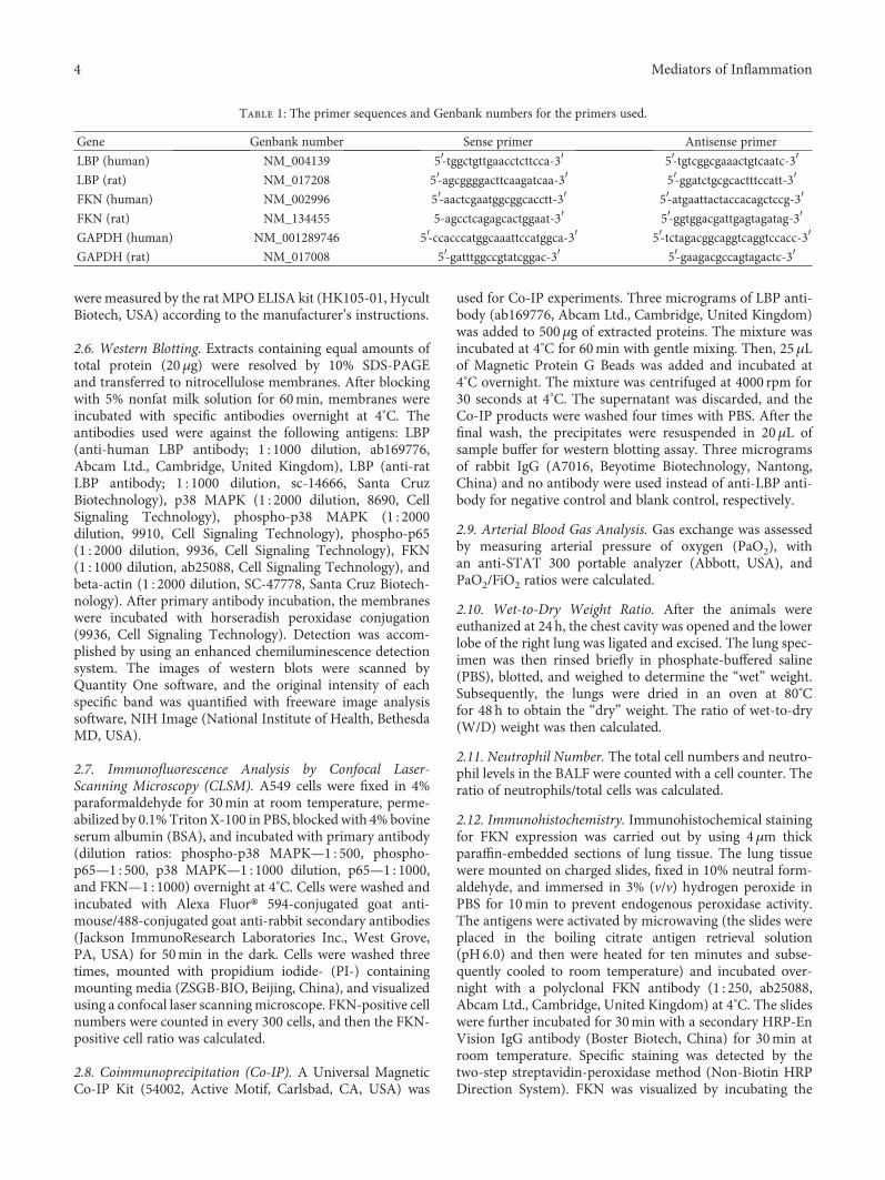
Table 1: The primer sequences and Genbank numbers for the primers used.
Gene Genbank number Sense primer Antisense primer
LBP (human) NM_004139 5′-tggctgttgaacctcttcca-3′ 5′-tgtcggcgaaactgtcaatc-3′LBP (rat) NM_017208 5′-agcggggacttcaagatcaa-3′ 5′-ggatctgcgcactttccatt-3′FKN (human) NM_002996 5′-aactcgaatggcggcacctt-3′ 5′-atgaattactaccacagctccg-3′FKN (rat) NM_134455 5-agcctcagagcactggaat-3′ 5′-ggtggacgattgagtagatag-3′GAPDH (human) NM_001289746 5′-ccacccatggcaaattccatggca-3′ 5′-tctagacggcaggtcaggtccacc-3′GAPDH (rat) NM_017008 5′-gatttggccgtatcggac-3′ 5′-gaagacgccagtagactc-3′
4 Mediators of Inflammation
0
1
2
3
4
⁎
⁎
⁎⁎
#⁎
#
LBP
mRN
A re
lativ
e qu
antifi
catio
n(fo
ld o
f con
trol)
CTL
LBP(
+)
LPS+
LBP(
+)
LPS+
LBP(
–)
LBP(–
)
LPS
Empt
y ve
ctor
0.0
0.5
1.0
1.5
2.0⁎ ⁎
⁎
#⁎
#
⁎LBP
(ng/
ml)
CTL
LBP(
+)
LPS+
LBP(
+)
LPS+
LBP(
–)
LBP(
–)
LPS
Empt
y ve
ctor
(a) (b)
LBP
�훽-actin
53 kD
43 kD
LPSCTL LPS+LBP(+)
LPS+LBP(–)
LBP(–)
LBP(+)
Emptyvector
⁎
0.0
0.5
1.0
1.5
⁎
⁎
⁎
⁎
#
#
LBP/�훽
-act
in re
lativ
e de
nsity
CTL
LBP(
+)
LPS+
LBP(
+)
LPS+
LBP(
–)
LBP(
–)
LPS
Empt
y ve
ctor
(c) (d)
0.0
0.5
1.0
1.5
2.0
FKN
mRN
A re
lativ
e qua
ntifi
catio
n(fo
ld o
f con
trol
)
CTL
LPS+
LBP(
+)
LPS+
LBP(
–)
LPS
LPS+
SB
LPS+
SC
TNF-�훼
⁎
⁎
#
⁎#⁎# ⁎#
⁎#
0.0
0.5
1.0
1.5
2.0
FKN
(ng/
mL)
CTL
LPS+
LBP(
+)
LPS+
LBP(
–)
LPS
LPS+
SB
LPS+
SC
TNF-�훼
⁎#
⁎#⁎#
⁎⁎
#
⁎#
(e) (f)
FKN
�훽-actin
90 kD
43 kD
LPSCTL LPS+LBP(+)
LPS+LBP(–)
LPS+SB
LPS+SC
TNF-�훼
0.0
0.5
1.0
1.5
⁎⁎#
⁎# ⁎#⁎#
⁎#
FKN
/�훽-a
ctin
rela
tive
dens
ity
CTL
LPS+
LBP(
+)
LPS+
LBP(
–)
LPS
LPS+
SB
LPS+
SC
TNF-�훼
(g) (h)
Figure 1: Continued.
5Mediators of Inflammation
sections with a solution of 3,3′-diaminobenzidine for 10min.Negative controls were incubated with normal rabbit seruminstead of the primary antibody. Then counterstaining wasperformed with hematoxylin. The immunoreactive cells werecounted in at least eight fields and expressed as a positive cellratio to the alveolar epithelium.
2.13. Statistical Analysis. Values were expressed asmeans ± standard errors of the means. Statistical analysiswas performed using SPSS for windows (version 16.0,Chicago, USA). Differences between the 2 groups werecompared by Student’s t-test. When multiple groups werecompared, analysis of variance was used and, if significance
was observed, post hoc comparisons between the groupsusing the method of Games-Howell were performed.pvalue < 0 05 was considered significant.
3. Results
3.1. LBP Gene Overexpression Further Decreased LPS-InducedDownregulation of FKN mRNA and Protein Expression;LBP Gene Silencing, SB203580, and SC-514 SuppressedLPS-Induced Downregulation of FKN mRNA and ProteinExpression, Respectively, in A549 Cells. RT-PCR, westernblotting, and ELISA were used to analyze the LBP and FKNmRNA and protein expression, respectively, in A549 cells.
20 �휇m20 �휇m
LPSCTL LPS+LBP(+)
LPS+LBP(–) LPS+SB LPS+SC
20 �휇m
20 �휇m 20 �휇m 20 �휇m 20 �휇m
TNF-�훼
0.0
0.2
0.4
0.6
0.8
1.0
⁎
⁎#
⁎# ⁎#⁎#
⁎#
FKN
-pos
itve c
ell r
atio
CTL
LPS+
LBP(
+)
LPS+
LBP(
–)
LPS
LPS+
SB
LPS+
SC
TNF-�훼
(i) (j)
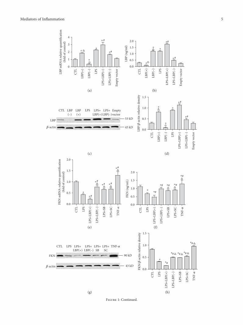
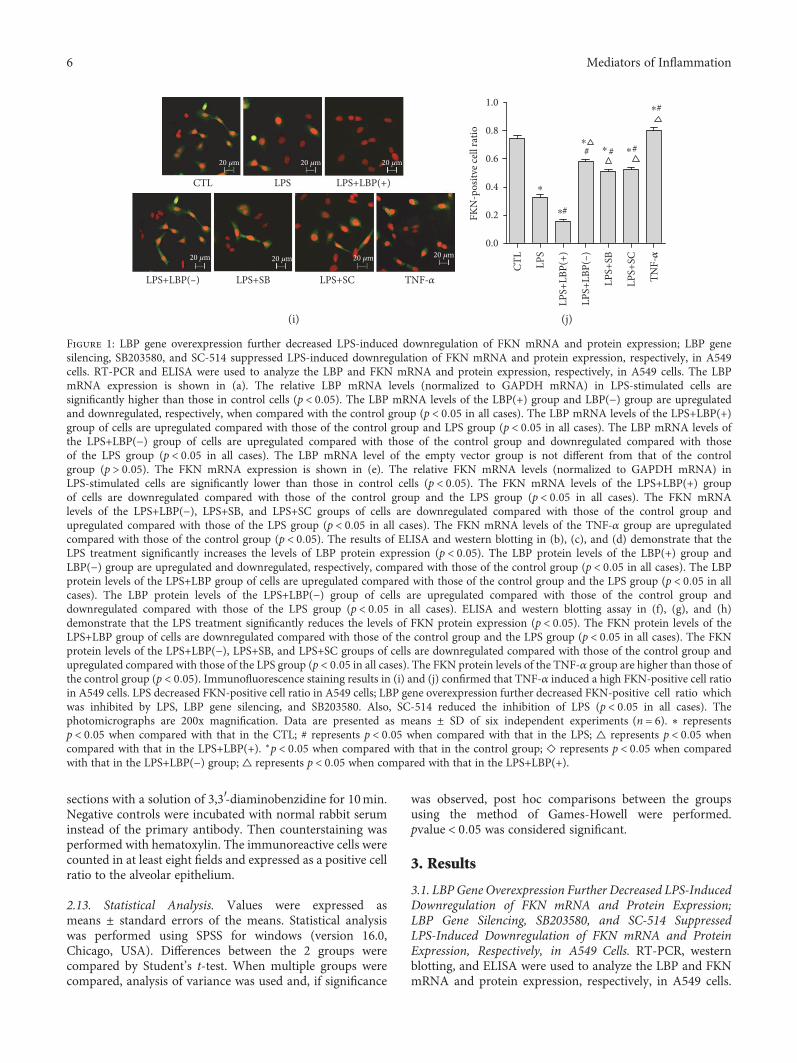
Figure 1: LBP gene overexpression further decreased LPS-induced downregulation of FKN mRNA and protein expression; LBP genesilencing, SB203580, and SC-514 suppressed LPS-induced downregulation of FKN mRNA and protein expression, respectively, in A549cells. RT-PCR and ELISA were used to analyze the LBP and FKN mRNA and protein expression, respectively, in A549 cells. The LBPmRNA expression is shown in (a). The relative LBP mRNA levels (normalized to GAPDH mRNA) in LPS-stimulated cells aresignificantly higher than those in control cells (p < 0 05). The LBP mRNA levels of the LBP(+) group and LBP(−) group are upregulatedand downregulated, respectively, when compared with the control group (p < 0 05 in all cases). The LBP mRNA levels of the LPS+LBP(+)group of cells are upregulated compared with those of the control group and LPS group (p < 0 05 in all cases). The LBP mRNA levels ofthe LPS+LBP(−) group of cells are upregulated compared with those of the control group and downregulated compared with thoseof the LPS group (p < 0 05 in all cases). The LBP mRNA level of the empty vector group is not different from that of the controlgroup (p > 0 05). The FKN mRNA expression is shown in (e). The relative FKN mRNA levels (normalized to GAPDH mRNA) inLPS-stimulated cells are significantly lower than those in control cells (p < 0 05). The FKN mRNA levels of the LPS+LBP(+) groupof cells are downregulated compared with those of the control group and the LPS group (p < 0 05 in all cases). The FKN mRNAlevels of the LPS+LBP(−), LPS+SB, and LPS+SC groups of cells are downregulated compared with those of the control group andupregulated compared with those of the LPS group (p < 0 05 in all cases). The FKN mRNA levels of the TNF-α group are upregulatedcompared with those of the control group (p < 0 05). The results of ELISA and western blotting in (b), (c), and (d) demonstrate that theLPS treatment significantly increases the levels of LBP protein expression (p < 0 05). The LBP protein levels of the LBP(+) group andLBP(−) group are upregulated and downregulated, respectively, compared with those of the control group (p < 0 05 in all cases). The LBPprotein levels of the LPS+LBP group of cells are upregulated compared with those of the control group and the LPS group (p < 0 05 in allcases). The LBP protein levels of the LPS+LBP(−) group of cells are upregulated compared with those of the control group anddownregulated compared with those of the LPS group (p < 0 05 in all cases). ELISA and western blotting assay in (f), (g), and (h)demonstrate that the LPS treatment significantly reduces the levels of FKN protein expression (p < 0 05). The FKN protein levels of theLPS+LBP group of cells are downregulated compared with those of the control group and the LPS group (p < 0 05 in all cases). The FKNprotein levels of the LPS+LBP(−), LPS+SB, and LPS+SC groups of cells are downregulated compared with those of the control group andupregulated compared with those of the LPS group (p < 0 05 in all cases). The FKN protein levels of the TNF-α group are higher than those ofthe control group (p < 0 05). Immunofluorescence staining results in (i) and (j) confirmed that TNF-α induced a high FKN-positive cell ratioin A549 cells. LPS decreased FKN-positive cell ratio in A549 cells; LBP gene overexpression further decreased FKN-positive cell ratio whichwas inhibited by LPS, LBP gene silencing, and SB203580. Also, SC-514 reduced the inhibition of LPS (p < 0 05 in all cases). Thephotomicrographs are 200x magnification. Data are presented as means ± SD of six independent experiments (n = 6). ∗ representsp < 0 05 when compared with that in the CTL; # represents p < 0 05 when compared with that in the LPS; △ represents p < 0 05 whencompared with that in the LPS+LBP(+). ∗p < 0 05 when compared with that in the control group; ◇ represents p < 0 05 when comparedwith that in the LPS+LBP(−) group; △ represents p < 0 05 when compared with that in the LPS+LBP(+).
6 Mediators of Inflammation
The LBP mRNA expression is shown in Figure 1(a). Therelative LBP mRNA levels (normalized to GAPDH mRNA)in LPS-stimulated cells were 1.25± 0.18 times higher thanthose in control cells (p < 0 05, n = 6). The LBP mRNA levelsof the LBP(+) group are 1.46± 0.27 times higher than thoseof the control group (p < 0 05, n = 6).The LBP mRNA levelsof the LBP(−) group were decreased by 62.67± 15.55% com-pared with those of the control group (p < 0 05, n = 6). TheLBP mRNA levels of the LPS+LBP(+) group of cells were2.96± 0.79 and 1.30± 0.28 times higher than those of thecontrol group and the LPS group, respectively (p < 0 05,n = 6). The LBP mRNA levels of the LPS+LBP(−) group ofcells were 2.65± 0.21 times higher and were decreased by70.22± 11.25% compared with those of the control groupand the LPS group, respectively (p < 0 05, n = 6, in all cases;Figure 1(a)). There is no difference between the empty vectorgroup and the control group (p > 0 05, n = 6). The FKNmRNA expression is shown in Figure 1(e). The relativeFKN mRNA levels (normalized to GAPDH mRNA) inLPS-stimulated cells decreased by 56.87± 16.42% comparedwith those seen in control cells (p < 0 05). The FKNmRNA levels of the LPS+LBP(+) group of cells decreasedby 77.52± 9.05% and 20.65± 15.10% compared with thoseof the control group and the LPS group, respectively(p < 0 05, n = 6). The FKN mRNA levels of the LPS+LBP(−),LPS+SB, and LPS+SC groups of cells decreased by 23.09± 22.07%, 34.22± 21.53%, and 32.75± 20.86% compared withthose of the control group and increased by 33.78± 19.80%,22.65± 7.57%, and 24.12± 10.58% compared with those ofthe LPS group, respectively (p < 0 05, n = 6). The FKNmRNA levels of the TNF-α group increase by 30.07± 4.36%compared with those of the control group (p < 0 05, n = 6).The results of western blotting and ELISA in Figures 1(b),1(c), and 1(d) demonstrate that after the LPS treatment,LBP plasmid DNA transfection increased 2.01± 1.33 and2.20± 1.01 times of LBP protein expression, respectively, incells (p < 0 05) and increased 3.58± 1.45 and 3.56± 1.45times of LBP protein expression, respectively, in superna-tants (p < 0 05). The LBP protein levels of the LBP(−) groupwere downregulated by 66.44± 15.04% and 61.44± 21.21%compared with those of the control group in cells andsupernatants, respectively (p < 0 05). The LBP protein levelsof the LPS+LBP(+) group of cells were upregulated by28.85± 23.11% and 48.22± 19.2% compared with thoseof the LPS group in cells and supernatants, respectively(p < 0 05). The LBP protein levels of the LPS+LBP(−)group of cells were downregulated by 46.88± 19.63% and60.70± 11.37% in cells and supernatants, respectively, com-pared with those of the LPS group (p < 0 05, n = 6). Westernblotting and ELISA assay results demonstrate that TNF-αincreased by 15.18± 22.36% and 15.05± 12.28% of FKNprotein expression in cells and supernatants, respectively;however, the LPS treatment significantly reduced by65.48± 8.45% and 38.25± 7.38% of FKN protein expressionin cells and supernatants, respectively (p < 0 05, n = 6). Com-pared with the control group and the LPS group, the FKNprotein levels of the LPS+LBP group of cells were down-regulated by 87.28± 6.32% and 64.36± 12.06% in cells and10.24± 6.52% and 47.37± 23.0% in supernatants (p < 0 05,
n = 6). The FKN protein levels of the LPS+LBP(−), LPS+SB, and LPS+SC groups of cells were downregulated by37.66± 14.55%, 40.37± 18.34%, and 31.77± 10.97%, respec-tively, in cells and downregulated by 57.47± 7.54%, 20.05± 8.25%, and 18.01± 5.92%, respectively, in supernatantscompared with those of the control group. The FKN proteinlevels of the LPS+LBP(−), LPS+SB, and LPS+SC groups ofcells were upregulated by 87.20± 52.05%, 84.43± 77.470%,and 102.2± 25.80%, respectively, in cells and upregulated by30.28± 14.69%, 30.03± 10.57%, and 33.74± 12.5%, respec-tively, in supernatants compared with those of the LPS group(p < 0 05, n = 6, in all cases). FKN positive cell ratios werecalculated to further verify the expression of FKN protein inthe cells of each group. In order to be a positive control, TNF-α induced a high FKN-positive cell ratio in A549 cells. LPSdecreased 55.49± 5.48% of the FKN-positive cell ratio inA549 cells. LBP gene overexpression further decreasedby 54.36± 21.25% of the FKN-positive cell ratio thatwas able to be inhibited by LPS. In addition, LBP genesilencing, SB203580, and SC-514 reduced by 84.36± 27.77%,54.84± 28.58%, and 58.48± 44.65% of the inhibition of LPS,respectively, by the different groups (p < 0 05, n = 6). BothFKN mRNA and protein expression were downregulatedfollowing LPS treatment with or without pretreatment of cul-tured A549 cells with LBP gene overexpression or silencing,SB203580, and SC-514 when compared with control cells.
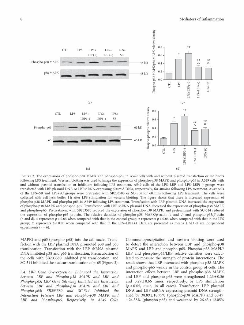
3.2. LBP Gene Overexpression Further Increased LPS-Induced p38 MAPK and p65 NF-κB Activation; LBP GeneSilencing, SB203580, and SC-514 Suppressed LPS-Inducedp38 MAPK and p65 NF-κB Activation, Respectively, inA549 Cells. Western blotting was used to image theexpression of phospho-p38 MAPK and phospho-p65 inA549 cells with and without plasmid transfection or inhib-itors following LPS treatment. The results show that therewere increases of 2.52± 0.86 and 2.73± 1.01 times of theexpression of phospho-p38 MAPK and phospho-p65,respectively, in A549 following LPS treatment (p < 0 05,n = 6, in all cases). Transfection with LBP plasmid DNAincreased the expression by 29.31± 13.83% and 38.73± 23.47% of phospho-p38 MAPK and phospho-p65,respectively (p < 0 05, n = 6, in all cases). Transfection withLBP shRNA plasmid DNA decreased the expression ofphospho-p38 MAPK by 37.20± 7.60% and the expressionof phospho-p65 by 30.14± 25.94% (p < 0 05, n = 6). Pretreat-ment with SB203580 reduced the expression of phospho-p38MAPK by 43.58± 7.90%, and pretreatment with SC-514reduced the expression of phospho-p65 protein by 24.03± 21.82% (p < 0 05, n = 6) (Figure 2).
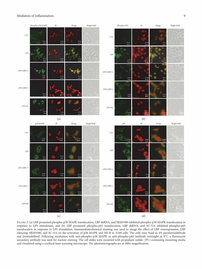
3.3. LBP Gene Overexpression Promoted Phospho-p38 MAPKand Phospho-p65 Translocation, LBP Gene SilencingInhibited Both Phospho-p38 MAPK and Phospho-p65Translocation, and SB203580 and SC-514 InhibitedPhospho-p38 MAPK and Phospho-p65 Translocation,Respectively, in Response to LPS Stimulation. Confocal imag-ing revealed that p38 MAPK, p65, phospho-p38 MAPK, andphospho-p65 were detectable within A549 cells. LPS inducedthe translocation of activated p38 MAPK (phospho-p38
7Mediators of Inflammation
MAPK) and p65 (phospho-p65) into the cell nuclei. Trans-fection with the LBP plasmid DNA promoted p38 and p65translocation. Transfection with the LBP shRNA plasmidDNA inhibited p38 and p65 translocation. Preincubation ofthe cells with SB203580 inhibited p38 translocation, andSC-514 inhibited the nuclear translocation of p-65 (Figure 3).
3.4. LBP Gene Overexpression Enhanced the Interactionbetween LBP and Phospho-p38 MAPK and LBP andPhospho-p65; LBP Gene Silencing Inhibited the Interactionbetween LBP and Phospho-p38 MAPK and LBP andPhospho-p65; SB203580 and SC-514 Inhibited theInteraction between LBP and Phospho-p38 MAPK andLBP and Phospho-p65, Respectively, in A549 Cells.
Coimmunoprecipitation and western blotting were usedto detect the interaction between LBP and phospho-p38MAPK and LBP and phospho-p65. Phospho-p38 MAPK/LBP and phospho-p65/LBP relative densities were calcu-lated to measure the strength of protein interactions. Theresult shows that LBP interacted with phospho-p38 MAPKand phospho-p65 weakly in the control group of cells. Theinteraction effects between LBP and phospho-p38 MAPKand LBP and phospho-p65 were strengthened 1.26± 0.36and 3.29± 0.66 times, respectively, by LPS stimulation(p < 0 05, n = 6, in all cases). Transfection LBP plasmidDNA and LBP shRNA-expressing plasmid DNA strength-ened by 38.89± 18.75% (phospho-p38 MAPK) and 50.49± 24.38% (phospho-p65) and weakened by 26.63± 12.05%
LPSCTL LPS+LBP(+)
LPS+LBP(–)
LPS+SB
Phospho-p38 MAPK
p38 MAPK
43 kD
43 kD
0.0
0.2
0.4
0.6
0.8
⁎
Phos
pho-
p38
MA
PK/p
38 M
APK
rela
tive
dens
ity
⁎#
CTL
LPS+
LBP(
+)
LPS+
LBP(
–)
LPS
LPS+
SB
⁎# ⁎#
(a) (b)
Phospho-p65
p65
65 kD
65 kD
LPSCTL LPS+LBP(+)
LPS+LBP(–)
LPS+SB
0.0
0.2
0.4
0.6
0.8
⁎
Phos
pho-
p65/
p65
rela
tive d
ensit
y ⁎#
CTL
LPS+
LBP(
+)
LPS+
LBP(
–)
LPS
LPS+
SC
⁎#⁎#
(c) (d)
Figure 2: The expressions of phospho-p38 MAPK and phospho-p65 in A549 cells with and without plasmid transfection or inhibitorsfollowing LPS treatment. Western blotting was used to image the expression of phospho-p38 MAPK and phospho-p65 in A549 cells withand without plasmid transfection or inhibitors following LPS treatment. A549 cells of the LPS+LBP and LPS+LBP(−) groups weretransfected with LBP plasmid DNA or LBPshRNA-expressing plasmid DNA, respectively, for 48mins following LPS treatment. A549 cellsof the LPS+SB and LPS+SC groups were pretreated with SB203580 or SC-514 for 60mins following LPS treatment. The cells werecollected with cell lysis buffer 1 h after LPS stimulation for western blotting. The figure shows that there is increased expression ofphospho-p38 MAPK and phospho-p65 in A549 following LPS treatment. Transfection with LBP plasmid DNA increased the expressionof phospho-p38 MAPK and phospho-p65. Transfection with LBP shRNA plasmid DNA decreased the expression of phospho-p38 MAPKand phospho-p65. Pretreatment with SB203580 reduced the expression of phospho-p38 MAPK, and pretreatment with SC-514 reducedthe expression of phospho-p65 protein. The relative densities of phospho-p38 MAPK/β-actin (a and c) and phospho-p65/β-actin(b and d). ∗ represents p < 0 05 when compared with that in the control group; # represents p < 0 05 when compared with that in the LPSgroup; △ represents p < 0 05 when compared with that in the LPS+LBP(+). Data are presented as means ± SD of six independentexperiments (n = 6).
8 Mediators of Inflammation
20 μm20 μm20 μm20 μm
20 μm
20 μm20 μm
20 μm
20 μm
Phospho-p38 MAPK PI Merge Bright field
CTL
LPS
LPS+LBP(+)
LPS+LBP(‒)
LPS+SC
20 μm 20 μm 20 μm
20 μm 20 μm 20 μm
20 μm 20 μm
20 μm 20 μm20 μm
Phospho-P65 PI Merge
CTL
Bright field
LPS
LPS+LBP(+)
LPS+LBP(‒)
LPS+SC
20 �휇m 20 �휇m 20 �휇m 20 �휇m
20 �휇m20 �휇m20 �휇m20 �휇m
20 �휇m 20�휇m20 �휇m 20�휇m
20 �휇m 20 �휇m 20 �휇m20 �휇m
20 �휇m 20 �휇m 20 �휇m 20 �휇m
(a) (b)
20 �휇m
20 �휇m
p38 MAPK PI Merge Bright field
CTL
LPS
LPS+LBP(+)
LPS+LBP(‒)
LPS+SB
20 �휇m 20 �휇m 20 �휇m
20 �휇m 20 �휇m 20 �휇m
20 �휇m 20 �휇m 20 �휇m 20 �휇m
20 �휇m 20 �휇m 20 �휇m 20 �휇m
20 �휇m 20 �휇m20 �휇m 20 �휇m
p65 PI Merge Bright field
CTL
LPS
LPS+LBP(+)
LPS+LBP(‒)
LPS+SC
20 �휇m20 �휇m20 �휇m 20 �휇m
20 �휇m 20 �휇m 20 �휇m 20 �휇m
20 �휇m20 �휇m 20 �휇m20 �휇m
20 �휇m20 �휇m 20 �휇m 20 �휇m
20 �휇m 20 �휇m 20 �휇m 20 �휇m
(c) (d)
Figure 3: (a) LBP promoted phospho-p38 MAPK translocation, LBP shRNA, and SB203580 inhibited phospho-p38 MAPK translocation inresponse to LPS stimulation, and (b) LBP promoted phospho-p65 translocation, LBP shRNA, and SC-514 inhibited phospho-p65translocation in response to LPS stimulation. Immunohistochemical staining was used to image the effect of LBP overexpression, LBPsilencing, SB203580, and SC-514 on the activation of p38 MAPK and NF-B in A549 cells. The cells were fixed in 4% paraformaldehydeand permeabilized. Following incubation with anti-phospho-p38 MAPK or anti-phospho-p65 antibody overnight at 4°C, a fluorescentsecondary antibody was used for nuclear staining. The cell slides were mounted with propidium iodide- (PI-) containing mounting mediaand visualized using a confocal laser scanning microscope. The photomicrographs are at 400x magnification.
9Mediators of Inflammation
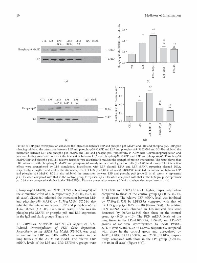
(phospho-p38 MAPK) and 29.95± 3.65% (phospho-p65) ofthe stimulation effect of LPS, respectively (p < 0 05, n = 6, inall cases). SB203580 inhibited the interaction between LBPand phospho-p38 MAPK by 31.78± 7.51%; SC-514 alsoinhibited the interaction between LBP and phospho-p65 by43.62± 8.35% (p < 0 05, n = 6, in all cases). There was nophospho-p38 MAPK or phospho-p65 and LBP expressionin the IgG and blank groups (Figure 4).
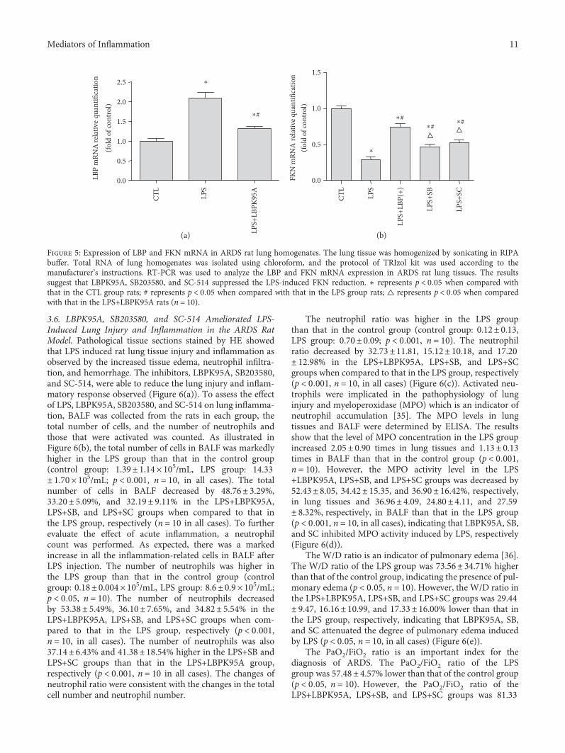
3.5. LBPK95A, SB203580, and SC-514 Suppressed LPS-Induced Downregulation of FKN Gene Expression,Respectively, in the ARDS Rat Model. RT-PCR was usedto analyze the LBP and FKN mRNA expression in thelung tissues of the ARDS rat model. The relative LBPmRNA levels of the LPS and LPS+LBPK95A groups were
2.09± 0.34 and 1.322± 0.12-fold higher, respectively, whencompared to those of the control group (p < 0 05, n = 10,in all cases). The relative LBP mRNA level was inhibitedby 77.10± 41.52% by LBPK95A compared with that ofthe LPS group (p < 0 05, n = 10) (Figure 5(a)). The relativeFKN mRNA levels observed in LPS-induced rats weredecreased by 70.72± 12.34% than those in the controlgroup (p < 0 05, n = 10). The FKN mRNA levels of thelung tissue in the LPS+LBPK95A, LPS+SB, and LPS+SCgroups of rat were downregulated by 25.90± 19.98%,53.47± 19.05%, and 47.387± 13.69%, respectively, comparedwith those in the control group and upregulated by44.82± 8.20%, 17.25± 3.32%, and 23.34± 12.01%, respec-tively, compared with those in the LPS group (p < 0 05,n = 10, in all cases) (Figure 5(b)).
LPSCTL LPS+LBP(+)
LPS+LBP(–)
IgGLPS+SB
Blank
53 kD
43 kDPhospho-p38 MAPK
LBP0.0
0.2
0.4
0.6
Phos
pho-
p38
MA
PK/L
BP re
lativ
e de
nsity
CTL
LPS+
LBP(
+)
LPS+
SB
LPS+
LBP(
–)
LPS
⁎
⁎#
⁎#
⁎#
(a) (b)
LBP
Phospho-p65
LPSCTL LPS+LBP(+)
LPS+LBP(–)
IgGLPS+SC
Blank
53 kD
65 kD
0.0
0.1
0.2
0.3
0.4
Phos
pho-
p65/
LBP
rela
tive d
ensit
y
CTL
LPS+
LBP(
+)
LPS+
SC
LPS+
LBP(
–)
LPS
⁎
⁎#
⁎ #
⁎#
(c) (d)
Figure 4: LBP gene overexpression enhanced the interaction between LBP and phospho-p38 MAPK and LBP and phospho-p65. LBP genesilencing inhibited the interaction between LBP and phospho-p38 MAPK and LBP and phospho-p65. SB203580 and SC-514 inhibited theinteraction between LBP and phospho-p38 MAPK and LBP and phospho-p65, respectively, in A549 cells. Coimmunoprecipitation andwestern blotting were used to detect the interaction between LBP and phospho-p38 MAPK and LBP and phospho-p65. Phospho-p38MAPK/LBP and phospho-p65/LBP relative densities were calculated to measure the strength of protein interactions. The result shows thatLBP interacted with phospho-p38 MAPK and phospho-p65 weakly in the control group of cells (p < 0 05 in all cases). The interactioneffects were strengthened by LPS stimulation. Transfection with LBP plasmid DNA and LBP shRNA-expressing plasmid DNA,respectively, strengthen and weaken the stimulatory effect of LPS (p < 0 05 in all cases). SB203580 inhibited the interaction between LBPand phospho-p38 MAPK; SC-514 also inhibited the interaction between LBP and phospho-p65 (p < 0 05 in all cases). ∗ representsp < 0 05 when compared with that in the control group; # represents p < 0 05 when compared with that in the LPS group; △ representsp < 0 05 when compared with that in the LPS+LBP(+). Data are presented as means ± SD of six independent experiments (n = 6).
10 Mediators of Inflammation
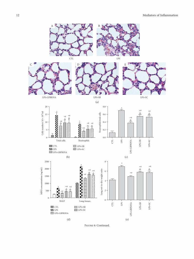
3.6. LBPK95A, SB203580, and SC-514 Ameliorated LPS-Induced Lung Injury and Inflammation in the ARDS RatModel. Pathological tissue sections stained by HE showedthat LPS induced rat lung tissue injury and inflammation asobserved by the increased tissue edema, neutrophil infiltra-tion, and hemorrhage. The inhibitors, LBPK95A, SB203580,and SC-514, were able to reduce the lung injury and inflam-matory response observed (Figure 6(a)). To assess the effectof LPS, LBPK95A, SB203580, and SC-514 on lung inflamma-tion, BALF was collected from the rats in each group, thetotal number of cells, and the number of neutrophils andthose that were activated was counted. As illustrated inFigure 6(b), the total number of cells in BALF was markedlyhigher in the LPS group than that in the control group(control group: 1.39± 1.14× 10⁵/mL, LPS group: 14.33± 1.70× 10⁵/mL; p < 0 001, n = 10, in all cases). The totalnumber of cells in BALF decreased by 48.76± 3.29%,33.20± 5.09%, and 32.19± 9.11% in the LPS+LBPK95A,LPS+SB, and LPS+SC groups when compared to that inthe LPS group, respectively (n = 10 in all cases). To furtherevaluate the effect of acute inflammation, a neutrophilcount was performed. As expected, there was a markedincrease in all the inflammation-related cells in BALF afterLPS injection. The number of neutrophils was higher inthe LPS group than that in the control group (controlgroup: 0.18± 0.004× 10⁵/mL, LPS group: 8.6± 0.9× 10⁵/mL;p < 0 05, n = 10). The number of neutrophils decreasedby 53.38± 5.49%, 36.10± 7.65%, and 34.82± 5.54% in theLPS+LBPK95A, LPS+SB, and LPS+SC groups when com-pared to that in the LPS group, respectively (p < 0 001,n = 10, in all cases). The number of neutrophils was also37.14± 6.43% and 41.38± 18.54% higher in the LPS+SB andLPS+SC groups than that in the LPS+LBPK95A group,respectively (p < 0 001, n = 10 in all cases). The changes ofneutrophil ratio were consistent with the changes in the totalcell number and neutrophil number.
The neutrophil ratio was higher in the LPS groupthan that in the control group (control group: 0.12± 0.13,LPS group: 0.70± 0.09; p < 0 001, n = 10). The neutrophilratio decreased by 32.73± 11.81, 15.12± 10.18, and 17.20± 12.98% in the LPS+LBPK95A, LPS+SB, and LPS+SCgroups when compared to that in the LPS group, respectively(p < 0 001, n = 10, in all cases) (Figure 6(c)). Activated neu-trophils were implicated in the pathophysiology of lunginjury and myeloperoxidase (MPO) which is an indicator ofneutrophil accumulation [35]. The MPO levels in lungtissues and BALF were determined by ELISA. The resultsshow that the level of MPO concentration in the LPS groupincreased 2.05± 0.90 times in lung tissues and 1.13± 0.13times in BALF than that in the control group (p < 0 001,n = 10). However, the MPO activity level in the LPS+LBPK95A, LPS+SB, and LPS+SC groups was decreased by52.43± 8.05, 34.42± 15.35, and 36.90± 16.42%, respectively,in lung tissues and 36.96± 4.09, 24.80± 4.11, and 27.59± 8.32%, respectively, in BALF than that in the LPS group(p < 0 001, n = 10, in all cases), indicating that LBPK95A, SB,and SC inhibited MPO activity induced by LPS, respectively(Figure 6(d)).
The W/D ratio is an indicator of pulmonary edema [36].The W/D ratio of the LPS group was 73.56± 34.71% higherthan that of the control group, indicating the presence of pul-monary edema (p < 0 05, n = 10). However, the W/D ratio inthe LPS+LBPK95A, LPS+SB, and LPS+SC groups was 29.44± 9.47, 16.16± 10.99, and 17.33± 16.00% lower than that inthe LPS group, respectively, indicating that LBPK95A, SB,and SC attenuated the degree of pulmonary edema inducedby LPS (p < 0 05, n = 10, in all cases) (Figure 6(e)).
The PaO2/FiO2 ratio is an important index for thediagnosis of ARDS. The PaO2/FiO2 ratio of the LPSgroup was 57.48± 4.57% lower than that of the control group(p < 0 05, n = 10). However, the PaO2/FiO2 ratio of theLPS+LBPK95A, LPS+SB, and LPS+SC groups was 81.33
0.0
0.5
1.0
1.5
2.0
2.5LB
P m
RNA
rela
tive
quan
tifica
tion
(fold
of c
ontr
ol)
CTL
LPS+
LBPK
95A
LPS
⁎
⁎#
0.0
0.5
1.0
1.5
FKN
mRN
A re
lativ
e qua
ntifi
catio
n(fo
ld o
f con
trol
)
CTL
LPS+
LBP(
+)
LPS+
SC
LPS+
SBLPS
⁎#⁎#
⁎
⁎#
(a) (b)
Figure 5: Expression of LBP and FKN mRNA in ARDS rat lung homogenates. The lung tissue was homogenized by sonicating in RIPAbuffer. Total RNA of lung homogenates was isolated using chloroform, and the protocol of TRIzol kit was used according to themanufacturer’s instructions. RT-PCR was used to analyze the LBP and FKN mRNA expression in ARDS rat lung tissues. The resultssuggest that LBPK95A, SB203580, and SC-514 suppressed the LPS-induced FKN reduction. ∗ represents p < 0 05 when compared withthat in the CTL group rats; # represents p < 0 05 when compared with that in the LPS group rats; △ represents p < 0 05 when comparedwith that in the LPS+LBPK95A rats (n = 10).
11Mediators of Inflammation
LPSCTL
LPS+LPBK95A LPS+SB LPS+SC
A
C D E
B
(a)
0
5
10
15
20
CTLLPSLPS+LBPK95A
LPS+SBLPS+SC
Cells
num
bers
10
× 10
5 /ml
Total cells Neutrophils
⁎
⁎
⁎#
⁎#
⁎#
⁎#
⁎#
⁎#
0.0
0.2
0.4
0.6
0.8
Neu
trop
hils/
tota
l cel
ls
⁎#
⁎
⁎
#
⁎#CT
L
LPS+
LBPK
95A
LPS+
SC
LPS+
SBLPS
(b) (c)
BALF Lung tissues0
500
1000
1500
2000
2500
CTLLPSLPS+LBPK95A
LPS+SBLPS+SC
MPO
conc
entr
atio
ns (n
g/m
L)
⁎
⁎#
⁎# ⁎#
⁎
⁎#
⁎# ⁎#
0
2
4
6
8
Lung
wet
-to-d
ry w
eigh
t rat
io
CTL
LPS+
LBPK
95A
LPS+
SC
LPS+
SBLPS
⁎#
⁎
⁎
#
⁎#
(d) (e)
Figure 6: Continued.
12 Mediators of Inflammation
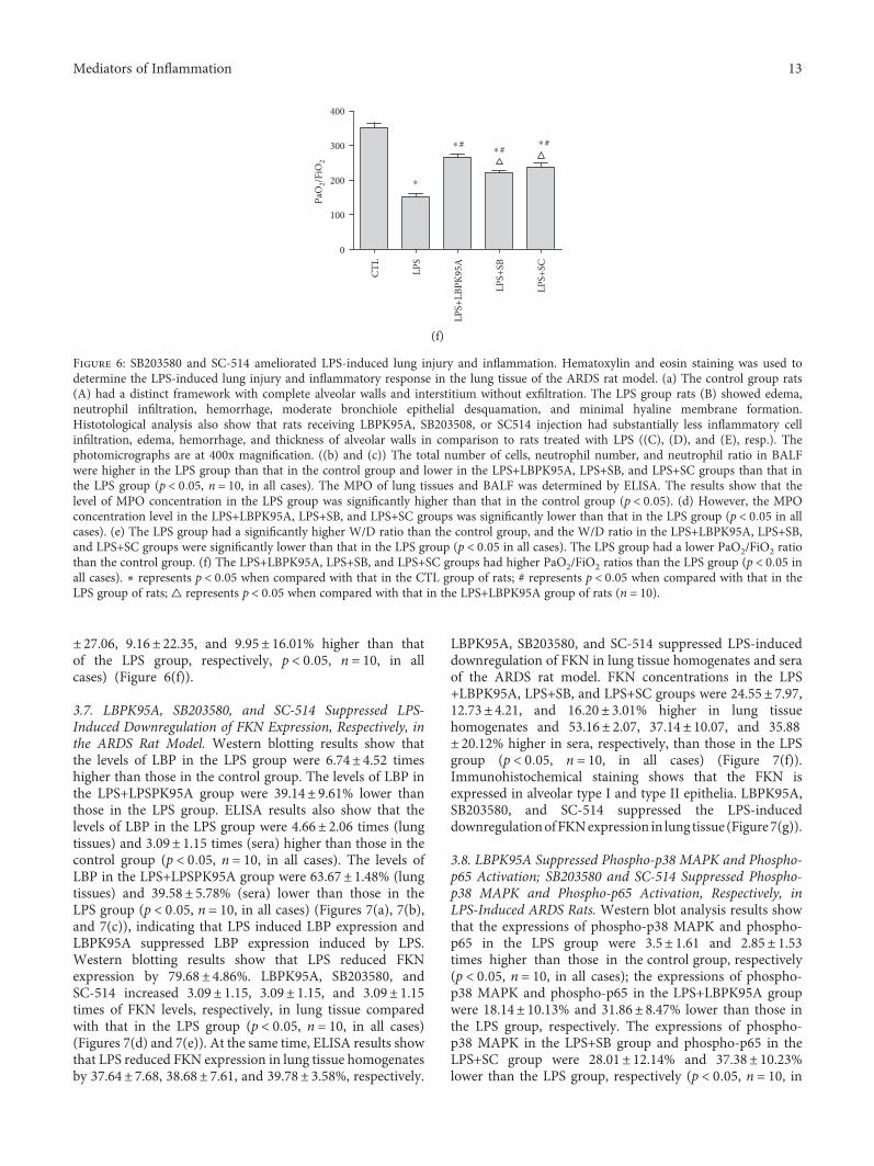
± 27.06, 9.16± 22.35, and 9.95± 16.01% higher than thatof the LPS group, respectively, p < 0 05, n = 10, in allcases) (Figure 6(f)).
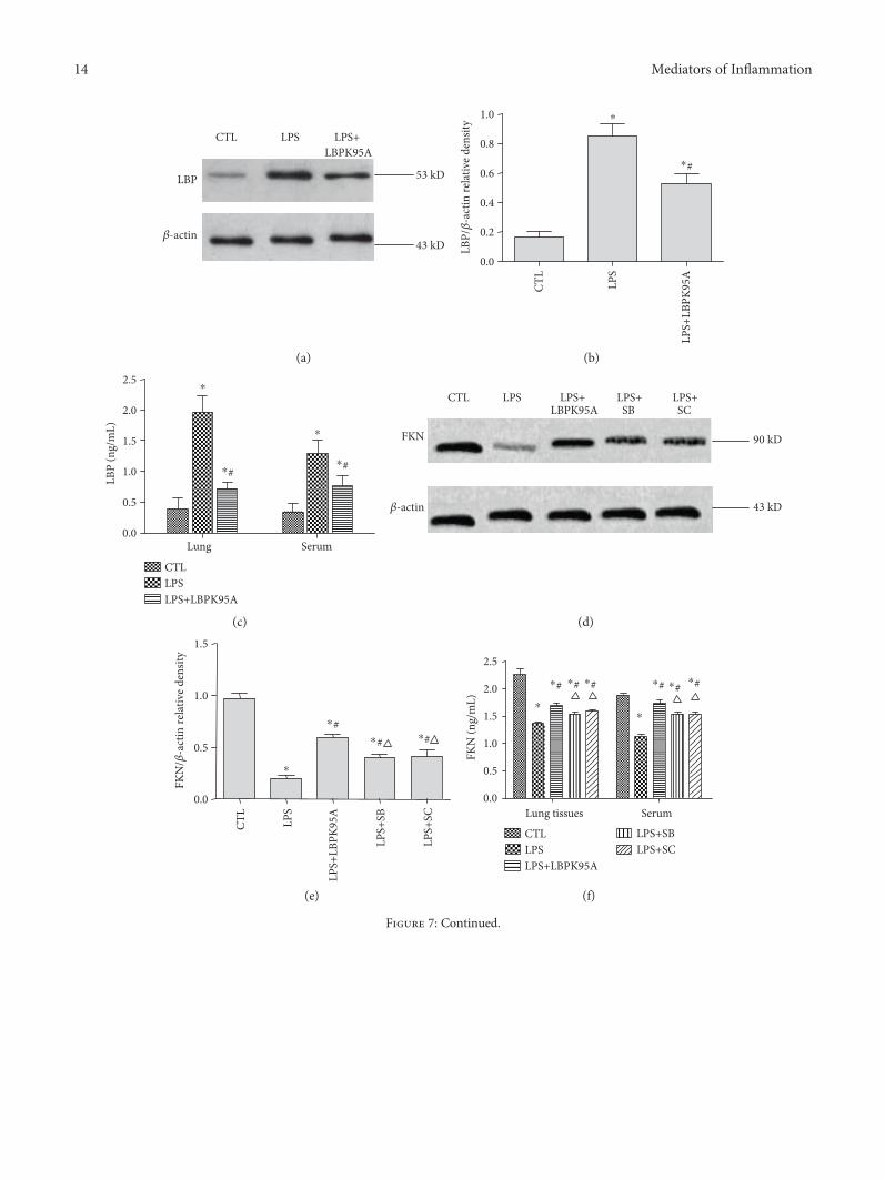
3.7. LBPK95A, SB203580, and SC-514 Suppressed LPS-Induced Downregulation of FKN Expression, Respectively, inthe ARDS Rat Model. Western blotting results show thatthe levels of LBP in the LPS group were 6.74± 4.52 timeshigher than those in the control group. The levels of LBP inthe LPS+LPSPK95A group were 39.14± 9.61% lower thanthose in the LPS group. ELISA results also show that thelevels of LBP in the LPS group were 4.66± 2.06 times (lungtissues) and 3.09± 1.15 times (sera) higher than those in thecontrol group (p < 0 05, n = 10, in all cases). The levels ofLBP in the LPS+LPSPK95A group were 63.67± 1.48% (lungtissues) and 39.58± 5.78% (sera) lower than those in theLPS group (p < 0 05, n = 10, in all cases) (Figures 7(a), 7(b),and 7(c)), indicating that LPS induced LBP expression andLBPK95A suppressed LBP expression induced by LPS.Western blotting results show that LPS reduced FKNexpression by 79.68± 4.86%. LBPK95A, SB203580, andSC-514 increased 3.09± 1.15, 3.09± 1.15, and 3.09± 1.15times of FKN levels, respectively, in lung tissue comparedwith that in the LPS group (p < 0 05, n = 10, in all cases)(Figures 7(d) and 7(e)). At the same time, ELISA results showthat LPS reduced FKN expression in lung tissue homogenatesby 37.64± 7.68, 38.68± 7.61, and 39.78± 3.58%, respectively.
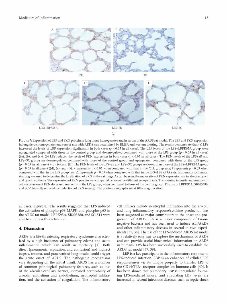
LBPK95A, SB203580, and SC-514 suppressed LPS-induceddownregulation of FKN in lung tissue homogenates and seraof the ARDS rat model. FKN concentrations in the LPS+LBPK95A, LPS+SB, and LPS+SC groups were 24.55± 7.97,12.73± 4.21, and 16.20± 3.01% higher in lung tissuehomogenates and 53.16± 2.07, 37.14± 10.07, and 35.88± 20.12% higher in sera, respectively, than those in the LPSgroup (p < 0 05, n = 10, in all cases) (Figure 7(f)).Immunohistochemical staining shows that the FKN isexpressed in alveolar type I and type II epithelia. LBPK95A,SB203580, and SC-514 suppressed the LPS-induceddownregulationofFKNexpression in lung tissue (Figure7(g)).
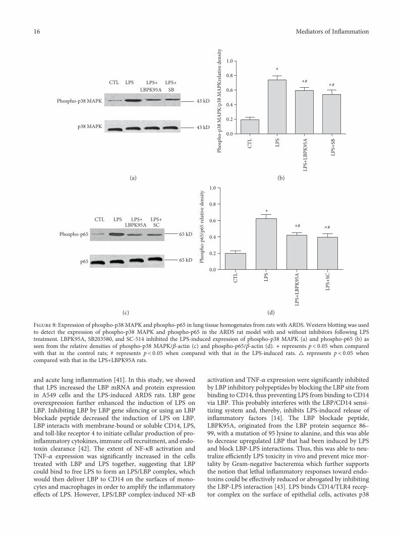
3.8. LBPK95A Suppressed Phospho-p38 MAPK and Phospho-p65 Activation; SB203580 and SC-514 Suppressed Phospho-p38 MAPK and Phospho-p65 Activation, Respectively, inLPS-Induced ARDS Rats. Western blot analysis results showthat the expressions of phospho-p38 MAPK and phospho-p65 in the LPS group were 3.5± 1.61 and 2.85± 1.53times higher than those in the control group, respectively(p < 0 05, n = 10, in all cases); the expressions of phospho-p38 MAPK and phospho-p65 in the LPS+LBPK95A groupwere 18.14± 10.13% and 31.86± 8.47% lower than those inthe LPS group, respectively. The expressions of phospho-p38 MAPK in the LPS+SB group and phospho-p65 in theLPS+SC group were 28.01± 12.14% and 37.38± 10.23%lower than the LPS group, respectively (p < 0 05, n = 10, in
0
100
200
300
400
PaO
2/FiO
2
CTL
LPS+
LBPK
95A
LPS+
SC
LPS+
SBLPS
⁎#⁎
⁎
# ⁎#
(f)
Figure 6: SB203580 and SC-514 ameliorated LPS-induced lung injury and inflammation. Hematoxylin and eosin staining was used todetermine the LPS-induced lung injury and inflammatory response in the lung tissue of the ARDS rat model. (a) The control group rats(A) had a distinct framework with complete alveolar walls and interstitium without exfiltration. The LPS group rats (B) showed edema,neutrophil infiltration, hemorrhage, moderate bronchiole epithelial desquamation, and minimal hyaline membrane formation.Histotological analysis also show that rats receiving LBPK95A, SB203508, or SC514 injection had substantially less inflammatory cellinfiltration, edema, hemorrhage, and thickness of alveolar walls in comparison to rats treated with LPS ((C), (D), and (E), resp.). Thephotomicrographs are at 400x magnification. ((b) and (c)) The total number of cells, neutrophil number, and neutrophil ratio in BALFwere higher in the LPS group than that in the control group and lower in the LPS+LBPK95A, LPS+SB, and LPS+SC groups than that inthe LPS group (p < 0 05, n = 10, in all cases). The MPO of lung tissues and BALF was determined by ELISA. The results show that thelevel of MPO concentration in the LPS group was significantly higher than that in the control group (p < 0 05). (d) However, the MPOconcentration level in the LPS+LBPK95A, LPS+SB, and LPS+SC groups was significantly lower than that in the LPS group (p < 0 05 in allcases). (e) The LPS group had a significantly higher W/D ratio than the control group, and the W/D ratio in the LPS+LBPK95A, LPS+SB,and LPS+SC groups were significantly lower than that in the LPS group (p < 0 05 in all cases). The LPS group had a lower PaO2/FiO2 ratiothan the control group. (f) The LPS+LBPK95A, LPS+SB, and LPS+SC groups had higher PaO2/FiO2 ratios than the LPS group (p < 0 05 inall cases). ∗ represents p < 0 05 when compared with that in the CTL group of rats; # represents p < 0 05 when compared with that in theLPS group of rats; △ represents p < 0 05 when compared with that in the LPS+LBPK95A group of rats (n = 10).
13Mediators of Inflammation
LPSCTL LPS+LBPK95A
53 kD
43 kD
LBP
�훽-actin
0.0
0.2
0.4
0.6
0.8
1.0
LBP/�훽
-act
in re
lativ
e de
nsity
CTL
LPS+
LBPK
95A
LPS
⁎
⁎#
(a) (b)
SerumLung
CTLLPSLPS+LBPK95A
0.0
0.5
1.0
1.5
2.0
2.5
LBP
(ng/
mL)
⁎
⁎#
⁎
⁎#
LPS+SC
FKN
�훽-actin
LPSCTL LPS+LBPK95A
LPS+SB
90 kD
43 kD
(c) (d)
0.0
0.5
1.0
1.5
FKN
/�훽-a
ctin
rela
tive
dens
ity
CTL
LPS+
LBPK
95A
LPS+
SC
LPS+
SBLPS
⁎#
⁎
⁎
#⁎#
SerumLung tissues
CTLLPSLPS+LBPK95A
LPS+SBLPS+SC
0.0
0.5
1.0
1.5
2.0
2.5
FKN
(ng/
mL) ⁎
⁎# ⁎# ⁎# ⁎
⁎
# ⁎#⁎#
(e) (f)
Figure 7: Continued.
14 Mediators of Inflammation
all cases; Figure 8). The results suggested that LPS inducedthe activation of phospho-p38 MAPK and phospho-p65 inthe ARDS rat model. LBPK95A, SB203580, and SC-514 wereable to suppress this activation.
4. Discussion
ARDS is a life-threatening respiratory syndrome character-ized by a high incidence of pulmonary edema and acuteinflammation which can result in mortality [1]. Bothdirect (pneumonia, aspiration, and contusion) and indirect(sepsis, trauma, and pancreatitis) lung insults could triggerthe acute onset of ARDS. The pathogenic mechanismsvary depending on the initial insult. ARDS has a numberof common pathological pulmonary features, such as lossof the alveolar-capillary barrier, increased permeability ofalveolar epithelium and endothelium, neutrophil infiltra-tion, and the activation of coagulation. The inflammatory
cell influxes include neutrophil infiltration into the alveoli,and lung inflammatory response/cytokine production hasbeen suggested as major contributors to the onset and pro-gression of ARDS. LPS is a major component of Gram-negative bacteria and has been used to induce ALI/ARDSand other inflammatory diseases in several in vivo experi-ments [37, 38]. The use of the LPS-induced ARDS rat modelis a relatively easy way to explore the mechanisms of ARDSand can provide useful biochemical information on ARDSin humans. LPS has been successfully used to establish theARDS rat model [37, 39].
LBP is a key participant in the inflammatory response toLPS-induced infection. LBP is an enhancer of cellular LPSresponsiveness via its unique property to transfer LPS tothe CD14/TLR4 receptor complex on immune cells [40]. Ithas been shown that pulmonary LBP is upregulated follow-ing LPS-mediated injury, and circulating LBP levels areincreased in several infectious diseases, such as septic shock
CTL LPS
LPS+LBPK95A LPS+SB LPS+SC
A
C D E
B
(g)
Figure 7: Expression of LBP and FKN protein in lung tissue homogenates and in serum of the ARDS rat model. The LBP and FKN expressionin lung tissue homogenates and sera of rats with ARDS was determined by ELISA and western blotting. The results demonstrate that (a) LPSincreased the levels of LBP expression significantly in both cases (p < 0 05 in all cases). The LBP levels of the LPS+LBPK95A group wereupregulated compared with those of the control group and downregulated compared with those of the LPS group (p < 0 05 in all cases)((a), (b), and (c)). (b) LPS reduced the levels of FKN expression in both cases (p < 0 05 in all cases). The FKN levels of the LPS+SB andLPS+SC groups are downregulated compared with those of the control group and upregulated compared with those of the LPS group(p < 0 05 in all cases) ((d), (e), and (f)). The FKN levels of the LPS+SB and LPS+SC groups are lower than those of the LPS+LBPK95A group(p < 0 05 in all cases) ((d), (e), and (f)). ∗ represents p < 0 05 when compared with that in the CTL group rats; # represents p < 0 05 whencompared with that in the LPS group rats.△ represents p < 0 05 when compared with that in the LPS+LBPK95A rats. Immunohistochemicalstaining was used to determine the localization of FKN in the rat lungs. As can be seen, the major sites of FKN expression are in alveolar type Iand type II epithelia. The expression of FKN protein was compared between the different groups of rats. The staining intensity and number ofcells expression of FKN decreased markedly in the LPS group, when compared to those of the control group. The use of LBPK95A, SB203580,and SC-514 partly reduced the reduction of FKN seen (g). The photomicrographs are at 400x magnification.
15Mediators of Inflammation
and acute lung inflammation [41]. In this study, we showedthat LPS increased the LBP mRNA and protein expressionin A549 cells and the LPS-induced ARDS rats. LBP geneoverexpression further enhanced the induction of LPS onLBP. Inhibiting LBP by LBP gene silencing or using an LBPblockade peptide decreased the induction of LPS on LBP.LBP interacts with membrane-bound or soluble CD14, LPS,and toll-like receptor 4 to initiate cellular production of pro-inflammatory cytokines, immune cell recruitment, and endo-toxin clearance [42]. The extent of NF-κB activation andTNF-α expression was significantly increased in the cellstreated with LBP and LPS together, suggesting that LBPcould bind to free LPS to form an LPS/LBP complex, whichwould then deliver LBP to CD14 on the surfaces of mono-cytes and macrophages in order to amplify the inflammatoryeffects of LPS. However, LPS/LBP complex-induced NF-κB
activation and TNF-α expression were significantly inhibitedby LBP inhibitory polypeptides by blocking the LBP site frombinding to CD14, thus preventing LPS from binding to CD14via LBP. This probably interferes with the LBP/CD14 sensi-tizing system and, thereby, inhibits LPS-induced release ofinflammatory factors [14]. The LBP blockade peptide,LBPK95A, originated from the LBP protein sequence 86–99, with a mutation of 95 lysine to alanine, and this was ableto decrease upregulated LBP that had been induced by LPSand block LBP-LPS interactions. Thus, this was able to neu-tralize efficiently LPS toxicity in vivo and prevent mice mor-tality by Gram-negative bacteremia which further supportsthe notion that lethal inflammatory responses toward endo-toxins could be effectively reduced or abrogated by inhibitingthe LBP-LPS interaction [43]. LPS binds CD14/TLR4 recep-tor complex on the surface of epithelial cells, activates p38
Phospho-p38 MAPK
p38 MAPK
LPSCTL LPS+LBPK95A
LPS+SB
43 kD
43 kD0.0
0.2
0.4
0.6
0.8
1.0
Phos
pho-
p38
MA
PK/p
38 M
APK
rela
tive d
ensit
y
⁎
⁎#⁎#
CTL
LPS+
LBPK
95A
LPS+
SBLPS
(a) (b)
Phospho-p65
p65
LPSCTL LPS+LBPK95A
LPS+SC
65 kD
65 kD
0.0
0.2
0.4
0.6
0.8
1.0
Phos
pho-
p65/
p65
rela
tive
dens
ity⁎
⁎#⁎#CT
L
LPS+
LBPK
95A
LPS+
SCLPS
(c) (d)
Figure 8: Expression of phospho-p38 MAPK and phospho-p65 in lung tissue homogenates from rats with ARDS. Western blotting was usedto detect the expression of phospho-p38 MAPK and phospho-p65 in the ARDS rat model with and without inhibitors following LPStreatment. LBPK95A, SB203580, and SC-514 inhibited the LPS-induced expression of phospho-p38 MAPK (a) and phospho-p65 (b) asseen from the relative densities of phospho-p38 MAPK/β-actin (c) and phospho-p65/β-actin (d). ∗ represents p < 0 05 when comparedwith that in the control rats; # represents p < 0 05 when compared with that in the LPS-induced rats. △ represents p < 0 05 whencompared with that in the LPS+LBPK95A rats.
16 Mediators of Inflammation
MAPK and NF-κB signal transduction pathways through themyeloid differentiation factor 88- (MyD88-) dependent path-way, and then induces the expression of many inflammatorymarkers [44, 45]. p38 MAPK and NF-κB pathways have beenreported to be involved in the release of proinflammatorymediators in ARDS. In vivo, treatment with SB203580substantially inhibited LPS-induced neutrophil recruitmentinto the lungs and changes in lung injury parameters, suchas total protein content in BAL fluid and apoptosis of neu-trophils and macrophages [46, 47]. Inhibition of p38MAPK decreased injury to the lung through attenuatedproduction of TNF-α and nitric oxide in the rat modelof pancreatitis-induced ARDS [48]. SB203580 is a classicalinhibitor of p38 MAPK that prevented the activation andphosphorylation of MAPKAP kinase-2 and the phosphor-ylation of hsp27 by cellular stresses, LPS, and IL-1 [49].NF-κB is an important transcription factor and mediatesthe expression of many inflammatory cytokines and celladhesion molecules during inflammation. The transcrip-tional activity of NF-κB depends on the posttranslationalmodification of p65. SC-514 is a cell-permeable, potent, andselective ATP competitive inhibitor of NF-κB-2 (Iκκ-2).Iκκ-2 inhibition by SC-514 demonstrates a decreased levelof IKK-2 phosphorylation/degradation, and diminishedLPS induces p65 translocation into the nucleus [50]. In thisstudy, we show that LPS not only induced phospho-p38MAPK and phospho-p65 expression but also promotedphospho-p38 MAPK and phospho-p65 transfer to thenucleus and enhanced interaction between LBP andphospho-p38 MAPK and LBP and phospho-p65, respec-tively. LBP gene overexpression and LBP gene silencingstrengthened and weakened the effect induced by LPS,respectively. The LBP blockade peptide, LBPK95A, inhibitedphospho-p38 MAPK and phospho-p65 increases in lung tis-sues of the LPS-induced ARDS rats. These results suggestthat LPS induced p38 MAPK and NF-κB signaling pathwaysactivation by LBP.
FKN has been recognized as a proinflammatory cytokine.FKN contributed to several inflammatory disorders, suchas acute necrotizing pancreatitis, tuberculosis, and sepsis[16, 51, 52]. FKN binds to its receptor CX3CR1, promotingneutrophil adhesion to epithelial cells, migration and tissueinvasion, production of oxygen free radicals, and generalexpansion of the inflammatory reaction [53]. FKN can beinduced by inflammatory mediators such as IFN-γ andTNF-α but is reduced by some inflammatory mediators suchas TGF-β [54]. FKN has been reported to be decreased insome inflammatory diseases, such as urogenital tract inflam-mation, which therefore implies an anti-inflammatory effectof FKN [55]. Recent research has shown that FKN attenuatedthe LPS-induced production of IL-1, IL-6, and TNF-α byrat and mouse microglia, which are phagocytotic cells thatare responsible for cytokine production in the CNS [56].Exogenous FKN injected into the mice did not increasethe expression of TNF-α, while the use of anti-FKN antibod-ies enhanced the inflammatory effect of LPS [57]. However,the anti-FKN antibody alone demonstrated similar toxicityto LPS and, in combination with anti-FKN antibody, wasable to enhance LPS toxicity.
In this study, we showed that LPS increased the LBPmRNA and protein expression and reduced the FKN produc-tion both in cultured A549 cells and in an ARDS rat model.Overexpression of LBP gene by transfection with LBP plas-mid further decreased LPS-induced downregulation of FKNmRNA and protein expression whereas silencing of the LBPgene inhibited LPS-induced downregulation of FKN.SB203580 and SC-514 pretreated A549 cells resulted in theinhibition of p38 MAPK and p65 NF-κB transduction,respectively, and the suppressive effect on the LPS-inducedreduction of FKN. These results suggest that both p38 MAPKand NF-κB are involved in the release of FKN in A549 cells.LBP increased LPS-induced reduction of FKN by activatingboth p38 MAPK and p65 NF-κB signaling pathwaysin vitro. Immunohistochemical staining results indicated thatpretreatment of the ARDS rats with LBPK95A, SB203580,and SC-514 reduced lung injury caused by LPS. The resultsof both in vitro and in vivo experiments demonstrated thatLPS reduced FKN expression and caused rat lung injuryand that LBPK95A, SB203580, and SC-514 inhibited theseeffects and reduced lung injury in a p38 MAPK- andNF-κB-dependent manner. These results suggest an anti-inflammatory and protective effect of FKN in ARDS. FKNcould therefore be considered as a potential therapeutictarget for regulating the inflammatory response in ARDS.
In this study, we show that LPS inhibited FKN expressionthrough p38 MAPK and NF-κB signaling pathways, butsome studies have demonstrated an increased expression ofFKN in chronic respiratory disease such as asthma andchronic obstructive pulmonary disease through p38 MAPKor NF-κB signaling pathways. Previous reports have shownthat FKNmainly upregulated expression in the airway tissuessuch as airway epithelium, submucosa, and smooth muscle ofchronic respiratory disease, but we show that LPS inhibitedFKN expression in lung tissues especially in alveolar epithe-lial cells of acute inflammatory lung disease. Some studiesshow that TNF-α-stimulated VSMC fractalkine mRNA andprotein expression are attenuated by pharmacologic inhibi-tors of PKC and p42/44 MAPK kinase, but not p38 MAPK,indicating that the intracellular signals mediating TNF-α-stimulated fractalkine expression involve the activation ofPKC and p42/44 MAPK, rather than p38 MAPK pathway[31]. In most studies where FKN is shown to be increased,NF-κB is activated by a classical heterodimer of NF-κB(p65-p50) formation [58]. However, in this study, FKNdecreased when induced by LPS and this was inhibited bySC-514 which is a selective ATP competitive inhibitor ofNF-κB-2 (Iκκ-2, p52) and it is able to block p65-p52 hetero-dimers but not p65-p50.
Although we were able to show the mechanism of LBPregulation of FKN and its related signaling pathways in acultured cell line and in an ARDS rat model, furtherresearch is required to investigate the anti-inflammatoryeffects of FKN and its potential role in ARDS treatment.
5. Conclusions
In conclusion, the data presented here show that LBP furtherdecreased LPS-induced reduction of FKN and LPS-induced
17Mediators of Inflammation
lung injury and these effects can be prevented by transfectionwith LBP shRNA plasmid or using LBP inhibitory peptideand inhibition of the activation of p38 MAPK and NF-κB signaling transduction pathways, suggesting that LBPdownregulates FKN expression through the activation ofp38 MAPK and NF-κB in the progression of ARDS.
Abbreviations
ARDS: Acute respiratory distress syndromeBALF: Bronchoalveolar lavage fluidCo-IP: CoimmunoprecipitationFKN: FractalkineGAPDH: Glyceraldehyde-3-phosphate dehydrogenaseLBP: LPS-binding proteinLPS: LipopolysaccharideMAPK: Mitogen-activated protein kinasesMPO: MyeloperoxidaseNF-κB: Nuclear factor-κBTNF-α: Tumor necrosis factor-α.
Ethical Approval
The study was approved by the Committee of Animal Careand Use of Youjiang Medical University for Nationalities,and all procedures were performed according to the NationalInstitute of Health Guidelines.
Conflicts of Interest
The authors declare that they have no competing interest.
Authors’ Contributions
Xia Huang, Yi Zeng, Yujie Jiang, Yueqiu Qin, WeiguiLuo, and Shuling Xiang were involved with acquisition,analysis, or interpretation of the data. Liao Pinhu andSuren R. Sooranna contributed to the design and the concep-tion of the study and interpretation of the data. Xia Huangand Yi Zeng contributed equally to this work. All authorswere involved with the drafting/revising and approving themanuscript.
Acknowledgments
This study was supported by grants from the National Natu-ral Science Foundation of China, nos. 81160013, 81060007,and 81560321, the Science and Technique Research Projectsof Guangxi, no. 1140003B-93, the Key Programs of NaturalScience Foundation of Guangxi, no. 2011GXNSFD018039,the Youth Programs of Natural Science Foundation ofGuangxi, no. 2015GXNSFBA139144, and Guangxi Univer-sities Science and Technology Research Programs, no.KY2015YB236.
References
[1] R. Blank and L. M. Napolitano, “Epidemiology of ARDS andALI,” Critical Care Clinics, vol. 27, no. 3, pp. 439–458, 2011.
[2] R. Santa Cruz, L. V. Alvarez, R. Heredia, and F. Villarejo,“Acute respiratory distress syndrome: mortality in a singlecenter according to different definitions,” Journal of IntensiveCare Medicine, 2015, Epub ahead of print.
[3] S. P. Lakshmi, A. T. Reddy, M. U. Naik, U. P. Naik, and R. C.Reddy, “Effects of JAM-A deficiency or blocking antibodies onneutrophil migration and lung injury in a murine model ofALI,” American Journal of Physiology. Lung Cellular andMolecular Physiology, vol. 303, no. 9, pp. L758–L766, 2012.
[4] N. D. Ferguson, E. Fan, L. Camporota et al., “The Berlindefinition of ARDS: an expanded rationale, justification, andsupplementary material,” Intensive Care Medicine, vol. 38,no. 10, pp. 1573–1582, 2012.
[5] M. Tang, Y. Tian, D. Li et al., “TNF-alpha mediatedincrease of HIF-1alpha inhibits VASP expression, whichreduces alveolar-capillary barrier function during acute lunginjury (ALI),” PloS One, vol. 9, no. 7, article e102967, 2014.
[6] G. Wang, D. Han, Y. Zhang et al., “A novel hypothesis: up-regulation of HO-1 by activation of PPARgamma inhibitsHMGB1-RAGE signaling pathway and ameliorates the devel-opment of ALI/ARDS,” Journal of Thoracic Disease, vol. 5,no. 5, pp. 706–710, 2013.
[7] W. J. Dai, Z. W. Dong, X. C. Yang, and Y. F. Yuan,“Significance of lipopolysaccharide detection in childrenwith pulmonary infections,” European Review for Medicaland Pharmacological Sciences, vol. 19, no. 12, pp. 2254–2260,2015.
[8] H. Mohanram and S. Bhattacharjya, “Resurrecting inactiveantimicrobial peptides from the lipopolysaccharide trap,”Antimicrobial Agents and Chemotherapy, vol. 58, no. 4,pp. 1987–1996, 2014.
[9] T. A. Coon, A. C. McKelvey, T. Lear et al., “The proinflamma-tory role of HECTD2 in innate immunity and experimentallung injury,” Science Translational Medicine, vol. 7, no. 295,p. 295ra109, 2015.
[10] Y. Zhang, D. Liang, L. Dong et al., “Anti-inflammatory effectsof novel curcumin analogs in experimental acute lung injury,”Respiratory Research, vol. 16, no. 1, p. 43, 2015.
[11] V. Regueiro, M. A. Campos, P. Morey et al., “Lipopolysaccha-ride-binding protein and CD14 are increased in the broncho-alveolar lavage fluid of smokers,” The European RespiratoryJournal, vol. 33, no. 2, pp. 273–281, 2009.
[12] Q. Yang, U. Doshi, N. Li, and A. P. Li, “Effects of cultureduration on gene expression of P450 isoforms, uptake andefflux transporters in primary hepatocytes cultured in theabsence and presence of interleukin-6: implications forexperimental design for the evaluation of downregulatoryeffects of biotherapeutics,” Current Drug Metabolism, vol. 13,no. 7, pp. 938–946, 2012.
[13] T. R. Martin, G. D. Rubenfeld, J. T. Ruzinski et al., “Relation-ship between soluble CD14, lipopolysaccharide binding pro-tein, and the alveolar inflammatory response in patientswith acute respiratory distress syndrome,” American Journalof Respiratory and Critical Care Medicine, vol. 155, no. 3,pp. 937–944, 1997.
[14] L. Fang, Z. Xu, G. S. Wang et al., “Directed evolution of anLBP/CD14 inhibitory peptide and its anti-endotoxin activity,”PloS One, vol. 9, no. 7, article e101406, 2014.
[15] M. A. Taddonio, V. Dolgachev, M. Bosmann et al., “Influenceof lipopolysaccharide-binding protein on pulmonary inflam-mation in gram-negative pneumonia,” Shock, vol. 43, no. 6,pp. 612–619, 2015.
18 Mediators of Inflammation
[16] C. Raspe, K. Hocherl, S. Rath, C. Sauvant, and M. Bucher,“NF-kappaB-mediated inverse regulation of fractalkine andCX3CR1 during CLP-induced sepsis,” Cytokine, vol. 61,no. 1, pp. 97–103, 2013.
[17] S. Maeda, K. Ohno, K. Uchida et al., “Intestinal protease-activated receptor-2 and fecal serine protease activity areincreased in canine inflammatory bowel disease and may con-tribute to intestinal cytokine expression,” The Journal of Veter-inary Medical Science, vol. 76, no. 8, pp. 1119–1127, 2014.
[18] S. E. Boag, R. Das, E. V. Shmeleva et al., “T lymphocytes andfractalkine contribute to myocardial ischemia/reperfusioninjury in patients,” The Journal of Clinical Investigation,vol. 125, no. 8, pp. 3063–3076, 2015.
[19] H. W. Zimmermann, C. Trautwein, and F. Tacke, “Functionalrole of monocytes and macrophages for the inflammatoryresponse in acute liver injury,” Frontiers in Physiology, vol. 3,p. 56, 2012.
[20] J. Zhang, H. Hu, N. L. Palma et al., “Hypoxia-induced endo-thelial CX3CL1 triggers lung smooth muscle cell phenotypicswitching and proliferative expansion,” American Journal ofPhysiology. Lung Cellular and Molecular Physiology, vol. 303,no. 10, pp. L912–L922, 2012.
[21] Y. Zhang, B. A. Luxon, A. Casola, R. P. Garofalo, M. Jamalud-din, and A. R. Brasier, “Expression of respiratory syncytialvirus-induced chemokine gene networks in lower airway epi-thelial cells revealed by cDNA microarrays,” Journal of Virol-ogy, vol. 75, no. 19, pp. 9044–9058, 2001.
[22] T. Isozaki, K. Otsuka, M. Sato et al., “Synergistic induction ofCX3CL1 by interleukin-1beta and interferon-gamma inhuman lung fibroblasts: involvement of signal transducer andactivator of transcription 1 signaling pathways,” TranslationalResearch, vol. 157, no. 2, pp. 64–70, 2011.
[23] J. Zhang, W. Yang, B. Luo, B. Hu, A. Maheshwari, and M. B.Fallon, “The role of CX(3)CL1/CX(3)CR1 in pulmonaryangiogenesis and intravascular monocyte accumulation in ratexperimental hepatopulmonary syndrome,” Journal of Hepa-tology, vol. 57, no. 4, pp. 752–758, 2012.
[24] S. A. Boehme, F. M. Lio, D. Maciejewski-Lenoir, K. B. Bacon,and P. J. Conlon, “The chemokine fractalkine inhibits Fas-mediated cell death of brain microglia,” Journal of Immunol-ogy, vol. 165, no. 1, pp. 397–403, 2000.
[25] S. Vitale, A. Schmid-Alliana, V. Breuil et al., “Soluble fractalk-ine prevents monocyte chemoattractant protein-1-inducedmonocyte migration via inhibition of stress-activated proteinkinase 2/p38 and matrix metalloproteinase activities,” Journalof Immunology, vol. 172, no. 1, pp. 585–592, 2004.
[26] X. Deng, K. Jin, Y. Li, W. Gu, M. Liu, and L. Zhou, “Platelet-derived growth factor and transforming growth factor beta1regulate ARDS-associated lung fibrosis through distinct sig-naling pathways,” Cellular Physiology and Biochemistry,vol. 36, no. 3, pp. 937–946, 2015.
[27] E. Zhou, Y. Li, Z. Wei et al., “Schisantherin A protectslipopolysaccharide-induced acute respiratory distress syndromein mice through inhibiting NF-kappaB and MAPKs signalingpathways,” International Immunopharmacology, vol. 22, no. 1,pp. 133–140, 2014.
[28] W. Li, X. Qiu, H. Jiang, Y. Zhi, J. Fu, and J. Liu, “Ulinasta-tin inhibits the inflammation of LPS-induced acute lunginjury in mice via regulation of AMPK/NF-kappaB path-way,” International Immunopharmacology, vol. 29, no. 2,pp. 560–567, 2015.
[29] T. Blank and M. Prinz, “NF-kappaB signaling regulatesmyelination in the CNS,” Frontiers in Molecular Neuroscience,vol. 7, p. 47, 2014.
[30] Y. Wang, B. Lin, J. Wu, H. Zhang, and B. Wu, “Metformininhibits the proliferation of A549/CDDP cells by activatingp38 mitogen-activated protein kinase,” Oncology Letters,vol. 8, no. 3, pp. 1269–1274, 2014.
[31] Y. M. Chen, C. J. Tu, K. Y. Hung, K. D. Wu, T. J. Tsai, andB. S. Hsieh, “Inhibition by pentoxifylline of TNF-alpha-stimulated fractalkine production in vascular smooth musclecells: evidence for mediation by NF-kappa B down-regula-tion,” British Journal of Pharmacology, vol. 138, no. 5,pp. 950–958, 2003.
[32] Z. G. Luan, M. Naranpurev, and X. C. Ma, “Treatment of lowmolecular weight heparin inhibits systemic inflammation andprevents endotoxin-induced acute lung injury in rats,” Inflam-mation, vol. 37, no. 3, pp. 924–932, 2014.
[33] H. Fang, A. Liu, J. Sun, A. Kitz, O. Dirsch, and U. Dahmen,“Granulocyte colony stimulating factor induces lipopolysac-charide (LPS) sensitization via upregulation of LPS bindingprotein in rat,” PloS One, vol. 8, no. 2, article e56654, 2013.
[34] T. D. Schmittgen and K. J. Livak, “Analyzing real-time PCRdata by the comparative C(T) method,” Nature Protocols,vol. 3, no. 6, pp. 1101–1108, 2008.
[35] X. X. Liu, D. D. Yu, M. J. Chen et al., “Hesperidin ameliorateslipopolysaccharide-induced acute lung injury in mice by inhi-biting HMGB1 release,” International Immunopharmacology,vol. 25, no. 2, pp. 370–376, 2015.
[36] A. Goraca and G. Jozefowicz-Okonkwo, “Protective effects ofearly treatment with lipoic acid in LPS-induced lung injuryin rats,” Journal of Physiology and Pharmacology, vol. 58,no. 3, pp. 541–549, 2007.
[37] R. J. McKallip, H. Ban, and O. N. Uchakina, “Treat-ment with the hyaluronic acid synthesis inhibitor 4-methylumbelliferone suppresses LPS-induced lung inflamma-tion,” Inflammation, vol. 38, no. 3, pp. 1250–1259, 2015.
[38] L. Pinhu, Y. Qin, B. Xiong, Y. You, J. Li, and S. R. Sooranna,“Overexpression of Fas and FasL is associated with infectiouscomplications and severity of experimental severe acute pan-creatitis by promoting apoptosis of lymphocytes,” Inflamma-tion, vol. 37, no. 4, pp. 1202–1212, 2014.
[39] W. Shen, J. Gan, S. Xu, G. Jiang, and H. Wu, “Penehyclidinehydrochloride attenuates LPS-induced acute lung injuryinvolvement of NF-kappaB pathway,” PharmacologicalResearch, vol. 60, no. 4, pp. 296–302, 2009.
[40] H. Wang, Y. Wei, Y. Zeng et al., “The association of polymor-phisms of TLR4 and CD14 genes with susceptibility to sepsisin a Chinese population,” BMC Medical Genetics, vol. 15,no. 1, p. 123, 2014.
[41] A. Liu, S. Weiss, H. Fang et al., “Lipopolysaccharide-bindingprotein (LBP) blockade augments the protective effect of gran-ulocyte colony-stimulating factor (G-CSF) in a rat sepsismodel,” Shock, vol. 43, no. 5, pp. 497–503, 2015.
[42] L. Ma, X. Y. Wu, L. H. Zhang et al., “Propofol exerts anti-inflammatory effects in rats with lipopolysaccharide-inducedacute lung injury by inhibition of CD14 and TLR4 expression,”Brazilian Journal of Medical and Biological Research, vol. 46,no. 3, pp. 299–305, 2013.
[43] J. Arana Mde, M. G. Vallespi, G. Chinea et al., “Inhibitionof LPS-responses by synthetic peptides derived from LBPassociates with the ability of the peptides to block LBP-
19Mediators of Inflammation
LPS interaction,” Journal of Endotoxin Research, vol. 9,no. 5, pp. 281–291, 2003.
[44] C. Xu, G. Chen, W. Yang et al., “Hyaluronan amelioratesLPS-induced acute lung injury in mice via Toll-like receptor(TLR) 4-dependent signaling pathways,” InternationalImmunopharmacology, vol. 28, no. 2, pp. 1050–1058, 2015.
[45] L. Lai, Y. Chen, X. Tian et al., “Artesunate alleviates hepaticfibrosis induced by multiple pathogenic factors and inflamma-tion through the inhibition of LPS/TLR4/NF-kappaB signalingpathway in rats,” European Journal of Pharmacology, vol. 765,pp. 234–241, 2015.
[46] H. J. Han, M. Li, J. K. Son et al., “Sauchinone, a lignan fromSaururus chinensis, attenuates neutrophil pro-inflammatoryactivity and acute lung injury,” International Immunopharma-cology, vol. 17, no. 2, pp. 471–477, 2013.
[47] W. C. Lin, C. F. Lin, C. L. Chen, C. W. Chen, and Y. S. Lin,“Inhibition of neutrophil apoptosis via sphingolipid signalingin acute lung injury,” The Journal of Pharmacology and Exper-imental Therapeutics, vol. 339, no. 1, pp. 45–53, 2011.
[48] W. Denham, J. Yang, H. Wang, G. Botchkina, K. J. Tracey,and J. Norman, “Inhibition of p38 mitogen activate kinaseattenuates the severity of pancreatitis-induced adult respira-tory distress syndrome,” Critical Care Medicine, vol. 28,no. 7, pp. 2567–2572, 2000.
[49] C. A. Hazzalin, E. Cano, A. Cuenda, M. J. Barratt, P. Cohen,and L. C. Mahadevan, “p38/RK is essential for stress-inducednuclear responses: JNK/SAPKs and c-Jun/ATF-2 phosphory-lation are insufficient,” Current Biology, vol. 6, no. 8,pp. 1028–1031, 1996.
[50] C. Dawson, A. Dhanda, B. Conway-Campbell, A. Dimambro,S. Lightman, and C. Dayan, “NFkappaB and glucocorticoidreceptor activity in steroid resistance,” Journal of Receptorand Signal Transduction Research, vol. 32, no. 1, pp. 29–35,2012.
[51] F. Li, H. Zhang, K. Y. Xu, Q. Wei, and G. X. Zhou,“Role of the chemokine fractalkine in a rat model ofacute necrotizing pancreatitis and the interventional effectof ulinastatin,” Archives of Iranian Medicine, vol. 16,no. 2, pp. 83–87, 2013.
[52] S. M. Hingley-Wilson, D. Connell, K. Pollock et al.,“ESX1-dependent fractalkine mediates chemotaxis andmycobacterium tuberculosis infection in humans,” Tuberculo-sis (Edinburgh, Scotland), vol. 94, no. 3, pp. 262–270, 2014.
[53] L. Wang, Z. Sun, L. Liu, and B. Peng, “Expression of CX3CL1and its receptor, CX3CR1, in the development of periapicallesions,” International Endodontic Journal, vol. 47, no. 3,pp. 271–279, 2014.
[54] M. B. Sukkar, R. Issa, S. Xie, U. Oltmanns, R. Newton, and K.F. Chung, “Fractalkine/CX3CL1 production by human air-way smooth muscle cells: induction by IFN-gamma andTNF-alpha and regulation by TGF-beta and corticosteroids,”American Journal of Physiology. Lung Cellular and MolecularPhysiology, vol. 287, no. 6, pp. L1230–L1240, 2004.
[55] S. Tsiporenko, “Influence of immunocorrection on the inflam-matory process in the urogenital tract in men with infertility,”Fiziologicheskiĭ Zhurnal, vol. 59, no. 6, pp. 72–80, 2013.
[56] A. D. Bachstetter, J. M. Morganti, J. Jernberg et al.,“Fractalkine and CX 3 CR1 regulate hippocampal neurogen-esis in adult and aged rats,” Neurobiology of Aging, vol. 32,no. 11, pp. 2030–2044, 2011.
[57] V. Zujovic, J. Benavides, X. Vige, C. Carter, and V. Taupin,“Fractalkine modulates TNF-alpha secretion and neurotoxic-ity induced by microglial activation,” Glia, vol. 29, no. 4,pp. 305–315, 2000.
[58] J. Ren, Q. Wang, S. Morgan et al., “Protein kinase C-delta(PKCdelta) regulates proinflammatory chemokine expressionthrough cytosolic interaction with the NF-kappaB subunitp65 in vascular smooth muscle cells,” The Journal of BiologicalChemistry, vol. 289, no. 13, pp. 9013–9026, 2014.
20 Mediators of Inflammation

Submit your manuscripts athttps://www.hindawi.com
Stem CellsInternational
Hindawi Publishing Corporationhttp://www.hindawi.com Volume 2014
Hindawi Publishing Corporationhttp://www.hindawi.com Volume 2014
MEDIATORSINFLAMMATION
of
Hindawi Publishing Corporationhttp://www.hindawi.com Volume 2014
Behavioural Neurology
EndocrinologyInternational Journal of
Hindawi Publishing Corporationhttp://www.hindawi.com Volume 2014
Hindawi Publishing Corporationhttp://www.hindawi.com Volume 2014
Disease Markers
Hindawi Publishing Corporationhttp://www.hindawi.com Volume 2014
BioMed Research International
OncologyJournal of
Hindawi Publishing Corporationhttp://www.hindawi.com Volume 2014
Hindawi Publishing Corporationhttp://www.hindawi.com Volume 2014
Oxidative Medicine and Cellular Longevity
Hindawi Publishing Corporationhttp://www.hindawi.com Volume 2014
PPAR Research
The Scientific World JournalHindawi Publishing Corporation http://www.hindawi.com Volume 2014
Immunology ResearchHindawi Publishing Corporationhttp://www.hindawi.com Volume 2014
Journal of
ObesityJournal of
Hindawi Publishing Corporationhttp://www.hindawi.com Volume 2014
Hindawi Publishing Corporationhttp://www.hindawi.com Volume 2014
Computational and Mathematical Methods in Medicine
OphthalmologyJournal of
Hindawi Publishing Corporationhttp://www.hindawi.com Volume 2014
Diabetes ResearchJournal of
Hindawi Publishing Corporationhttp://www.hindawi.com Volume 2014
Hindawi Publishing Corporationhttp://www.hindawi.com Volume 2014
Research and TreatmentAIDS
Hindawi Publishing Corporationhttp://www.hindawi.com Volume 2014
Gastroenterology Research and Practice
Hindawi Publishing Corporationhttp://www.hindawi.com Volume 2014
Parkinson’s Disease
Evidence-Based Complementary and Alternative Medicine
Volume 2014Hindawi Publishing Corporationhttp://www.hindawi.com