Embed Size (px)
Citation preview
ORIGINAL RESEARCHpublished: 13 October 2016
doi: 10.3389/fnins.2016.00458
Frontiers in Neuroscience | www.frontiersin.org 1 October 2016 | Volume 10 | Article 458
Edited by:
Martin Ingelsson,
Uppsala University, Sweden
Reviewed by:
Clevio Nobrega,
University of Coimbra, Portugal
Sami Abu Hamdeh,
Uppsala University, Sweden
*Correspondence:
Emanuela Esposito
Specialty section:
This article was submitted to
Neurodegeneration,
a section of the journal
Frontiers in Neuroscience
Received: 10 March 2016
Accepted: 22 September 2016
Published: 13 October 2016
Citation:
Impellizzeri D, Campolo M,
Bruschetta G, Crupi R, Cordaro M,
Paterniti I, Cuzzocrea S and
Esposito E (2016) Traumatic Brain
Injury Leads to Development of
Parkinson’s Disease Related
Pathology in Mice.
Front. Neurosci. 10:458.
doi: 10.3389/fnins.2016.00458
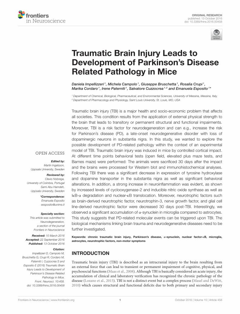
Traumatic Brain Injury Leads toDevelopment of Parkinson’s DiseaseRelated Pathology in Mice
Daniela Impellizzeri 1, Michela Campolo 1, Giuseppe Bruschetta 1, Rosalia Crupi 1,
Marika Cordaro 1, Irene Paterniti 1, Salvatore Cuzzocrea 1, 2 and Emanuela Esposito 1*
1Department of Chemical, Biological, Pharmaceutical, and Environmental Sciences, University of Messina, Messina, Italy,2Department of Pharmacology and Physiology, Saint Louis University, St. Louis, MO, USA
Traumatic brain injury (TBI) is a major health and socio-economic problem that affects
all societies. This condition results from the application of external physical strength to
the brain that leads to transitory or permanent structural and functional impairments.
Moreover, TBI is a risk factor for neurodegeneration and can e.g., increase the risk
for Parkinson’s disease (PD), a late-onset neurodegenerative disorder with loss of
dopaminergic neurons in substantia nigra. In this study, we wanted to explore the
possible development of PD-related pathology within the context of an experimental
model of TBI. Traumatic brain injury was induced in mice by controlled cortical impact.
At different time points behavioral tests (open field, elevated plus maze tests, and
Barnes maze) were performed: The animals were sacrificed 30 days after the impact
and the brains were processed for Western blot and immunohistochemical analyses.
Following TBI there was a significant decrease in expression of tyrosine hydroxylase
and dopamine transporter in the substantia nigra as well as significant behavioral
alterations. In addition, a strong increase in neuroinflammation was evident, as shown
by increased levels of cyclooxygenase-2 and inducible nitric oxide synthase as well as
IκB-α degradation and nuclear-κB translocation. Moreover, neurotrophic factors such
as brain-derived neurotrophic factor, neurotrophin-3, nerve growth factor, and glial cell
line-derived neurotrophic factor were decreased 30 days post-TBI. Interestingly, we
observed a significant accumulation of α-synuclein in microglia compared to astrocytes.
This study suggests that PD-related molecular events can be triggered upon TBI. The
biological mechanisms linking brain trauma and neurodegenerative diseases need to be
further investigated.
Keywords: chronic traumatic brain injury, Parkinson’s disease, α-synuclein, nuclear factor-κB, microglia,
astrocytes, neurotrophic factors, non-motor symptoms
INTRODUCTION
Traumatic brain injury (TBI) is described as an intracranial injury to the brain resulting froman external force that can lead to transient or permanent impairment of cognitive, physical, andpsychosocial functions (Maas et al., 2008). Although TBI is basically considered an acute injury, theaccumulation of clinical and laboratory verification has recognized the chronic pathology of thedisease (Lozano et al., 2015). TBI is not a distinct event but a complex process (Masel and DeWitt,2010) which causes structural and functional deficits due to both primary and secondary injury

Impellizzeri et al. Chronic TBI Induces Parkinson’s Disease
mechanisms (Davis, 2000). The primary injury results fromthe immediate mechanical disruption of cerebral tissue (acutestage) that occurs at the time of external force and includescontusion and hemorrhage (Gaetz, 2004). Secondary injuryprogresses over minutes to months after the primary damageand is the consequence of cascades of molecular and cellularevents that lead to tissue damage, atrophy and brain celldeath (Marklund et al., 2006; Bramlett and Dietrich, 2007;Lozano et al., 2015). TBI is closely related to the developmentof chronic neuro-inflammation, sensory-motor, and cognitivecomplications which appear some 5–6 years after the traumaticevent, and corresponds to∼30 days in the mouse (chronic stage)(Ettenhofer and Abeles, 2009; Ozen and Fernandes, 2012; Acostaet al., 2013; Kirkwood and Yeates, 2013).
Few studies have investigated the possibility that chronicTBI causes secondary injury with pathophysiological changessimilar to those seen in neurodegenerative diseases such asdementia pugilistica, Alzheimer disease (Saing et al., 2012) andParkinson’s disease (PD) (Saing et al., 2012; Acosta et al., 2013;Xiong et al., 2013). Parkinson’s disease is a late-onset age-relatedneurodegenerative disorder of unknown etiology, characterizedby a progressive loss of cathecolaminergic neurons, especiallydopaminergic neurons within the substantia nigra pars compactaand the presence of α-syn-rich cytoplasmic neuronal inclusionsnamed Lewy bodies (Dauer and Przedborski, 2003; Brandt et al.,2008). Alpha-synuclein (α-syn) is a 14-kDa synaptic protein witha key role in PD progression (Maroteaux et al., 1988) and ithas a high propensity to misfold and aggregate as a response toan increase in its concentration, post-translational modifications,mutations, oxidative stress, pH, and metal ions (Hoyer et al.,2002; Jomova et al., 2010; Oueslati et al., 2010).
Neuroinflammation plays a fundamental role in thepathogenesis of TBI, involving cells of the innate immune systemsuch as microglia, astrocytes, pro-inflammatory cytokines, andchemokines that interfere with the brain’s endogenous abilityto self-repair, thereby exacerbating neuronal cell death (Tajiriet al., 2014). The transcription factor nuclear factor kappa B(NF-κB) is a key modulator in inflammatory gene expression inthe nervous system (Grilli and Memo, 1999), which is activatedin response to inflammatory mediators and oxidative stress(Baldwin, 1996). Brains from patients with dementia pugilisticaexhibit an abnormal accumulation of α-synuclein (α-syn) inaxonal swellings and dystrophic neurites, suggesting a potentiallink between brain trauma, and the development of α-synpathology in PD (Newell et al., 1999). The mediators betweenbrain trauma and PD pathophysiology are unknown, but mayinvolve oxidative and/or nitrative stress (Mésenge et al., 1998).
Abbreviations: DAPI, 4′, 6′-diamidino-2-phenylindole; BDNF, Brain-derived
neurotrophic factor; CCI, Controlled cortical impact; Cox-2, Cyclooxygenase-2;
DAT, Dopamine transporter; EPM, Elevated plus-maze; GDNF, Glial cell line-
derived neurotrophic factor; GFAP, Glial fibrillary acidic protein; iNOS, Inducible
nitric oxide synthase expression; Iba1, Ionized calcium-binding adapter molecule
1; NeuN, Neuronal nuclear antigen; NT3, Neurotrophin-3; IκBα, Nuclear factor
of kappa light polypeptide gene enhancer in B-cells inhibitor α; NF-κB, Nuclear
factor -κB; OF, Open field test; PD, Parkinson’s disease; PBS, Phosphate-buffered
saline; SEM, Standard error of the mean; SN, Substantia nigra; TBI, Traumatic
brain injury; TH, Tyrosine hydroxylase; α-syn, α-synuclein.
The present study was carried out to better understandthe molecular mechanisms that associate the long-termconsequences of TBI with the development of parkinsonism,focusing on the role of neuroinflammation and its impact onbehavior in mice.
MATERIALS AND METHODS
AnimalsMale CD1 mice (25–30 g, Harlan, Milan, Italy), aged between10 and 12 weeks, were used for all studies. Mice were housedfive per cage and maintained under a 12:12 h light/dark cycleat 21 ± 1◦C and 50 ± 5% humidity. Standard laboratory dietand tap water were available ad libitum. The University ofMessina Review Board for the care of animals approved thestudy. Animal care was in compliance with Italian regulations onprotection of animals used for experimental and other scientificpurposes (Ministerial Decree 16192) as well as with the CouncilRegulation (EEC) (Official Journal of the European Union L358/1 12/18/1986).
Controlled Cortical Impact (CCI)Experimental TBITBI was induced in mice by a controlled cortical impactor (CCI)as previously described (Campolo et al., 2014). A craniotomywas induced in the right hemisphere, with a Micro motorhand piece and drill (UGO Basile SRL, Comerio Varese, Italy),encompassing bregma and lambda, and among the sagittal sutureand the coronal ridge. The consequential bone flap was removedand the craniotomy enlarged additionally with cranial rongeurs(New Adalat Garh, Roras Road, Pakistan). A cortical contusionwas made on the exposed cortex using the controlled impactordevice Impact OneTM Stereotaxic impactor for CCI (Leica,Milan, Italy). Concisely, the impacting shaft was extended, andthe impact tip was lowered over the craniotomy site until ittouched the dura mater. Subsequently, the rod was retracted,and the impact tip was advanced farther to produce a braininjury of moderate severity for mice (tip diameter: 4mm;cortical contusion depth: 3mm; impact velocity: 1.5m/sec).Instantaneously after injury, the skin incision was secure withnylon sutures, and 2% lidocaine jelly was spread to the lesion siteto reduce pain.
Experimental GroupsMice were randomly allocated into the following groups:
Sham group: mice were subjected to equal surgical proceduresexcept for TBI and were kept under anesthesia for the durationof the experiment (n= 10).TBI group: mice were subjected to CCI (n= 10).
Mice were sacrificed 30 days after TBI, the brains removed andthe midbrain dissected out for study. Immunohistochemicallocalization of PD markers (TH, α-syn, and dopaminetransporter (DAT) was assessed. Western blot was used todetermine the translocation of NF-κB and degradation of IκBα.In addition, over the 30-day experimental period behavioralchanges at 1, 7, 14, and 30 days were evaluated.
Frontiers in Neuroscience | www.frontiersin.org 2 October 2016 | Volume 10 | Article 458

Impellizzeri et al. Chronic TBI Induces Parkinson’s Disease
Behavioral TestingAll animals were subjected to the same battery of behavioraltests at 1, 7, 14, and 30 days post-CCI. All behavioral testingwas conducted during the light cycle phase and in enclosedbehavior rooms (50–55 dB ambient noise) within the housingroom. The mice were placed in behavior rooms 5min for 2 daysfor acclimation prior to the onset of behavioral testing.
The behavioral tests were conducted by three different reliableexpert observers blinded to the injury status of the animals. Testsare described below:
Open field test (OF): Locomotor activity and anxiety-likebehavior were monitored for 5 min using the open field test,a white Plexiglas box 50 × 50 cm with its floor divided into16 squares. Four squares were defined as the center and 12squares along the walls as the periphery. Each mouse wasgently placed in the center of the box and activity was scoredas a line crossing when a mouse removed all four paws fromone square and entered another. Before each trial, the chamberwas cleaned with water containing a detergent. The animals’behavior was videotaped. The line crossings and the time spentin the center were counted and scored (Prut and Belzung,2003).Elevated pluz-maze (EPM): The EPM test was used to measureanxiety-like behavior, as described previously (Pellow et al.,1985). The EPM apparatus consisted in two open arms (30× 5 × 0.25 cm) and two enclosed arms (30 × 5 × 15 cm)extended from a common central platform (5 × 5 cm) andthe entire apparatus was elevated by a single central supportto a height of 60 cm above floor level. The behavioral modelwas based on rodents’ aversion of open spaces that leads tothigmotaxis. Anxiety reduction was indicated by an increase inthe proportion of time spent in the open arms and an increasein the proportion of entries into the open arms. The totalnumbers of arm entries and number of closed-arm entrieswere used as measures of general activity.Barnes maze: The Barnes maze is a less stressful alternativethan the Morris water maze, and is a validated test oftenused to assess spatial learning and memory in rodents. Thetest consists of a circular surface with up to 20 circularholes around its circumference. Under one of the holes is anescape box which can be reached by the rodent through thecorresponding hole on the table top. The model was basedon rodents’ aversion of open spaces, which motivates the testsubject to seek shelter in the escape box. The performance wasmeasured by number of errors the rodent makes and the rateof decline in the number of errors per trial was calculated torepresent a learning curve (Barnes, 1979).
Western Blot AnalysesCytosolic and nuclear extracts were prepared as previouslydescribed (Esposito et al., 2012), with slight modifications.The ipsilateral hemisphere from each mouse after injury wascollected and suspended in Buffer A containing proteaseinhibitors, homogenized for 2min, then centrifuged at1000× g for 10min at 4◦C. Supernatants contained thecytosolic fraction. The pellets, containing enriched nuclei,
were resuspended in Buffer B containing 1% Triton X-100,150mM NaCl, 10mM Tris-HCl pH 7.4, 1mM, ethylene glycoltetra-acetic acid, 1mM, ethylenediaminetetraacetic acid, 0.2mM phenylmethanesulfonylfluoride and protease inhibitors.After centrifugation for 30min at 15,000× g at 4◦C, thesupernatants containing nuclear proteins were stored at −80◦Cfor further analysis. The expression of inducible nitric oxidesynthase (iNOS), cyclooxygenase (COX)-2 and IκBα werequantified in cytosolic fractions. NF-κBp65 was quantifiedin nuclear fractions collected 30 days after TBI. The filterswere probed with specific antibodies for iNOS (1:1000; BDBiosciences, Milan, Italy), COX-2 (1:1000; Cayman Chemicals,Tallinn Estonia), glial fibrillary acidic protein (GFAP) (1:2000;Santa Cruz Biotechnology, Heidelberg, Germany), NFκBp65(1:500; Santa Cruz Biotechnology) and IκBα (1:500; Santa CruzBiotechnology) at 4◦C overnight in 1 × phosphate-bufferedsaline (PBS), 5% (w/v), non-fat dried milk and 0.1% Tween-20.Membranes were incubated with peroxidase-conjugated bovineanti-mouse IgG secondary antibody or peroxidase-conjugatedgoat anti-rabbit IgG (1:2000; Jackson ImmunoResearch, WestGrove, PA, USA) for 1 h at room temperature. To ascertainthat blots were loaded with equal amounts of protein, then theywere incubated in the presence of antibodies against β-actinor lamin A/C (1:5000; Santa Cruz Biotechnology). The signalswere detected with enhanced a chemiluminescence detectionsystem reagent according to the manufacturer’s instructions(Super Signal West Pico Chemiluminescent Substrate, PierceThermo Scientific, Rockford, IL. USA). Relative expression ofbands for IκBα (approximately 37 kDa), NF-κB (approximately65 kDa), iNOS (approximately 130 kDa), COX-2 (approximately72 kDa) and GFAP (approximately 55 kDa) were imported toanalysis software (Image Quant TL, v2003) and standardizedto β-actin and lamin A/C levels. The relative expression of theprotein bands was calculated by densitometry with Bio-RadChemiDocTM XRS+software. Molecular weight standards (10–250 kDa) were used to define molecular weight positions, and asreference concentrations for each protein.
ImmunofluorescenceAfter deparaffinization and rehydration, detection of TH, α-syn,NeuN, GFAP, Iba1, and neurotrophin-3 (NT-3) was carried outafter boiling the tissue sections in 0.1M citrate buffer for 1 min.Non-specific adsorption was minimized by incubating in 2%(vol/vol) normal goat serum in PBS for 20min. Sections wereincubated with one of the following primary antibodies: mousemonoclonal anti-TH (1:100, Millipore), mouse monoclonal anti-α-syn (1:100, Santa Cruz Biotechnology), rabbit polyclonal anti-GFAP (1:100, Santa Cruz Biotechnology), rabbit polyclonal anti-Iba1 (1:100, Santa Cruz Biotechnology), rabbit polyclonal anti-NeuN (1:100, Santa Cruz Biotechnology) or rabbit polyclonalanti-NT3 (1:100, Millipore) in a humidified oxygen and nitrogenchamber overnight at 37◦C. Sections were then incubated withsecondary antibody: fluorescein isothiocyanate-conjugated anti-mouse Alexa Fluor-488 (1:2000, Molecular Probes, Monza, Italy)or Texas Red-conjugated anti-rabbit Alexa Fluor-594 (1:1000,Molecular Probes) for 1 h at 37◦C. For nuclear staining, 2 µg/ml4′, 6′-diamidino-2-phenylindole (DAPI; Hoechst, Frankfurt,
Frontiers in Neuroscience | www.frontiersin.org 3 October 2016 | Volume 10 | Article 458
Impellizzeri et al. Chronic TBI Induces Parkinson’s Disease
Germany) in PBS was added. Sections were observed at 20×and 40x magnification using a Leica DM2000 microscope (Leica,Milan, Italy). TH+, a-syn+, NeuN+, GFAP+, Iba1+, and NT-3+cells were counted stereologically on sections cut at a 40µmthickness and every 4th section was counted using a grid of100 × 100µm (Mallajosyula et al., 2008). Optical sectionsof fluorescence specimens were obtained using a HeNe laser(543 nm), an ultraviolet laser (361–365 nm) and an argon laser(458 nm) at a one-mi, 2 s scanning speed with up to eightaverages; 1.5µm sections were obtained using a pinhole of 250.Examining the most brightly labeled pixels and applying settingsthat allowed clear visualization of structural details, while keepingthe highest pixel intensities close to 200, established contrast andbrightness. The same settings were used for all images obtainedfrom the other samples that had been processed in parallel.Digital images were cropped and figure montages prepared usingAdobe Photoshop 7.0 (Adobe Systems; Palo Alto, California,United States). The co-localization of images was examined withImage J software (National Institutes of Health) as describedpreviously (Zhou et al., 2011).
ImmunohistochemistryBrain tissues were fixed in 10% (w/v) buffered formaldehyde30 days after TBI, and 7-µm sections were prepared fromparaffin-embedded tissues. After deparaffinization, endogenousperoxidase was quenched with 0.3% H2O2 in 60% methanolfor 30min. The sections were permeabilized with 0.1% TritonX-100 (Sigma-Aldrich, Milan, Italy) in PBS (pH 7.4) for 20min. Non-specific adsorption was minimized by incubatingthe section in 2% normal goat serum in PBS for 20min.Endogenous biotin- or avidin-binding sites were blockedby sequential incubation for 15min with avidin and biotin.The sections were then incubated overnight with one of thefollowing primary antibodies diluted in PBS: anti-TH (1:500,Millipore-monoclonal or polyclonal), anti-DAT (1:500, SantaCruz Biotechnology polyclonal), polyclonal anti-glial cellline-derived neurotrophic factor (GDNF) (1:500, Santa CruzBiotechnology), polyclonal anti-brain-derived neurotrophicfactor (BDNF) (1:500, Santa Cruz Biotechnology), anti-α-syn(1:250, Santa Cruz Biotechnology-polyclonal), and anti-NT-3 (1:500, Millipore-polyclonal). The immunohistochemicalpictures were collected by Zeiss microscope using Axio Visionsoftware. For graphic representation of densitometric analyses,we measured the intensity of positive staining (brown staining)by computer-assisted color image analysis (Leica QWin V3,UK). The percentage area of immunoreactivity (determinedby the number of positive pixels) was expressed as percent oftotal tissue area (red staining). Photomicrographs were assesseddensitometrically with Optilab software (Graftek, Mirmande,France) on a MacBook Pro computer (Apple, Cupertino, CA,USA). Analysis was carried out by assigning quantitative differentcriteria for staining intensity as described by Ding et al. (2012),which included assignment of staining intensity using a scale of0–10 (with 0 indicating a lack of brown immunoreactivity and 10reflecting intense dark brown staining) by three different reliableexpert observers. The mean was then calculated and resultsconverted into grades: a score of 1–3 was assigned “+,” 4–6 was
“++,” more than 7 was “+ + +”(Ding et al., 2012). Scores fromall sections of each brain were averaged to give a final score foreach mouse. All histological studies were performed in a blindedfashion.
Tissue Processing and HistologyAn experienced histopathologist evaluated sagittal sections of 5-µm thickness from the perilesional brain area of each animal.Histopathologic changes of the gray matter were scored on a six-point scale (Kawai and Akira, 2007) as described by Campoloet al. (2014). Scores from all sections of each brain were averagedto give a final score for each mouse. All histological studies wereperformed in a blinded fashion.
Statistical AnalysisAll values in the figures and text are expressed as mean ±
standard error of the mean (SEM) of N observations. For invivo studies N represents the number of animals. In thoseexperiments involving histology or immunohistochemistry, thefigures shown are representative of at least three experimentsperformed on different days. Results were analyzed by one-wayANOVA followed by a Bonferroni post-hoc test for multiplecomparisons. A F-value was shown, p-value of less than 0.05 wasconsidered significant.
RESULTS
Changes of PD Markers in the SNc afterChronic TBITo examine if chronic TBI can modify PD-like markers, brainsections from control and TBI mice 30 days after surgery werestained with dopaminergic-specific markers (TH and DAT) andα-syn. Midbrain expression of TH-positive neurons and DATwas significantly decreased 30 days after TBI (Figures 1A,B,respectively, see densitometric analysis, Figure 1D; F = 3 forTH and for DAT). Western blot analysis confirmed a significantreduction of TH and DAT protein expression (Figure 1A1, P <
0.005; and Figure 1B1, respectively, P < 0.05; F = 1.111 for THand 4.88 for DAT). In contrast, chronic TBI resulted in a visibleincrease in α-syn staining compared to control group (Figure 1C,see densitometric analysis, Figure 1D, F = 3) and protein asshown byWestern blot analysis (Figure 1C1 P< 0.01; F= 0.078).
To confirm these data, TH and α-syn were double-stained with antibodies against the neuronal marker NeuN,in the midbrain. Thirty days after TBI there was an evidentaccumulation of α-syn in neurons compared with sham mice(Figure 2B, see corresponding cell count analysis; F = 0.428).On the other hand, TH-positive neuron counts were significantlydecreased (Figure 2A, see corresponding cell count analysis; F =
7). Also, to evaluate the α-syn accumulation specifically in thedopaminergic neurons we assessed a double staining between THand α-syn (Figure 2C). We noticed a basal level of α-syn into thedopaminergic neurons TH-positive in sham group while 30 daysafter TBI there was an increasing accumulation of α-syn in theTH positive neurons (Figure 2C).
Frontiers in Neuroscience | www.frontiersin.org 4 October 2016 | Volume 10 | Article 458

Impellizzeri et al. Chronic TBI Induces Parkinson’s Disease
FIGURE 1 | Effect of chronic TBI on Parkinsonian markers. Midbrain was stained with antibodies against tyrosine hydroxylase (A), dopamine transporter (B) and
α–synuclein (C). Immunohistochemical analysis of midbrain obtained from mice subjected to TBI revealed a positive staining for TH, DAT, and α-syn [TBI panels TBI
(A–C) respectively; see densitometric analysis, D] compared with sham-operated mice [Sham panels TBI (A–C) respectively; see densitometric analysis, D]. Data are
expressed as a percentage of total tissue area and are means ± SE of 5 mice/group. **P < 0.005 vs. Sham; *P < 0.05 vs. sham (Student’s t-test). Western blot
analysis confirmed our data (A1–C1 respectively). Each data are expressed as Mean ± SEM from N = 5 mice/group. *P < 0.01 vs. sham, **P < 0.005 vs. sham.
Microglia, but not Astrocytes in the SNcRegulate α-syn Expression after ChronicTBI
To evaluate microglia involvement and its correlation withPD markers, the midbrain was double-stained with antibodiesagainst Iba1 (red; Figure 3A) and α-syn (green; Figure 3A).
Microglial cells (Iba1-+cells) visibly expressed α-syn-positivestaining in chronic TBI tissue (Figure 3A) compared to sham.Further, there was a clear co-localization of α-syn and Iba1 in theTBI samples (merged, Figure 3A- see cell count analysis A1; F =
0.428).To analyze the activation of astrocytes and its involvement
in PD, contused brain tissue at the collision site 30 days after
Frontiers in Neuroscience | www.frontiersin.org 5 October 2016 | Volume 10 | Article 458

Impellizzeri et al. Chronic TBI Induces Parkinson’s Disease
FIGURE 2 | Effects of chronic TBI on Tyrosine hydroxylase (TH) and α-synuclein expression in NeuN positive cells. Midbrain was double-stained with
antibodies against NeuN (red), TH (green) (A), or α-syn (red) (B). The red spots indicate the co-localizations (merged) indicated by yellow arrows. TH-positive neurons
were significantly reduced after chronic TBI (TBI merged A). Midbrain sections revealed increased α-syn positive neurons 30 days after TBI (TBI merged B). Moreover,
midbrain sections showed a marked α-syn accumulation in TH+ cell (TBI merged C). Scale bar = 50 and 20 µm (particle). Data are expressed as Mean ± SEM from
N = 5 mice/group. Counting of colocalized cell confirmed our data. The co-localization of image was analyzed with image J software. *P < 0.05 vs. sham (Student’s
t-test), **P < 0.005.
Frontiers in Neuroscience | www.frontiersin.org 6 October 2016 | Volume 10 | Article 458

Impellizzeri et al. Chronic TBI Induces Parkinson’s Disease
FIGURE 3 | Effect of chronic TBI on α-synuclein expression in ionized calcium binding adaptor molecule 1 (Iba1) and glial fibrillary acidic protein
(GFAP) positive cells. Midbrain sections wer double-stained with antibodies against Iba1 (red) or GFAP (red) and α-syn (green). The red spots indicate the
co-localizations (merged) indicated by yellow arrows. There were increased microglia cells (Iba1+cells) in TBI mice (A) as compared to the control group (Sham A).
α-syn positive neurons were significantly increased in microglia after chronic TBI (TBI merged A). Considerable astrogliosis (GFAP+cells) was present in TBI panels (B).
α-syn positive neurons did not show any significant immunoreactivity in astrocytes after TBI (TBI merged B). Scale bar = 50 and 20 µm (particle). Each data are
expressed as Mean ± SEM from N = 5 mice/group. Counting of colocalized cell confirmed our data. The co-localization of image was analyzed with image J
software. *P < 0.05.
injury was double-stained with for GFAP (red; Figure 3B) andα-syn (green; Figure 3B) or TH (green; Figure 4). Midbrainsections showed an increased astrogliosis (GFAP+ cells) in TBIpanels (Figures 3B, 4). Furthermore, there was a non-significantco-localization of both α-syn and TH in GFAP+ cells afterchronic TBI [merged, Figures 3B, 4, respectively- see cell countanalysis in Figures 3B1 (F = 1) and Figure 4A, respectively(F = 1)].
Chronic TBI Induces a SignificantNeuroinflammatory Response Regulatedby NF-κBTo investigate the cellular substrate(s) that link PD with chronicTBI, Western blot analysis was assessed in midbrain tissue 30days post-TBI, using IκBα- and NF-κB- specific antibodies.There was a basal expression of IκBα in sham mice (Figure 5A,see densitometry analysis (P < 0.005), while IκBα expressionwas considerably reduced in mice subjected to chronic TBI(Figure 5A, see densitometry analysis, P < 0.005; F = 1.087).Moreover, p65 subunit translocation was increased in nuclearbrain homogenates after TBI, compared with sham (Figure 5B,see densitometry analysis P < 0.01; F = 0.0195). Translocation ofNF-κB is a critical phase in the coupling of extracellular stimuli
to the transcriptional stimulation of specific pro-inflammatorytarget genes such as iNOS and COX-2. To evaluate the role ofnitric oxide in TBI, iNOS expression was evaluated by Westernblot analysis. At 30 days post-TBI there was a significant increasein iNOS expression in midbrain of TBI mice (Figure 5C, seedensitometry analysis, P < 0.005, F = 0.0312). COX-2 expressionwas also stimulated by TBI compared to sham (Figure 5D, seedensitometry analysis, P < 0.001, F = 0.0019).
Chronic TBI Reduces Neurotrophic FactorExpression LevelsTo test whether chronic TBI modulates PD via regulationof neurotrophic factors, we used immunohistochemistry toexamine BDNF, GDNF, and NT-3 levels in the midbrain. Thirtydays post-trauma there was a reduction in immunostaining forall three neurotrophic proteins (Figures 6A–C, respectively,see densitometric analysis, Figure 6D; F = 3 for BDNF andGDNF, and F = 1.0002 for NT-3) in comparison to shamanimals. Moreover, Western blot analysis confirmed theseobservations (Figures 6A1–C1, respectively, see densitometricanalysis, Figure 6D: P < 0.005, P < 0.001, and P < 0.05,respectively; and F = 2.156, 1.318, 9.30 for BDNF, GDNF,and NT-3).
Frontiers in Neuroscience | www.frontiersin.org 7 October 2016 | Volume 10 | Article 458

Impellizzeri et al. Chronic TBI Induces Parkinson’s Disease
FIGURE 4 | Effect of chronic TBI on Tyrosine hydroxylase (TH) expression in glial fibrillary acidic protein (GFAP) positive cells. Midbrain tissues were
double-stained with antibodies against GFAP (red) and TH (green). The red spots indicate the co-localizations (merged) indicated by yellow arrows. Midbrain sections
revealed increased astrogliosis (GFAP+ cells) in TBI panels. TH positive neurons were significantly reduced after TBI (TBI merged panel). Scale bar = 50 and 20µm
(particle). Each data are expressed as Mean ± SEM from N = 5 mice/group. Counting of colocalized cell confirmed our data. The co-localization of image was
analyzed with image J software.
Chronic TBI Induces Depression- andAnxiety-Like Behaviors in the MouseTo examine anxiety-like behavior and locomotor function, micesubjected to chronic TBI were compared to sham mice at 1, 7,14, and 30 days on several behavioral tests, including the openfield test, EPM, and Barnes maze. Mice were tested in the EPMat all time points, TBI mice spent notably less time in the openarms as compared to sham animals (Figure 7A, see densitometricanalysis, Figure 7F, F = 1.333 at 1 days, F = 1 at 3 days, F = 1at 7 days, F = 4 at 30 days). The anxiety-like phenotype inducedby the lesion was confirmed in the open field test (Figures 7B,B1,see densitometric analysis, Figure 7F; ∗P < 0.05 and ∗∗P < 0.005;Figure 7B, F = 1.750 at 1 days, F = 4 at 3 days, F = 3 at 7 days, F= 3 at 30 days; Figure 7B1, F = 1.531 at 1 d, F = 3.226 at 3 days,F = 4 at 7 days, F = 4 at 30 days). Chronic TBI mice showeda pronounced increase in thigmotaxis, seen as the tendency toremain close to the wall (Simon et al., 1994), when compared toshammice, as specified by less time spent in the center of the openfield (Figure 7B, ∗P< 0.05 and ∗∗P< 0.005 vs. sham) and shorterdistance covered in the center of the open field (Figure 7B1, P< 0.05 vs. sham). Spatial learning and memory were assessed inTBI mice using the Barnes maze. In this test, sham mice quicklylearned to escape the open field and reach the black escape box,as revealed by the rapid decline in escape latency. In contrast,there was a statistically significant decrease in spatial learning
induced by TBI for all time points, with an increase in escapelatency and mean number of errors (Figures 7C,C1, respectively,see densitometric analysis, Figure 7F, ∗∗∗P < 0.001 vs. sham;Figure 7C) F = 12 at 1 days, F = 2.583 at 3 days, F = 7 at 7 days,F = 1.714 at 30 days): Figure C1) F = 1 at 1 days, F = 1 at 3 days,F = 2.333 at 7 days, F = 4 at 30 days).
Behavioral tests showed the degree of severity in our TBImodel 30 days post-trauma. Using hematoxylin and eosinstaining we failed to detect any frank cell loss or alterationof tissue morphology in the midbrain either 24 or 48 h afterTBI compared to sham groups (Figures 7D,E, respectively,see densitometric analysis, Figure 7F, see histological score∗P < 0.05).
DISCUSSION
A strong association of TBI with an increased risk of PD hasbeen documented (Bower et al., 2003; Goldman et al., 2006).The development of animal models of TBI that validate manyfundamental pathophysiological processes related to PD has beenan important advance in this field (Uryu et al., 2003; Xiong et al.,2013). Among themanymechanisms associated in PD pathology,α-syn accumulation coupled with TBI appears to synergisticallyimpact on PD symptoms (Shahaduzzaman et al., 2013; Ulusoyand Di Monte, 2013). In the present study we demonstrate
Frontiers in Neuroscience | www.frontiersin.org 8 October 2016 | Volume 10 | Article 458

Impellizzeri et al. Chronic TBI Induces Parkinson’s Disease
FIGURE 5 | Effects of chronic TBI on Nuclear factor κB (NF-κB) pathway and pro-inflammatory enzymes. Degradation of IκBα was significantly increased 30
days after TBI (A). Also, chronic TBI resulted in enhancednuclear translocation of p65 (B). A significant increase in inducible nitric oxide synthase (iNOS) and
cyclooxygenase (COX)-2 (C,D, respectively) was observed in the midbrain from TBI mice compared with the Sham mice (C,D, respectively). Data show one
representative blot from three independent experiments with similar results. Data are expressed as Mean ± SEM from N = 5 mice/group. * P < 0.01 vs. sham, **P <
0.005 vs. sham,***P < 0.005 vs. sham. (Student’s t-test).
that already 30 days after TBI PD-like markers are significantlyup regulated. In addition, midbrain tissue of chronic TBI micedisplayed a remarkable decrease in the immunohistochemicalexpression of dopaminergic markers, along with an evidentaccumulation of α-syn in neurons.
Inflammation plays an important role in PD (Engleret al., 2009). Dopaminergic neurons are highly susceptible toinflammation and oxidative damage and neuroinflammationmediated by glial cells has attracted much attention. Previousstudies revealed abnormalities in astrocytes and microglia in PDpatients, suggesting involvement of these two cell populations inthe development of early stage PD (Rappold and Tieu, 2010).
TBI is reported to lead to accumulation of α-syn in microglialcells and astrocytes11, leading to propagation of damage toneurons that perpetuates neurodegeneration. Our results show asignificant accumulation of α-syn in microglia but not astrocytes,which could represent an important link between TBI and PD.In addition, microglial infiltration into the substantia nigra hasbeen suggested to alter the inflammatory environment, and
promote the buildup of α-syn (Acosta et al., 2015). In somestudies microglia have been shown to activate NF-κB, leading todopamine neuron degeneration, possibly through apoptotic celldeath (Phani et al., 2012).
NF-κB is a cardinal transcriptional regulator of inflammationand apoptosis, neuronal cell survival, and signaling involvedin brain damage. NF-κB activation may persist for at least1 year following cortical injury, suggesting that chronic NF-κB activation may play a role in long-term inflammatorymechanism following brain trauma (Nonaka et al., 1999). Infact, our data show a clear and significant expression of NF-κB 30 days after TBI. In line with this, there was a significantincrease in the degradation of cytoplasmic IKB. Further, NF-κB activation in the midbrain was accompanied by increasedtranscription of the inflammatory markers COX-2 and iNOS.The latter may result in production of high levels of nitricoxide and superoxide radicals, which are neurotoxic. COX-2,which generates prostaglandins, represents a potential hazardfor dopamine neurons. In fact, increased COX-2 expression was
Frontiers in Neuroscience | www.frontiersin.org 9 October 2016 | Volume 10 | Article 458

Impellizzeri et al. Chronic TBI Induces Parkinson’s Disease
FIGURE 6 | Effect of chronic TBI on Neurotrophic factors. By immunohistochemical analysis, a basal level of BDNF (A) GDNF (B) and NT-3 (C) positive staining
was detected in midbrain samples from sham-operated mice (sham panels A–C respectively; see densitometric analysis, D). BDNF and GDNF and NT-3 expression
was significantly reduced in midbrain samples from TBI mice (TBI panels A–C respectively; see densitometric analysis, D). Data are expressed as a percentage of total
tissue area and are means ± SE of 5 mice/group. **P < 0.005 vs. Sham; *P < 0.05 vs. sham (Student’s t-test). Western blot analysis confirmed our data showing a
significant decrease of neurotrophic factor following TBI (A1–C1 respectively, see correspond densitometric analysis). Each data are expressed as Mean ± SEM from
N = 5 mice/group. **P < 0.005 vs. sham; *P < 0.05 vs. sham (Student’s t-test).
localized to the substantia nigra pars compacta in post-mortembrains of PD patients (Przedborski, 2007). We hypothesizethat NF-κB activity in the midbrain could play a key role indetermining inflammatory processes in the lesioned nigrostriatalpathway, which consequently influence the neurodegenerativeoutcome.
Neurons in the substantia nigra show up-regulateddopaminergic markers in response to increases in BDNF,GDNF, and NT-3 (Peterson and Nutt, 2008). BDNF elevationhas been associated to better performance into the motor cortex(Zhao et al., 2016). GDNF has been linked to the survival ofdopaminergic cells in the substantia nigra through increases
Frontiers in Neuroscience | www.frontiersin.org 10 October 2016 | Volume 10 | Article 458

Impellizzeri et al. Chronic TBI Induces Parkinson’s Disease
FIGURE 7 | Effect of chronic TBI on anxiety-like behaviors. The degree of non-motor impairment was assessed in a blinded manner at different time points
(1,7,14, and 30d) by elevated plus maze (EPT), open field (OF), and Barnes maze. EPM tests showed that TBI animals spent less time exploring the open arms and
head dipping than Sham animals (A). No significant differences in rearing or grooming were observed. OF tests showed that TBI mice traveled significantly shorter
distances and didn’t stay in the center area for longer (B,B1). Barnes maze revealed a decrease of spatial learning induced by TBI in all time point increasing the
latency for escape (C) and the mean number errors (C1) Data are means ± SE of 10 mice/group. *P < 0.05 vs. sham; **P < 0.005; ***P < 0.001 vs. sham (ANOVA
and Bonferroni test). H&E staining: (D–E). As compared with sham groups at 24 and 48 h (D,E respectively, see histological score F), after TBI wasn’t shown a
significant alteration of tissue morphology (D,E respectively, see histological score F) in the midbrain. Each data are expressed as Mean ± SEM from N = 5 Mice for
each group. Scale bar = 50 and 20µm (higher magnification).
in synaptic excitability (Bourque and Trudeau, 2000) and theinhibition of apoptosis (Burke et al., 1998). BDNF, NT-3, andGDNF, by binding to their respective high-affinity receptorsstimulate the survival and morphological differentiation ofmidbrain dopaminergic neurons and increase dopamine uptake(Baquet et al., 2005; von Bohlen und Halbach et al., 2005;Grandoso et al., 2007; Stahl et al., 2011) Interestingly, our studyindicates a significant reduction in expression of BDNF, GDNF,and NT-3 in the midbrain 30 days after TBI.
Parkinson’s disease does not only feature motor symptoms,but also non-motor symptoms which can lead to cognitiveand psychiatric disturbances (Bonito-Oliva et al., 2014). Thedepressive-like phenotype observed in our TBI-PD-inducedmouse model was paralleled by increased thigmotaxis andreduced time spent in the open arms of the EPM, two standard
behavioral parameters of anxiety. These data are in agreementwith the frequent co-morbidity between anxiety and depressionobserved in PD patients (Menza et al., 1993) and with anumber of observations in experimental models. Also, humanstudies comparing neurobehavioral outcomes after TBI suggestthat cognitive and emotional impairments increase with injuryseverity (Washington et al., 2012). Little is known about themechanistic role, direct or indirect, of the substantia nigraon learning processes, and our Barnes maze test in chronicTBI mice could reveal early dysfunction within the substantianigra (Li et al., 2013) or inability of the mice to effectivelyutilize a search strategy. Furthermore, our findings demonstratethat acute-to-chronic TBI could cause long-term deficits incognitive function that accompanies gradual lesion expansionand continuing neurodegeneration evolving in PD.
Frontiers in Neuroscience | www.frontiersin.org 11 October 2016 | Volume 10 | Article 458

Impellizzeri et al. Chronic TBI Induces Parkinson’s Disease
The present observations provide the first demonstration thata cortical lesion can expand to the midbrain and culminate in theexpression of PD-like elements 30 days post-trauma. Specifically,our study indicates specific cellular mechanisms that may linkchronic TBI and PD, in particular neuroinflammatory reactionsvia the NF-κB pathway. Moreover, microglia appear to occupy aprominent position as a link between TBI and the developmentof PD-like pathology with aberrant α-syn accumulation, whichcould play an important role in the neurodegenerative process.In terms of the PD-related non-motor symptoms, chronic TBIenhanced anxiety-like behavior and spatial learning. Based onprevious findings (DeKosky et al., 2013), our study also suggeststhat such PD-like pathology and symptoms could represent anetiology for other neurodegenerative diseases such as chronictraumatic encephalopathy (dementia pugilistica). Although thereare limitations and challenges with animal models for mild TBI,these should still be relevant for the study of behavioral sequelaeof single and repetitive mild TBI and how they relate to thenumerous complex histopathological pathways. For example,the safety, and efficacy identified in animal studies may notgenerally translate to human trials, since one cannot excludethe possibility that some residual unmeasured confounders exist(e.g., a behavior thatmay lead a person to bemore likely to sustaina TBI and/or that may also be an independent risk factor for
PD). Also, this study is unable to provide information about theneuro-functional assessment of chronic TBI.
Regardless of its limitations, this study has investigatedan under-appreciated biological mechanism that indicates acausal relationship between brain injury and neurodegenerativedisease; In particular, our findings suggest that sustainedneuroinflammatory processes following a single moderate TBIcan lead to a PD-like pathophysiology in the midbrain.
AUTHOR CONTRIBUTIONS
Study concept and design: DI, SC, and EE. Acquisition ofdata and statistical analysis: GB, RC, MaC, and IP. Analysisand interpretation of the data: DI, MiC, SC, EE. Drafting ofpaper: MiC and EE. Critical revision of the manuscript forintellectual content: SC, EE. All authors read and approved thefinal manuscript.
ACKNOWLEDGMENTS
The authors would like to thank Maria Antonietta Medici forexcellent technical assistance, Mr. Francesco Soraci for secretarialand administrative assistance and Miss Valentina Malvagni foreditorial assistance with the manuscript.
REFERENCES
Acosta, S. A., Tajiri, N., de la Pena, I., Bastawrous, M., Sanberg, P. R., Kaneko, Y.,
et al. (2015). Alpha-synuclein as a pathological link between chronic traumatic
brain injury and Parkinson’s disease. J. Cell. Physiol. 230, 1024–1032. doi:
10.1002/jcp.24830
Acosta, S. A., Tajiri, N., Shinozuka, K., Ishikawa, H., Grimmig, B., Diamond, D. M.,
et al. (2013). Long-term upregulation of inflammation and suppression of cell
proliferation in the brain of adult rats exposed to traumatic brain injury using
the controlled cortical impact model. PLoS ONE 8:e53376. doi: 10.1371/journal.
pone.0053376
Baldwin, A. S. Jr. (1996). The NF-kappa B and I kappa B proteins: new
discoveries and insights. Annu. Rev. Immunol. 14, 649–683. doi: 10.1146/
annurev.immunol.14.1.649
Baquet, Z. C., Bickford, P. C., and Jones, K. R. (2005). Brain-derived neurotrophic
factor is required for the establishment of the proper number of dopaminergic
neurons in the substantia nigra pars compacta. J. Neurosci. 25, 6251–6259. doi:
10.1523/JNEUROSCI.4601-04.2005
Barnes, C. A. (1979). Memory deficits associated with senescence: a
neurophysiological and behavioral study in the rat. J. Comp. Physiol. Psychol.
93, 74–104. doi: 10.1037/h0077579
Bonito-Oliva, A., Masini, D., and Fisone, G. (2014). A mouse model of
non-motor symptoms in Parkinson’s disease: focus on pharmacological
interventions targeting affective dysfunctions. Front. Behav. Neurosci. 8:290.
doi: 10.3389/fnbeh.2014.00290
Bourque, M. J., and Trudeau, L. E. (2000). GDNF enhances the synaptic efficacy
of dopaminergic neurons in culture. Eur. J. Neurosci. 12, 3172–3180. doi:
10.1046/j.1460-9568.2000.00219.x
Bower, J. H., Maraganore, D. M., Peterson, B. J., McDonnell, S. K., Ahlskog, J. E.,
and Rocca, W. A. (2003). Head trauma preceding PD: a case-control study.
Neurology 60, 1610–1615. doi: 10.1212/01.WNL.0000068008.78394.2C
Bramlett, H. M., and Dietrich, W. D. (2007). Progressive damage after brain and
spinal cord injury: pathomechanisms and treatment strategies. Prog. Brain Res.
161, 125–141. doi: 10.1016/S0079-6123(06)61009-1
Brandt, I., Gérard, M., Sergeant, K., Devreese, B., Baekelandt, V., Augustyns,
K., et al. (2008). Prolyl oligopeptidase stimulates the aggregation of alpha-
synuclein. Peptides 29, 1472–1478. doi: 10.1016/j.peptides.2008.05.005
Burke, R. E., Antonelli, M., and Sulzer, D. (1998). Glial cell line-derived
neurotrophic growth factor inhibits apoptotic death of postnatal substantia
nigra dopamine neurons in primary culture. J. Neurochem. 71, 517–525. doi:
10.1046/j.1471-4159.1998.71020517.x
Campolo, M., Esposito, E., Ahmad, A., Di Paola, R., Paterniti, I., Cordaro,
M., et al. (2014). Hydrogen sulfide-releasing cyclooxygenase inhibitor
ATB-346 enhances motor function and reduces cortical lesion volume
following traumatic brain injury in mice. J. Neuroinflammation 11:196. doi:
10.1186/s12974-014-0196-1
Dauer, W., and Przedborski, S. (2003). Parkinson’s disease: mechanisms and
models. Neuron 39, 889–909. doi: 10.1016/S0896-6273(03)00568-3
Davis, A. E. (2000). Mechanisms of traumatic brain injury: biomechanical,
structural and cellular considerations. Crit. Care Nurs. Q. 23, 1–13. doi:
10.1097/00002727-200011000-00002
DeKosky, S. T., Blennow, K., Ikonomovic, M. D., and Gandy, S. (2013). Acute and
chronic traumatic encephalopathies: pathogenesis and biomarkers. Nat. Rev.
Neurol. 9, 192–200. doi: 10.1038/nrneurol.2013.36
Ding, W., Zhang, W., Hui, F. M., Zhang, Y. H., Zhang, F. F., Li, X. M.,
et al. (2012). Cell-specific expression and immunolocalization of nitric oxide
synthase isoforms and soluble guanylyl cyclase alpha1 and beta1 subunits in
the ovary of fetal, neonatal and immature pigs.Anim. Reprod. Sci. 131, 172–180.
doi: 10.1016/j.anireprosci.2012.02.013
Engler, H., Doenlen, R., Riether, C., Engler, A., Niemi, M. B., and Besedovsky, H.
O., et al (2009). Time-dependent alterations of peripheral immune parameters
after nigrostriatal dopamine depletion in a rat model of Parkinson’s disease.
Brain Behav. Immun. 23, 518–526. doi: 10.1016/j.bbi.2009.01.018
Esposito, E., Impellizzeri, D., Mazzon, E., Fakhfouri, G., Rahimian, R., Travelli, C.,
et al. (2012). The NAMPT inhibitor FK866 reverts the damage in spinal cord
injury. J. Neuroinflammation 9:66. doi: 10.1186/1742-2094-9-66
Ettenhofer, M. L., and Abeles, N. (2009). The significance of mild traumatic
brain injury to cognition and self-reported symptoms in long-term
recovery from injury. J. Clin. Exp. Neuropsychol. 31, 363–372. doi:
10.1080/13803390802175270
Gaetz, M. (2004). The neurophysiology of brain injury. Clin. Neurophysiol. 115,
4–18. doi: 10.1016/S1388-2457(03)00258-X
Goldman, S. M., Tanner, C. M., Oakes, D., Bhudhikanok, G. S.,
Gupta, A., and Langston, J. W. (2006). Head injury and Parkinson’s
Frontiers in Neuroscience | www.frontiersin.org 12 October 2016 | Volume 10 | Article 458

Impellizzeri et al. Chronic TBI Induces Parkinson’s Disease
disease risk in twins. Ann. Neurol. 60, 65–72. doi: 10.1002/ana.
20882
Grandoso, L., Ponce, S., Manuel, I., Arrúe, A., Ruiz-Ortega, J. A., Ulibarri, I., et al.
(2007). Long-term survival of encapsulated GDNF secreting cells implanted
within the striatum of parkinsonized rats. Int. J. Pharm.. 343, 69–78. doi:
10.1016/j.ijpharm.2007.05.027
Grilli, M., and Memo, M. (1999). Nuclear factor-kappaB/Rel proteins: a point
of convergence of signalling pathways relevant in neuronal function and
dysfunction. Biochem. Pharmacol. 57, 1–7. doi: 10.1016/S0006-2952(98)0
0214-7
Hoyer, W., Antony, T., Cherny, D., Heim, G., Jovin, T. M., and Subramaniam,
V. (2002). Dependence of alpha-synuclein aggregate morphology on solution
conditions. J. Mol. Biol. 322, 383–393. doi: 10.1016/S0022-2836(02)00775-1
Jomova, K., Vondrakova, D., Lawson, M., and Valko, M. (2010). Metals, oxidative
stress and neurodegenerative disorders. Mol. Cell. Biochem. 345, 91–104. doi:
10.1007/s11010-010-0563-x
Kawai, T., and Akira, S. (2007). Signaling to NF-kappaB by Toll-like receptors.
Trends Mol. Med. 13, 460–469. doi: 10.1016/j.molmed.2007.09.002
Kirkwood, M., and Yeates, K. (2013).Mild Traumatic Brain Injury in Children and
Adolescents. New York, NY: Guilford Press.
Li, X., Redus, L., Chen, C., Martinez, P. A., Strong, R., Li, S., et al. (2013).
Cognitive dysfunction precedes the onset of motor symptoms in the
MitoPark mouse model of Parkinson’s disease. PLoS ONE 8:e71341. doi:
10.1371/journal.pone.0071341
Lozano, D., Gonzales-Portillo, G. S., Acosta, S., de la Pena, I., Tajiri, N., Kaneko,
Y., et al. (2015). Neuroinflammatory responses to traumatic brain injury:
etiology, clinical consequences, and therapeutic opportunities. Neuropsychiatr.
Dis. Treat. 11, 97–106. doi: 10.2147/NDT.S65815
Maas, A. I., Stocchetti, N., and Bullock, R. (2008). Moderate and severe
traumatic brain injury in adults. Lancet Neurol. 7, 728–741. doi: 10.1016/S1474-
4422(08)70164-9
Mallajosyula, J. K., Kaur, D., Chinta, S. J., Rajagopalan, S., Rane, A., Nicholls, D. G.,
et al. (2008). MAO-B elevation in mouse brain astrocytes results in Parkinson’s
pathology. PLoS ONE 3:e1616. doi: 10.1371/journal.pone.0001616
Marklund, N., Bakshi, A., Castelbuono, D. J., Conte, V., and McIntosh, T. K.
(2006). Evaluation of pharmacological treatment strategies in traumatic brain
injury. Curr. Pharm. Des. 12, 1645–1680. doi: 10.2174/138161206776843340
Maroteaux, L., Campanelli, J. T., and Scheller, R. H. (1988). Synuclein: a neuron-
specific protein localized to the nucleus and presynaptic nerve terminal. J.
Neurosci. 8, 2804–2815.
Masel, B. E., and DeWitt., D. S. (2010). Traumatic brain injury: a disease process,
not an event. J. Neurotrauma 27, 1529–1540. doi: 10.1089/neu.2010.1358
Menza, M. A., Robertson-Hoffman, D. E., and Bonapace, A. S. (1993). Parkinson’s
disease and anxiety: comorbidity with depression. Biol. Psychiatry 34, 465–470.
doi: 10.1016/0006-3223(93)90237-8
Mésenge, C., Charriaut-Marlangue, C., Verrecchia, C., Allix, M., Boulu, R. R., and
Plotkine, M. (1998). Reduction of tyrosine nitration after N(omega)-nitro-L-
arginine-methylester treatment of mice with traumatic brain injury. Eur. J.
Pharmacol. 353, 53–57. doi: 10.1016/S0014-2999(98)00432-4
Newell, K. L., Boyer, P., Gomez-Tortosa, E., Hobbs, W., Hedley-Whyte, E. T.,
Vonsattel, J. P., et al. (1999). Alpha-synuclein immunoreactivity is present in
axonal swellings in neuroaxonal dystrophy and acute traumatic brain injury. J.
Neuropathol. Exp. Neurol. 58, 1263–1268. doi: 10.1097/00005072-199912000-
00007
Nonaka, M., Chen, X. H., Pierce, J. E., Leoni, M. J., McIntosh, T. K., Wolf,
J. A., et al. (1999). Prolonged activation of NF-kappaB following traumatic
brain injury in rats. J. Neurotrauma 16, 1023–1034. doi: 10.1089/neu.1999.
16.1023
Oueslati, A., Fournier, M., and Lashuel, H. A. (2010). Role of post-
translational modifications in modulating the structure, function and toxicity
of alpha-synuclein: implications for Parkinson’s disease pathogenesis and
therapies. Prog. Brain Res. 183, 115–145. doi: 10.1016/S0079-6123(10)
83007-9
Ozen, L. J., and Fernandes, M. A. (2012). Slowing down after a mild traumatic
brain injury: a strategy to improve cognitive task performance? Arch. Clin.
Neuropsychol. 27, 85–100. doi: 10.1093/arclin/acr087
Pellow, S., Chopin, P., File, S. E., and Briley, M. (1985). Validation of open:closed
arm entries in an elevated plus-maze as a measure of anxiety in the rat. J.
Neurosci. Methods 14, 149–167. doi: 10.1016/0165-0270(85)90031-7
Peterson, A. L., and Nutt, J. G. (2008). Treatment of Parkinson’s disease with
trophic factors. Neurotherapeutics 5, 270–280. doi: 10.1016/j.nurt.2008.02.003
Phani, S., Loike, J. D., and Przedborski, S. (2012). Neurodegeneration and
inflammation in Parkinson’s disease. Parkinsonism Relat. Disord. 18(Suppl. 1),
S207–S209. doi: 10.1016/S1353-8020(11)70064-5
Prut, L., and Belzung, C. (2003). The open field as a paradigm tomeasure the effects
of drugs on anxiety-like behaviors: a review. Eur. J. Pharmacol. 463, 3–33. doi:
10.1016/S0014-2999(03)01272-X
Przedborski, S. (2007). Neuroinflammation and Parkinson’s disease. Handb. Clin.
Neurol. 83, 535–551. doi: 10.1016/S0072-9752(07)83026-0
Rappold, P. M., and Tieu, K. (2010). Astrocytes and therapeutics for Parkinson’s
disease. Neurotherapeutics 7, 413–423. doi: 10.1016/j.nurt.2010.07.001
Saing, T., Dick, M., Nelson, P. T., Kim, R. C., Cribbs, D. H., and Head, E. (2012).
Frontal cortex neuropathology in dementia pugilistica. J. Neurotrauma 29,
1054–1070. doi: 10.1089/neu.2011.1957
Shahaduzzaman, M., Acosta, S., Bickford, P. C., and Borlongan, C. V. (2013).
Alpha-Synuclein is a pathological link and therapeutic target for Parkinson’s
disease and traumatic brain injury. Med. Hypotheses 81, 675–680. doi:
10.1016/j.mehy.2013.07.025
Simon, P., Dupuis, R., and Costentin, J. (1994). Thigmotaxis as an index of anxiety
in mice. Influence of dopaminergic transmissions. Behav. Brain Res. 61, 59–64.
doi: 10.1016/0166-4328(94)90008-6
Stahl, K., Mylonakou, M. N., Skare, Ø., Amiry-Moghaddam, M., and Torp, R.
(2011). Cytoprotective effects of growth factors: BDNF more potent than
GDNF in an organotypic culture model of Parkinson’s disease. Brain Res. 1378,
105–118. doi: 10.1016/j.brainres.2010.12.090
Tajiri, N., Hernandez, D., Acosta, S., Shinozuka, K., Ishikawa, H., Ehrhart, J., et al.
(2014). Suppressed cytokine expression immediatey following traumatic brain
injury in neonatal rats indicates an expeditious endogenous anti-inflammatory
response. Brain Res. 1559, 65–71. doi: 10.1016/j.brainres.2014.02.041
Ulusoy, A., and Di Monte, D. A. (2013). Alpha-Synuclein elevation in human
neurodegenerative diseases: experimental, pathogenetic, and therapeutic
implications.Mol. Neurobiol. 47, 484–494. doi: 10.1007/s12035-012-8329-y
Uryu, K., Giasson, B. I., Longhi, L., Martinez, D., Murray, I., Conte, V., et al. (2003).
Age-dependent synuclein pathology following traumatic brain injury in mice.
Exp. Neurol. 184, 214–224. doi: 10.1016/S0014-4886(03)00245-0
von Bohlen und Halbach, O., Minichiello, L., and Unsicker, K. (2005).
Haploinsufficiency for trkB and trkC receptors induces cell loss and
accumulation of alpha-synuclein in the substantia nigra. FASEB J.19,
1740–1742. doi: 10.1096/fj.05-3845fje
Washington, P. M., Forcelli, P. A., Wilkins, T., Zapple, D. N., Parsadanian, M., and
Burns, M. P. (2012). The effect of injury severity on behavior: a phenotypic
study of cognitive and emotional deficits after mild, moderate, and severe
controlled cortical impact injury in mice. J. Neurotrauma 29, 2283–2296. doi:
10.1089/neu.2012.2456
Xiong, Y., Mahmood, A., and Chopp,M. (2013). Animal models of traumatic brain
injury. Nat.Rev. Neurosci. 14, 128–142. doi: 10.1038/nrn3407
Zhao, S., Gao, X., Dong, W., and Chen, J. (2016). The role of 7,8-dihydroxyflavone
in preventing dendrite degeneration in cortex after moderate traumatic brain
injury.Mol. Neurobiol. 53, 1884–1895. doi: 10.1007/s12035-015-9128-z
Zhou, D., Masliah, E., and Spector, S. A. (2011). Autophagy is increased in
postmortem brains of persons with HIV-1-associated encephalitis. J. Infect. Dis.
203, 1647–1657. doi: 10.1093/infdis/jir163
Conflict of Interest Statement: The authors declare that the research was
conducted in the absence of any commercial or financial relationships that could
be construed as a potential conflict of interest.
The reviewer SAH and handling Editor declared their shared affiliation, and
the handling Editor states that the process nevertheless met the standards of a fair
and objective review.
Copyright © 2016 Impellizzeri, Campolo, Bruschetta, Crupi, Cordaro, Paterniti,
Cuzzocrea and Esposito. This is an open-access article distributed under the terms
of the Creative Commons Attribution License (CC BY). The use, distribution or
reproduction in other forums is permitted, provided the original author(s) or licensor
are credited and that the original publication in this journal is cited, in accordance
with accepted academic practice. No use, distribution or reproduction is permitted
which does not comply with these terms.
Frontiers in Neuroscience | www.frontiersin.org 13 October 2016 | Volume 10 | Article 458