Embed Size (px)
Citation preview
Title Velopharyngeal Insufficiency after Palatoplasty with or withoutPharyngeal Flap : Fiberscopic Assessment.
Author(s) Harita, Yutaka; Isshiki, Nobuhiko; Goto, Mayuki; Kawano,Michio
Citation 音声科学研究 = Studia phonologica (1985), 19: 1-10
Issue Date 1985
URL http://hdl.handle.net/2433/52523
Right
Type Departmental Bulletin Paper
Textversion publisher
Kyoto University

STUDIA PHONOLOGICA XIX (1985)
Velopharyngeal Insufficiency after Palatoplastywith or without Pharyngeal Flap:
Fiberscopic Assessment
Yutaka HARITA, Nobuhiko ISSHIKI, Mayuki GOTO
and Michio KAWANO
Various types of the surgical procedures for velopharyngeal incompetence
have been reported, including pharyngeal flap, velopharyngoplasty and so on.l,2)
At present, a surgeon has to make his own choice from those methods, for no standard
one haS' been established yet. At our clinic, a folded pharyngeal flap devised by
Isshiki3) in 1975 has been mostly used, occasionally with some modification
depending on the surgeon. Comparative study was made of the pharyngeal flap
performed as a secondary procedure for cleft palate at our hospital before and after
1975 and those at other hospitals. Through examination of the correlation between
the surgical technique and the result, an attempt was made to improve the diagnostic
procedure and surgical technique for pharyngeal flap. Six cases of velopharyngeal
incompetence after palatoplasty combined with or without pharyngeal flaps are
first described to illustrate the problem.
Case 1.
A 33-year-old female with cleft palate, who underwent a primary palatoplasty
with pharyngeal flap at 22 years of age, and secondary pharyngeal flap at 32
years of age. She visited our clinic after these 2 operations performed in other
hospital, with apparent hypernasality still remaining.
Fig. I shows a view by nasopharyngofiberscope on the first visit to our clinic.
A. shows the primary pharyngeal flap.
B. shows the one performed secondarily.
In spite of about six months of speech therapy, no improvement in nasality
was recognized. As we have never had such experience, we are now in the process
of searching for some adequate treatment to be instituted.
Yutaka HARITA (5:lHE 1ft): Assistant, Department of Plastic and Reconstructive Surgery, Facultyof Medicine, Kyoto University.Nobuhiko ISSHIKI (-B1~Eit): Professor, Department of Plastic and Reconstructive Surgery,Faculty of Medicine, Kyoto University.Mayuki GOTO (&]i~ \1J ~): Department of Plastic and Reconstructive Surgery, Kobe Central
City Hospital.Michio KAWANO (JII!Y:®~): Assistant, Department of Otorhinolaryngology, Faculty of Medicine,Kyoto University.

2 Ylilaka HMUTA. :'\obubiko IS."IIIKI. :\I:tyuki GOTO and ~Ii(':hio "'-.... \\".-\......0
v
A
Fig. 1-1
Two flaps in the nasopharynx.
Fig. 1-2
A : primary pharyngeal flap
B : secondary pharyngeal flap
P. W : posterior pharyngeal wall
Pr : right-port
PI : left· port
V : velum
Case 2.
}\ 30-year-old female with complete del"t palate, who underwent a primary
palaLOplasly at 2 years or age. Secondary palatoplasty combined with surgical
procedures to narrow the \'clopharyngeal port followed at the age of 20. in othcl
hospital.
Fig. 2 demonstrates {he fibcrscopic finding of' the \'clopharynx on her first \·isil.
On producing the \'owels and frical.i,·c sounds, movemCnlS of neither the bilateral
v
Fig. 2-1
Before operation:
prominent scar contracture
noticed on the pharyngeal wall
and velum.
Fig. 2-2
V : velum
L : lateral pharyngeal wall
P. W : posterior pharyngeal wall

..-'1
"elopharyngt'al InsuA-lcieno' after Palatorla~ty with or without Pharyngeal Flap 3
Fig. 3-1
After operation:
adequate apertures beside
a wide pharyngeal flap.
Fig. 3-2
f : pharyngeal flap
P : port
P. W : posterior pharyngeal wall
wall nor \'clulTI \vcre noted with complete velopharyngeal incompetence. On the
basis 01' these local findings and her operation history, NeLlner'sIO) operation or
similar to that was supposed to ha\'e been pedormed.
Generally speaking the local finding after ~ellner's operation seems to be
featured by seemingly short v·p distance and sufficient V-P closure on peroral
inspection. I\asopharyngofiberscopic examination, however, revealed wide scar
contracture spread all over the vclopharyngeal area, greatly reducing the mobility
of the velopharynx. i\ remarkable hypernasalily was accompained as a matter of
course. Pharyngeal nap operation was indicated for the velopharyngeal incompet
ence and the one of Owsley type was employed for the following reason.
The wide scar in the posterior pharyngeal wall and unpredicability of the
postoperative improvement of the lateral wall movement prevented the use of the
folded pharyngeal flap. Restoration in mobility of the lateral wall cannot be
assured by this simple incisions alone, and abundant scar was remaining, whether
postoperative or 110l, in the postcrior \vall. which prevented thc usc of folded Hap.
The \<elopharyngeal port after operation as seen by fiberscope is illustrated in Fig.
3; the mobility or velum and bilateral pharyngeal wall was remarkably improved,
with adequate \'elopharyng-eal function postoperatively.
Cast' 3.
.\ 24-year-old male with cleft palate underwent a primary palatoplasty at
8 ycars of agc, and pharyngeal nap operation at II years of age, both in other
hospital. The nasopharyngofiberscopic finding on his first visit to our clinic showed
a narrow nap shifting sliRhtly toward the len, as in Fig. 4. The mobility of the
len lateral wall was greater than (he right. After excision of the old flap) a new

4 Ylllab. HARITA. XObllhiko !ssllno. :vlayuki COTO and Michio K..."'.'ANO
lalded pharyngeal flap was reconstructed at the position slightly deviated toward
the right to match the mobility of the lateral wall. The fihe,-scopic finding of the
\Tlopharynx after surgery indicated adequate velopharyngeal function (Fig. 5).
;/
v
Pr J. f
~~
Fig. 4-1
Narrow flap with too large apertures.
Fig. 5-1
A sufficiently wide flap
with the right aperture
larger than the left.
Fig. 4-2
P. W : posterior pharyngeal wall
Pr : right port
PI : left port
f : flap
V : velum
Fig. 5-2
P. W : posterior pharyngeal wall
Pr : right port
PI : left port
f : pharyngeal flap
V : velum
Case 4.
A 21-year-old male with complete c1ert lip and palate underwent a primary
palatoplasty combined with pharyngoplasly at the ag-e or one year. Fiberscopic

\'("!OI>han'nMcal Insufficl('Ill", al!('r Palaloplastv \\ilh or Wilholll I)haryngeal Flap .5
examination rC\'ealed lOO wide a pharyngeal flap adhering with the laleral pha
ryngeal wall almost in an appearance of choanal atresia. The mobililies of both
the \'durn <lnd the laleral wall were naturally very poor, and nasal obstruction
was extreme. The wide Hap was excised and a new folded flap was reconstructed
to make an adequate palency of the \'e1opharynx for brealhing (Fig. 6).
Fig. 6-1
After operation
Case 5.
v
Fig. 6-2
P. W : posterior pharyngeal wall
Pr : right port
PI : left port
f : pharyngeal flap
V : velum
i\ 6-ycar-old boy with submucous cleft palate underwenl an inferior based
pharyngeal flap about one year ago in mhe!" hospilal. The flap was locaLCclloo low
to contribute to the effective vclophal'yngeal closure and so wide as to cause de
nasality and \'clophar'yngcal obstruction (Fig. 7 I.

6 YUI:lka HARITA. :"obuhiko ISSlllKI. \Iayuki GOTO and \Iichio KAW",,,,O
Fig. 7-1
Inferiorly based pharyngeal
flap. The flap is low in
position and wide.
ff PW
"L \, 1-/ L
VV
Fig. 7-2
P. W : posterior pharyngeal wall
L : lateral pharyngeal wall
V : velum
I : pharyngeal flap
/L
PWL
IV
Fig. 8-1
Phonation /a;:
the velum and bilateral pharyngeal
wall are well mobile.
Fig. 8-2
p. W : posterior pharyngeal wall
L : lateral pharyngeal wall
V : velum
Fig. 8 shows the velopharynx in motion on phonation lat- .\ narrower
and higher-positioned Rap seemed preferable to achieve greater eAect. :\t present.
the patient is under obsen:ation, though planned rOl- folded pharyngeal Rap. because
his family docs not consent to additional surgery only one year ancl" the prc\-iolls
one.
Case 6.
.\ 13-year-old girl with eleCt palate underwent pnmary palatoplasty at one

\'dopharyngeal Insufficiclln aftcr Palatoplastv with or without Pharyngeal Flap 7
yeal Or agC' elsewhere. .-\ loldcd pharyngeal nap opel·alion was done at our
clinic at 4 yeani of age. ,\s shown Fi~. 9. three apenures were found in the \'clo
pharyn.x. two it beside the Oap and one in the middle t The lateral
pharyngeal wall was mobile cnou~h to close the bilateral apertures, but hypcrnasality
still remained due to the patent middle apenure (Fi~, 10). Surgical closure of the
PW
f
Fig. 9-1
Three apertures
around the pharyngeal
flap at rest.
Fig. 9-2
P. W : posterior pharyngeal wall
P II : port
f : pharyngeal flap
V : velum
P t : median port
Fig. 10-1
On phonation la,:the bilateral apertures
are closed with the median
one remaining open.
Fig. 10-2
P. W : posterior pharyngeal wall
Pr II : right port
PI ll: left port
P t : median port
f : pharyngeal flap
V : velum

8 Yutaka HARITA, Nobuhiko ISSHlKI, Mayuki GOTO and Michio KAWANO
middle aperture resulted in normal speech.
DISCUSSION
Case No. 1 exemplifies how important the fiberoptic and videofluorographic
examin<;ttions of the velopharynx are for treating the V-P incompetence. With
those assessments, two uneffective flaps would have been avoided. Case No. 2
exemplifies the results of Neuner's operation. On per-oral inspection, the V-P
distance was apparently short, and an adequate V-P function was suggested.
Fiberscopy, however, revealed almost no mobility of both the velum and the lateral
wall. The immobility was greatly restored by secondary operation to release the
scar contracture. This case was instructive in that any operation to greatly interfere
with the velar mobility should be avoided, and preoperative evaluation of the
velopharyngeal function by fiberscopy is essential. Case No.3, the narrow flap
and its malposition were responsible for the velopharyngeal imcompetence. Further
slight shift of the flap to the right would have been more effective. Fiberscopy or
fluorovideoscopy occasionally revealed asymmetrical mobility of the lateral pharyn
geal wall, which would not be detected otherwise. As shown by case No.4 too
wide a flap often adheres with the posterior palatal arches, resulting in difficulty of
nasal breathing and denasality. Surgeons tend to be negative towards the reopening
operation, being afraid of hypernasality. Too much conservativeness in surgery
may, however, result in readhesion. Bilateral apertures should be reconstructed
fairly large and be covered with mucosa flap as much as possible, with a silicon tube
retaining in the aperture for a month to prevent readhesion. Case No.5 represents
too wide a flap which is located too low. It should be attached at the level where
the closing mobility of the velopharynx is greatest in order to make the flap most
effective.
Preoperative assessments of velopharyngeal function 4,5) cannot be over
emphasized, which is made either by (1) listener's judgement, (2) inspection per
oral, (3) aerodynamic measurement, (4) rhinometric mirror, (5) still lateral X-rays,
(6) nasopharyngofiberscopy, or (7) fluorovideoscopy. Diagnostic methods (1)-(5)
are indirect while the ones (6)-(7) are direct. The cases where ve10pharyngeal
insufficiency was suspected indirectly and pharyngeal flap operation seemed to be
indicated must be subjected for further direct examination before the flap is per
formed. The purpose of pharyngeal flap operation is to obtain an adequate velo
pharyngeal function for speech and easy nasal breathing at rest. None of the cases
here described demonstrated the adequate dual functions. Too wide a flap, which
have been utilized with too much eagerness to attain the velopharyngeal closure may
result in denasality and nasal obstruction, as actually demonstrated in some cases.
In order to achieve the dual antagonistic function by reconstructing the flap,
sufficient information on the dynamic function of the velopharynx is essential before
operation.
Velopharyngeal Insufficiency after Palatoplasty with or without Pharyngeal Flap 9
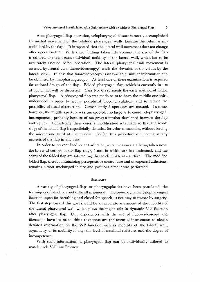
After pharyngeal flap operation, velopharyngeal closure is mostly accomplished
by medial movement of the bilateral pharyngeal walls, because the velum is im·
mobilized by the flap. It is reported that the lateral wall movement does not change
after operation. 6)-8) With these findings taken into account, the size of the flap
is tailored to match each individual mobility of the lateral wall, which has to be
accurately assessed before operation. The lateral pharyngeal wall movement is
assessed by frontal-view fluorovideoscopy,9) while the elevation of the velum by the
lateral view. In case that fluorovideoscopy is unavailable, similar information can
be obtained by nasopharyngoscopy. At least one of these examinations is required
for rational design of the flap. Folded pharyngeal flap, which is currently in use
at our clinic, will be discussed. Case No.6 represents the early method of folded
pharyngeal flap. A pharyngeal flap was made so as to have the middle one third
undenuded in order to secure peripheral blood circulation, and to reduce the
possibility of nasal obstruction. Consequently 3 apertures are created. In some,
however, the middle aperture was unexpectedly so large as to cause velopharyngeal
incompetence, probably because of too great a tension developed between the flap
and velum. Considering these cases, a modification was made so that the whole
ridge of the folded flap is superficially denuded for velar connection, without leaving
the middle one third of the mucosa. So far, this procedure did not cause any
necrosis of the flap in any case.
In order to prevent inadvertent adhesion, some measures are being taken now:
the bilateral corners of the flap ridge, I mm in width, are left undenued, and the
edges of the folded flap are sutured together to eliminate raw surface. The modified
folded flap, thereby minimizing postoperative contracture and unexpected adhesions,
remains almost unchanged in size and positions after it was performed.
SUMMARY
A variety of pharyngeal flaps or pharyngoplasties have been postulated, the
techniques of which are not difficult in general. However, dynamic velopharyngeal
function, open for breathing and closed for speech, is not easy to restore by surgery.
The first step toward this goal should be an accurate assessment of the mobility of
the lateral pharyngeal wall which plays the major role in dynamic V-P function
after pharyngeal flap. Our experiences with the use of fluorovideoscope and
fiberscope have led us to think that these are the essential instruments to obtain
detailed information on the V-P function such as mobility of the lateral wall,
asymmetry of its mobility if any, the level of maximal stricture, and the degree of
incompetence.
With such information, a pharyngeal flap can be individually tailored to
match each V-P insufficiency.

10 Yutaka HARITA, Nobuhiko ISSHIKI, Mayuki GOTO and Michio KAWANO
BIBLIOGRAPHY
1) Shprintzen, R.J., Lewin, M. L.. Croft, C. B., et al.: A Comprehensive Study of PharyngealFlap Surgery: Tailor made flaps. Cleft Palate J. 16: 46-55, 1979.
2) Owsley,J. Q.Jr., Lawson, L. I., and Miller, E. R., et al.: Experience with the High AttachedPharyngeal Flap. Plast. Reconstr. Surg. 39: 232-242, 1966.
3) Isshiki, N., and Morimoto, M.: A New Folded Pharyngeal Flap: Preliminary Report.Plast. Reconstr. Surg. 55: 461-465, 1975.
4) Honjo, I., Isshiki, N.: Diagnosis of Velopharyngeal Function. Practica Otologica. 64(8):853-861, 1971 (in Japanese).
5) Kawano, M., Isshiki, N., Harita, Y., et al.: Treatment and Result of Slight VelopharyngealIncompetence. Studia Phonologica XVII: 15-26, 1983.
6) Shprintzen, R. J., McCall, G. N., and Skolnick, M. L.: The Effect of Pharyngeal Flap Surgeryon the Movements of the Lateral Pharyngeal Walls. Plast. Reconstr. Surg. 66: 570-573, 1980.
7) Argamaso, R. V., Shprintzen, R.J., and Strauch, B. et al.: The Role of Lateral PharyngealWall Movement in Pharyngeal Flap Surgery. Plast. Reconstr. Surg. 66: 214-219, 1980.
8) Harita, Y., Isshiki, N.~ Hayashi, 0., et al.: Lateral Pharyngeal Walls Movement AfterPharyngeal Flap Surgery. Logop. Phoniatr. 26: 97, 1985. (in Japanese)
9) Skolnick, M. L., Shprintzen, R.J., McCall, G. N., et al.: Patterns ofVelopharyngeal Closurein Subjects with Repaired Cleft Palate and Normal Speech: A multi-view videofluoroscopicanalysis. Cleft Palate J. 12: 369-376, 1975.
10) Neuner,O.: A New Method for the Velopharyngeal Operation. Plast. Reconstr. Surg. 37:111-116, 1966.
(Aug. 31, 1985, received)