Embed Size (px)
Citation preview


A STUDY OF VELOPHARYNGEAL CLOSURE
IN CHILDREN WITH VOCAL
NODULES
by
Bonnie Wagoner Amos
A Thesis Submitted to the Faculty of the Graduate School at
The University of North Carolina at Greensboro in Partial Fulfillment
of the Requirements for the Degree Master of Arts
Greensboro 1972
Approved by
^e Ccd-as- Thesis Adviser

AMOS, BONNIE WAGONER. A Study of Velopharyngeal Closure in Children with Vocal Nodules. (1972) Directed by: Dr. Mariana Newton. Pp. 60.
The etiology of vocal nodules has eluded speech pathologists and
physicians alike. The literature reporting incidence and etiology has
been inconclusive. Few studies have suggested a physiological disorder
as the etiology of vocal nodules. However, McWilliams, Bluestone, and
Musgrave (1969), in noting the high frequency of vocal nodules in a
population of cleft palate children, have suggested that velopharyngeal
inadequacy may be a cause of vocal nodules in these children. The
possibility that non-cleft palate children with vocal nodules also have
minimal velopharyngeal inadequacy was proposed.
The method for data collection consisted of obtaining airflow
measures on nine children with vocal nodules. These children ranged
in age from eight to twelve years. In addition, an individual record
was compiled on each child. This record contained information regarding
the child's medical history, onset and development of hoarseness, vari-
ables affecting hoarseness, and history of vocal use. In addition, a
space was provided on the record to note the occurrence of other speech
disorders in the families of the subjects.
The results of the study revealed that all nine subjects had
adequate velopharyngeal closure as measured by the airflow procedure.
Therefore the hypothesis that these non-cleft palate children with vocal
nodules also have velopharyngeal inadequacy must be rejected. The in-
dividual record results supported the literature in suggesting that
vocal abuse, particularly during a time when the vocal cords are in-
flamed, is related etiologically to vocal nodules. The results on two
subjects supported the theory that tension may be related to vocal abuse.
.

APPROVAL PAGE
This thesis has been approved by the following committee of the
Graduate School at The University of North Carolina at Greensboro.
Thesis Adviser L 4.fk
Oral Examination Committee Members i_
Jlrviv^v Afci
3 «*««, //. ,-1 1 -7
Date of Examination
ii

ACKNOWLEDGMENTS
The writer wishes to acknowledge with gratitude the extra-
ordinary talent and patience of her thesis adviser, Dr. Mariana Newton.
The writer further wishes to acknowledge the wisdom of the oral exam-
ination committee: Dr. Herman Middleton and Dr. Nancy White.
The writer is particularly grateful to Dr. Doris Bradley and to
Dr. Donald Warren for their assistance with the research and for making
the facilities at the Dental Research Center at Chapel Hill available
for this study. Appreciation is extended to Dr. Bradley, who generously
gave her counsel in the planning of the study, and to Dr. Warren, who
directed the administration of the airflow measures and supervised the
data analysis. A special thanks is due to Jeannine A. Mashburn, who
conducted the hearing tests, and to Karen B. Barker, who assisted with
the designing of the illustrations used in the text. Finally, the writer
wishes to express gratitude to her family and friends for their encour-
agement and patience.
iii

TABLE OF CONTENTS
Page
LIST OF TABLES v
LIST OF FIGURES _ vi
CHAPTER I. INTRODUCTION 7
CHAPTER II. REVIEW OF THE LITERATURE 9
Definition of Vocal Nodules 9 Incidence of Vocal Nodules 10 Etiology of Vocal Nodules 13
CHAPTER III. PROCEDURES 22
Subjects 22 Testing Procedures , 24 Data Analysis 32
CHAPTER IV. RESULTS AND DISCUSSION 36
Velopharyngeal Area Results 36 Individual Records Results 37 Discussion 44
CHAPTER V. SUMMARY AND CONCLUSIONS 52
BIBLIOGRAPHY 54
APPENDIX 57
iv
-
LIST OF TABLES
Table Page
1. Area Measurements, Reported in Square Millimeters, on Nine Subjects Saying Ten Phrases 38
LIST OF FIGURES
Fi8ure Page
1. Schematic Representation of the Equipment 26
2. Schematic Diagram of the Printout , 28
3. Schematic Representation of Procedure Use In Recording Airflow Data 31
4. Airflow Data Indicating a Velopharyngeal Orifice Area Too Small to be Computed 34
5. Airflow Data Indicating an Adequate and Yet Measureable Velopharyngeal Orifice Area 34
6. Calibration Data Indicating the Baseline and an Area Equivalent to .5cm2 34
vi

■
CHAPTER I
INTRODUCTION
Vocal nodules in children are a problem for speech pathologists.
Although the etiology of vocal nodules has not been specifically deter-
mined, much of the literature supports the theory that such nodules are
the direct result of excessive vocal use. Therefore, vocal rest
followed by voice training is the treatment which is generally recom-
mended by speech pathologists. However, the usual play habits of child-
ren make vocal rest difficult, if not impossible, before or after
surgical intervention.
Few studies have explored the possibility of a physiological
factor in the etiology of vocal nodules in children. However,
McWilliams, Bluestone, and Musgrave (1969) noted an unusually high
incidence of vocal nodules in a population of children with congenital
clefts of the palate. It was proposed that these children, with inad-
equate velopharyngeal closure, may attempt to use vocal cord valving
activity to compensate for the poor closure. Thus, this compensatory
valving movement could cause stress on the vocal cords and, therefore,
could result in the development of vocal nodules. In view of the
frequency of vocal nodules in children with palatal clefts, the
possibility that vocal nodules in non-cleft palate children may be due
to inadequate velopharyngeal closure is proposed.
Various techniques have been used in the past to determine
velopharyngeal closure. For example, by looking in the mouth of the

8
subject, the closure can be indicated through observation of palatal
elevation. Others have placed a mirror under the subject's nose while
the subject was producing non-nasal speech sounds. Any clouding on the
mirror revealed nasal emission of air and, thus, indicated poor velo-
pharyngeal closure. Another technique has involved the use of a dry
spirometer. The spirometer is designed to measure airflow through the
mouth. Therefore, low measures of airflow through the mouth could be
indicative of poor velopharyngeal closure. However, these techniques
fail to provide a precise measure of the velopharyngeal orifice area.
During the last decade, there have been significant improvements in the
techniques for measuring vocal tract configurations, particularly
velopharyngeal closure. For example, various X-ray techniques have
been used to determine the degree of velopharyngeal inadequacy. Another
method, the airflow procedure, was the technique selected for the
purposes of this study. This procedure, which subtracts nasal pressure
from oropharyngeal pressure, yields a precise measurement of small
velopharyngeal gaps.
The purpose of this study was to investigate the extent of
velopharyngeal closure in non-cleft palate children with a diagnosis
of vocal nodules. It was hoped that such a study might provide some
information of significance to the understanding of the precipitation
and development of vocal nodules in children. Improved understanding
of this disorder could lead to more effective clinical management of
such cases and, possibly, to preventative measures.

CHAPTER II
REVIEW OF THE LITERATURE
The diagnosis of vocal nodules in children has often followed
when hoarseness was the chief complaint. The medical literature has
provided information about the nature of this condition; however, the
investigations concerning the incidence and etiology of nodules have
been few and inconclusive.
Definition of Vocal Nodules
Vocal nodules are described as growths which do not spread or
effect general health but which may cause pain or hamper functioning
(Brodnitz, 1953). Froschels and Jellinek (1941, p. 158) have stated
that vocal nodules appear to be an ". . . epithelial alteration of the
vocal cords in response to the irritation caused through exaggerated
pressure of one cord against the other." More specifically, there are
two major types of nodules: the reddish, soft, young nodule composed
of normal squamous epithelium and the white, hard, mature nodule com-
posed of thickened epithelium (Arnold, 1962). The development of nodules
is continuous. Ash and Swartz (1944) have described four stages of
epithelial alteration of nodules: fibroid, polypoid, varicose, and
hyalin. The irritation of the young blood vessels extending toward the
epithelium surface have characterized the fibroid state. Scattered
growth of fibroblasts, connective tissue cells, have characterized the
polypoid state. Varicose, the state often diagnosed as polypoid, is
recognized by the presence of interstitial hyalin. Finally, the hyalin

10
state was referred to as the degeneration of connective tissue. Most
authorities have agreed that vocal nodules are usually bilateral and
occur at the point of greatest stress, which is generally on the anter-
ior third of the cord (Arnold, 1962; Ash and Swartz, 1944; Harris, 1948;
and Wilson, 1965). Although nodules are considered benign, they do
hamper the functioning of the vocal cords, resulting in audible symptoms.
Hoarseness is reported as the major voice symptom (Wilson, 1966).
Physiologically, vocal nodules prevent adequate approximation of the
vocal cords and interfere with the free vibration of the cords (Grey,
England, and Mahoney, 1965). The resulting dysphonias may be classified
as hoarseness and breathiness. In addition to these symptoms, low pitch,
glottal plosive attack, narrow pitch range, and stress patterns may
accompany vocal nodules (Berry and Eisenson, 1965). In summary, vocal
nodules are considered to be benign lesions of the vocal cords resulting
in hoarseness. These nodules are usually bilateral and generally appear
on the anterior third of the cords.
Incidence of Vocal Nodules
The literature concerning the incidence of voice disorders in
general has been sketchy; information about the incidence of vocal
nodules is practically nonexistent. Johnson, et^ al. (1967) estimated
that the incidence of voice problems in children is from one to two
per cent of the population. Moore (1957) added that voice disorders
occur in five to fifteen per cent of the defective speech population.
In a survey to determine the number of speech handicapped persons in
New England, Pronovost (1951) found that 6.6 per cent of a population
of 12,565 had voice disorders. The data, collected by sending

11
questionnaires to those institutions offering speech and hearing ser-
vices, failed to define voice disorders. A study to determine the need
of a speech therapist in the Holyoke, Massachusetts schools indicated
that 1.5 per cent of a population of 4,685 had voice disorders, with a
greater incidence in grades one through three. An amusing aspect of
this study was that the school system later employed the therapist who
assisted in the initial screening for the survey (Mills and Streit,
1942). In a study of the incidence of chronic hoarseness in the Willow
Run Public School System in Michigan, Baynes (1966) found an incidence
of 7.1 per cent in a population of 1,012, with a greater incidence in
the first grade. The procedure for this study involved three screening
surveys at one month intervals. If the patient manifested hoarseness
during all three surveys, the judges diagnosed the case as chronic
hoarseness. Baynes, who excluded those cases of mild hoarseness, con-
sidered his results to be conservative.
Several studies have focused on the incidence of voice disorders
in persons with congenital clefts of the palate. In a study of 1,061
clinic records on persons with cleft palate, Takagi, McGlone, and
Millard (1965) reported the incidence of voice disorders other than
nasality as .5 per cent of the males and .7 per cent of the females.
Using 12 normal children as the control group and 76 children with cleft
palate as the experimental group, Brooks and Shelton (1963) found that
ten per cent of those with cleft palate have voice deviations such as
hoarseness, breathiness, and inappropriate pitch. The between-judge
reliability was .95. A possible explanation for the discrepancy in
these two studies may be that one relied on clinic records, while the
other resulted from experimental research.

■ 12
McWilliams, Bluestone, and Musgrave (1969), studying minimal
velopharyngeal inadequacy in children with congenital clefts of the
palate, found that, in a population of 32 children with hoarse voices,
71 per cent had bilateral nodules. In addition, three children demon-
strated left unilateral nodules, four demonstrated right unilateral
nodules, and four had atypical vocal cords other than nodules. In-
cluded in these last four children was one child with edema of the
vocal cords, who developed vocal nodules four months later.
In relating hoarseness to the occurrence of nodules, McCall
(1970) states, "It has been my experience that 80-90 per cent of
children with variable hoarse voices (i.e. degree of hoarseness varies)
exhibit laryngeal pathology. The most frequent pathology observed is
vocal nodules." In a study of 300 cases of benign vocal cord lesions,
Fitz-Hugh, Smith and Chiong (1958) found 134 cases of nodules. In
addition, of these 300 cases, 68.3 per cent occurred in males and 31.7
per cent occurred in females. No sex ratio was reported in relation to
nodules specifically. Similarly, Arnold (1962) said that vocal nodules
are the most common laryngeal lesion treated surgically; the occurrence
is greater among males. However, others have reported a greater inci-
dence of vocal nodules in females. In a study of 1,160 cases of func-
tional voice disorders, Garde (1961) found 97 cases of unilateral nodules
and 168 cases of bilateral nodules. Included in the 97 cases of uni-
lateral nodules, were 92 women, 4 men, and 1 child. Among the 168 cases
of bilateral nodules were 164 women, 3 children, and 1 man. Perhaps the
greater incidence among women may be explained in part by the fact that
93 of the cases studied were school teachers. Although no specific

13
numbers are reported, Zerffi (1935) reported a greater incidence of
nodules among females. Zerffi based his observations on singers. In
summary, although none of these findings were conclusive, the informa-
tion to date appeared to concur with the opinion that the incidence of
vocal nodules is relatively infrequent among children (Wilson, 1965).
Etiology of Vocal Nodules
In turning to the causes of vocal nodules and related voice dis-
orders, the literature appeared repetitive and scarce. Van Riper and
Irwin (1958, p. 279) have offered a possible reason.
The literature is scanty and scattered. Except for certain occupations such as the ministry, teaching, and entertaining, the average voice defect is not a handicap, since communica- tion is still possible, a factor which does not hold true in stuttering or articulatory defects.
However, the literature concerning the causes of vocal nodules and re-
lated voice disorders does lend itself to grouping into four major
categories: vocal abuse, physical causes, psychological causes, and
mixed causes.
Some authorities have expressed the opinion that vocal nodules
are the result of vocal abuse. Greene (1957, p. 78) has written, "The
nodules which form on the outer edge of the cords and cause severe
dysphonia are the direct outcome of vocal abuse and the individual's
habitual method of forcing the voice." In referring specifically to
vocal nodules in children, Arnold (1962, p. 214) has said, "In screaming
children, bilateral vocal nodules result from and demonstrate the pres-
ence of excessive and uncontrolled vocal expression." To clarify this
point, Arnold (1962, p. 214) stated that "In children, nodules result
from excessive yelling, singing, or vociferous outdoor play."
I
14
Furthermore, in a study of 138 persons with vocal nodules, Ash and
Swartz (1944) found that all but 9 cases were due to excessive, loud
talking as determined by case history reports. In this study, a spe-
cific instance of vocal abuse was the vocal habits of the drill ser-
geant. Seventy-five of the 138 persons were Army personnel.
However, there are those (West, et al., 1947, pp. 140-41) who
have said that "No amount of vigorous vocalization can damage the edges
of the vocal folds if the voice is properly used." Zerffi (1935,
pp. 552-53), in discussing singer's nodules, related the improper
physiological production of high pitches to a theoretical cause of
singer's nodules.
The action of the arytenoid and lateral cricoaryte- noids, and the thyroarytenoids is that of bringing the poster- ior edges of the cords closely together and thus shortening the vibrating length. This approximation helps to raise the pitch of the tone, and high tones are thus sung with the anterior two-thirds or even with the anterior half of the cords. Since nodules occur at the junction of the anterior third of the cords, high tones sung as described could not possibly produce an irritation of the anterior third. Nor are there any laryn- geal muscles which act in such a manner as to provide sufficient pressure at this point to result in the formation of nodules. It is therefore obvious that other muscles than those of the larynx proper are concerned in this action. In the opinion of this writer, the muscles which bring about this pressure are those which are concerned in deglutition. These muscles, nota- bly those of the tongue, the action of which can be detected by means of finger palpation, assist those of the larynx in bring- ing about complete closure of the larynx when deglutition is about to be performed. Forced production of the voice is brought about by a similar action as that employed by deglutition. A partial contraction of the muscles is used to force the vocal cords into approximation and only by generation of considerable energy can the breath be driven through the glottis.
McWilliams, Bluestone, and Musgrave (1969, p. 3), in a study of
32 children with hoarse voices and congenital clefts of the palate,
found that 84 per cent demonstrated pathology of the vocal cords. Of
this 84 per cent, 59 per cent had borderline velopharyngeal inadequacy
15
as determined by radiographic tapes. Therefore, these writers
suggested:
. . .inadequate velarpharyngeal valving mechanisms might be related to vocal cord nodules in a logical if not a readily demonstrable manner. This idea was reinforced by information to the effect that 16 of the 22 children with vocal cord nodules had had speech therapy prior to their laryngeal diagnoses. It appeared to us that compensatory valving activities, even in the absence of glottal frica- tives and plosives, might be one means by which children would attempt to handle problems high in the tract thus subjecting the vocal cords to stress.
An interesting aspect of this study was that one child developed nodules
following therapy sessions designed to evaluate his velopharyngeal clo-
sure (McWilliams, Bluestone, and Musgrave, 1969, p. 4).
A third group of studies have concentrated on the psychological
causes of nodules and associated voice disorders. In this group is a
study of reciprocal inhibition as a treatment for nodules. In describ-
ing a method of treatment Grey, England, and Mahoney (1965, p. 188)
wrote:
The basic premise of the present therapy is that there are certain people in whom benign functional vocal nodules de- velop as a result of pervasive anxiety. This type of patient is seen as a person who is in a more or less con- stant state of anxiety or stress. This anxiety is mani- fested in emotional over-reaction to situations and misuse of the vocal apparatus due to psychological and physiologi- cal stress. Originally, isolated situations may have been responsible for high anxiety states—the anxiety reaction being an unconditioned response to the stimulus situation. However, as more and more cues became associated with the stimulus situation the anxiety reaction became a conditioned reaction to a variety of conditioned stimuli which formerly did not evoke anxiety—the result being pervasive or free floating anxiety. Thus, the patient is seen as moving from isolated situations of anxiety to a more or less constant state of anxiety.
This report implied the opinion that vocal nodules appear to be related
etiologically to anxiety states. The authors have suggested a treatment
program which includes the patient's acknowledgment of feared

16
situations, followed by practiced non-anxiety reactions to these
situations (Grey, England, and Mahoney, 1965).
Although Aronson, Peterson, and Litin have dealt primarily
with voice disorders in adults which are due to psychological factors,
their research was interesting to note. In one study (Aronson, Peter-
son, and Litin, 1964), the authors classified vocal nodules as a direct
result of misuse and described voice disorders, such as spastic dyspho-
nias and ventricular dysphonias, as being characteristic of psycho-
neurotic or psychotic mental states. These disorders manifest audible
symptoms similar to those of vocal nodules. In 1966 Aronson, Peterson,
and Litin administered the Minnesota Multiphasic Personality Inventory
to 27 persons with voice disorders (spastic and ventricular dysphonias).
These persons ranged in age from 14 to 72. As a result of this study,
Aronson, Peterson, and Litin (1966, p. 126) concluded that "Acute or
chronic situational conflicts were causally related to the voice dis-
orders in the overwhelming majority of patients, regardless of type of
voice symptomatology." Furthermore, Alfaro (1960) reported that voice
disorders due to psychogenic influences were more common in adults than
in children. Contrary to these studies, Coodstein (1958, p. 364),
in a review of psychological causes of voice disorders, concluded that
"... the relationship between voice disorders and personality is yet
to be empirically demonstrated."
By far, the bulk of literature has concentrated on a combination
of factors as causing vocal nodules and associated voice disorders.
That is, when speaking of vocal abuse, physical factors, and psycho-
logical factors as causes of vocal nodules, some cases may be due to a
single cause, but the majority have been the result of a combination of
r*.
17
all three (Withers, 1961). Several writers have discussed these three
causal factors while others consider only two factors to be interacting.
In discussing all three aspects, Brodnitz (1958, p. 112) stated
that the fact that nodules occur on the vocal cords, "... does not
mean that faulty vocal cord function alone is to be blamed." Brodnitz
(1958) listed eight factors to consider: the breathing mechanism, the
resonation cavities, mutational changes, tenseness during speech, ex-
cessive talking or shouting, faulty voice production during singing,
daily emotional tenseness, and hormonal or metabolic imbalance.
Froschels and Jellinek (1941, p. 194) discussed the role of
vocal abuse.
. . . . Professional singers and speakers such as teachers, politicians, lecturers, lawyers, preachers, etc. who have to stand great professional strain without sufficient technical preparation often try to overcome fatigue or decrease of their vocal capacities through some acute disease, for instance, a cold by augmented use of force. They tighten muscles which should work with ease. The effect is an ever increasing over- strain of these muscles, which may even result in some organic alteration, such as bursting of small vessels, irritating sen- sations with resulting coughing. The nodules of the vocal cords, so greatly feared by singers, are among the effects of such overstrain.
In describing types of physical hyperfunctions, Froschels and Jellinek
(1941) listed six hyperfunctions: violent forcing of air through the
vocal cords, 'coup de glotte,' contraction of pharyngeal muscles, re-
traction of the tongue, excessive elevation of the velum, and tenseness
of the lips. Moreover, a study of 1000 persons revealed that contrac-
tion of pharyngeal muscles and 'coup de glotte' were the factors which
occurred most often in children speaking under stressful situations.
In addition, Froschels and Jellinek (1941, p. 195) offered the following
consideration of the psychological factor:
18
The psychic phenomena accompaning every decay of the vocal qualities in a singer are striking, and greatly increase his difficulties. These difficulties in singing are usually far greater than we should expect after examination of the vocal cords. They originate in part from the exaggerated attention which the patient—singer or professional speaker— directs toward his own phonetic function.
Besides faulty usage, Arnold (1962, pp. 205-6) cited three
additional causative factors: 'predisposing,* 'precipitating,' and
'aggravating.' Included in the 'predisposing' factors were persons
with aggressive personality structure, persons inclined to allergies,
and persons with poorly constructed vocal mechanisms. The 'precipi-
tating' factors were tobacco and alcohol, accompanied by vocal abuse.
That is, during social gatherings, alcohol may promote mucosal hyperemia
(a concentration of blood cells) of the vocal cords, and then tobacco
may irritate the vocal cords. This irritation, resulting in hoarseness,
could cause hyperkinetic efforts to talk louder.
Wilson's discussions of vocal nodules and laryngeal dysfunction
were of interest. Regarding vocal nodules in children, Wilson (1961)
listed seven causitive factors: inappropriate pitch, excessive air
force during phonation, excessive talking, loud phonation, abrupt phon-
ation, excessive strained phonation during play, and emotional factors.
Later Wilson (1966, pp. 76-78) listed the following suggested goals
of voice therapy which explained the above factors in more detail and
mentioned the additional factors of posture and rate of speaking.
1. Vocal Abuse Attention must be given to vocal abuse in all benign voice path- ology patients. Abusive habits include the frequent use of such traumatizing vocal practices as vigorous throat clearing, ex- cessive and hard coughing, vocalizing on intake of air, scream- ing and shouting, prolonged vigorous use of the voice and exces- sive talking, and emitting strained vocalizations. . . .

19
2. Easy Initiation of Tones Patients with benign vocal pathology may speak with sudden, abrupt initiation of sounds resulting in an irregular and staccato type of speaking. . . . This practice, when force- ful and traumatizing, may result in nodules or polyps on the vocal cords, contact ulcers on the vocal processes or the arytenoids, or nonspecific laryngitis.
3. Desirable Pitch Change .... That is, some patients may use an abnormally high pitch when they speak loudly under noisy conditions, talk under emotional strain, or give a public speech. Other patients may use an abnormally low pitch in certain situa- tions, such as when talking on the telephone, giving a sales pitch, or participating in small group discussions and con- ferences. . . .
4. Appropriate Loudness of the Voice The habitual speech of some patients is so loud that irrita- tion of the vocal mechanism results. . . .
5. Relaxation, Correct Breathing Patterns, and Good Posture . . . . Specific relaxation is especially necessary when undue tension of certain laryngeal muscles is contributing to the pathology or when continued overuse has led to hypofunction and weakness. Efficient breathing patterns and good posture may also need attention.
6. Rate of Speaking Any deviations from a desirable rate of speaking should be corrected. An excessively rapid rate of speaking is charac- teristic of many patients with vocal pathology and may indi- cate faulty use of the vocal mechanism. . . .
Rather than emphasizing the psychological aspect, others concen-
trated more on the vocal abuse and medical or physical aspects. For
example, Berry and Eisenson (1956) reported vocal abuse as the probable
cause of vocal nodules in 90 per cent of the cases. Included as types
of vocal abuse were loud phonation and inappropriate, low pitch. Con-
tinuing, Berry and Eisenson (1956, p. 212) gave attention to the aspect
of tension in the following statement.
The effect of the tension, whether external or internal, is to restrict the freedom of movement, particularly of the anterior one-third section of the vocal folds. As a result of tension and contact, the area becomes degraded, and in- creased layers of epithelium are built up forming the nodule.

20
West, Kennedy, and Carr (1947) cited that repeated misuse of the vocal
mechanism and chronic laryngitis or tonsillitis can lead to nodules.
Similarly, Alfaro (1960, p. 6) said that "Abnormalities of phonation in
the child are usually on an organic basis such as vocal nodules, from
misuse of the voice, or laryngitis of either infectious or allergic
et iology."
In clarifying the meaning of vocal abuse, Van Riper and Irwin
(1958) mentioned vocal abuse in its relationship to physical factors.
That is, vocal abuse may include excessive use of the voice, particu-
larly if the vocal cords were inflamed, speaking 'on residual air,'
speaking loudly over a continuous masking noise, speaking with inappro-
priate pitch and loudness, and hypertension of the laryngeal muscles
used in swallowing while speaking.
Placing less emphasis on the physical aspect, Rubin and
Lehrhoff (1962, p. 153) saw the causes of vocal nodules as being ". . .
an extended spectrum ranging from simple loud phonating at one end to
unadulterated emotional tension at the other, with varying combinations
of the two between." In clarifying the importance of vocal abuse as a
cause, Rubin and Lehrhoff stress the relativity of vocal abuse to in-
dividual speakers. That is, what constitutes vocal abuse for one
speaker might not necessarily be detrimental to another speaker (Rubin
and Lehrhoff, 1963).
In summary, vocal nodules are defined as benign lesions of the
vocal cords resulting in hoarseness. Although the reports were incon-
clusive, the information indicated a relatively infrequent incidence
of nodules in children. Concerning etiology, the literature has con-
centrated on four types: vocal abuse, physical causes, psychological

21
causes, and mixed causes. The majority of the literature emphasized
the interaction of several factors to produce vocal nodules.

22
CHAPTER III
PROCEDURES
The etiology of vocal nodules has eluded speech pathologists
and physicians alike. The etiological studies have been few and in-
conclusive. Most of these studies conclude that nodules result from
loud, excessive vocal use. Few studies investigate the possibility
of a physiological inadequacy in relation to vocal nodules. However,
McWilliams, Bluestone, and Musgrave (1969) have reported that children
with inadequate velopharyngeal closure, related to congenital clefts of
the palate, frequently develop nodules. Therefore this study was de-
signed to investigate velopharyngeal adequacy, not related to palatal
clefts, in children with vocal nodules. The subjects, the instrumen-
tation, and the procedure will be discussed below.
Subjects
The nine subjects used in this study consisted of six males and
three females. Six of the subjects were selected from the client records
at the University of North 'arolina at Greensboro Speech and Hearing
Center. The remaining three subjects were referrals from the Guilford
County Health Department, a practicing laryngologist in Greensboro, and
the Dental Research Center at Chapel Hill, North Carolina.
With regard to the previous treatment of the subjects at the
time of this study, three of the subjects had had voice therapy fol-
lowing the surgical removal of the nodules. The remaining subjects
reportedly had had voice therapy which concentrated on general
23
relaxation, alterations in vocal pitch, and a decrease in vocal abuse.
No therapy had been centered on velopharyngeal adequacy or nasality
problems.
The initial contact with the parents of the subjects involved
sending a Letter of Introduction. (See Appendix I, page 57). The
purpose of the letter was to provide the parents with a general concept
regarding the nature of the study. After the letter, each parent was
contacted by telephone. This telephone contact served to answer any
questions the parents had regarding the study and to further solicit
their cooperation.
Prior to being selected as a subject for the study, a confirma-
tion of the diagnosis of vocal nodules in each subject was needed.
Clinic records revealed that each of the subjects had had a laryngeal
examination by practicing laryngologists in Greensboro or Chapel Hill.
These records showed that the three female subjects and five of the
male subjects had bilateral nodules. One male subject had a unilateral
nodule.
The subjects were limited to those children between the ages of
eight and twelve years. The upper age limit was specified hopefully to
exclude those children experiencing voice mutation. However, due to the
physical appearance and voice quality of one twelve year old child, it
was decided that this child's voice was possibly undergoing mutation.
Therefore, to avoid contamination of the data, this child was excluded
from the study.
In addition, the parents were required to sign a Statement of
Informed Consent. (See Appendix II, page 58). This statement explained
the requirements for participation in the study. These requirements
24
stated that the parents were to supply information for an individual
record and that each child was to have hearing and airflow evaluations.
The Individual Record was designed to collect information con-
cerning the child's medical history, the onset and development of
hoarseness, and a history of the child's vocal use. Also, a section
was included to note the occurrence of other speech disorders in the
subject's family. (See Appendix III, pages 59-60).
The literature cites inappropriate use of the vocal mechanism
as a possible cause of vocal nodules. Since faulty voice production
can occur in those persons with a hearing loss, a pure-tone audio-
metric sweep test was administered in a sound proof room to each sub-
ject. The procedure for the sweep test, suggested by Newby (1964),
was used.
The parents were informed that their children would be taken
to the Dental Research Center in Chapel Hill for the airflow evaluation.
Transportation to and from Chapel Hill was provided as a convenience to
the parents.
Testing Procedures
Individual Record
The literature has cited upper respiratory infection and vocal
abuse as possible causes of vocal nodules. Therefore, to supplement
the data, an individual record was kept on each subject. This record
included questions regarding: (1) the child's medical history;
(2) description and management of the child's vocal nodules; and (3)
the development of the child's voice problem. Additional sections of
the record provided spaces for identification information and the

*<*»
25
results of the hearing test. Information for the Individual Record was
acquired in a parent interview conducted by a graduate clinician.
Equipment
The equipment used for the study included a tape recorder,
microphone, differential pressure transducer, heated pneumotachograph,
and a Honeywell Visicorder and analogue computer arranged as shown in
Figure 1, page 26. This equipment recorded velopharyngeal orifice
differential pressure, volume rate of airflow through the orifice, and
computed velopharyngeal area according to the formula given below.
Speech was recorded through the use of a microphone placed under the
subject's chin. Thus, speech appeared on the visicorder printout and
helped to identify points on the record that should be measured. A
tape recorder was placed approximately three feet from the subject so
that a record could be made of the test phrases. This record served
to verify, during analysis, the accurate order of presentation.
Velopharyngeal orifice differential pressure was determined
through the use of two catheters, one in the subject's mouth and one
in the nose, leading to the differential pressure transducer. So that
the catheters would pick up only lateral pressure, both catheters were
plugged at the end with wax and small holes were drilled along the sides
of the catheters. Utilizing these two measures, the transducer sub-
tracted the nasal pressure from oropharyngeal pressure to yield the
velopharyngeal orifice differential pressure (Warren, 1964). The trans-
ducer then directed this electrical information into the computer.
The aspect of velopharyngeal airflow was detected by a flow-
meter attached to plastic tubing placed in the subject's right nostril

Differential Pressure Transducer
Tape Recorder
Left Nostril
Mouth
Right Nostril
Heated Pneumotachograph
Fig. 1. Schematic Representation of the Equipment

27
(Warren, 1964). A transducer changed this flow measurement Into an
electrical signal and fed this signal into the computer.
The analogue computer was programmed to determine velopharyngeal
area through use of the measurements received regarding the differential
pressure and airflow. The program was based on the following equation:
Area = Volume rate of airflow through the orifice
.65 M2(0rifice differential pressure) Density of Air
This equation, which is a modification of one reported by Gorlin and
Gorlin (1951), was adapted by Warren and Dubois (1964) to measure velo-
pharyngeal area. The formula was modified through the use of a correc-
tion factor (.65). It was believed that this factor was needed to ac-
count for the variations in the size of the oropharyngeal orifice due
in part to the turbulent nature of the airflow during speech. Sixteen
experiments were conducted to ascertain the effect which alterations
in oropharyngeal size would have on the amount of the correction factor
needed. However, the changes in the correction factor, resulting from
various oropharyngeal sizes, were so slight that the decision was made
to compute the arithmetical mean of the factors and to treat this mean
as a constant (Warren and Dubois, 1964).
The visicorder graphically recorded four parameters: speech,
differential pressure, airflow, and area. This information was printed
on photosensitive paper. The first line of the graph indicated the
occurrence of speech. The next graphic plot reflected the alterations
in the velopharyngeal differential pressure. Variations in the volume
rate of airflow through the velopharyngeal orifice were recorded as the
third plot. The fourth line indicated velopharyngeal area. A schematic
diagram of the printout appears in Figure 2, page 28.

28
Speech
Airflow
Pressure
Area
Fig. 2. Schematic Diagram of the Printout
29
Calibration
The apparatus was calibrated following the evaluation of each
subject. The calibration procedure consisted of attaching the two
catheters to a model of a velopharyngeal orifice and the flowmeter to
a model "nose." The models were constructed of plastic tubings and
joints. The simulated airflow of speech was furnished by an air pump.
This cylinder introduced a known quantity of air into the model. The
differential pressure transducer and flow transducer fed electrical
impulses into the computer which yielded a graphic plot of an area
equal to ,5cm2.
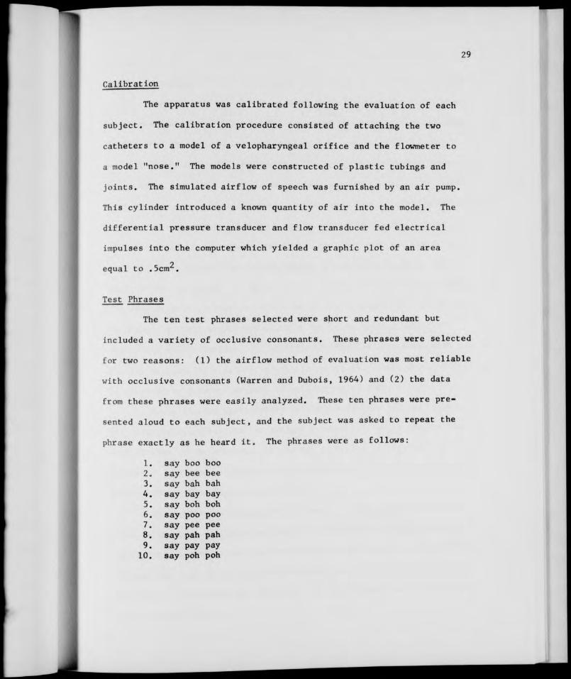
Test Phrases
The ten test phrases selected were short and redundant but
included a variety of occlusive consonants. These phrases were selected
for two reasons: (1) the airflow method of evaluation was most reliable
with occlusive consonants (Warren and Dubois, 1964) and (2) the data
from these phrases were easily analyzed. These ten phrases were pre-
sented aloud to each subject, and the subject was asked to repeat the
phrase exactly as he heard it. The phrases were as follows:
1. say boo boo 2. say bee bee 3. say bah bah 4. say bay bay 5. say boh boh 6. say poo poo 7. say pee pee 8. say pah pah 9. say pay pay
10. say poh poh

30
Airflow Procedure
Each subject was seated on a stool in front of the table where
the transducers were affixed. The height of the stool was adjustable
so that the child could be positioned in a manner suitable for the in-
sertion of the catheters and tubing. The catheters and tubing were
inserted as described above. Each subject was instructed to say sample
test phrases in order to determine if the tubes were properly positioned
and to familiarize the child with the test procedure. One problem
associated with the procedure existed in the fact that the children
were reluctant to use their usual manner of articulation while the
catheter was in place. It should be noted that erratic printouts from
the visicorder were usually indicative of the presence of fluid in the
catheter. In this event, the catheter had to be cleaned before the
evaluation could continue. In addition, deflection of the catheter by
the subject usually resulted in erratic printouts. These printouts did
not provide an accurate measure of velopharyngeal area.
Three persons participated in the procedure: (1) one to hold
the catheter and tubing in the subject's nose; (2) one to present the
test phrases to the subject; and (3) one to manage and monitor the
computer. The tape recorder was turned on prior to the evaluation of
each subject. (See Figure 3, page 31).
Each child was instructed to repeat the test phrases one at a
time. With three syllables per test phrase, there resulted thirty
velopharyngeal orifice area measurements for each subject. One subject
was found to have an articulation disorder; he distorted occlusive
consonants. Therefore, two trials of the test phrases were recorded to
insure adequate data.

A-Pressure Transducer B and C-Pneumotachograph
Microphone
Fig. 3. Schematic Representation of Procedure Used in Recording Airflow Data
Analogue Computer
w

32
Data Analysis
The data consisted of airflow measures on the ten test phrases
and calibration printouts for each subject. The airflow data were in
the form of a graphic representation of four parameters: speech,
differential pressure, airflow, and area. The first line of the graph
indicated the occurrence of speech. The next graphic plot reflected
the alterations in the differential pressure. Variations in the volume
rate of airflow were recorded as the third plot. The fourth line in-
dicated area. The calibration printout for each subject indicated a
baseline and a linear area equivalent to .5cm^.
The purpose of the analysis was to determine the velopharyngeal
closure during speech for the nine subjects. Analysis of the airflow
data consisted of recording the speech samples appropriately on the
graphic plot from the visicorder, marking the baseline on the graph,
and computing the velopharyngeal area during each repetition of the ten
test phrases for the individual subjects.
The purpose in indicating the speech samples at appropriate
points on the graph was twofold: to facilitate discussion of the data
and to assist in comparing the velopharyngeal area measurements in the
same subject during varying speech samples. While playing the tape
recording taken during the test situation, the appropriate test phrases
were written above the first line on the graph which indicated that
speech had occurred. Therefore, casual examination of the graph re-
vealed the test phrase being used during a particular measurement of
velopharyngeal orifice area.
The baseline revealed the area on the graph below which the
velopharyngeal orifice area was too small to be computed. That is,

33
when the line indicating velopharyngeal area fell below this baseline,
the orifice area could be regarded as adequate. (See Figure 4, page 34).
Furthermore, when the area line appeared above the baseline, the velo-
pharyngeal area could be computed to determine its adequacy. (See
Figure 5, page 34). Since this baseline appeared only on the calibra-
tion sheet for each subject, the line had to be drawn in manually on
each graph.
The calibration showed a graphic plot of an area equal to .5cm .
(See Figure 6, page 34). Therefore, in order to compute the velo-
pharyngeal orifice area, a metric ruler was used to make two measure-
ments: the linear equivalent to .5cm2 and the distance in centimeters
between the baseline and the area line. This second measurement was
made only on the sections of the baseline and area line which appeared
directly below peaks in the orifice differential pressure line. That is,
as differential pressure increased, orifice area decreased. Furthermore,
this second measurement was required only if the orifice area line
appeared above the baseline. No measurement was necessary when the
orifice area line fell below the baseline. To compute the orifice area,
the two measurements were used in the following simple relationship
equation:
9 9 . Scm^ or SOmm^ The linear measure- ment on the calibra- tion sheet equal to
Velopharyngeal orifice area The distance in centimeters between the baseline and the area line
.5cm^
The results to the equation were expressed in square millimeters. Using
subject B. M. as an example, if the linear equivalent to .5cm2 were
5.2cm and if the distance between the baseline and the area line were
.2cm, the area would be computed as follows:
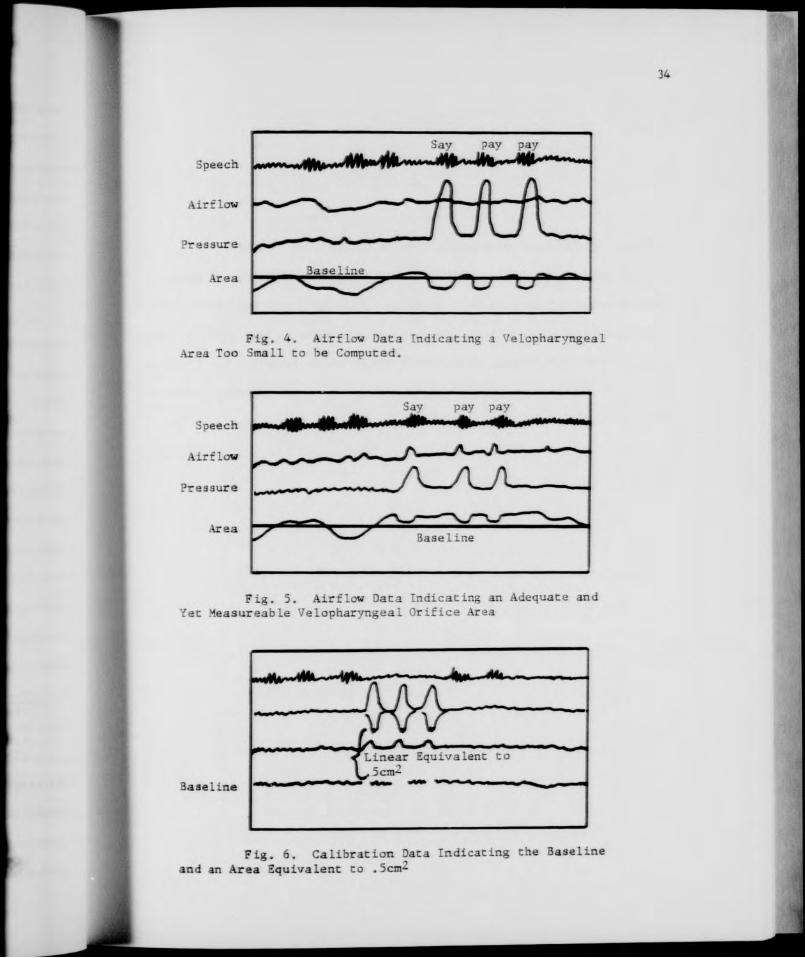
34
Fig. 4. Airflow Data Indicating i Velopharyngeal Area Too Small to be Computed.
Fig. 5. Airflow Data Indicating an Adequate and Yet Measureable Velopharyngeal Orifice Area
3aseline
Linear Equivalent to 5cm^
Fig. 6. Calibration Data Indicating the Baseline and an Area Equivalent to .5cm--

35
SOmnr Area 5.2cm .2cm
(5.2)(Area) = (.2)(50)
5.2 Area = 10
Area = 1.9mm2
In summary, five measures were obtained for each of the nine
subjects in this study. Four were recorded by the computer: speech,
differential pressure, airflow, and area. The area of the velopharyn-
geal orifice, was calculated using the procedure described above. An
analysis and interpretation of the findings is presented in the
following chapter.

36
CHAPTER IV
RESULTS AND DISCUSSION
Data on nine subjects with vocal nodules were obtained in order
to determine if these subjects manifested minimally inadequate velo-
pharyngeal closure during speech. The primary method of data collec-
tion consisted of airflow measures on each subject while repeating ten
phrases. In addition, an individual record was kept on each subject.
With parents serving as informants, information was recorded regarding
each subject's medical history, the onset and development of hoarseness,
the variables affecting hoarseness, and the occurrence of other speech
disorders in the families of the subjects. These measures provided the
data which are presented and discussed in this chapter.
Velopharyngeal Area Results
A summary of the velopharyngeal area measurements on each sub-
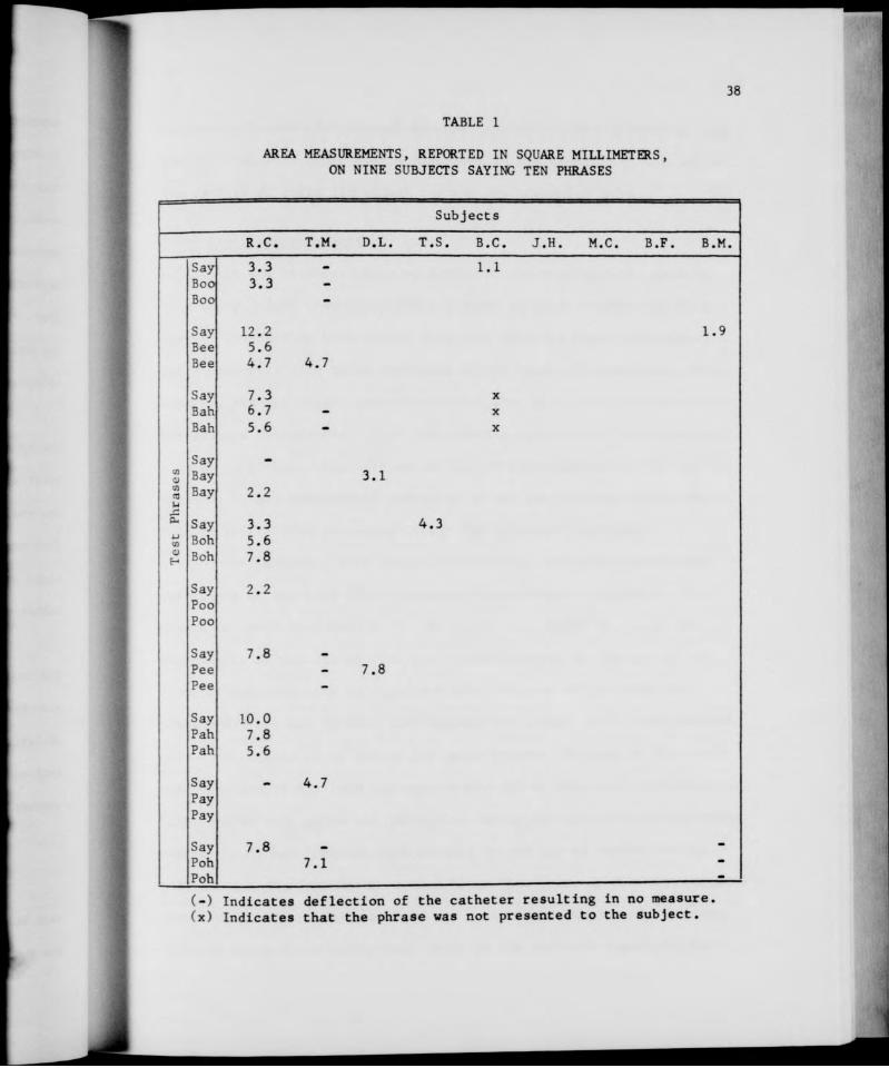
ject is presented in Table 1, page 38. As is indicated on this table,
27 measurements out of a possible 252 instances were calculated. These
27 measurements were conducted on six of the subjects. Three subjects,
B. M. , B. C, and T. S., each had one syllable out of the ten test
phrases during which their velopharyngeal area could be computed. The
results, expressed in square millimeters, were (Say) 4.3, (Say) 1.1,
and (Bee) 1.9. The results on D. L. indicated a measurable velopharyn-
geal area on two syllables out of the ten test phrases. These syllables
were (Bay) 3.1 and (Pee) 7.8. Three velopharyngeal area measurements,
(Bee) 4.7, (Say) 4.7, and (Poh) 7.1, were computed on T. M.

37
Interestingly, one subject, R. C. , had measurable orifice areas ranging
in size from 2.2 to 1.2mm on nineteen syllables. However, due to the
fact that this subject had an articulation disorder which involved poor
production of the consonants in the test phrases, caution should be
exercised in interpreting his area results. That is, his manner of
articulation may have affected his velopharyngeal port area. Nonethe-
less, the airflow results on these 27 measurements failed to approach
the established critical level of 20mm2 (Warren, 1964) for inadequate
velopharyngeal closure.
No calculations were necessary or could even be conducted on
the remaining 225 syllables because the graphic printout from the com-
puter showed the area line below the baseline. That is, the area was
too small to be computed and, therefore, was regarded as adequate. In
fact three subjects, J. H., M. C, and B. F., required no area calcula-
tions at all. The other six subjects had a combined total of 117 syl-
lables on which no calculations were necessary.
In summary, 27 velopharyngeal measurements out of a possible
252 could be conducted on six subjects. These measurements ranged in
size from 1.1mm2 to 12.2mm2, with a mean of 5.5mm2 and a standard
deviation of 2.59mm2. None of the measurements approached the signifi-
cant velopharyngeal port size of 20mm2. Such an area would have been
indicative of inadequate velopharyngeal closure.
Individual Record Results
The individual records were forms on which information regarding
each subject's medical history, the onset and development of hoarseness,
the variables affecting hoarseness, and a history of vocal use was
38
TABLE 1
AREA MEASUREMENTS, REPORTED IN SQUARE MILLIMETERS, ON NINE SUBJECTS SAYING TEN PHRASES
Subjects
R.C, T.M. D.L. T.S. B.C. J.H, M.C. B.F, B.M.
Say 3.3 Boo 3.3 Boo
Say 12.2 Bee 5.6 Bee 4.7
Say 7.3 Bah 6.7 Bah 5.6
Say - Ul 41 Bay CO Bay 2.2 U A — Say 3.3 u Boh 5.6 4) Boh 7.8
Say 2.2 Poo Poo
Say 7.8 Pee Pee
Say 10.0 Pah 7.8 Pah 5.6
Say — Pay Pay
Say 7.8 Poh Poh
1.1
1.9
4.7
x x X
3.1
4.3
7.8
4.7
7.1
(-) Indicates deflection of the catheter resulting in no measure, (x) Indicates that the phrase was not presented to the subject.
39
recorded. In addition, questions were included regarding etiology and
the occurrence of other speech disorders in the families of the subjects.
The results of these individual records are presented below.
Upper Respiratory Infections
It was interesting to note that of the nine subjects used in
this study, eight reportedly had a history of upper respiratory infec-
tions. Included in these eight were four subjects whose respiratory
infections were said to be unrelated to the onset of hoarseness. For
example, the individual records revealed that R. C. had frequent sore
throats and tonsillitis. J. H. had chronic colds as an infant and an
allergy to pollen. In addition, he had an adenoidectomy at the age of
six. T. S. had symptoms of pneumonia at the age of five. B. M. was
said to suffer from allergies during the Spring of each year.
The remaining four subjects reportedly had upper respiratory
infections at the time when hoarseness first became noticeable. In-
cluded in these four were B. F., D. L., T. M., and M. C. B. F. had
tonsillitis at the age of five and a tonsillectomy at the age of six.
Then, between the ages of eight and nine, the age of the onset of
hoarseness, she had chronic sore throats and colds. D. L. had frequent
mild sore throats as an infant and again between the ages of five and
six. The age of five was the approximate age of the onset of hoarseness.
T. M. had chronic colds and laryngitis during the period when hoarseness
began. M. C. had frequent sore throats at the age of eight, the age
when hoarseness first became noticeable. Therefore, of the nine subjects
under study, eight subjects or 88 per cent of the sample had a history
of upper respiratory infections. Four of the subjects reportedly had
40
respiratory infections which appeared to be related to the onset of
hoarseness.
Medication
Three of the subjects, who had experienced upper respiratory
infections, had taken certain prescribed medications. For example,
B. F. took Dimetapp during her frequent attacks of sore throats and
colds. J. H. reportedly had been on Triaminic for his allergy to
pollen. Also, this subject has been taking Dexedrlne for hyperactivity.
The third subject, B. M., has taken Histalet to control conjestion due
to allergy. In summary, both subjects who were said to have allergies
were taking a prescribed medication. However, only one of the subjects
who had had chronic colds and sore throats had taken a prescribed medi-
cation to control such infections.
Age of the Onset of Hoarseness
When the mothers of each subject were questioned regarding the
age of the onset of hoarseness, four mothers placed the onset at age
eight, one at age nine, and one at age ten. R. C, serving as his own
informant,1 and the two remaining mothers identified the onset at much
younger ages. R. C., who developed a hoarse voice at the age of three,
had the earliest reported age of onset. D. L.'s hoarseness became
noticeable at the age of five. B. C.'s mother recalled that the
TJL, C. was recently placed in a foster home for reasons not specified to the examiner. However, clinic records and information acquired from the staff at the Dental Research Center in Chapel Hill assisted in assuring the reliability of the information received from R. C.
41
hoarseness developed at the age of six. Therefore, the ages of onset
ranged from three to ten years. However, six subjects reported the on-
set to be between the ages of eight and ten years.
Development of Hoarseness
When asked to describe the course of the development of their
children's hoarseness, six mothers recalled that the hoarseness grad-
ually progressed from slight to more severe. Two subjects had hoarse-
ness which was described as being constant in severity since the initial
occurrence. M. C.'s mother reported that the severity of the hoarseness
has varied from time to time. Therefore, although three subjects had
hoarseness which was described as either constant or varying, six of
the subjects had hoarseness which was judged as following a gradual
developmental course.
Variables Affecting Hoarseness
Except for R. C., whose hoarseness reportedly was at a constant
state at all times, the remaining subjects listed a number of variables
which negatively affected the hoarseness. The most common variables
reported were prolonged talking and vocal abuse. These factors were
relative to seven subjects. The individual records of four subjects
indicated additional variables. For example, B. F.'s hoarseness wor-
sened during periods of tension. T. S., T. M. , and D. L.'s hoarseness
became more noticeable after they had been singing. B. M.'s hoarseness
was judged by her mother as being more severe in the morning and evening
than during the midday. This judgement was contrary to that given by
five other mothers, who stated that their children's hoarseness was less
severe in the morning and more severe at night.

42
Excessive Vocal Use
The mothers of two subjects, B. F. and B. C, reported excessive
crying during infancy in their children. The mothers of four subjects,
B. F., J. H., B. M., and M. C., stated that their children were much
more talkative and noisy than other children. Seven subjects, B. F.,
J. H., D. L., B. C, B. M. , T. M., and M. C. reportedly yelled exces-
sively while playing with other children out of doors. The individual
records revealed that three of the subjects, B. F., T. S., and T. M.,
were involved in excessive singing prior to the diagnosis of vocal
nodules. R. C. stated that he did not use his voice differently from
other children. That is, eight of the subjects had a history of vocal
abuse.
Statements of Etiology
Of the eight mothers interviewed, four stated that they could
think of no factor which may have had a causal relationship with the
development of nodules in their children. R. C. stated that he did not
know of any reason why he developed vocal nodules. The remaining four
mothers did make statements regarding cause. One mother reported that
J. H. screams during periods of emotional stress. B. C.'s mother stated
that he talks loud to compensate for his small physical size. Another
mother said that B. M. is an outgoing child who abuses her voice fre-
quently. Although the specific reasons varied, these three mothers
agreed that vocal strain caused the hoarseness and subsequent vocal
nodules. However, T. M.'s mother expressed the opinion that the chronic
colds led to the development of her child's vocal nodules.
43
Other Speech Disorders in the Family
Surprisingly, in this sample of nine subjects, seven of the
individual records revealed the occurrence of other speech disorders in
the families of the subjects. R, C. had a twin brother and sister with
articulation disorders. B. F.'s sister and first cousins reportedly
had articulation disorders. T. M.'s brother is presently attending
speech therapy in the public schools; however, the mother stated that
she did not know why the child was enrolled in therapy. J. H. has a
brother who "use to stutter." The records on two subjects indicated
other incidences of hoarseness in their families. T. S.'s father and
B. M.'s mother complained of hoarseness. In summary seven of the
subjects' families reportedly had speech disorders. Five of the seven
had speech disorders other than hoarseness, and two had other occur-
rences of hoarseness.
In summary, the individual records yielded information on each
subject. This information included a medical history, a description of
the onset and development of hoarseness, the variables affecting hoarse-
ness, the mothers' statements of etiology, and a history of vocal use.
The individual records also offered information regarding the occurrence
of other speech disorders in the families of the subjects. The results
indicated that eight of the nine subjects had a history of upper res-
piratory infections and that four of these eight had these infections
during the time when the hoarseness reportedly became noticeable.
Surprisingly, only three of the subjects were said to have taken any
prescribed medications to control the infections. The age of onset of
hoarseness reportedly ranged from three to ten years with six subjects
having an onset between the ages of eight and ten years. In describing
44
the course of development of hoarseness, six of the mothers stated that
their children's hoarseness gradually developed from mild to more severe.
In addition, several variables were given which were said to have an
adverse effect on the hoarseness. These factors included prolonged
vocal use such as talking, vocalizing while tense, and excessive sing-
ing. When questioned about possible etiology, three of the mothers
agreed that vocal strain caused the hoarseness and subsequent vocal
nodules. One mother felt that chronic colds led to the development of
vocal nodules in her child. Eight subjects had a history of excessive
vocal use. Interestingly, the individual records indicated the presence
of other speech disorders in the families of seven subjects. Two of
these seven families had other occurrences of hoarseness.
Discussion
The results of the airflow evaluations indicated that six
9 subjects had measurable velopharyngeal areas ranging from 1.1mm to
12.2mm2. For the purpose of this study, velopharyngeal orifice areas
of 20mm2 and greater were considered excessive and indicative of in-
adequate velopharyngeal closure. This figure was suggested by Warren
(1964) as being the area size which separates adequate velopharyngeal
closure from inadequate closure. Warren determined this standard
measurement in an experiment with ten prosthetically treated cleft
palate subjects. In that study, it was determined that nasality in
speech becomes apparent to the listener when the orifice area exceeds
20mm2. Using this standard as a base, all nine subjects in the present
study had adequate velopharyngeal closure. Examination of Table 1,
page 38, revealed that R. C. had the greatest number of measurable

45
instances of any of the subjects. Also, during those instances when
airflow measurements were made, R. C. had the largest velopharyngeal
gaps. However, this subject did manifest an articulation disorder
which may have affected the airflow results. That is, while repeating
the test phrases, R. C. demonstrated very weak lip closure on the stop
consonants [p] and [b] and excessive lateral air leakage on the affri-
cate [s]. This method of articulation may have affected the air pres-
sure in the oropharynx and, therefore, may have affected the airflow
measurement of the velopharyngeal port.
Excluding R. C. because of these unusual circumstances, the
results on the remaining eight subjects may be viewed with the 20mm
criterion in mind. An obvious conclusion would be that the criterion
level may be too large. Since this level was based on the perception
of nasality and since the present study was investigating only minimal
velopharyngeal inadequacy not necessarily resulting in a perceivable
nasal leakage, it is conceivable that the 20mm2 level was too generous
for the purposes of this study. That is, excessive vocal effort may
be necessary long before nasality is perceived in order to achieve
acceptable loudness and quality. With this in mind, if the criterion
level were lowered by one-fourth and set at 15mm2, there still would
be no measurements on the remaining eight subjects which even approached
this new critical level. If the level were lowered by fifty per cent
and set at lOmm2, again none of the eight subjects would manifest velo-
pharyngeal gaps larger than the criterion. If the level were lowered
by seventy-five per cent and set at 5mm2, only one measurement each on
D. L. and T. M. in the remaining eight subjects would fall above the
critical level. Therefore, it is certain that none of the eight subjects

46
had inadequate velopharyngeal closure, even with a very stringent
criterion.
However, the results do not negate the possibility that these
children may have initially utilized excessive effort to achieve this
closure. The tension created by this effort may have overflowed through-
out the intrinsic muscles of the larynx, thus causing undue strain on
the vocal cords and eventually causing vocal nodules. It is possible
that such a strain over a prolonged period of time may have served to
strengthen the muscles needed to achieve adequate velopharyngeal closure.
Tf such were the case, the subjects would have had residual vocal
nodules but good velopharyngeal closure at the time of the airflow eval-
uation. However, this reasoning would not seem logical when applied to
those four subjects who reportedly had upper respiratory infections at
the time when hoarseness first became noticeable. Such infections are
usually accompanied by edema of the tissue in the nasopharynx and
oropharynx. This swelling could only serve to improve velopharyngeal
closure. Therefore, it is not likely that these four subjects would
have had to exert excessive effort to achieve adequate closure.
Therefore, the proposition that children with vocal nodules
also have inadequate velopharyngeal closure, as measured by the airflow
procedure, must be rejected. However, there is a possibility that
inadequate velopharyngeal closure, as measured by the airflow procedure,
may be related to individual children with vocal nodules not used in
this study.
For the purpose of this study, an individual record was kept on
each subject. This record aided in acquiring information regarding

47
medical history, onset and development of hoarseness, types of vocal
usage, and etiology. In addition, this record was used to note the
occurrence of other speech disorders in the families of the subjects.
Upper Respiratory Infections
The individual records indicated that eight of the nine sub-
jects reportedly had a history of upper respiratory infections. This
finding tended to support some of the literature on the etiology of
vocal nodules. It has been suggested that vocal nodules may be caused
by excessive use of the voice while the vocal cords are inflamed. For
example, West, Kennedy, and Carr (1947) have stated that repeated mis-
use of the vocal mechanism and chronic laryngitis or tonsillitis can
be related to vocal nodules. Similarly, Alfaro (1960) has credited
the development of vocal nodules to vocal abuse or infections or
allergic laryngitis.
Medication
In addition to those subjects taking medications for allergic
or infectious conditions discussed above, J. H. was said to be taking
medication to control hyperactivity. Relating this hyperactivity to
vocal usage, the mother of this child recalled that J. H. frequently
screams while under tension. This finding appeared to support those
who have mentioned excessive vocal use and tension as being related
etiologically to the occurrence of hoarseness and vocal nodules. In
discussing vocal abuse, Froschels and Jellinek (1958) observed 1000
persons and concluded that contraction of the pharyngeal muscles and
'coup de glotte' were the factors which occurred most often in children
speaking under stressful situations. Furthermore, Wilson (1966) has

48
recommended relaxation of the laryngeal muscles in the treatment of
children who have been diagnosed as having vocal nodules as a result
of excessive tension.
Age of the Onset of Hoarseness
Six of the subjects reportedly developed hoarse voices between
the ages of eight and ten years, and one subject's voice became notice-
ably hoarse at the age of six. Only two of the subjects were said to
have developed hoarseness at a preschool age. Therefore, the results
indicated that, in these children, hoarseness did not become noticeable
until the children reached school age. One possible explanation for
the development of hoarseness during the school years may be that child-
ren of school age usually have a greater opportunity to participate in
activities where vocal abuse may occur.
Development of Hoarseness
Six mothers recalled that their children's hoarseness developed
gradually from slight to more severe. These reports appeared to reflect
the literature's description of the gradual process involved in the
development of vocal nodules. That is, as the nodule advances from a
soft epithelial lesion to a hard growth composed of thickened epithe-
lium, the degree to which the nodule interferes with the approximation
of the vocal cords may increase. Therefore, a gradual increase in
hoarseness would seem to reflect the gradual nature of the development
of vocal nodules.

49
Variables Affecting Hoarseness
The individual records on eight subjects indicated that prolonged
talking and vocal abuse negatively affected hoarseness, a result which
supports the findings of Wilson (1966). R. C. failed to report such
variation. The records on two subjects, B. F. and J. H. stated that
emotional tension adversely affected the hoarseness, supporting the pos-
sibility that tension may be a cause for vocal nodules (Arnold, 1962).
Excessive Vocal Use
Eight of the nine subjects had a history of excessive vocal use.
This excessive use took the form of excessive crying in infancy, talk-
ing, yelling, and singing. Although highly subjective, this information
did seem to reinforce the concept of vocal abuse as a cause of vocal
nodules. R. C, the subject who served as his own informant, was the
only subject who did not report a history of excessive vocal use.
Statements of Etiology
Three mothers expressed the opinion that vocal strain was the
cause of their children's vocal nodules. This opinion is strongly
supported by the literature. Greene (1957, p. 78) has referred to this
vocal strain as the "... individual's habitual method of forcing the
voice." Arnold (1962, p. 214) has called the factor ". . . excessive
and uncontrolled vocal expression." Wilson (1967, p. 19) has written
about "... excessive strained phonation during play. . . ." In addi-
tion, one mother attributed the cause of the nodules to chronic colds
and laryngitis. Therefore, this study has found additional support for
the opinion that vocal strain and upper respiratory infections are caus-
ative factors in the development of vocal nodules.

50
Other Speech Disorders In the Families
Seven of the nine subjects reportedly had other members of their
families who had speech disorders. It was difficult to account for this
relatively high percentage in a sample of nine. No information was
available on the nature of the diagnosis or treatment of the speech dis-
orders in five of the families. The records did reveal that the two
parents who complained with hoarseness themselves had not sought medical
treatment or speech therapy for the hoarseness. The explanation of a
multiple occurrence of hoarseness in a family would seem to be dependent
on various theories of etiology. It could well be that vocal habits
among family members are similar. Therefore, if vocal abuse were the
cause of hoarseness, then it is highly conceivable that a child could
learn poor vocal habits from a parent. Moreover, there could be phys-
iological factors involved. As Rubin and Lehrhoff (1962) have suggested,
perhaps vocal abuse is relative to the speaker. That is, what consti-
tutes vocal abuse for one speaker may not necessarily be detrimental to
another speaker. Therefore, a weakness in the parent's vocal mechanism
may be inherited by the child. Another explanation may lie in the
possibility of multiple occurrences of upper respiratory infections in
the same family. Also, the presence of hoarseness in a child and parent
may possibly be unrelated etiologically.
In summary, the airflow procedure failed to reveal inadequate
velopharyngeal closure in any of the subjects. In fact, the compiled
information from the individual records appeared to confirm the theories
set forth in the literature on the etiology of vocal nodules. That is,
the high incidence of upper respiratory infections and vocal abuse in
the sample supported the theory that poor vocal habits, particularly

51
during a period when the vocal cords are inflamed, can lead to hoarse-
ness and subsequently to vocal nodules. In addition, two subjects
reporting a history of tension supported the theory that tension can
be related to vocal abuse. The multiple occurrence of hoarseness in
the same family could be explained as being due to similar etiological
factors such as poor vocal habits, inherently weak vocal mechanisms,
or chronic upper respiratory infections.
1 52
CHAPTER V
SUMMARY AND CONCLUSIONS
The etiology of vocal nodules is of interest to speech patholo-
gists. However, the literature reporting the incidence and etiology
of vocal nodules is inconclusive. McWilliams, Bluestone, and Musgrave
(1969) have suggested that the high frequency of vocal nodules in
children with congenital clefts of the palate may be related etiologi-
cally to velopharyngeal inadequacy in these children. Therefore, the
present study was conducted to determine if non-cleft palate children
with vocal nodules also manifest minimal velopharyngeal inadequacy.
The procedure for the study involved conducting airflow evaluations on
nine children, ranging in age from eight to twelve years, with vocal
nodules. In addition, an individual record was compiled on each sub-
ject's medical history, the onset and development of hoarseness, the
management of the vocal nodules, the variables affecting hoarseness,
and a history of vocal use. An additional section provided space to
record the occurrence of other speech disorders in the subject's family.
The results of the study revealed that all nine subjects had
adequate velopharyngeal closure during speech as measured by the airflow
procedure. Therefore, the hypothesis that these non-cleft palate child-
ren with vocal nodules also have velopharyngeal inadequacy must be
rejected. The individual record results supported the literature in
suggesting that vocal abuse, particularly during a time when the vocal
cords are inflamed, is related etiologically to vocal nodules. The

53
results on two subjects supported the theory that tension may be
related to vocal abuse.

54
BIBLIOGRAPHY
Alfaro, Victor H. "Psychogenic Influences in Otolaryngology." Archives of Otolaryngology, LXXI (January, 1960), 1-7.
Arnold, Godfrey E. "Vocal Nodules and Polyps: Laryngeal Reaction to Habitual Hyperkinetic Dysphonia." Journal of Speech and Hearing Disorders, XXVII (August, 1962), 205-15.
Aronson, Arnold E.; Peterson, Herbert W., Jr.; and Litin, Edward M. "Voice Symptomatology in Functional Dysphonia and Aphonia." Journal of Speech and Hearing Disorders, XXIX (November, 1964), 367-80.
Aronson, Arnold E.; Peterson, Herbert W., Jr.; and Litin, Edward M. "Psychiatric Symptomatology in Functional Dysphonia and Aphonia." Journal of Speech and Hearing Disorders, XXXI (May, 1966), 115-27.
Ash, J. E., and Swartz, Leo. "The Laryngeal (Vocal Cord) Node." American Academy of Opthalmology and Otolaryngology Transact ions, XLVIII (May-June, 1944), 323-32.
Baynes, Robert A. "An Incidence Study of Chronic Hoarseness Among Children." Journal of Speech and Hearing Disorders, XXXI (May, 1966), 172-75.
Berry, Mildred F., and Eisenson, Jon. Speech Disorders: Principles and Practices of Therapy. New York: Appleton-Century-Crofts, 1956.
Brodnitz, Friedrich. Keep Your Voice Healthy. New York: MacMillan Co., 1957.
Brodnitz, Friedrich. "Vocal Rehabilitation in Benign Lesions of the Vocal Cords." Journal of Speech and Hearing Disorders, XXIII (February, 1958), 112-17.
Brooks, Alta R. , and Shelton, Ralph L., Jr. "Incidence of Voice Dis- orders Other Than Nasality in Cleft Palate Children." Cleft Palate Bulletin, XIII (1963), 63-64.
Fitz-Hugh, G. S.; Smith, David E.; and Chiong, A. T. "Pathology^of Three Hundred Clinically Benign Lesions of the Vocal Cords." Laryngoscope, LXVIII (1958), 855-75.
Froschels, Emil, and Jellinek, Auguste. Practices of Voice and Speech Therapy. Boston: Expression Co., 1941.
55
Garde, E. J. "Nodules et Polypes des Cords Vocales. Considerations Nouvelles." Annales d'oto-laryngologie, LXXVIII (1961), 378-98.
Greene, Margaret. The Voice and Its Disorders. New York: MacMillan Co. 1957.
Grey, Burl B.; England, Gene; and Mahoney, Jack. "Treatment of Benign Vocal Nodules by Reciprocal Inhibition." Behavior Research and Therapy, III (November, 1965), 186-93.
Goodstein, Leonard D. "Functional Speech Disorders and Personality: A Survey of the Research." Journal of Speech and Hearing Research, XXX (December 1958), 359-76.
Gorlin, R., and Gorlin, S. G. "Hydraulic Formula for Calculation of the Area of the Stenoic Mitral Valve, Other Cardiac Valves, and Central Circulatory Shunts." Plastic and Reconstructive Surgery, XLI (1951), 1-29.
Harris, Herbert H. "Benign Lesions of the True Vocal Cords." Annals of Otolaryngology, LVII (March, 1948), 187-96.
Johnson, Wendell; Brown, Spencer; Curtis, James; Edney, Clarence; and Keaster, Jacqueline. Speech Handicapped School Children. New York: Harper & Row, 1967.
McCall, Gerald. Associate Professor at Upstate Medical Center, Syracuse, New York. Personal Letter. November 2, 1970.
McWilliams, Betty Jean; Bluestone, Charles D.; and Musgrave, Ross H. "Diagnostic Implications of Vocal Cord Nodules in Children with Cleft Palate." The Laryngoscope, LXXIX (December, 1969), 1-9.
Mills, Alice W., and Streit, Helen. "Reports of a Speech Survey, Holyoke, Massachusetts." Journal of Speech and Hearing Disorders, VII (June, 1951), 148-56
Moore, Paul. "Voice Disorders Associated with Organic Abnormalities." Handbook of Speech Pathology. Edited by Lee Edward Travis. New York: Appleton-Century-Crofts, 1957.
Newby, Hayes A. Audiology. New York: Appleton-Century-Crofts, 1964.
Pronovost, William. "A Survey of Services for the Speech and Hearing Handicapped in New England." Journal of Speech and Hearing Dis- orders, XVI (June, 1951), 148-56.
Rubin, Henry J., and Lehrhoff, Irwin. "Pathogenesis and Treatment of Vocal Nodules." Journal of Speech and Hearing Disorders, XXVII (May, 1962), 150-61.

56
Takagi, Yasuaki; McGlone, Robert E.; and Millard, Robert T. "A Survey of the Speech Disorders of Individuals with Clefts." Cleft Palate Journal, II (1965), 28-31.
Van Riper, Charles, and Irwin, John W. Voice and Articulation. Englewood Cliffs, N. J.: Prentice-Hall, Inc., 1957.
Warren, Donald, W. and Dubois, Arthur B. "A Pressure-Flow Technique for Measuring Velopharyngeal Orifice Area During Continuous Speech." The Cleft Palate Journal, I (January, 1964), 54-71.
Warren, Donald, W. "Velopharyngeal Orifice Size and Upper Pharyngeal Pressure-Flow Patterns in Cleft Palate Speech: A Preliminary Study." Plastic and Reconstructive Surgery, XXXIV (July, 1964), 15-26.
West, Robert; Kennedy, Lou; Carr, Anna; and Backus, Ollie, L. The Rehabilitation of Speech. New York: Harper & Bros., 1947.
Wilson, D. Kenneth. "Voice Re-Education in Benign Laryngeal Pathology." Eye, Ear, Nose, and Throat Monthly, XLV (November, 1966), 76-80.
Wilson, D. Kenneth. "Children with Vocal Nodules." Journal of Speech and Hearing Disorders, XXVI (February, 1961), 19-26.
Withers, Ben T. "Vocal Nodules." Eye, Ear, Nose, and Throat Monthly, XL (January, 1961), 35-38.
Zerffi, Williams. "Vocal Nodules and Crossed Arytenoids." The Laryngoscope, XLV (July, 1935), 532-34.

57
APPENDIX I
LETTER OF INTRODUCTION
Date
Dear
A study is being conducted at the University of North Carolina at Greensboro in an attempt to better understand the causes of vocal nodules. As your child previously obtained services at (the Speech and Hearing Center, the Health Department, from Dr. L. L. Patseavouras), your names are listed in (our, their, his) files. Therefore, we are contacting you in the hope that you and your child would be willing to participate.
The study is designed to measure how far the soft palate raises toward the back of the throat during speech. We will contact you by telephone in a few days to tell you more about the extent of your participation and to answer any questions you may have.
It is our hope that this study may benefit children with vocal nodules. You and your child's participation would be an extremely helpful contri- bution and would be sincerely appreciated.
Very truly yours,
Bonnie W. Amos
Mariana Newton, Ph. D. Assistant Professor of Speech Pathology

58
APPENDIX II
STATEMENT OF INFORMED CONSENT
We would like you and your child to participate in a research project which is designed to measure how much a person's soft palate raises toward the back of the throat during speech. We would like for you to come to the University of North Carolina at Greensboro Speech and Hear- ing Center for two days. On one day we will take your child to the Dental Research Center in Chapel Hill, North Carolina. On that day, we will conduct an airflow procedure on your child. On the second day, we will ask you questions about your child and conduct a hearing eval- uation. The hearing and airflow examinations will be conducted by or under the supervision of a licensed clinician who does these procedures as a clinically acceptable part of his practice.
The hearing and airflow evaluations are safe and are currently in clinical use in the United States by certified clinicians. There are no side effects.
The speech samples, which are necessary for the airflow procedure, will be recorded for our records. These tapes will be considered confiden- tial, and you and your child's identity will not be revealed.
The above statement has been read aloud to me. I understand the nature and extent of my participation and my child's participation in the study, and by my signature I consent to participate as outlined above.
Signature
Bonnie W. Amos
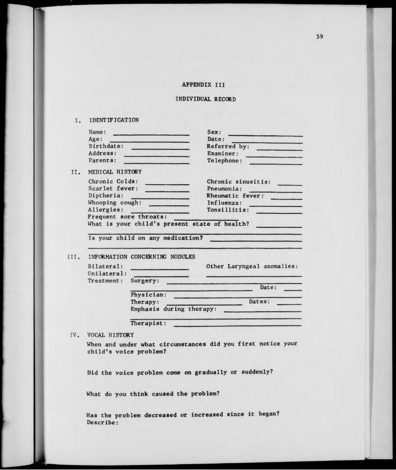
59
III.
APPENDIX III
INDIVIDUAL RECORD
I. IDENTIFICATION
Name: Sex: Age: Date: Birthdate: Referred by: Address: Examiner: Parents: Telephone:
MEDICAL HISTORY
Chronic Colds: Chronic sinusitis: Scarlet fever: Pneumonia: Diptheria: Rheumatic fever: Whooping cough: Influenza: Allergies: Tonsillitis: Frequent sore throats: What is your child's pi resent sta te of health?
Is your child on any m< >dication1
INFORMATION CONCERNING
Bilateral:
NODULES
Other Laryngeal anomalies: Unilateral: Treatment: Surgery:
Date: Physician: Therapy: Dates:
Emphasis di iring ther apy:
Therapist:
IV. VOCAL HISTORY When and under what circumstances did you first notice your child's voice problem?
Did the voice problem come on gradually or suddenly?
What do you think caused the problem?
Has the problem decreased or increased since it began? Describe:

60
What time of the day is your child's voice best? Worse?
Does the problem vary with the season of the year or the weather?
Does the problem vary with the amount of talking that your child does?
Does the problem vary with any other conditions?
Is your child's voice similar to some other person's in your family? Whose?
Do you know of any speech disorders in your family?
Amount and kind of vocal usage: As an infant, did your child cry or scream a lot? Is your child very talkative or noisy? Does your child yell excessively on the play- ground or in competitive sports? Does your child sing in a choir? What activities does your child participate in which involve loud or continuous talking?
V. HEARING
Results of pure-tone screening test: