Embed Size (px)
Citation preview
Formatted for: Experimental Brain Research
Tilt and Translation Motion Perception
During OffVertical Axis Rotation
Scott J. Wood 1, 2 , Millard F. Reschke 2 , Gilles Clément 3
1 Universities Space Research Association, Houston, TX, USA 2 Neuroscience Laboratory, NASA Johnson Space Center, Houston, TX, USA
3 Centre de Recherche Cerveau et Cognition, UMR 5549 CNRS/UPS, Toulouse, France
Running title: Motion perception during OVAR
Address for Correspondence:
Scott J. Wood, Ph.D.
NASA JSC, Mail Code SK272 Tel (281) 4837294
2101 NASA Parkway Fax (281) 2445734
Houston, TX 77058 Email: [email protected]
Acknowledgements: Philippe Tauzin of Service Commun Multimedia at the University Paul
Sabatier in Toulouse provided the artwork. We thank Pierre Denise for his contributions to the
questions used for verbal reports of selfmotion perception. This research was funded by NASA
(DSO 499) and Centre National d'Etudes Spatiales.
https://ntrs.nasa.gov/search.jsp?R=20080026153 2018-06-05T03:07:36+00:00Z
Motion perception during OVAR
2
ABSTRACT
The effect of stimulus frequency on tilt and translation motion perception was studied during
constant velocity offvertical axis rotation (OVAR), and compared to the effect of stimulus
frequency on eye movements. Fourteen healthy subjects were rotated in darkness about their
longitudinal axis 10° and 20° offvertical at 0.125 Hz, and 20° offvertical at 0.5 Hz. Oculomotor
responses were recorded using videography, and perceived motion was evaluated using verbal
reports and a joystick with four degrees of freedom (pitch and roll tilt, mediallateral and
anteriorposterior translation). During the lower frequency OVAR, subjects reported the
perception of progressing along the edge of a cone. During higher frequency OVAR, subjects
reported the perception of progressing along the edge of an upright cylinder. The modulation of
both tilt recorded from the joystick and ocular torsion significantly increased as the tilt angle
increased from 10° to 20° at 0.125 Hz, and then decreased at 0.5 Hz. Both tilt perception and
torsion slightly lagged head orientation at 0.125 Hz. The phase lag of torsion increased at 0.5 Hz,
while the phase of tilt perception did not change as a function of frequency. The amplitude of
both translation perception recorded from the joystick and horizontal eye movements was
negligible at 0.125 Hz and increased as a function of stimulus frequency. While the phase lead of
horizontal eye movements decreased at 0.5 Hz, the phase of translation perception did not vary
with stimulus frequency and was similar to the phase of tilt perception during all conditions.
During dynamic linear acceleration in the absence of other sensory input (canal, vision) a change
in stimulus frequency alone elicits similar changes in the amplitude of both self motion
perception and eye movements. However, in contrast to the eye movements, the phase of both
perceived tilt and translation motion is not altered by stimulus frequency. We conclude that the
neural processing to distinguish tilt and translation linear acceleration stimuli differs between eye
movements and motion perception.
Keywords: OVAR, otolith, acceleration, orientation, VOR
Motion perception during OVAR
3
INTRODUCTION
The otolith organs of the vestibular system transduce linear acceleration from both
translation motion and head tilt relative to gravity. The ambiguity between these two types of
linear acceleration must be resolved for both the accurate perception of motion and generation of
compensatory eye movements during different types of head movement (Mayne 1974). Constant
velocity rotation of head and body around an axis tilted with respect to gravity (OffVertical
Axis Rotation, OVAR) is one means of providing a dynamic linear acceleration stimulus. During
OVAR, the angle of tilt determines the amplitude of the linear acceleration stimulus, whereas the
velocity of rotation determines its frequency content. The sinusoidally varying linear acceleration
during OVAR in darkness elicits a modulation of horizontal, torsional and vertical eye
movements (Guedry 1965; Benson and Bodin 1966; Darlot et al. 1988; Haslwanter et al. 2000;
Yagi et al. 2000). Recent work in our laboratory has demonstrated that lower frequency
responses (< 0.3 Hz) during OVAR are principally characterized by the modulation of tilt
position dependent ocular reflexes (e.g., torsion) whereas the modulation of translational ocular
reflexes (e.g., horizontal) are predominant at higher frequencies (Wood 2002).
Although motion perception has been studied during OVAR (Guedry 1965; Graybiel and
Miller 1970; Denise et al. 1988), these studies have typically employed lower frequency stimuli
below 0.3 Hz. The motion perception commonly reported during OVAR at these low frequencies
is a progression along the circumferential edge of a downward oriented cone (Denise et al.
1988). The angle of the perceived conical body path increases with the actual tilt angle of the
rotation axis, indicating that the perception process depends on inputs signaling head and body
orientation with respect to gravity. Few studies have been performed using OVAR stimuli
greater than 0.3 Hz. In one study, Miller and Graybiel (1973) found that subjects reported “being
at or near upright” when rotating at 0.66 Hz (240°/s). Unfortunately, it was unclear from Miller
and Graybiel’s report whether this reduced sense of tilt motion at higher frequencies was
accompanied by an increased sense of translation analogous to the transition from tilt to
translation ocular reflexes (Wood 2002).
The purpose of this research was therefore to examine the effect of stimulus frequency on
the perception of motion during OVAR. Specifically, we compared the sensation of whole body
tilt versus whole body translation at frequencies above and below the crossover range of tilt and
Motion perception during OVAR
4
translation ocular reflexes (~0.3 Hz, Wood 2002). The emphasis on wholebody motion is
important to provide insight into how the central nervous system is resolving the ambiguity
between tilt and translation. The perception of a downward oriented cone described in earlier
studies (Denise et al. 1988) involves translation along the edge of the cone, although clearly this
results from the perception that one is tilting about an axis below the head. It is important
therefore to distinguish translation of the head associated with tilt about an eccentric axis from
the perception of whole body translation.
We predicted that the sensation of progressing along a cone reported during low
frequency OVAR would be replaced by a more pronounced sense of mediallateral and anterior
posterior translation similar to progression along a cylindrical path at high frequency. In addition
to comparing responses at low (0.125 Hz) and high (0.5 Hz) frequencies during OVAR,
responses were obtained at two angles of tilt (10 and 20°) in order to compare responses to
different magnitudes of linear acceleration. These small angles of tilt were chosen to be within
the range of natural behavior. We introduce a new technique for simultaneous recording of
perception of both tilt and translation that reflects how the central nervous system distinguishes
these two types of linear acceleration for motion perception.
MATERIALS AND METHODS
Subjects
Fourteen subjects (7 females, 7 males), ranging in age from 2451 yrs (mean 34 yrs)
participated in this study. All subjects were required to pass a medical examination, including an
audiogram, and have no history of vestibular or oculomotor abnormalities. Normal vestibular
(canal) function was also confirmed by postrotatory responses to velocity step profiles obtained
following each experiment trial (gain 0.41 ± 0.04, time constant 12.5 ± 2.0 s). The experiment
was under taken with the understanding and written consent of each subject. The test procedures
were approved by and in compliance with the standards of the NASA institutional review board
for human testing and were performed in accordance with the ethical standards laid down in the
1964 Declaration of Helsinki.
Motion perception during OVAR
5
Equipment
Subjects were rotated about their longitudinal axis in the OffVertical Axis Rotator
System at the NASA Johnson Space Center Neurosciences Laboratory. This system utilizes a
directdrive 80 ftlb DC motor and high precision tachometerbased servo controller (Neuro
Kinetics Inc., Pittsburgh, PA) for motor control and stability during rotation. An
electromechanical linear actuator was used to tilt the axis of rotation between upright and 20°
offvertical. A photodetector circuit provided onceperrevolution pulses in the noseup
orientation and a tachometer provided rotator velocity.
The rotation was performed in complete darkness, and operator instructions as well as
background masking noise were provided via a chairfixed speaker to minimize any extraneous
spatial orientation cues. Subjects were restrained in an upright seated position. The Reid's
baseline was approximately in the plane of rotation, with the horizontal canals and utricular
macula inclined up approximately 20° relative to this plane (Curthoys et al. 1977). This
orientation was used to maintain a natural headerect position.
Body support was provided by straps at the shoulders, waist, thighs, knees, and feet.
Adjustable pads applied pressure to formable VacPacs® (Olympic Medical, Seattle, WA) to
immobilize the midtorso and uniformly distribute the pressure. The head position was stabilized
with adjustable clamps coupled to customfitted thermoplastic pads positioned over the forehead
and sides of the head with the aid of a skull cap.
Eye movements were recorded on video cassette tapes with a chairfixed binocular video
camera system, which used dichroic mirrors to provide a full field of view (120° horizontal by
90° vertical). Small monochrome video cameras were used with nearinfrared emitting diodes to
allow eye recording in darkness. A timecode was digitally overlaid on each video field (60 Hz
sample rate) to permit offline processing and synchronization with the rotation stimuli signals,
joystick perception signals and calibration target information. Torsional, horizontal and
vertical eye position were processed offline using an eye tracking system previously
described (Wood et al. 1998; Wood 2002).
Pre and posttest eye measurement calibrations were made by having subjects fixate a
series of wall targets positioned 5° apart over a range of ± 25° horizontally and ± 20° vertically
at a distance of 1.7 m. A lowpower laser crosshair pointer was mounted to the chair at height of

Motion perception during OVAR
6
the outer canthus and projected toward the calibration array to align the horizontal calibration
targets with eye level.
A joystick using four singleaxis potentiometers was mounted on the chair directly in
front of the subject at midtorso. Subjects used one hand on the upper stage to indicate the
direction and amplitude of perceived body tilt in roll (coronal plane) and pitch (sagittal plane).
Subjects used their other hand on the lower stage to indicate direction and amplitude of
perceived body translation along mediallateral (ML) and anteriorposterior (AP) axes (Fig. 1).
Subjects were instructed to report ‘wholebody’ tilt and translation, limiting translation to
perceived cylindrical motion path. Given the limited range of the joystick, subjects were
instructed to scale their perceived amplitude such that the length of the joystick handle
represented one body length. The data were then normalized as a percentage of the maximum
peaktopeak range.
< Insert Figure 1 about here>
Procedure
The experimental protocol included 2 velocities of rotation, 45°/s and 180°/s
corresponding to 0.125 Hz and 0.5 Hz, respectively, during OVAR at 20° of tilt. A tilt angle of
10° was also tested during 0.125 Hz rotation, which allowed a direct comparison of our motion
perception results with those previously obtained by Denise et al. (1988).
Subjects were instructed to keep their eyes open in the dark and look straight ahead at an
imaginary horizon. Data collection started with the chair tilted 10° and the subject in the noseup
position. The chair accelerated at 25°/s 2 up to 45°/s constant velocity. Following 60 s to allow
the primary perrotatory nystagmus to decay, eye movements were recorded during constant
velocity at 0.125 Hz for 80 s (i.e., 10 cycles). The subjects were then requested to reproduce with
the joystick the direction and amplitude of their perceived motion (Fig. 1). The joystick signals
were recorded for a minimum of 10 cycles before the following systematic questions were asked
by the operator: (1) do you perceive moving along the edges of a cone and/or cylinder, (2) is the
path circular or elliptical, (3) if cone, quantify the amplitude of tilt and location of its apex (tip)
relative to the head, (4) quantify the degree to which the long axis of the cone or cylinder is tilted
Motion perception during OVAR
7
relative to vertical, and (5) what direction do you feel you are moving (clockwise, CW or
counterclockwise, CCW, as viewed from above).
The chair was then tilted to 20° in less than 5 s. After obtaining eye movements (10
cycles), joystick (10 cycles) and verbal reports at 0.125 Hz during 20° OVAR, the chair was
accelerated at 25°/s 2 up to 0.5 Hz constant velocity. Following 60 s to allow the primary canal
response to decay, eye movements, joystick data and verbal reports were acquired during
approximately 15 cycles each. The rotation axis was then brought back to Earthvertical. At this
point none of the subjects perceived rotation, and the chair was stopped (deceleration 25°/s 2 ),
thereby eliciting a horizontal postrotatory nystagmus. After several minutes of rest, rotation in
the opposite direction was performed using the same protocol. The direction of rotation (CW or
CCW) was counterbalanced across subjects. The test was terminated early at the onset of motion
sickness (mild nausea) reported by the subjects.
Data Analysis
Eye position, joystick, and rotator data were filtered with a predictive finiteimpulse
response (FIR), median hybrid filter (Engelken and Stevens 1990). Conjugate eye position
(average of left and right) was differentiated to calculate eye velocity. Fast phase components of
the eye movements were identified using acceleration and velocity thresholds and verified
manually to be excluded from the analysis.
Nonlinear least squares sinusoidal curve fits were used to describe the modulation of eye
and joystick responses as a function of the sinusoidalvarying linear acceleration stimulus. The
curve fits were used to determine the amplitude and phase of torsion position, horizontal slow
phase velocity (SPV), pitch tilt, roll tilt, ML translation and AP translation. Phases for torsion
position and joystick data were expressed relative to tilt position with positive leading and
negative lagging. Phases for horizontal SPV were offset by 90° to account for the fact that they
are compensatory for change in head position.
These response parameters were analyzed with repeated measures ANOVAs using a
commercial statistics program (JMP, SAS Institute Inc.). Using an alpha error of 0.05 as the
decision rule, the null hypothesis that there is no difference across tilt angle and frequency was
tested with Wilke's lambda serving as the critical statistic.
Motion perception during OVAR
8
RESULTS
Seven out of the 14 subjects completed the entire protocol including both directions of
rotation at both velocities and both tilt angles (indicated by asterisk in Table 1). For the
remaining subjects, the experiment was stopped at the onset of mild nausea. Four subjects did
not develop symptoms until the completion of the first run or during the second run in the
opposite direction. Three developed symptoms during the first direction of rotation, one at the
lower speed of rotation. The modulation of eye movement and joystick data were not
significantly different across rotation direction for the seven subjects who completed the entire
protocol. Therefore, data from the first run was used for the remainder of the analyses below.
Figure 2 shows samples of eye and joystick responses for one typical subject during 20° OVAR
at both 0.125 Hz (A) and 0.5 Hz (B).
< Insert Figure 2 about here>
Verbal reports
Verbal reports of the perceived motion path were consistent across CW and CCW
directions of rotation, and are therefore combined in Table 1. During OVAR at 10° tilt, all
subjects reported the pattern of body motion previously described by Denise et al. (1988):
progression along a conical path while facing the same direction. For 20° tilt at 0.125 Hz, 79% of
subjects continued to perceive a conical path. The average amplitude of perceived tilt was
reported to be 19.1° (± 1.9° sem) for 10° OVAR and 34.4° (± 2.3° sem) for 20° OVAR.
However, at 0.5 Hz 86% of subjects reported a cylindrical path with no sense of tilt, and the
remaining two reported dramatically reduced sense of tilt (5°). At both frequencies and tilt
angles, the majority of subjects reported a circular path with the long axis of the cone or cylinder
upright.
< Insert Table 1 about here>

Motion perception during OVAR
9
The apex of the cone at 0.125 Hz was perceived below the head at an average estimated
distance of 2.7 m (± 0.5 m sem) for 10° OVAR and 1.6 m (± 0.4 m sem) for 20° OVAR. It is
interesting to note that based on this average distance and tilt amplitude, the radius of the
perceived conical path was the same for both tilt angles (~0.9 m). The subjects generally felt
progressing along the cone or cylinder in the opposite direction of the actual spinning.
Eye movement responses
The torsion amplitude doubled from 10° to 20° OVAR whereas the phase remained the
same across tilt angles at 0.125 Hz (Figure 3). However; consistent with our previous study
(Wood 2002), both amplitude and phase of torsion and horizontal slow phase velocity (SPV)
were significantly affected by stimulus frequency. At the high frequency, the torsion amplitude
decreased while there was a greater phase lag relative to the low frequency (Figures 2 and 3).
The amplitude of the horizontal SPV was negligible at both 10° to 20° OVAR at 0.125 Hz, but
significantly increased at the high frequency. The horizontal SPV phase was leading the linear
stimulus at the low frequency, and became more in phase at the high frequency.
< Insert Figure 3 about here>
Joystick responses
The joystick settings were consistent with the perceived motion path verbally reported by
the subjects. The amplitude of perceived roll and pitch tilt was larger than perceived ML and AP
translation, respectively, at the low frequency for both 10° and 20° OVAR (Figure 4A and 4B).
The effect of stimulus frequency on the amplitude of the joystick measures was similar to the
effect observed for the eye movement amplitude described above. As the stimulus increased
from 0.125 Hz to 0.5 Hz, the amplitude of tilt significantly decreased while the amplitude of
translation increased. In contrast to the eye movements; however, the average phase of the
perceived motion was not affected by stimulus frequency (Figure 4C and 4D), consistently
lagging the stimulus by <30°. In fact, the phase of the perceived tilt was not significantly
Motion perception during OVAR
10
different from the phase of the corresponding perceived translation (roll versus ML, pitch versus
AP) in any of the tested conditions.
< Insert Figure 4 about here>
DISCUSSION
One major contribution of this study is that, as already shown for eye movements (Wood
2002), the amplitude of both tilt and translation motion perception during OVAR varies as a
function of linear acceleration frequency in the absence of visual and canal cues. However; in
contrast with eye movements, the phase of motion perception does not vary with stimulus
frequency. We infer that the neural processing required to distinguish between tilt and translation
differs for eye movements and motion perception (Merfeld et al. 2005). While the tilt and
translation ocular reflexes appear to operate in a more independent fashion, the timing of
perceived tilt and translation influence each other. We conclude that the perceived motion path
during linear acceleration results from a composite representation of tilt and translation. While
increasing stimulus frequency results in striking decreases in the amplitude of tilt (Glasauer
1995; Merfeld et al. 2005), the concomitant increase in sense of translation serves to maintain the
phase of the perceived motion relative to the stimulus.
Figure 5 illustrates the ambiguous motion paradigm used in the current study. During
OVAR a CW rotation results in a sequence of nosedown, leftear down, noseup, and rightear
down orientations (Fig. 5A). However, one would need to tilt the head along a CCW motion path
to result in the same sequence of orientations without rotating about a longitudinal axis. This
explains why the perceived direction of rotation during OVAR at low velocities is generally
opposite to the actual direction of rotation (Graybiel and Miller 1970; Denise et al. 1988). The
principal ambiguity during constant velocity OVAR is whether the sinusoidally varying linear
acceleration is due to tilt or translation. For example, the maximal leftward interaural
acceleration due to gravity occurs in the righteardown (RED) orientation. This can be
interpreted as either resulting from a rightward roll tilt, or resulting from translation acceleration
in the leftward direction. Since acceleration is 180° out of phase from position, the maximal
leftward acceleration during sinusoidal translation occurs when one is at the extreme right
Motion perception during OVAR
11
position. Therefore, at the RED orientation, our subjects reported either the maximal rightward
tilt (Fig. 5B) or maximal rightward translation position (Fig. 5C). If one perceives the linear
acceleration during OVAR as tilt, the motion path is described as moving along the edge of a
cone, while always facing the same direction (Fig. 5B). If one perceives linear acceleration
during OVAR as translation, the motion path is moving along the edge of an upright cylinder,
again in a direction opposite to true rotation (Fig. 5C).
< Insert Figure 5 about here >
Our results are consistent with the hypothesis that the vestibular system resolves
ambiguous linear acceleration information from the otolith afferent information, at least in part,
on the basis of the head motion frequency content (Paige 1996). However, frequency segregation
does not completely resolve the ambiguity (Wood 2002). First, the phase of the perceived motion
does not vary as a function of frequency, as one would predict from high and lowpass filtering
alone. Second, there will be at some point a crossover of low and highpass information where
the resolution of tilt and translation information by frequency content is more complex. Several
studies have demonstrated that the central nervous system utilizes other sensory systems, e.g.,
semicircular canals and vision, to resolve the ambiguity of tilt and translational linear
acceleration stimuli (Angelaki et al. 2004; Merfeld et al. 2005). Neurophysiological evidence for
this stems from the observation that neurons in the vestibular nuclei that respond to tilt or
translation often receive canal input (Angelaki and Dickman 2003).
The large majority of previous human OVAR studies have been limited to frequencies
below 0.3 Hz. These studies have demonstrated the perception of a conical motion path with the
amplitude dependent on the tilt angle (Graybiel and Miller 1970; Denise et al. 1988). Our results
were consistent in that the amplitude of tilt perception approximately doubled when moving
from 10° to 20° tilt angle. Except for some limited recordings using a goggle device by Graybiel
and Miller (1970), previous studies have relied on verbal reports. During OVAR at high
frequency, Miller and Graybiel (1973) reported their subjects had the sensation of “being at or
near upright.” However, there was no indication whether subjects felt any sense of translation.
Noteworthy in the present study is that the perception of reduced tilt at the high frequency is
accompanied with increased translation.

Motion perception during OVAR
12
The effects of stimulus frequency on perceived tilt amplitude are not specific to OVAR,
but should be present in other motion paradigms involving linear acceleration without visual and
canal inputs. For example, similar results have been demonstrated during variable radius
centrifugation and/or during translations along a linear track (Glasauer 1995; Merfeld et al.
2005). Although perception of translation has never been reported in earlier studies, the
modulation of horizontal SPV during OVAR has been observed by numerous investigations
(Guedry 1965; Benson and Bodin 1966; Correia and Money 1970; Young and Henn 1975;
Raphan et al. 1981; Cohen et al. 1983; Hain 1986; Wall and Furman 1989; Furman et al. 1992;
Clément et al. 1995; Angelaki and Hess 1996). It is generally accepted that this modulation of
horizontal SPV represents a translational otolithocular response to the modulation of interaural
acceleration generated during OVAR (Angelaki and Hess 1996). Therefore, the increase in the
amplitude of translation perception as a function of stimulus frequency is consistent with
increasing amplitude of translational ocular reflexes.
Testing otolith responses induced by both low and highfrequency linear accelerations is
of interest for assessing vestibular disorders (Furman et al. 1992) or astronauts returning from
spaceflight (Clément et al. 1995). One advantage of OVAR over other types of linear
acceleration paradigms is that the amplitude of linear acceleration can be precisely maintained
across a large frequency range, including low frequencies. For example, linear acceleration of
0.34 g at 0.125 Hz, such as used in the present study during OVAR at 20° tilt angle, would
require a 5.4m long linear sled. Variable radius centrifugation has been used to overcome the
limitations of track length that often preclude the investigation of low frequency linear
acceleration using linear sleds (Glasauer 1995; Seidman et al. 1998; Merfeld et al. 2005).
Unfortunately, the linear coriolis forces during variable radius centrifuge become greater at
higher frequencies. Although OVAR has been used to test motion sickness sensitivity, higher
frequency OVAR has the advantage of being less provocative (Denise et al. 1996; Wood 2002).
Perception of translation has not been studied during high frequency linear acceleration, although
there is a lot of high frequency content during normal activities (MacDougall and Moore 2005).
One could argue that testing at high frequency would be more relevant for evaluating functional
deficits in dynamic vestibular responses (Curthoys 2000).
One implication of our findings is that techniques that rely on visual references for self
motion reports during linear acceleration stimuli are likely to be confounded because of the
Motion perception during OVAR
13
underlying differences in phase between eye movements and perception as a function of stimulus
frequency. Wade and Curthoys (1997) have shown the influence of torsion on the amplitude of
subjective visual vertical during static tilts. During dynamic linear stimuli, similar effects on the
phase of visual vertical would be expected as a function of frequency. Similarly, visual
psychophysical measures to determine perceived translation would also be confounded by high
pass filtering of horizontal eye movements, especially at frequencies below 0.3 Hz where there
are large phase leads (Wood 2002; Merfeld et al. 2005). Nevertheless, translation perception
reporting such as the one in this study is a complementary technique to eye measurement
recording that will provide additional information on the cognitive processing of highfrequency
otolith information.
REFERENCES
Angelaki DE, Dickman JD (2003) Gravity or translation: central processing of vestibular signals
to detect motion or tilt. J Vestib Res 13: 245253
Angelaki DE, Hess BJ (1996) Threedimensional organization of otolithocular reflexes in rhesus
monkeys. I. Linear acceleration responses during offvertical axis rotation. J
Neurophysiol 75: 24052424
Angelaki DE, Shaikh AG, Green AM, Dickman JD (2004) Neurons compute internal models of
the physical laws of motion. Nature 430: 560564
Benson AJ, Bodin MA (1966) Interaction of linear and angular accelerations on vestibular
receptors in man. Aerosp Med 37: 144154
Clément G, Darlot C, Petropoulos A, Berthoz A (1995) Eye movements and motion perception
induced by offvertical axis rotation (OVAR) at small angles of tilt after spaceflight. Acta
Otolaryngol (Stockh) 115: 603609
Cohen B, Suzuki JI, Raphan T (1983) Role of the otolith organs in generation of horizontal
nystagmus: effects of selective labyrinthine lesions. Brain Res 276: 159164
Correia MJ, Money KE (1970) The effect of blockage of all six semicircular canal ducts on
nystagmus produced by dynamic linear acceleration in the cat. Acta OtoLaryngologica
69: 716
Curthoys IS (2000) Vestibular compensation and substitution. Curr Opin Neurol 13: 2730
Motion perception during OVAR
14
Curthoys IS, Blanks RH, Markham CH (1977) Semicircular canal functional anatomy in cat,
guinea pig and man. Acta Otolaryngol (Stockh) 83: 258265
Darlot C, Denise P, Droulez J, Cohen B, Berthoz A (1988) Eye movements induced by off
vertical axis rotation (OVAR) at small angles of tilt. Exp Brain Res 73: 91105
Denise P, Darlot C, Droulez J, Cohen B, Berthoz A (1988) Motion perceptions induced by off
vertical axis rotation (OVAR) at small angles of tilt. Exp Brain Res 73: 106114
Denise P, Etard O, Zupan L, Darlot C (1996) Motion sickness during offvertical axis rotation:
prediction by a model of sensory interactions and correlation with other forms of motion
sickness. Neurosci Lett 203: 183186
Engelken EJ, Stevens KW (1990) A new approach to the analysis of nystagmus: an application
for orderstatistic filters. Aviation Space & Environmental Medicine 61: 859864
Furman JM, Schor RH, Schumann TL (1992) Offvertical axis rotation: a test of the otolith
ocular reflex. Ann Otol Rhinol Laryngol 101: 643650
Glasauer S (1995) Linear acceleration perception: frequency dependence of the hilltop illusion.
Acta Otolaryngol Suppl 520: 3740.
Graybiel A, Miller EFI (1970) The otolith organs as a primary etiological factor in motion
sickness: with a note on "offvertical" rotation. In: Fourth Symposium on the Role of the
Vestibular Organs in Space Exploration, vol SP187. NASA, Naval Aerospace Medical
Research Laboratory; Pensacola, FL, pp 5366
Guedry FE, Jr. (1965) Orientation of the rotationaxis relative to gravity: its influence on
nystagmus and the sensation of rotation. Acta Otolaryngol (Stockh) 60: 3048
Hain TC (1986) A model of the nystagmus induced by off vertical axis rotation. Biological
Cybernetics 54: 337350
Haslwanter T, Jaeger R, Mayr S, Fetter M (2000) Threedimensional eyemovement responses to
offvertical axis rotations in humans. Exp Brain Res 134: 96106.
MacDougall HG, Moore ST (2005) Marching to the beat of the same drummer: the spontaneous
tempo of human locomotion. J Appl Physiol 99: 11641173
Mayne R (1974) A systems concept of the vestibular organs. In: Kornhuber HH (ed) Handbook
of Sensory Physiology, vol VI/2. Springer Verlag, Berlin Heidelberg New York, pp 493
580
Motion perception during OVAR
15
Merfeld DM, Park S, GiannaPoulin C, Black FO, Wood S (2005) Vestibular perception and
action employ qualitatively different mechanisms. I. Frequency response of VOR and
perceptual responses during Translation and Tilt. J Neurophysiol 94: 186198
Miller EFI, Graybiel A (1973) Perception of the upright and susceptibility to motion sickness as
functions of angle of tilt and angular velocity in offvertical rotation. In: Fifth
Symposium on the Role of the Vestibular Organs in Space Exploration, vol SP314.
NASA, Naval Aerospace Medical Research Laboratory; Pensacola, FL, pp 99103
Paige GD (1996) How does the linear vestibuloocular reflex compare with the angular
vestibuloocular reflex? In: Baloh RW, Halmagyi GM (eds) Disorders of the Vestibular
System. Oxford University Press, New York, pp 93104
Raphan T, Cohen B, Henn V (1981) Effects of gravity on rotatory nystagmus in monkeys.
Annals of the New York Academy of Sciences 374: 4455
Seidman SH, Telford L, Paige GD (1998) Tilt perception during dynamic linear acceleration.
Exp Brain Res 119: 307314
Wade SW, Curthoys IS (1997) The effect of ocular torsional position on perception of the roll
tilt of visual stimuli. Vision Res 37: 10711078
Wall Cd, Furman JM (1989) Nystagmus responses in a group of normal humans during earth
horizontal axis rotation. Acta OtoLaryngologica 108: 327335
Wood SJ (2002) Human otolithocular reflexes during offvertical axis rotation: Effect of
frequency on tilttranslation ambiguity and motion sickness. Neurosci Lett 323: 4144
Wood SJ, Paloski WH, Reschke MF (1998) Spatial coding of eye movements relative to
perceived head and earth orientations during static rolltilt. Exp Brain Res 121: 5158
Yagi T, Kamura E, Shitara A (2000) Three dimensional eye movement analysis during off
vertical axis rotation in human subjects. Arch Ital Biol 138: 3947
Young LR, Henn VS (1975) Nystagmus produced by pitch and yaw rotation of monkeys about
nonvertical axes. Fortschr Zool 23: 235246
Motion perception during OVAR
16
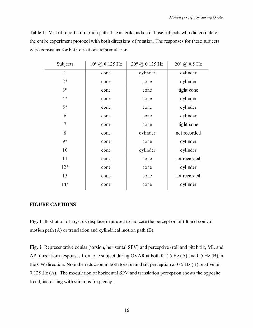
Table 1: Verbal reports of motion path. The asteriks indicate those subjects who did complete
the entire experiment protocol with both directions of rotation. The responses for these subjects
were consistent for both directions of stimulation.
Subjects 10° @ 0.125 Hz 20° @ 0.125 Hz 20° @ 0.5 Hz
1 cone cylinder cylinder
2* cone cone cylinder
3* cone cone tight cone
4* cone cone cylinder
5* cone cone cylinder
6 cone cone cylinder
7 cone cone tight cone
8 cone cylinder not recorded
9* cone cone cylinder
10 cone cylinder cylinder
11 cone cone not recorded
12* cone cone cylinder
13 cone cone not recorded
14* cone cone cylinder
FIGURE CAPTIONS
Fig. 1 Illustration of joystick displacement used to indicate the perception of tilt and conical
motion path (A) or translation and cylindrical motion path (B).
Fig. 2 Representative ocular (torsion, horizontal SPV) and perceptive (roll and pitch tilt, ML and
AP translation) responses from one subject during OVAR at both 0.125 Hz (A) and 0.5 Hz (B).in
the CW direction. Note the reduction in both torsion and tilt perception at 0.5 Hz (B) relative to
0.125 Hz (A). The modulation of horizontal SPV and translation perception shows the opposite
trend, increasing with stimulus frequency.
Motion perception during OVAR
17
Fig. 3 A. The average modulation amplitude (± sem) for the ocular responses at each of the
three stimulus conditions. Note that the amplitude of torsion (solid circles) is indicated by the left
ordinate axis, while the horizontal SPV amplitude (open circles) is indicated by the right ordinate
axis. Significant differences (p<0.05) between 10° and 20° OVAR at 0.125 Hz is indicated by ‘*’
and significant differences (p<0.05) between 0.125 Hz and 0.5 Hz at 20° OVAR is indicated
with ‘**.’ B. The average phase (± sem) for the ocular responses at each of the three stimulus
conditions. Note that the average torsion phase (solid circles) was lagging for all conditions, and
is indicated with increasing phase lag in the upward direction indicated by the left ordinate axis.
The horizontal SPV phase (open circles) was leading in all conditions, and is shown with
increasing phase lead in the upward direction indicated by the right ordinate axis. Note there was
a significant effect of frequency as indicated by the apparent crossover in both amplitude and
phase. Significant differences are shown as in A.
Fig. 4 A, B. The average (± sem) roll tilt (solid circles) and mediallateral (ML, open circles)
translation perception amplitude and phase at each of the three stimulus conditions. C, D. The
average (± sem) pitch tilt (solid circles) and anteriorposterior (AP, open circles) translation
perception amplitude and phase at each of the three stimulus conditions. Significant differences
are shown as in Fig. 3.
Fig. 5 A. The change in orientation during CW OVAR from nosedown (ND), righteardown
(RED), noseup (NU) and lefteardown (LED). B. This sequence of orientation will result in the
sensation of tilt following a conical motion path in a CCW direction during rotation at low
frequency (Adapted from Denise et al. 1988). C. At high frequency of rotation, it will result in
the sensation of translating along a cylindrical motion path in a CCW direction.
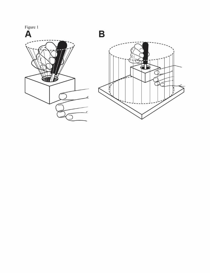
Figure 1
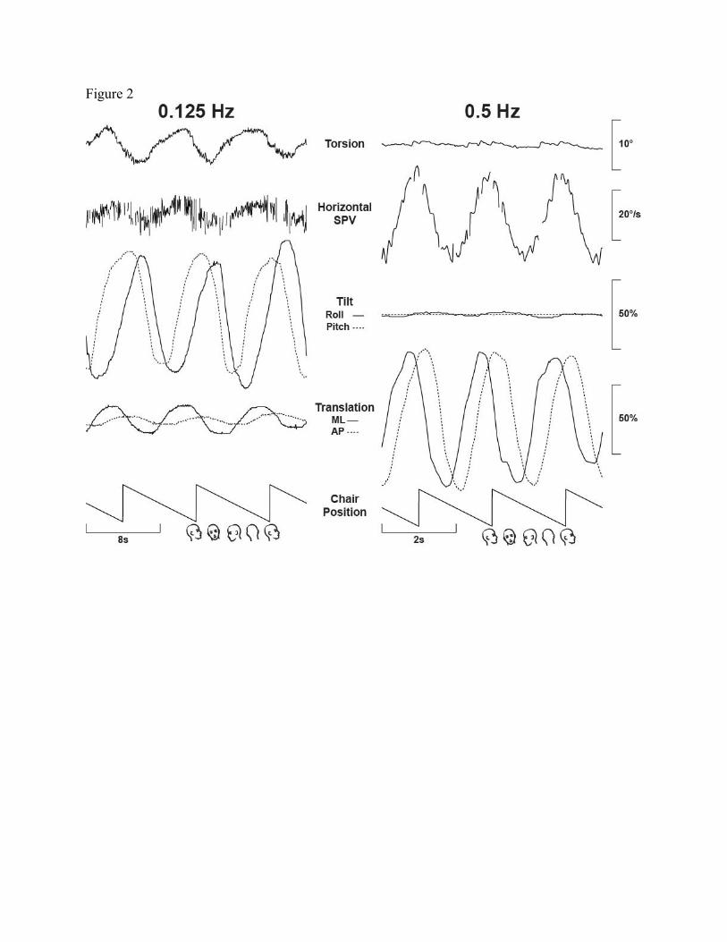
Figure 2
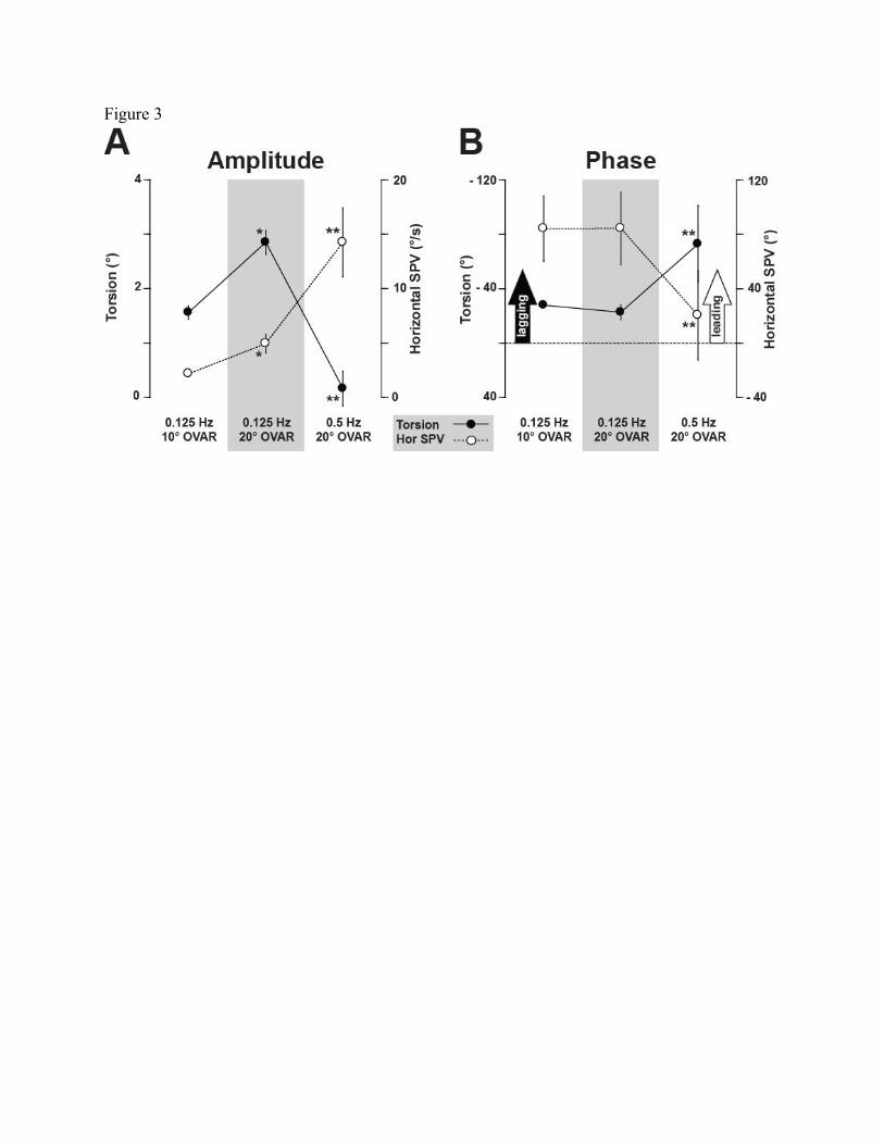
Figure 3
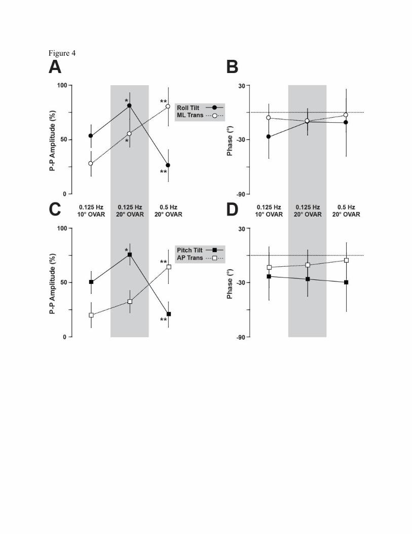
Figure 4
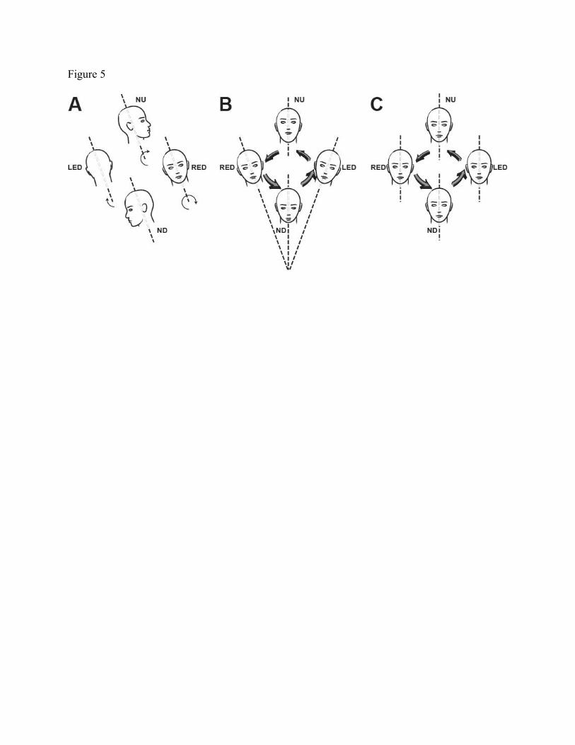
Figure 5