Embed Size (px)
Citation preview
Tomado con permiso de emedicine.medscape.com
eMedicine Specialties > Emergency Medicine >Hematology & Oncology
Thrombotic Thrombocytopenic PurpuraDeborrah Symonette, MD, MPH, Healthcare Consultant, DSKSD, IncDeepak Sharma, Touro College of Osteopathic Medicine, New York; Priya SAbraham, Touro College of Osteopathic Medicine, New York; Vanessa Yoo, TouroCollege of Osteopathic Medicine, New York; Wafa Qamar, Touro College ofOsteopathic Medicine, New York
Updated: Sep 16, 2009
Introduction
Background
Thrombotic thrombocytopenic purpura (TTP) is a rare life-threatening multisystemdisorder that is considered a true medical hematological emergency. Moschcowitz firstdescribed TTP in 1924 when he observed that a 16-year-old girl had anemia, petechiae,and microscopic hematuria. She died of multiorgan failure, and, at autopsy,disseminated microvascular hyaline platelet thrombi were prevalent in terminalarterioles and capillaries of the heart and kidneys. These platelet-rich thrombi remainthe hallmark of the pathologic diagnosis. Microangiopathic hemolytic anemia alongwith the aggregation of platelet thrombi are present in a setting of microvascular injuryand high fluid shear stress.
Varying degrees of organ ischemia due to the vascular occlusion occur. In this life-threatening disease, recognizing the clinical presentation and initiating medicalintervention early are critical. This is difficult at times because there is a range ofoverlapping signs and symptoms with other microangiopathic diseases, suchas hemolytic uremic syndrome (HUS). Both TTP and HUS have thrombocytopenia andmicroangiopathic hemolytic anemia but different medical management pathways. Thepatient may not present with all of the signs and symptoms of TTP, and there can be agray zone as to which microangiopathy truly exists. TTP is a medical emergency withdiagnostic criteria for treatment that is not always definitive but, if TTP is suspected,then the first-line treatment of plasma exchange should be initiated to save the patient'slife.
Prior efforts to identify this disease clinically lead to the development of a diagnosticcriteria classifying thrombotic thrombocytopenic purpura as a syndrome in 1966 byAmorosi and Ultmann. They reviewed 255 patients previously reported and 16 otherpatients. They outlined a pentad of clinical features including microangiopathic
hemolytic anemia, thrombocytopenia, neurologic abnormalities, fever, and renaldysfunction.1 Later studies show this pentad is seen in 40% of cases. A triad ofmicroangiopathic hemolytic anemia, thrombocytopenia, and neurologic abnormalitiesare seen in 74%.2 Clinically, if a patient presents with thrombocytopenia (withoutcoagulopathy), red cell fragmentation, elevated lactate dehydrogenase (LDH) level, andmuscle and organ ischemia, consider TTP urgently in the differential diagnosis.3
Thrombotic microangiopathic diseases
Thrombotic thrombocytopenic purpura (TTP) is categorized into acquired (idiopathic)TTP and congenital (familial) TTP. Acquired TTP is mainly idiopathic, but there areother conditions and comorbidities besides idiopathic. Congenital TTP is a rareautosomal recessive disease present in childhood. Acquired and congenital TTP areboth part of the larger spectrum of thrombotic microangiopathic diseases. Thesediseases have microvascular thrombosis and hemolysis with fragmented red blood cells.
TTP and hemolytic uremic syndrome (HUS) were once thought to have shared thepathophysiological etiology. Hemolytic uremic syndrome is usually found in children,and renal involvement is significant. Hemolytic uremic syndrome is caused by Shiga-like toxin-producing E coli O157:H7 in 90% of the cases.4 Hemolytic uremic syndromeis characterized by a triad of hemolytic anemia, thrombocytopenia, and acute renalfailure. TTP is usually found in adults and characterized by hemolytic anemia,thrombocytopenia, and, to a lesser extent, neurological manifestations. Symptoms aresimilar for TTP and HUS based solely on their clinical presentation. Microthrombi inTTP are mainly platelets, but microthrombi in HUS have more fibrin deposition.
Various terms have been used for other thrombotic microangiopathies such as TTP-likediseases, secondary TTP, or nonidiopathic TTP. In addition, TTP may also be drug-induced.5 At times, distinguishing TTP from other thrombotic microangiopathies thathave similar overlapping clinical presentations is very difficult. The complexity ofcategorizing these comorbidities or other conditions has not been resolved. Thromboticmicroangiopathies have varying causes and pathology but present with clinicalmanifestations that are TTP-like. These include drugs such as quinine, ticlopidine,clopidogrel, and cyclosporine; cancers; vasculitis; hematopoietic stem celltransplantation; infections such as human immunodeficiency virus infection; andpregnancy, especially in the third trimester.5 Further investigation is needed to classifythis group.6
In 1977, a breakthrough in treatment was reported by Bukowski et al using whole-bloodexchange transfusion, also known as plasmapheresis and fresh frozen plasma (FFP).Shortly after, Byrnes and colleagues used plasma infusion. During the plasma exchange,the entire plasma volume is replaced with normal human plasma and the largemolecular inhibitory antibodies are removed and the plasma is replenished with thedeficient protease. Delay in starting the plasma exchange is correlated with treatmentfailure. If a delay is unavoidable, begin plasma infusion until the plasma exchange isavailable. Intravenous (IV) plasma exchange is the present standard of treatment forthrombotic thrombocytopenic purpura. Fresh frozen plasma has become the standardreplacement fluid with its plasma protein levels closely paralleling physiologicallevels.3,7
Plasma infusion and plasma exchange have had a significant impact on the lifeexpectancy of patients. With the introduction of plasma exchange, the survival rate hasimproved from approximately 3% prior to the 1960s to 82%. By 1991, a landmarkclinical trial by Rock et al presented evidence of the efficacy of plasma exchangetreatment.8 Early recognition of the clinical features and intervention with plasmaexchange can reduce the mortality rate associated with TTP from 90% to approximately10-20%. Early recognition and management are essential for patient survival. Plasmainfusion is a temporary measure, and its use is limited by volume overload. Plasmaexchange is the treatment of choice for patients with acquired TTP. Congenital TTPresponds to plasma infusion.
Pathophysiology
The TTP syndrome is characterized by microangiopathic hemolysis and plateletaggregation/hyaline thrombi whose formation is unrelated to coagulation systemactivity. Platelet microthrombi predominate; they form in the microcirculation (ie,arterioles, capillaries) throughout the body causing partial occlusion of vessels. Organischemia, thrombocytopenia, and erythrocyte fragmentation (ie, schistocytes) occur. Thethrombi partially occlude the vascular lumina with overlying proliferative endothelialcells. The endothelia of the kidneys, brain, heart, pancreas, spleen, and adrenal glandsare particularly vulnerable to TTP. The liver, lungs, gastrointestinal tract, gallbladder,skeletal muscles, retina, pituitary gland, ovaries, uterus, and testes are also affected to alesser extent. No inflammatory changes occur. The occlusion of the microthrombiaffects many organs, and a myriad of symptoms are presented.
Mechanism
von Willebrand factor (vWF) was observed in 1982 by Moake and his colleagues. Thisis a large, adhesive glycoprotein that mediates thrombus formation at sites of vascularinjury. vWF is synthesized in the endothelium and megakaryocytes, and it circulates inthe plasma. Various sizes of multimers were noted, and the large form, ultralarge vonWillebrand factor (ULVWF) multimers were secreted from the endothelium.9 These arethe largest soluble protein found in human plasma and are considered the majorpathogenic factor in TTP due to the platelet clumping in the microvasculature.
The ULVWF is the most active of the various-sized multimers and is found in platelets,endothelial cells, and subendothelium. They were seen in the plasma of 4 patients withrelapsing TTP.10,11 The plasma of normal individuals has much smaller vWF. Moakesuggested that there is a deficiency in an enzyme that reduces the large vWF to itsnormal size in plasma of patients with TTP. This large vWF appeared to have a greaterability to adhere with platelets mediating a thrombus formation. The large vWFcombine with platelets consumed from the arterioles and capillaries of organs in a high-shearing stress environment and cause endothelial injury leading to ischemia. The redblood cells collide with the thrombi, and fragment leads to hemorrhage. As a result, theorgan function is compromised.
The agitated endothelial cells are the main source of ULVWF multimer secretion intothe bloodstream where they bind to specific surface platelet receptors. The ULVWFmultimers adhere to the damaged endothelium or exposed subendothelium, with theplatelet receptor binding to the ULVWF. The sheer stress of fluid and platelet thrombi
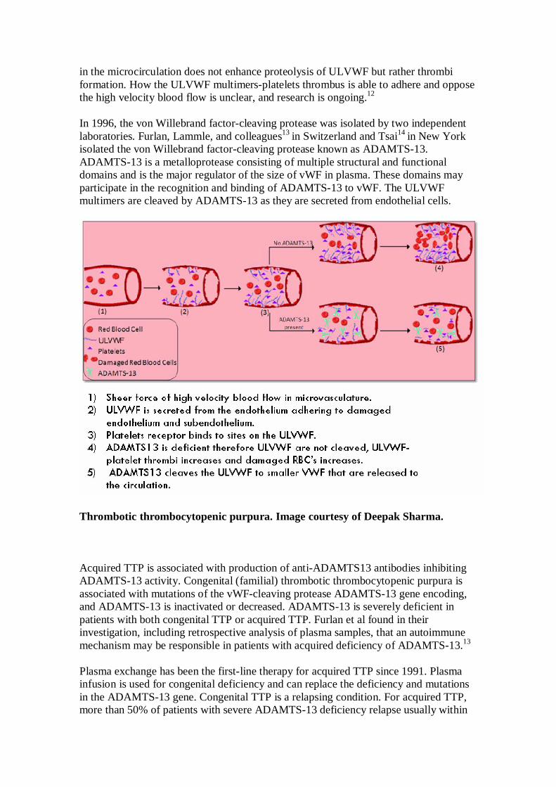
in the microcirculation does not enhance proteolysis of ULVWF but rather thrombiformation. How the ULVWF multimers-platelets thrombus is able to adhere and opposethe high velocity blood flow is unclear, and research is ongoing.12
In 1996, the von Willebrand factor-cleaving protease was isolated by two independentlaboratories. Furlan, Lammle, and colleagues13 in Switzerland and Tsai14 in New Yorkisolated the von Willebrand factor-cleaving protease known as ADAMTS-13.ADAMTS-13 is a metalloprotease consisting of multiple structural and functionaldomains and is the major regulator of the size of vWF in plasma. These domains mayparticipate in the recognition and binding of ADAMTS-13 to vWF. The ULVWFmultimers are cleaved by ADAMTS-13 as they are secreted from endothelial cells.
Thrombotic thrombocytopenic purpura. Image courtesy of Deepak Sharma.
Acquired TTP is associated with production of anti-ADAMTS13 antibodies inhibitingADAMTS-13 activity. Congenital (familial) thrombotic thrombocytopenic purpura isassociated with mutations of the vWF-cleaving protease ADAMTS-13 gene encoding,and ADAMTS-13 is inactivated or decreased. ADAMTS-13 is severely deficient inpatients with both congenital TTP or acquired TTP. Furlan et al found in theirinvestigation, including retrospective analysis of plasma samples, that an autoimmunemechanism may be responsible in patients with acquired deficiency of ADAMTS-13.13
Plasma exchange has been the first-line therapy for acquired TTP since 1991. Plasmainfusion is used for congenital deficiency and can replace the deficiency and mutationsin the ADAMTS-13 gene. Congenital TTP is a relapsing condition. For acquired TTP,more than 50% of patients with severe ADAMTS-13 deficiency relapse usually within

the year.15 For acquired TTP, the inhibitor of ADAMTS-13 is removed by plasmaexchange and is more effective than plasma infusion. Nevertheless, relapsing cases dooccur in those with severe ADAMTS-13 deficiency.11 The ULVWF is a marker found inthe plasma of patients most likely to have a recurrence of TTP in acquired TTP, and thisis also observed in congenital TTP.
ADAMST-13 multimers are abundant and fibrinogen/fibrin is minimal in TTP, whereasfibrinogen is abundant in disseminated intravascular coagulation (DIC). The life span ofADAMTS-13 is 2-4 days, and, if a relapse occurs after plasma exchange, then repeattreatment with plasma exchange is recommended. Certain immunosuppressive drugsand splenectomy are treatments for refractory cases of acquired TTP. Reasons forrelapsing after plasma exchange in patients with severe ADAMTS-13 deficiency areunclear. An immune regulation defect may play a role in patients with recurrentADAMTS-13 deficiency, but investigation is ongoing.
Future development in research
A greater focus on thrombotic thrombocytopenic purpura has emerged in recent yearswith advances in pathophysiology and diagnostic testing. Understanding thepathophysiology of thrombotic thrombocytopenic purpura is continuous and too early tohave clearly defined evidence-based standards applicable to patient management andtreatment.
Classification of thrombotic microangiopathies through better methods of assaysmeasuring the ADAMTS-13 activity rather than the present-day cumbersome method ofmeasuring the ADAMTS-13 proteolytic multimers, in addition to ways of detectingautoantibodies, and advances in our understanding of how ADAMTS-13 is regulatedare forthcoming.8
Further research into replacement therapy with recombinant ADAMTS-13 instead ofplasma16 and reliable standardized assays with rapid results to measure ADAMTS-13levels of activity will assist in diagnosis, leading to appropriate treatment plans. Forexample, differentiating TTP from HUS benefits the patient since plasma exchange isthe treatment of choice for TTP not HUS, and plasma exchange is not a benignintervention. It is known that TTP has a severe deficiency in ADAMTS-13 not seen inHUS. Clinical trials using immunosuppressive treatments or alternative replacementfluids along with better prognostic measures for treatment are for the future.
vWF plays a role in occlusive arterial thrombosis and the possibility of ADAMTS-13 asa therapeutic instrument to discover ways of treating and managing more commonplatelet-mediated illnesses such as myocardial infarction and ischemic stroke is abeneficial research challenge.
Frequency
United States
More than 80 years ago, the occurrence rate of this uncommon disorder was 1 case per 1million patients; however, the incidence rate is increasing, with the incidence rate adecade ago being 4-11 cases per 1 million patients. The incidence today is higher, with

greater awareness of this disorder and increasing reports of thromboticthrombocytopenic purpura (TTP) in patients with comorbidities, conditions, and drugtherapy.17
Incidence today is 6.5 cases per million per year, with a predominance in women. Lessthan 5% of cases are congenital.
Mortality/Morbidity
The mortality rate associated with thrombotic thrombocytopenic purpura (TTP)approached 100% until the 1980s; the drop in mortality rate since that time is attributedto earlier diagnosis and improvement in therapy with plasma exchange.
Presently, the mortality rate is approximately 95% for untreated cases. The survival rateis 80-90% with early diagnosis and treatment with plasma infusion and plasmaexchange.
Thirty percent of patients who survive the initial episode experience one or morerelapses within 2 years.
Race
No significant racial difference exists.
Sex
Thrombotic thrombocytopenic purpura is more common in women than in men, with afemale-to-male ratio of 2:1 to 3:1.18
Age
Thrombotic thrombocytopenic purpura is most common in adults, although it can occurin neonates to persons as old as 90 years. The peak occurs in the fourth decade of life,with a median age at diagnosis of 35 years.
Clinical
History
• The pentad of findings associated with thrombotic thrombocytopenic purpura(TTP) is rarely found. Patients with TTP present with nonspecific complaints,and the current clinical factors leading to the diagnosis include the following:
o Thrombocytopenia with petechial hemorrhages in the lower extremitiesand a lack of bleeding
o Schistocytosiso Anemia - Hemoglobin levels less than 10 g/dLo Serum lactate dehydrogenase (LDH) levels often markedly elevatedo Absence of other disease entities that could explain the
thrombocytopenia and microcytic hemolytic anemia
• Neurologic changeso Altered mental status (36%) - Patients can present with confusion,
generalized headaches, altered mental status, focal deficits, seizures,visual disturbances, and coma. Symptoms may wax and wane secondaryto the microhemorrhagic and microocclusive vascular changes in thebrain. CNS bleeding is an ominous sign.
o Seizures (16%)o Hemiplegia (12%)o Paresthesias (4%)
• Renal changes (88%) with gross hematuria (15%)• Fever (60%)• Abdominal pain (24%) - May be related to gastrointestinal ischemia or
pancreatitis2
• Cardiac changeso Heart failureo Arrhythmias
• Fatigue/generalized malaise• Viral, flulike illness• Arthralgias
Physical
Physical examination findings may be normal. Typical signs include the following:
• Fever• Purpura - Nonpalpable small purpuric spots or petechiae occur with
thrombocytopenia (ie, platelet count <50 X 109/L)• Petechial hemorrhages in the lower extremities• Retinal hemorrhages• Jaundice (ie, hemolysis)• Severe hypertension (ie, renal failure)• Neurologic deficits (eg, altered mental status, seizure)• Splenomegaly
Causes
• Pregnancy can trigger congenital and acquired thrombotic thrombocytopenicpurpura (TTP), especially second trimester and postpartum after delivery, andaccounts for 10-25% of cases of TTP.19
o TTP usually presents before 24 weeks' gestation and can be distinguishedfrom other thrombotic microangiopathic disorders in thatthrombocytopenia occurs without DIC.
o Central nervous system (CNS) findings occur early and aredisproportionate to alterations in blood pressure, renal dysfunction, orhepatic compromise.
o The course of the syndrome is not altered by termination of pregnancy.o Improvement in survival rate is due to aggressive treatment with
plasmapheresis or plasma transfusion.• Cancers are associated with TTP, especially adenocarcinoma of the breast,
gastrointestinal tract, and prostate cancer.2

o Anemia and thrombocytopenia occurring with TTP may be out ofproportion to that expected from cancer and chemotherapy reactions.
o LDH level is elevated, and the Coombs test result is negative.o In the cancer patient, coagulation factor consumption is often low.o Both TTP and DIC can be present in the same patient and may be
difficult to distinguish.o TTP is associated with various infections.
• HIV-related TTPo Thrombotic microangiopathic disorder is uncommon but occurs in
greater frequency in patients with HIV-1 infection; it may be the initialpresentation.
o The usual presentation is thrombocytopenia, MAHA, renalabnormalities, and neurologic dysfunction.
o Serum LDH level is extremely elevated (ie, >1000 U/L); LDH level alsois elevated with Pneumocystis carinii infection, high-grade B-celllymphoma, and sulfa drug reactions.
• Autoimmune diseases such as systemic lupus erythematosus and otherautoimmune diseases can present as idiopathic TTP with severe ADAMTS-13deficiency and thrombotic microangiopathy.20
• Medication-induced TTPo Heparin is the most common medication associated with
thrombocytopenia (3-7% of patients with IV heparin use).22
o Ticlopidine and clopidogrel are closely related antiplatelet agents. TTPdevelops after 1-2 weeks of therapy. The mechanism causing TTP is notclear. ADAMTS-13 is identified in some but not all of the patients.Plasma exchange should be initiated early to reduce mortality from 60%to 14-20%.20,21
o Quinine2 an ingredient in some "tonic" water and an agent used for "nightcramps of the legs" causes thrombocytopenia.22
o Cancer chemotherapeutic agents associated with TTP include mitomycinC, tamoxifen, bleomycin, cytosine arabinoside, and daunomycin.
o Noncancer chemotherapeutic and other drugs related to TTP includeimmunosuppressive agents (eg, cyclosporine A), crack cocaine, oralcontraceptives, penicillin, and rifampin.
• Toxins (eg, bee venoms23 ) are associated with TTP.• Autoimmune disorders• Infectious process and sepsis• Splenic sequestration• Transplant-associated TTP• Vasculitis• Vascular surgery postoperative 5-9 days2
• Infections -Streptococcus pneumonia, cytomegalovirus2
Differential Diagnoses
Disseminated Intravascular CoagulationHemolytic Uremic SyndromeIdiopathic Thrombocytopenic PurpuraPregnancy, EclampsiaStroke, HemorrhagicStroke, Ischemic
Other Problems to Be Considered
Autoimmune disordersCancer-associated TTPDrug-induced TTPHIV-related TTPInfectious process and sepsisSplenic sequestrationTransplant-associated TTPVasculitis
Workup
Laboratory Studies
Thrombotic thrombocytopenic purpura (TTP) is a clinical diagnosis with nopathognomonic laboratory test findings.
• Lactate dehydrogenase level is extremely elevated due, in part, to ischemic ornecrotic tissue cells rather than to hemolysis.
• Platelets less than 20,000/µ L• Plasma haptoglobin – Decreased• Complete blood count (CBC)
o Hemoglobin less than 10 g/dLo Thrombocytopenia - Evidence of thrombocytopenia may precede the
appearance of fragmented RBCs and LDH elevation by several days.• Peripheral blood smear - Fragmented RBCs (ie, schistocytes) are consistent with
hemolysis. Schistocytes on a blood smear is the morphologic hallmark of thedisease, but no guidelines exist as to the number of schistocytes required todifferentiate TTP from other thrombotic microangiopathies.
• LDH level - Indirect bilirubin level - Elevated• Direct bilirubin – Normal• Reticulocyte count - Elevated• Prothrombin time (PT) and activated partial thromboplastin time (aPTT) -
Normal• DIC panel (eg, fibrinogen, D-dimer) - The results are usually normal. Increasing
D-dimer levels are the most specific DIC parameter and reflect fibrinolysis ofcross-linked fibrin.
• Pregnancy test - This helps to identify the 10-25% of patients with TTP who arepregnant or postpartum.
• Creatinine level - Mildly elevated (46%)

• HIV testing - This test helps to identify patients with HIV in whom TTP is thepresenting symptom.
• Urinalysis - Proteinuria and microscopic hematuria• ADAMTS-13 activity assay is used to measure the activity level of the
ADAMTS-13 protease. Measuring ADAMTS-13 activity or ADAMTS antigenlevels via ELISA assay as a single test to distinguish TTP from HUS is notpractical at this time. The absence of in vitro tests capable of detectingabnormalities in all the molecular interactions required for the cleavage ofULVWF multimers by ADAMTS-13 in vivo is a limitation.
Imaging Studies
CT scan of the head may be indicated to assess for intracranial bleeding and infarcts.
Other Tests
Bone marrow or gingival biopsy samples yield diagnostic lesions (hyaline thrombi) in30-50% of cases. This is not a necessary test for diagnosis of TTP.
Treatment
Emergency Department Care
Practice diagnostic criteria for initiating therapy are thrombocytopenia, schistocytosis,and significant elevations in serum LDH levels.
• Thrombocytopenia - Platelets• Schistocytosis - Peripheral smear• Elevated serum LDH levels• Look for other disease entities that could explain the thrombocytopenia and
microcytic hemolytic anemia such as disseminated intravascular coagulation(DIC).
• Once thrombotic thrombocytopenic purpura (TTP) is included in the differentialdiagnosis and other causes are eliminated, contact a hematologist. As a team, thepatient is managed with plasma exchange, antiplatelet agents (eg, dipyridamole,aspirin, steroids), and supportive care for the various complaints. Splenectomyfor refractory cases is not an emergency medicine issue. Survival rate andprognosis are poor, and, in most instances, the chance for survival is time-specific.
Plasma exchange
Use a device with a wide-bore, 2-lumen catheter at the femoral site. Use blood-cellseparators so that the patient's plasma is removed and replaced by standard replacementfluid, fresh frozen plasma (FFP), to eliminate ADAMTS-13 autoantibodies. Start with asingle plasma volume and exchange FFP at a rate of 40 mL/kg of body mass. A plasmaexchange twice a day may be necessary for resolution of thrombocytopenia andneurologic complications if the response to the initial daily exchange is poor. Theprocedure may be repeated for days to weeks for effect. The target platelet level is

150,000/µ L, although this number is variable. A declining lactate dehydrogenase levelindicates a positive response to treatment. Complications include death, systemicinfections, allergic reaction, catheter or venous thrombosis, serum sickness, fever, andhypocalcemia from citrate.2
Infusion of high-dose FFP (30 mL/kg) is used as a temporizing measure until the patientcan be transferred to a facility where plasma exchange is available. Patients withcongenital TTP undergo infusion therapy using 10-15 mL of FFP per kg of body weightevery 2-3 weeks.24
Other treatments
Cryosupernatant is the residual plasma fraction after the separation of cryoprecipitatethat can be used in plasma exchange, but it has not been found to be better than FFP.
Antiplatelet agents aspirin and dipyridamole have been used since the 1970s, but theiruse is controversial. Hemorrhage is a concern, and these agents' benefit has not beenproven. Other antiplatelet agents (eg, ticlopidine, prostacyclin) have variable outcomes.
Platelet-depleted packed RBCs may be necessary for severe hemolytic anemia.
Splenectomy sequesters red blood cells, platelets, and B cells that produce antibodies toVWF-cleaving protease.2 Splenectomy is performed occasionally to treat patients whodo not respond to plasma exchange or who relapse chronically. Some patients benefitfrom splenectomy and others do not. The spleen is a major site of microvascularocclusive lesions in severe TTP.
Hemodialysis as supportive care for end-organ damage may be required.
Medications including angiotensin-converting enzyme (ACE) inhibitors, nitroprusside,or esmolol may be required to control severe hypertension. Anticonvulsants, such asphenytoin, may be required to control seizures.
Contraindications
Platelet transfusion is contraindicated because it is associated with rapid deterioration.The platelet aggregation worsens with platelet transfusions. In some studies, extensiveplatelet aggregates were found throughout the CNS on postmortem examination.
Heparin and fibrinolytic agents are contraindicated due to their increase bleeding riskand ineffectiveness.2
Desmopressin (DDAVP) is contraindicated because it acts by releasing ULVWF fromthe endothelium into the circulating blood.
Consultations
Early consultation with a hematologist is beneficial because of the diagnostic andmanagement complexity of TTP.
The differential diagnosis is extensive for thrombocytopenia, but early recognition ofTTP is essential for the patient's survival.
MedicationThe goal of therapy is to reduce destruction of platelets.
Glucocorticoids
These agents have immunosuppressant activity.
Prednisone (Sterapred)
Glucocorticoids inhibit phagocytosis of antibody-covered platelets. Treatment ofhemolytic anemia during pregnancy is conservative unless disease is severe (use lowestdose of glucocorticoids). In neonates, if platelet count drops below 50-75 X 109/L,consider prednisone and exchange transfusions of immune globulin.
Dosing
Adult
1-2 mg/kg/d PO divided bid/qid; until remission occurs
Pediatric
4-5 mg/m2/d PO; alternatively, 1-2 mg/kg PO divided bid/qid; taper over 2 wk assymptoms resolve
Interactions
Estrogens may decrease clearance; concurrent use with digoxin may cause digitalistoxicity secondary to hypokalemia; phenobarbital, phenytoin, and rifampin mayincrease metabolism (consider increasing maintenance dose); monitor for hypokalemiawith coadministration of diuretics
Contraindications
Documented hypersensitivity; viral, fungal, connective tissue, or tubercular skininfections; peptic ulcer disease; hepatic dysfunction; GI disease
Precautions
Pregnancy
B - Fetal risk not confirmed in studies in humans but has been shown in some studies inanimals

Precautions
Abrupt discontinuation of glucocorticoids may cause adrenal crisis; hyperglycemia,edema, osteonecrosis, myopathy, peptic ulcer disease, hypokalemia, osteoporosis,euphoria, psychosis, myasthenia gravis, growth suppression, and infections may occur
Immunosuppressant agents
These agents inhibit key factors involved in immune reactions. In addition to the drugslisted below, treatment of refractory or relapsing TTP includes vincristine, a second-linetherapy with an unknown mechanism of action. Vincristine is occasionally given to treatresistant cases, but it has no proven benefit. Dosing is 1 mg/m2, with a maximum doseof 2 mg, given weekly.
Rituximab (Rituxan)
Indicated to reduce signs and symptoms for moderately-to-severely active rheumatoidarthritis in combination with methotrexate. For use in adults who have experienced aninadequate response to one or more TNF antagonist therapies. Antibody geneticallyengineered. Chimeric murine/human monoclonal antibody directed against the CD20antigen found on surface of B lymphocytes.
Dosing
Adult
1000 mg IV infusion for 2 doses, separated by 2 wk; administer methylprednisolone100 mg IV (or its equivalent) 30 min before each infusion to reduce infusion relatedreactionsDo not exceed infusion rate of 50 mg/h initially; if hypersensitivity or infusion-relatedreactions do not occur, may escalate infusion rate by 50 mg/h increments q30min; not toexceed 400 mg/h
Pediatric
Not established
Interactions
Coadministration with cisplatin is known to cause severe renal toxicity including acuterenal failure; may interfere with immune response to live virus vaccine (MMR) andreduce efficacy (do not administer within 3 mo of vaccine)
Contraindications
Documented hypersensitivity; IgE-mediated reaction to murine proteins
Precautions
Pregnancy
C - Fetal risk revealed in studies in animals but not established or not studied inhumans; may use if benefits outweigh risk to fetus
Precautions
Use with caution in patients with dormant infections such as hepatitis B, hepatitis C, orCMV due to risk of reactivation; hypotension, bronchospasm, and angioedema mayoccur, premedication with acetaminophen and diphenhydramine may decreaseincidence; discontinue treatment if life-threatening cardiac arrhythmias occur; mustadminister by slow IV infusion, do not administer IV push or bolus
Cyclophosphamide (Cytoxan, Neosar)
Cyclic polypeptide that suppresses some humoral activity. Chemically related tonitrogen mustards. Activated in the liver to its active metabolite, 4-hydroxycyclophosphamide, which alkylates the target sites in susceptible cells in an all-or-none type reaction. As an alkylating agent, the mechanism of action of the activemetabolites may involve cross-linking of DNA, which may interfere with growth ofnormal and neoplastic cells.Biotransformed by cytochrome P-450 system to hydroxylated intermediates that breakdown to active phosphoramide mustard and acrolein. Interaction of phosphoramidemustard with DNA considered cytotoxic.When used in autoimmune diseases, mechanism of action is thought to involveimmunosuppression due to destruction of immune cells via DNA cross-linking.In high doses, affects B cells by inhibiting clonal expansion and suppression ofproduction of immunoglobulins. With long-term low-dose therapy, affects T-cellfunctions.
Dosing
Adult
500-750 mg/m2 IV qmo
Pediatric
Administer as in adults
Interactions
Allopurinol may increase risk of bleeding or infection and enhance myelosuppressiveeffects; may potentiate doxorubicin-induced cardiotoxicity; may reduce digoxin serumlevels and antimicrobial effects of quinolones; toxicity may increase withchloramphenicol; may increase effect of anticoagulants; coadministration with high
doses of phenobarbital may increase leukopenic activity; thiazide diuretics may prolongcyclophosphamide-induced leukopenia; coadministration with succinylcholine mayincrease neuromuscular blockade by inhibiting cholinesterase activity
Contraindications
Documented hypersensitivity; severely depressed bone marrow function
Precautions
Pregnancy
D - Fetal risk shown in humans; use only if benefits outweigh risk to fetus
Precautions
Regularly examine hematologic profile (particularly neutrophils and platelets) tomonitor for hematopoietic suppression; regularly examine urine for RBCs, which mayprecede hemorrhagic cystitis
Cyclosporine (Neoral, Sandimmune)
An 11-amino acid cyclic peptide and natural product of fungi. Acts on T-cell replicationand activity.Specific modulator of T-cell function and an agent that depresses cell-mediated immuneresponses by inhibiting helper T-cell function. Preferential and reversible inhibition of Tlymphocytes in G0 or G1 phase of cell cycle suggested.Binds to cyclophilin, an intracellular protein, which, in turn, prevents formation ofinterleukin 2 and the subsequent recruitment of activated T cells.Has about 30% bioavailability, but there is marked interindividual variability.Specifically inhibits T-lymphocyte function with minimal activity against B cells.Maximum suppression of T-lymphocyte proliferation requires that drug be presentduring first 24 h of antigenic exposure.Suppresses some humoral immunity and, to a greater extent, cell-mediated immunereactions (eg, delayed hypersensitivity, allograft rejection, experimental allergicencephalomyelitis, and graft-vs-host disease) for a variety of organs.
Dosing
Adult
Clinical and immunological effects correlate with serum concentration, and dose usuallyadjusted to achieve trough serum level of 100-200 ng/mL (as determined by HPLC)4-10 mg/kg/d PO in 2-3 divided doses has been used
Pediatric
Administer as in adults
Interactions
Carbamazepine, phenytoin, isoniazid, rifampin, and phenobarbital may decreasecyclosporine concentrations; azithromycin, itraconazole, nicardipine, ketoconazole,fluconazole, erythromycin, verapamil, grapefruit juice, diltiazem, aminoglycosides,acyclovir, amphotericin B, and clarithromycin may increase cyclosporine toxicity; acuterenal failure, rhabdomyolysis, myositis, and myalgias increase when taken concurrentlywith lovastatin; methylprednisolone and cyclosporine mutually inhibit one anotherresulting in increased plasma levels of each drug
Contraindications
Documented hypersensitivity; uncontrolled hypertension or malignancies; do notadminister concomitantly with PUVA or UVB radiation in psoriasis since it mayincrease risk of cancer
Precautions
Pregnancy
C - Fetal risk revealed in studies in animals but not established or not studied inhumans; may use if benefits outweigh risk to fetus
Precautions
Evaluate renal and liver functions often by measuring BUN, serum creatinine, serumbilirubin, and liver enzymes; may increase risk of infection and lymphoma; reserve IVuse only for those who cannot take PO
Follow-up
Further Inpatient Care
Emergency medicine is acute care management to stabilize the patient with thromboticthrombocytopenic purpura (TTP) for continued care. Follow-up is referred to internalmedicine.
Miscellaneous
Medicolegal Pitfalls
Thrombotic thrombocytopenic purpura (TTP) is a hematologic emergency. It is amultisystem disease that can cause rapid deterioration of the patient's neurologic, renal,and hematologic status. TTP is an uncommon disease with a high fatality rate ifuntreated or misdiagnosed. Rapid diagnosis and aggressive treatment by therapeuticplasma exchange are necessary to reduce the risk of a fatal outcome.
TTP is difficult to diagnose because the patient's presentation can be nonspecific and thecharacteristic pentad of symptoms may not occur together. Other disease entities can
have some of the same symptoms.
To avoid the devastating pitfall of misdiagnosing a patient include TTP in thedifferential diagnosis of diseases in a patient with new-onset thrombocytopenia,schistocytosis, and marked elevation of LDH value. Treatment with platelet infusioncan be fatal in patients with TTP but beneficial in DIC; therefore, including TTP in thedifferential diagnosis is critical.
References1. Amorosi El, Ultmann JE. Thrombocytopenic Purpura: report of 16 cases and
review of the literature. Medicine. 1966;45:139-59.2. Jaffey PB, Feldman HA. The Clinical Spectrum of Thrombotic
Thrombocytopenic Purpura. Emergency Medicine. 2005;36-44.3. Crowther MA, George JN. Thrombotic thrombocytopenic purpura: 2008
update. Cleve Clin J Med. May 2008;75(5):369-75. [Medline].4. Zheng XL, Sadler JE. Pathogenesis of thrombotic microangiopathies. Annu Rev
Pathol. 2008;3:249-77. [Medline].5. Lammle B, George JN. Thrombotic thrombocytopenic purpura: advances in
pathophysiology, diagnosis, and treatment--introduction. SeminHematol. Jan 2004;41(1):1-3. [Medline].
6. Zheng XL, Sadler JE. Pathogenesis of thrombotic microangiopathies. Annu RevPathol. 2008;3:249-77. [Medline].
7. Fontana S, Hovinga JAK, Lammle, Teleghani M. Treatment of thromboticthrombocytopenic purpura. Vox Sanguinis. 2008;90:245-254.
8. Rock GA, Shumak KH, Buskard NA, et al. Comparison of plasma exchangewith plasma infusion in the treatment of thrombotic thrombocytopenic purpura.Canadian Apheresis Study Group. N Engl J Med. Aug 8 1991;325(6):393-7. [Medline].
9. Martinelli I. von Willebrand factor and factor VIII as risk factors for arterial andvenous thrombosis. Semin Hematol. Jan 2005;42(1):49-55. [Medline].
10. Moake JL. Studies on the pathophysiology of thrombotic thrombocytopenicpurpura. Semin Hematol. Apr 1997;34(2):83-9. [Medline].
11. Moake JL. von Willebrand factor, ADAMTS-13, and thromboticthrombocytopenic purpura. Semin Hematol. Jan 2004;41(1):4-14. [Medline].
12. Manea M, Karpman D. Molecular basis of ADAMTS13 dysfunction inthrombotic thrombocytopenic purpura. Pediatr Nephrol. Mar 2009;24(3):447-58. [Medline].
13. Furlan M, Robles R, Galbusera M, et al. von Willebrand factor-cleavingprotease in thrombotic thrombocytopenic purpura and the hemolytic-uremicsyndrome. N Engl J Med. Nov 26 1998;339(22):1578-84. [Medline].
14. Tsai HM, Lian EC. Antibodies to von Willebrand factor-cleaving protease inacute thrombotic thrombocytopenic purpura. N Engl J Med. Nov26 1998;339(22):1585-94. [Medline].
15. Sadler JE, Moake JL, Miyata T, George JN. Recent advances in thromboticthrombocytopenic purpura. Hematology Am Soc Hematol EducProgram. 2004;407-23. [Medline].
16. Plaimauer B, Scheiflinger F. Expression and characterization of recombinanthuman ADAMTS-13. Semin Hematol. Jan 2004;41(1):24-33. [Medline].
17. George JN. Clinical practice. Thrombotic thrombocytopenic purpura. N Engl JMed. May 4 2006;354(18):1927-35. [Medline].
18. Tsai HM. Thrombotic thrombocytopenic purpura: a thrombotic disorder causedby ADAMTS13 deficiency. Hematol Oncol Clin NorthAm. Aug 2007;21(4):609-32, v. [Medline].
19. Esplin MS, Branch DW. Diagnosis and management of thromboticmicroangiopathies during pregnancy. Clin Obstet Gynecol. Jun 1999;42(2):360-7. [Medline].
20. Sadler JE. Thrombotic thrombocytopenic purpura: a moving target. HematologyAm Soc Hematol Educ Program. 2006;415-20. [Medline].
21. Zakarija A, Bandarenko N, Pandey DK, et al. Clopidogrel-associated TTP: anupdate of pharmacovigilance efforts conducted by independent researchers,pharmaceutical suppliers, and the Food and DrugAdministration. Stroke. Feb 2004;35(2):533-7. [Medline].
22. Rutherford CJ, Frenkel EP. Thrombocytopenia. Issues in diagnosis andtherapy. Med Clin North Am. May 1994;78(3):555-75. [Medline].
23. Gordon LI, Kwaan HC. Cancer- and drug-associated thromboticthrombocytopenic purpura and hemolytic uremic syndrome. SeminHematol. Apr 1997;34(2):140-7. [Medline].
24. Zhou W, Dong L, Ginsburg D, Bouhassira EE, Tsai HM. Enzymatically activeADAMTS13 variants are not inhibited by anti-ADAMTS13 autoantibodies: anovel therapeutic strategy?. J Biol Chem. Dec 2 2005;280(48):39934-41. [Medline].
25. Aster RH, Bougie DW. Drug-induced immune thrombocytopenia. N Engl JMed. Aug 9 2007;357(6):580-7. [Medline].
26. Bain BJ. Diagnosis from the blood smear. N Engl J Med. Aug4 2005;353(5):498-507. [Medline].
27. Bell WR. Thrombotic thrombocytopenic purpura/hemolytic uremic syndromerelapse: frequency, pathogenesis, and meaning. SeminHematol. Apr 1997;34(2):134-9. [Medline].
28. Bell WR, Braine HG, Ness PM, Kickler TS. Improved survival in thromboticthrombocytopenic purpura-hemolytic uremic syndrome. Clinical experience in108 patients. N Engl J Med. Aug 8 1991;325(6):398-403. [Medline].
29. Bridgman J, Witting M. Thrombotic thrombocytopenic purpura presenting as asudden headache with focal neurologic findings. Ann EmergMed. Jan 1996;27(1):95-7. [Medline].
30. Dierickx D, Rycke AD, Vanderschueren S, Delannoy A. New treatment optionsfor immune-mediated hematological disorders. European Journal of InternalMedicine. Aug 2007;19:579-586.
31. Elkins SL, Wilson PP Jr, Files JC, Morrison FS. Thrombotic thrombocytopenicpurpura: evolution across 15 years. J Clin Apher. 1996;11(4):173-5. [Medline].
32. Fontana S, Hovinga JA, Studt JD, Alberio L, Lammle B, Taleghani BM. Plasmatherapy in thrombotic thrombocytopenic purpura: review of the literature and theBern experience in a subgroup of patients with severe acquired ADAMTS-13deficiency. Semin Hematol. Jan 2004;41(1):48-59. [Medline].
33. Franchini M, Mannucci PM. Advantages and limits of ADAMTS13 testing inthrombotic thrombocytopenic purpura. Blood Transfus. Jul 2008;6(3):127-35. [Medline].

34. George JN. Evaluation and management of patients with thromboticthrombocytopenic purpura. J Intensive Care Med. Mar-Apr 2007;22(2):82-91. [Medline].
35. George JN, Vesely SK, Terrell DR. The Oklahoma ThromboticThrombocytopenic Purpura-Hemolytic Uremic Syndrome (TTP-HUS) Registry:a community perspective of patients with clinically diagnosed TTP-HUS. SeminHematol. Jan 2004;41(1):60-7. [Medline].
36. Hymes KB, Karpatkin S. Human immunodeficiency virus infection andthrombotic microangiopathy. Semin Hematol. Apr 1997;34(2):117-25. [Medline].
37. Kaplan RN, Bussel JB. Differential diagnosis and management ofthrombocytopenia in childhood. Pediatr Clin North Am. Aug 2004;51(4):1109-40, xi. [Medline].
38. Keusch GT, Acheson DW. Thrombotic thrombocytopenic purpura associatedwith Shiga toxins. Semin Hematol. Apr 1997;34(2):106-16. [Medline].
39. Kwaan HC, Ganguly P. Introduction: Thrombolytic thrombocytopenia purpuraand the hemolytic uremic syndrome. Semin Hematol. 1997;81-82.
40. Kwaan HC, Soff GA. Management of thrombotic thrombocytopenic purpura andhemolytic uremic syndrome. Semin Hematol. Apr 1997;34(2):159-66. [Medline].
41. Laurence J, Mitra D. Apoptosis of microvascular endothelial cells in thepathophysiology of thrombotic thrombocytopenic purpura/sporadic hemolyticuremic syndrome. Semin Hematol. Apr 1997;34(2):98-105. [Medline].
42. Lee GR, Paraskevas F, Lukens J, et al. Wintrobe's Clinical Hematology. 10th
ed. Lippincott Williams & Wilkins; 1999.43. Levandovsky M, Harvey D, Lara P, Wun T. Thrombotic thrombocytopenic
purpura-hemolytic uremic syndrome (TTP-HUS): a 24-year clinical experiencewith 178 patients. J Hematol Oncol. Dec 1 2008;1:23. [Medline].
44. Lopez JA, Dong JF. Cleavage of von Willebrand factor by ADAMTS-13 onendothelial cells. Semin Hematol. Jan 2004;41(1):15-23. [Medline].
45. Mannucci PM. von Willebrand factor: a prima ballerina on two differentstages. Semin Hematol. Jan 2005;42(1):1-4. [Medline].
46. Matsumoto M, Yagi H, Ishizashi H, Wada H, Fujimura Y. The Japaneseexperience with thrombotic thrombocytopenic purpura-hemolytic uremicsyndrome. Semin Hematol. Jan 2004;41(1):68-74. [Medline].
47. Matzdorf AC, George JN. Thrombotic Thrombocytopenia Purpura. NewEngland Journal of Medicine. 2006;355(6):630.
48. McCrae KR, Cines DB. Thrombotic microangiopathy during pregnancy. SeminHematol. Apr 1997;34(2):148-58. [Medline].
49. Mendolicchio GL, Ruggeri ZM. New perspectives on von Willebrand factorfunctions in hemostasis and thrombosis. Semin Hematol. Jan 2005;42(1):5-14. [Medline].
50. [Best Evidence] Michael M, Elliott EJ, Craig JC, Ridley G, HodsonEM. Interventions for hemolytic uremic syndrome and thromboticthrombocytopenic purpura: a systematic review of randomized controlledtrials. Am J Kidney Dis. Feb 2009;53(2):259-72. [Medline].
51. Moake JL, Rudy CK, Troll JH, et al. Unusually large plasma factor VIII:vonWillebrand factor multimers in chronic relapsing thrombotic thrombocytopenicpurpura. N Engl J Med. Dec 2 1982;307(23):1432-5. [Medline].

52. Murrin RJ, Murray JA. Thrombotic thrombocytopenic purpura: aetiology,pathophysiology and treatment. Blood Rev. Jan 2006;20(1):51-60. [Medline].
53. Nabhan C, Kwaan HC. Current concepts in the diagnosis and management ofthrombotic thrombocytopenic purpura. Hematol Oncol Clin NorthAm. Feb 2003;17(1):177-99. [Medline].
54. Rodeghiero F, Castaman G. Treatment of von Willebrand disease. SeminHematol. Jan 2005;42(1):29-35. [Medline].
55. Schneppenheim R, Budde U. Phenotypic and genotypic diagnosis of vonWillebrand disease: a 2004 update. Semin Hematol. Jan 2005;42(1):15-28. [Medline].
56. Schneppenheim R, Budde U, Hassenpflug W, Obser T. Severe ADAMTS-13deficiency in childhood. Semin Hematol. Jan 2004;41(1):83-9. [Medline].
57. Schriber JR, Herzig GP. Transplantation-associated thromboticthrombocytopenic purpura and hemolytic uremic syndrome. SeminHematol. Apr 1997;34(2):126-33. [Medline].
58. Smith BR, Rinder HM. Interactions of platelets and endothelial cells witherythrocytes and leukocytes in thrombotic thrombocytopenic purpura. SeminHematol. Apr 1997;34(2):90-7. [Medline].
59. Soejima K, Nakagaki T. Interplay between ADAMTS13 and von Willebrandfactor in inherited and acquired thrombotic microangiopathies. SeminHematol. Jan 2005;42(1):56-62. [Medline].
60. Tsai HM. Advances in the pathogenesis, diagnosis, and treatment of thromboticthrombocytopenic purpura. J Am Soc Nephrol. Apr 2003;14(4):1072-81. [Medline].
61. Veyradier A, Girma JP. Assays of ADAMTS-13 activity. SeminHematol. Jan 2004;41(1):41-7. [Medline].
Keywordsthrombocytopenic purpura, Moschcowitz disease, thrombotic thrombocytopenicpurpura, TTP, multisystem disorder, plasma exchange, fresh-frozen plasma, FFP,microangiopathic hemolytic anemia, hemolytic uremic syndrome, HUS, familialthrombotic thrombocytopenic purpura, familial TTP, acquired idiopathic thromboticthrombocytopenic purpura, acquired idiopathic TTP, von Willebrand factor multimers,vWF, vWF multimers, vWF-cleaving protease, anemia, petechiae, microscopichematuria, disseminated microvascular thrombi, thrombocytopenia, renal dysfunction,Escherichia coli, E coli O157:H7, Shigalike toxin, microangiopathic hemolysis, plateletmicrothrombi, ultralarge von Willebrand factor multimers, ULVWF multimers,ADAMTS-13 gene mutations, ULVWF multimer–induced platelet thrombosis,ULVWF-cleaving protease, petechial hemorrhages, seizures, CNS bleeding, heartfailure, arrhythmias, gross hematuria, purpuric spots, plasmapheresis, plasmatransfusion, thrombotic microangiopathic
Contributor Information and DisclosuresAuthor
Deborrah Symonette, MD, MPH, Healthcare Consultant, DSKSD, IncDeborrah Symonette, MD, MPH is a member of the following medical societies:
American College of Emergency PhysiciansDisclosure: Nothing to disclose.
Coauthor(s)
Deepak Sharma, Touro College of Osteopathic Medicine, New YorkDisclosure: Nothing to disclose.
Priya S Abraham, Touro College of Osteopathic Medicine, New YorkPriya S Abraham is a member of the following medical societies: Student OsteopathicMedical Association (SOMA)Disclosure: Nothing to disclose.
Vanessa Yoo, Touro College of Osteopathic Medicine, New YorkDisclosure: Nothing to disclose.
Wafa Qamar, Touro College of Osteopathic Medicine, New YorkWafa Qamar is a member of the following medical societies: Microscopy Society ofAmericaDisclosure: Nothing to disclose.
Medical Editor
Miguel C Fernández, MD, FAAEM, FACEP, FACMT, FACCT, Associate ClinicalProfessor; Medical and Managing Director, South Texas Poison Center, Department ofSurgery/Emergency Medicine and Toxicology, University of Texas Health ScienceCenter at San AntonioMiguel C Fernández, MD, FAAEM, FACEP, FACMT, FACCT is a member of thefollowing medical societies: American Academy of Emergency Medicine, AmericanCollege of Clinical Toxicologists, American College of Emergency Physicians,American College of Medical Toxicology, American College of Occupational andEnvironmental Medicine, Society for Academic Emergency Medicine, and TexasMedical AssociationDisclosure: Nothing to disclose.
Pharmacy Editor
Francisco Talavera, PharmD, PhD, Senior Pharmacy Editor, eMedicineDisclosure: eMedicine Salary Employment
Managing Editor
Jeffrey L Arnold, MD, FACEP, Chairman, Department of Emergency Medicine,Santa Clara Valley Medical CenterJeffrey L Arnold, MD, FACEP is a member of the following medical societies:American Academy of Emergency Medicine and American College of PhysiciansDisclosure: Nothing to disclose.
CME Editor
John D Halamka, MD, MS, Associate Professor of Medicine, Harvard MedicalSchool, Beth Israel Deaconess Medical Center; Chief Information Officer, CareGroupHealthcare System and Harvard Medical School; Attending Physician, Division ofEmergency Medicine, Beth Israel Deaconess Medical CenterJohn D Halamka, MD, MS is a member of the following medical societies: AmericanCollege of Emergency Physicians, American Medical Informatics Association, Phi BetaKappa, and Society for Academic Emergency MedicineDisclosure: Nothing to disclose.
Chief Editor
Barry E Brenner, MD, PhD, FACEP, Professor of Emergency Medicine, Professor ofInternal Medicine, Program Director, Emergency Medicine, University Hospitals, CaseMedical CenterBarry E Brenner, MD, PhD, FACEP is a member of the following medical societies:Alpha Omega Alpha, American Academy of Emergency Medicine, American Collegeof Chest Physicians, American College of Emergency Physicians, American College ofPhysicians, American Heart Association, American Thoracic Society, Arkansas MedicalSociety, New York Academy of Medicine, New York Academy of Sciences, andSociety for Academic Emergency MedicineDisclosure: Nothing to disclose.
Acknowledgments
The authors and editors of eMedicine gratefully acknowledge the contributions ofprevious author, Eric J Hoffman, DO, and of previous editor, Charles V Pollack, Jr,MD, to the development and writing of this article.
Further Reading
© 1994- 2009 by Medscape.All Rights Reserved(http://www.medscape.com/public/copyright)