Embed Size (px)
Citation preview

Thrombotic Thrombocytopenic Purpura
A Disseminated Disease of Arterioles
By (;ORDON C. MEACHAM, M.D., .J. LOWELL ORBIsoN, I’SI.D.,
ROBERT W. HEINLE, M.D., HOWARD .J. STEELE, M.D.
AND J. ALPERT SCHAEFER, M.D.
SYNDROME, characterized by the occurrence of w’idely disseminated
“hyalin thrombi” in the terminal arterioles and! capillaries of many or-
gans, was dlescrihed by Moschcow’it.z.’ From the study of 4 patients, Baehr,
Klemperer Iln(l Schifrin2 concluded that the syndrome was a clinical and patho-
logic entity characterized by endothelial proliferation and! the occurrence of
platelet thrombi in the terminal arterioles and capillaries, especially prominent
in the renal cortex, adrenals, myocardium and pancreas. It has been established
by several in�’estigators�9 that the clinical findings consist of an acute onset,
pyrexia, hemolytic anemia, thrombocytopenic purpura, and by bizarre mental
an(l neurologic manifestations which are the result of disseminated arteriolar
occlusion. In all cases studied thus far, with 2 possible exceptions,’#{176} the course
has been steadily progressive and fatal. It has occurred more often in females,
but has been observed in both sexes, from youth to old age.The clinical and pathologic findings of 2 cases are presented in this report. One
patient had a clinical course different from any other case reported.
CASE REPORTS
Case Report No. 1: Negro female, age 54 years, was admitted to the University Hospitals
of Cleveland on November 12, 1949, complaining of weakness and dizziness. Three weeks
before admission she experiende(l gradual onset of nausea, especially in the morning, aecom-
patsied by transient attacks of “light-headedness.” Shortly after the evening meal on the
day of admission she vomited undigested food with small particles of blood. There had been
no (Irug ingestion. She had had discoid lupus of the scalp for the past ten years and hyper-
tension for four years. The family and past history were otherwise noncontributory.
Examination revealed a blood pressure of 165/105 mm. Hg. There were irregular areas of
alopecia areata and vitiligo over the scalp. The sclerae were slightly icteric. A left lateral
strabismus, present for many years, was noted. There were scattered inspiratory rales at
the right lung base.
Laboratory findings: Urine specific gravity 1.012, 3+ albuminuria, large numbers of
erythrocvtes per H.P.F. Bile test (Harrison), negative. Hemoglobin, 10.1 Gm. per 100 ml.;ervthrocytes, 3,490,000 per cu. mm.; leukocytes, 13,000 pc” cu. mm. Hematocrit, 31. I)iffer-
ential count : 70 per ccitt neutrophils, 5 Per cent unsegmented neutrophils, 22 per cent
lymphocytes, 3 ier cent monocvtes. Icterus index, 18 units. Microprecipitation test, liega-
tive for syphilis. Guaiac test on stool negative. X-ray examinations of chest, abdomen, gall
bladder and kidney (ret rogra(le pyelograms) were negative.
The leukocvtes varied between 13,500 and 23,7(X) P�’ cu. mm. On the ninth hospital day
the temperature rose to 39.2 C., the patient vomited and became aphasic, confused, and
disoriente(l. The next day she had a convulsive seizure with tonic movements of the right
arm of fifteen minutes’ duration. The spinal fluid was clear and colorless, with a pressure
From the Departments of Mehcine and Pathology, Western Reserve University School
of Medicine and University Hospitals of (‘leveland, Cleveland, Ohio.
Submitted January �, 1951; accepted for pul)licat ion March 31, 1951.
706
For personal use only.on January 11, 2019. by guest www.bloodjournal.orgFrom

MEACHAM, ORBISOX, HEINLE, STEELE AND SCHAEFER 707
of 3D0 mni. of �vater, Contaillcd 5 neutrophils and S ei’vthi’ocvtes, audi iia�i a pI’oteill content
of 29 mg. per 100 ml. The hemoglobin fell to 6.2 Gm. per 100 ml., erythrocytes to 2,2(X),0()0
per cu. mm. and the hematocrit to 17. The leukocyte count was 29,2(X) pt’� Cu. mm. and the
blood filIn showed 22 normoblast.s per’ 100 leukocvtes. Heticulocvtes, 10.5 � cent . The
tourniquet. test was strongly positive. Bleeding tinie (Duke), 17 minutes; clotting time
(Lee-White), 7 minutes. No clot. retraction in twenty-four hours. Platelet count, 17,280 �cr
Cu. mm. Aspirated sternal marrow showed hyperactivity of the erythrogenic series. The
megakaryocytes were increased ill nun1I)er 101(1 Platelet. forniation (‘oUld not be seen. The
saline fragility test. demonstrated increased fragility of the ervthrocytes (patient 0.56 to
0.3S per cent saline, control 0.42 to 0.34 per cent saline). Two hour uritiary urobilinogen,
2.4 Ehrlich units. Fecal urobilinogen, 560 1’lrlich units ler 100 Gm. with a control of 192
Ehrlich units. Serum protein, 7.1 Gm. per 100 ml.; albumin, 4.0 Gm.; globulin, 3.1 Gm.
Serum bilirubin, 1.22 mg. �er 100 ml., of which 0.3 mg. ��as direct. The cephalin flocculation
test, negative on admission, became 4+ positive. Blood urea nitrogen, 16.9 mg. per 100
ml.; urea clearance, high normal (Cs = 138 and 111 Pr” cent). Prothrombin content, 65 l)C�
cent of normal (Quick). The Coombs developing test. was negative.
Repeated blood transfusions were given. Repeat reticulocyt.e counts were 11.4 and 11.3
per cent. The temperature varied from 37.5 to 39 C. The aphasia improved, but she remained
disoriented and intermittently hypomanic and stuporous. On the thirteenth hospital day
the urine became grossly bloody and the stools were guaiac positive. On the seventeenth
hospital day, splenectomy was performed, but the patient (lied during the operation.
Autopsy: Gross examination disclosed focal hemorrhage in the epicardium, myocardium,
endocardium, larynx, trachea, renal pelvis and mucosa of the urinary bladder. The sj)leen,
removed surgically, weighed 175 Gm. and appeared normal. The heart weighed 400 Gm.
Microscopic examination revealed widespread occlusion of arterioles, most frequent in
myocardium, adrenal, esophagus and lymph nodes, but. also present in lung, skin, skeletal
muscle, genital system and spleen. These consisted of amorphous and finely granular mate-
rial which stained pink with hemat.oxvlin and eosin. In every instance, the occlusive masswas adherent to and continuous with the arteriolar wall at some point (figs. 1 and 2). At
the point of adherence, the arteriolar wall was abnormal, being composed of the same
amorphous pink-staining material as that seers in the occlusive masses. The entire wall of
the affected vessels was often ext remelv thin and so distorted that it was possible to identify
the type of vessel only by examining serial sections and demonstrating continuity with a
normal segment of arteriole at either end of the occluded segment.. In some of the occluded
vessels t.he lumen was rel)resented by an endothelial-lined crescent.ic slit as a result of dis-
placement. by a mass projecting inward from the vessel wall (figs. 1 and 2). The absence offibrin within the occlusive masses was demonstrated by a negative phosphotungstic acid-
hematoxvlin stain. The periodic acid-Schiff reaction not only gave a positive reaction ii, the
occluding material, but also gave a similar staining reaction throughout the segment of
vessel wall in continuity with the mass in the lumen. The loss of both elastic tissues and
smooth muscle in the wall of the affected arterioles was demonstrated with the elastic and
Van Giesen stains.
Case Report No. 2: This white woman was 43 years 01(1 when first a(lmit ted to the Univer-
sity Hospitals of Cleveland on August 6, 1947. She had experienced gradual onset of weak-
ness and lethargy one month before, followed by intermittent “chilly sensat ions,” and, on
one occasion, fainting when arising from bed.
On admission t.he temperature was 38.1 C. She was acutely ill, clvspneic, and Pale. The
liver extended 2 cm. and the spleen 1 cm. below the cost.al margins.
Laboratory findings: Urinalysis was within normal limits. Ervthrocytes, 1,080,000 P(!�’
cu. mm.; hemoglobin, 3.8 Gm. per 100 ml.; heniatocrit, 13.5; leukocvtes, 10,450 per cu. mm.
Differential count.: 67.5 per cent neutrophils, 0.5 per cent basophils, 7.5 per cent unseg-
mented neutrophils, 0.5 per cent. metamyelocytes, 14.5 Ier cetit lymphocytes, 7.0 per cent
monocytes, 2.5 per cent unidentified mononuclears. There were 20 normoblasts pN’ 200
leukocytes. The plat.elet.s were slightly decreased in number as estimated from survey of a
blood film. Reticulocvtes, 14.5 per cent. Bleeding (Duke) and clotting (Lee-White) times,
normal. Aspirated sternal marrow revealed marked eryt.hroid hyperplasia. The megakaryo-
For personal use only.on January 11, 2019. by guest www.bloodjournal.orgFrom

708 THROMBOTIC THROMBOCYTOPENIC PURPURA
Hg. She remained asymptoniatic.
cvtes were considered to 1)e normal in number and appearance. The saline fragility test
showed increased! ervthrocyte fragility (patient 0.50 to 0.40 per cent saline, control 0.42 to
0.32 per cent.). Blood urea nitrogen, 35.3 mg. per’ 100 ml. Serum protein, 7.4 Gm. per 100
ml., with albumin 3.5 Gm., globulin 3.9 Gni. Serum hilirubin, 0.25 mg. per 100 ml. Cephalin
flocculation test, 4+ positive. Guaiac test on feces, negative. Gastric analysis showed 30
units of free HC1 after histamine.
There was sustained fever of 38.0 to 40.5 C. (luring the first twenty-four hospital days.
Despite administrat ion of 7000 ml. of whole blood, there was no improvenient in erythrocyte
or hemoglobin levels. Heticulocyte counts varied from 28 to 54 Pr” cent. Splenectomy was
FIG. 1.-Case 1. Arteriole in myocardium. In the lower l)ortion, the wall of the vessel is
thin and the lumen occluded by a mass continuous with the wall at one site. Its the upper
portion, the arteriolar wall shows focal degeneration and marked thickening. X280.
performed on the twenty-fourth hospital day because of the spherocytic hemolyt.ic anemia.The Sl)leeli weighed 500 Gm. Gross and microscopic examination revealed multiple recent
and old infarcts, and hyperemia. An organized thrombus was found in a small artery.
The patient had a stormy, febrile course, but had recovered by the twenty-first post-
operative (lay, at which time the blood findings were: er�-throcvtes, 3,100,000 per cu. mm.;
hemoglobin, 9.0 Gm., � 100 ml.; leukocytes, 13,800 per cu. mm. She received 3,300 ml. ofwhole 1)100(1 during the postoperative period.
During the next two years the ervthrocytes varied between 3,000,000 and 4,080,000 per
cu. mm. Three months after splenectomy the blood pressure was recorded at 190/130 mm.
For personal use only.on January 11, 2019. by guest www.bloodjournal.orgFrom

Fin. 2-Case 1. Arteriole in myocardium. The upper right vessel has a partially degener-
ated wall without change in thickness whereas the lower one has a degenerated wall with
prominent focal thickening and narrowing of the lumen. X280.
MEACHAM, ORBISON, HEIXLE, STEELE, AND SCHAEFER 709
She was re-admitted to the hospital on June 30, 1950, in a semi-stuporous condition. Six
months before the admission there had been gradual and progressive diminution of intelli-
gence, poor memory, slovenly habits, and slowness of speech with subsequent apathy and
lethargy.
On admission she was unresponsive and dyspneic with Cheyne-Stokes respiration. The
blood pressure was 215/125 mm. Hg, the pulse rate 100 p(’� minute. Cyanosis was present.
The liver was palpable 5 cm. below- the costal margin. The right extremities were flaccid,
l)ut capable of small motion on stimulation. The deep tendon reflexes were hyperactive. A
Babinski sign was present. bilaterally. The right pupil was larger than the left and hot Is
respotided sluggishly atid I ticoi,iplet ely to light . Futiduscopic exansi tiat iou rev(’al(’d 1 )lurri tug
of the nasal margins of both discs, arterio-venous Ilicking, and a striate hemorrhage below
the left disc. There was flattening of the right naso-lahial fold and saggilsg of the angle of the
right side of the mouth.
Gross hematuria and 3+ albuminuria were present. The hemoglobin was 13.3 Gm. P�’�
100 ml.; ervthrocvtes, 3,900,000 per cu. mm.; leukocvtes, 11,500 per cu. mm. The differ-
ential count showed a predominance of adult neutrophils. Blood urea nitrogen was 46 rug.
per 100 ml. The spinal fluid had an initial pressure of 270 mm. of water, 26 cells per cu.
mm., of which 24 were erythrocytes, and protein cont.ent of 176 mg. � 100 nil.
Therapy included digitalization and oxygen administration. Four (lays after admission
the temperature rose to 39 C. On the fifth day blood studies show-ed: erythrocytes,4,560,000
For personal use only.on January 11, 2019. by guest www.bloodjournal.orgFrom

FIG. 3.-Case 2. Section of a healed occlusion in a splenic arteriole. Note extensive altera-
tion in the vessel wall. X255.
710 THROMBOTIC THROMBOCYTOPENIC PURPURA
per cu. mm.; hemoglobin, 12.8 Gm. Pr” 100 nil.; leukocytes, 25,450 ier cu. mm. of which 83
per cent were neutrophils, 6 per cent unsegmented neutrophils, 6 per cent lymphocytes and
5 pe� cent. monocvtes. Heticulocytes, 6.9 per cent. Platelets, 59.0(X) per cu. mm. Bleedingtime (Duke), 9 minutes. Clotting time (Lee-White), 10 minutes. There was ISO clot retrac-
tion in twenty-four hours. The Rumpel-Leed test was strongly positive. Sternal marrow
obtained by aspirat ion was h�percellular, with a left shift of t lie granulocyte series and a
granulocyte to nucleated red cell ratio of 7.5:1.0. The megakaryocytes were increased in
number. The Coonihs developing test was negative. Serum hihiruhin was 0.56 mg. per 100
ml.; cephalin flocculation, 4+; thvmol turbidity 1.8 units.
Sections of the spleen. removed three yeal’s previously, wei’e re-examined and multiple
thromboses of arterioles, previously disregarded, were tlemonst rated (figs. 3 and 4).
On the sixth hospital (lay, whets the patient was completely unresponsive, a twenty-three
da� course of ACTH (Armour) was begun, with a daily dose of 20 mg. During the first ten
days the temperature became normal. she responded by nod(ling :10(1 smiling, and dyspnea
an(1 cyanosis disappeared, allowing the discontinuance of oxygen therapy.
The saline fragility test was normal at this time. Ret iculocytes decreased to 2.2 per cent.
For personal use only.on January 11, 2019. by guest www.bloodjournal.orgFrom
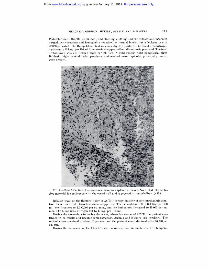
FIG. 4.-Case 2. Section of a recent occlusion in a sl)lenic arteriole. Note that the occlu-
sive material is continuous with the vessel wall and is covered by endothehum. X255.
Relapse l)egan on the thirteenth day of ACTII therapy, in spite of cant inued administra-
tion. Fever recurred. Gross hematuria reappeared. The hemoglobin fell to 8.3 Gm. per 100
nil., ervthrocytes to 2,900,000 p(’� cu. mm., anti the leukocvtes increased to 36,000 per cu.mm. The 1)100(1 urea nitrogen fell to 44 mg. per 100 ml.
During the seven days following the twenty-three day course of ACTH the 1)atient con-
tinued to be febrile :uuid became semi-comatose. Anemia and leukocytosis persisted. The
reticuloc�-tes remained at about 10 per cent and the platelet count diminished to 86,520 �
cu. mm.
During the last seven weeks of her life, she remained stuporous and fehrile wit hi tempera-
MEACHAM, ORBISOX, HEINLE, STEELE AND SCHAEFER 711
Platelets rose to 166,800 pcI’ cu. mm., and bleedilig, clotting, and clot i’etraction times were
normal. Erythrocytes anal hemoglobin remained at normal levels, h)ut a leukocytosis of
22,850 persisted. The Runipel-Leed test was only slightly positive. The 1)100(1 urea nitrogen
had riseti t.o 115 rug. per 100 ml. Flematuria disap�)eared 1)ut albuminuria I)ersisted. The fecal
urohilinogen was 240 Ehrlich units pes’ 100 Gm. A mild spastic right hemiplegia, rightBabinski, right. central facial pai’alysis, and marked mixed aphasia, principally motor,
were pI’esent.
..a-. i”f, �-
8 ‘�
1’ %#{149}�)�‘ �� ��db�’
� #{149}i�l�t� . “u.’ -
)��I,1 �#- a � � .,
� � .-�
�%r: �. r...�
1%
;i’�� �� --‘I-
� �tr.
� � .,, .:
(‘ �#%, u�
For personal use only.on January 11, 2019. by guest www.bloodjournal.orgFrom

�;�#{149}p�_� �
� .;�
., ,1�’A
.�-...--.,. C,
-�
-, - .�,r �
� ‘r� �. _-.1
0.,
“5
FIG. 5.-Case 2. Section of a healed occlusions in a meningeal arteriole. X255.
712 THROMBOTIC THROMBOCYTOPENIC PURPURA
ture ranging from 37 to 39.8 C. Nine days before death 1)100(1 values were: erythrocytes,
3,960,0(X) per cu. mm.; hemoglobin, 12.7 Cm. per 100 ml. (no blood transfusions were given);
leukocvtes, 18,400 per cu. cm. Platelets, 97,000 p�’� cu. mm. wit Is normal bleeding, clotting
and clot retraction times. Heticulocvtes, 5.9 pe�� cent. I)eath occurred on September 25,
1950, eighty-seven days after hospital admission.
Autopsy: Hemorrhage was a prominent gross feature, especially in the skin and brain.
In the right frontal lobe a hemorrhagic area measured 2 x 2 x 1 cm. and its the left occipital
lobe another measure(1 3 x 3 x 2 cm. Each was confined to the medulla, extending to the
cortex. In the skits, hemorrhages were present as ecchymoses on the neck, trunk, and ex-
tremities, the largest measuring 3 x 5 cm. Petechiae and ecchymoses w-ere visible in the
cortex arid pelvis of 1)0th kidneys and in the mucosa of the urinary bladder.
Old insfarcts occupied large areas of the left. pariet.al and right occipital lobe of the cere-
brum. A more recent infarct, of the upper lobe of the left lung had undergone liquefaction
necrosis and appeared as an abscess 1.2 cm. in diameter. Another old infarct of the terminal
ileum resulted its a chronic ulcer measuring 1 x 1.5 cm. Occluded vessels w-ere identified by
gross examination in the last. two instances.
The spleen, removed three years previously, weighed 500 Gm. and contained old and
For personal use only.on January 11, 2019. by guest www.bloodjournal.orgFrom

MEACHAM, ORBISON, HEINLE, STEELE AND SCHAEFER 713
recent infarcts. The heart weighed 480 Gm. The anterior and posterior leaflets of the mitral
valve both had firmly attached, pink, verrucous vegetations measuring 1.4 x 0.5 x 0.2 cm.
An anatomically patent foramen ovale had a few- small pink, firmly attached vegetations
along the margin in the right atrium. In addition, there were hyperemia anal edema of the
lungs, hyperplasia of the femoral marrow-, a small prepyloric gastric ulcer, irregular cortical
atrophy of the kidneys arid nodular cortical hyperplasia of the adrenals.
The significant microscopic finding was w-idespread occlusion of arterioles and small ar-
teries involving nearly every tissue of the b)ody. In sonic instances the occlusive material
�‘�j�” ,�: �-- - �“‘�“�‘ ‘ ‘� E”�. ‘� �
- I � � .�“ -
-� . .�-. ;�. , � ,,) 4*�,
v�”� �#{149}
2 - ‘ � t, - �“- �. , -
�4�# � � #{149}
� -��1
“S
.,‘ 4
#{234}d
‘ .,‘
‘4.
, � ‘4 4’.
-- , ‘C-� ,,�_‘,.‘ .
a, - ‘� � ,�‘. -
td �
� ____ �
FIG. 6,-Case 2. Section of a relatively recent occlusion of a myocardial arteriole. Note
th:tt occlusive material is continuous with vessel u-all aid s c.)ver(’d by euunlot Iseliuni. X255.
was eosinophilic, amorphous and finely granular alsd covered with, etsdotheli:tl cells. In
many sites, it could he show-n by serial sectioning that the material occluding the lumeti
was continuous with similar material located subendot heliallv in the vessel wall, as was true
in the first case (fig. 6). The Van Giesen stain demonstrated the absence ofbothelastic and
muscle fibers of the vessels in involved areas. The occlusive material and degenerated areas
of the vessel staitsed identically with the periodic acid-Schiff reagent. In some instances,
the occlusion contained numerous stnall mononuclear cells, 1)ut collagen formation u-as
absent. The normal structure of the vessel wall was absent, frequently, at these sites, with
For personal use only.on January 11, 2019. by guest www.bloodjournal.orgFrom

I
r �
Fir;. 7.-Case 2. Low power magnification of vegetation on mitral valve. X 15.
714 THROMBOTIC ‘rHROMBoCYTOPENI(’ PURPURA
marked t lii titii tsg of t he walls and dilat at ions of t he vessel wit Ii format ion of o(’cluded an-
curvstiis. There was tio I tsflamtiat otv react tori associated wit h t isese lesiotis.
Th’� changes were especially prominent its the vessels of the cerebrum, leptomeninges
(fig. 5), tii�-ocardium (fig. lb and spleeti (figs. 3 and 4). Its t lie cerebrum, there were asso-
ciated suhcortical hemorrhages wit Ii coit ical :tnd medulla,’�- infarct ions. Focal fibrosis was
l)resetst its the myocardium. Occlusive vascular lesiotss were foutid its 4 of 6 groups of lymphnodes examined. Other sites wit Ii deniotsst rable lesi otis included t he p:tticreits, adrenals,
skin, mesenter�-, kidney, liver, femoral l.)one marrow and stomachs. Otslv one lesiots was
foutid irs the lungs and notse could be (lemotsstrate(1 in skeletal muscle.
The vegetatiots on the mitral valve (figs. 7 and 8) consisted of hyalinized fibrous tissue
u-it Is superimposed amorphous, gratiular, neut rophihic material. Bacteria were tiot present.
Its additiots to the occlusive lesiotis, there was a diiffuse, moderate, interstitial nephritis,
an(1 moderate arteriolar tsephrosclerosis. The islets of Langerhans were hyperplastic and
the adretial cortex was tiodular and thicker than usual. Basophils were tiumerous in the
pituitary atid sonic were etslarged �asd had granular :ttid vacuolated cytoplasm.
DISCUSSION
As pointed out by Singer et al.,9 this syndrome and its clinical manifestations
are the result of three dlist.inCt phenomena which do not occur together in any
For personal use only.on January 11, 2019. by guest www.bloodjournal.orgFrom

I’
‘a
�‘ a’
-. I
/ a a
\ ‘a
S a’_.- ‘a -5
‘a � � � I
-� � � ,� ,\a’-�’a’.� ‘0�� -F-, ‘�‘��‘ - - - .� a� . ‘aS - a , a,�5j .� -
--
V
-a-.
‘a �
.‘e�.�ip’a
‘a�#{149} ‘#{149}‘a
.‘
-�:.I-
FIG. 8.-Case 2. Base of vegetation sisowis its figure 7 demotist rates fibrosis and hvalitiiza-
tion without. inflammatory exudate. Fibrin was l)resent ots the surface of thse vegetat ion
but cellular exudate was tiot present. in any part of the sect ions. X80.
coma appear as the coliditioli becomes more fulminating. Low-gt’ade fever and
pallor are present. early, followed! by icterus, petechiae, ecchymoses and epistaxis.Moderate splenomegaly and hepatomegaly are usually present. Mental and
neurologic signs are constant and develop as the fever increases. There may 1)e
signs of focal involvement such as hemiplegia, aphasia or cranial nerve paralysis,
as w’ell as those of generalized cerebral invol�’ement with dlelinium, muttering
and hypomanic activity. Some of these signs may be transitory, but- their oc-
currence has been associated with the terminal st.age of the disease.
Laboratory examination reveals the combination of hemolvtic anemia and
MEA(HAM, ORBISON, HEINLE, STEELE AND SCHAEFER 715
other clinical entity: (1) hemolytic anemia, (2) thromhocvtopenie puirputra, (3)
signs (tue to occlusion of small arterioles, usually manifested chnicallv by focal
neurologic changes and mental aberrations.
In cases studied! previously, the onset has been abnipt, and the course violent,
short and fatal. The onset has occasionally been precede(! by an u��pei’ respiratory
infection. The first complaints, often vague, include malaise, weakness, nausea,
vomiting, headache, dizziness, arthralgia and myalgia. Convulsions, stupor and
For personal use only.on January 11, 2019. by guest www.bloodjournal.orgFrom

716 THROMBOTIC THROMBOCYTOPENIC PURPURA
thrombocytopenic purpura. The hemolytic anemia is accompanied by the usual
findings of hemolysis, including reticulocytosis, nucleated red cells in the pe-
ripheral blood, erythroid hyperplasia of the marrow, and increased output of
urobilinogen. It was not possible to demonstrate abnormal agglutinins in the
blood nor was the Coombs test positive, findings in agreement with those of
Singer.9 Similarly, while spherocytosis w’as demonstrated by the one fragility
test performed on Case 1 , it was variable from time to time in Case 2, a variation
observed by others.9 The ultimate mechanism of the hemolytic process has not
been delineated.
The thrombocytopenia is accompanied by the laboratory findings considered
to be diagnostic of so-called idiopathic thrombocytopenic purpura. In these 2
patients, the numbers of megakaryocytes in the marrow were increased, im-
mature forms were present, and, unlike the observations of Singer,’ platelet
formation was inhibited. It is the authors’ belief, in fact, that the thrombocyto-
penia is not the result of deposition of large numbers of platelets in the occluded
vessels, but may be the result of a process similar to that producing idiopathic
thrombocytopenic purpura. There is no universal agreement concerning the
mechanism of t.his process, although there is little doubt that the spleen is in-
volved in some manner. It seems likely to us that the abnormal splenic mechanism
responsible for the production of idiopathic thrombocytopenic purpura was
also operating in these patients.
Singer et al.’ have discussed the possible relationship of the syndrome to
polyarterit.is nodosa, lupus erythematosus and to certain clinical and experi-
mental hypertensive states. From a study of 2 patients, Beigelman’#{176} has also
proposed that the platelet thrombosis syndrome be placed in t.he same category
as the collagen diseases.
Our histologic observations on the present 2 cases support the concept that
the degenerative lesion of vessels is primary and in this respect the syndrome
is similar to the so-called collagen diseases. This raises the question of whether
t.he occlusive material in the involved vessels actually consists of agglutinated
platelets. Review of the evidence upon which such an impression is based reveals
that the absence of erythrocytes, hemoglobin, leukocytez and fibrin in the lesions
led to the conclusion that the material must consist of platelets, by exclusion.
The presence of platelets at the margins of the occlusions and Gore’s observa-
tion3 that both the occlusive material and platelets gave positive reactions with
t.he periodic acid-Schiff t.est supported t.his impression furt.her. Since t.his reaction
is positive in many types of tissues involving degenerative changes in vessels,
it cannot, however, be considered specific. Thus, there is no conclusive positive
evidence that the occlusions are composed of platelets.
In an attempt to identify or exclude platelets specifically, comparative cyto-
chemical reactions were performed on centrifuged platelets, the vascular oc-
elusions of Case 1, and on certain degenerative vascular lesions (arteriolar
necrosis, polyarteritis and amyloid in vessels). Since no specific reaction is
known for platelets in tissues, these tests were performed to compare the react.ion
of platelets w’ith that of the other tissues. The Feulgen reaction and methyl
green stain were negative in all of the tested tissues, indicating the absence of
nucleic acids. The fast green stain, which identifies a basic substance, and the
For personal use only.on January 11, 2019. by guest www.bloodjournal.orgFrom

MEACHAM, ORBISON, HEINLE, STEELE AND SCHAEFER 717
periodic acid-Schiff reaction, which indicates the presence of a polysaccharide,
were positive in all the tissues tested. The toluidine blue test, to demonstrate
metachromasy, w’as positive in the case of the platelets, the vascular occlusions
of Case 1 and amyloid in �‘essel walls, but was negative in arteriolar necrosis
and polyarteritis. Thus, none of these tests are specific, and in themselves, do
not prove or disprove that platelet material is present in the occlusive lesions.
Nevertheless, it should 1)e stated that, insofar as studied, platelets and the
material in the vessels showed parallel reactions.
Evaluation of certain prominent histologic features of these 2 cases, how’ever,
make it seem unlikely t.hat the occlusive lesions were composed only of plate-
lets. The amorphous material present in these vessels was almost always covered
by endothelium, suggesting that it ��‘as degenerated and swollen material en-
croaching upon the lumens from the vessel wall and not an intralumenal throm-
bus. Further support for this view is provided by the fact that the same material
was seen frequently throughout all layers of the vessel wall and occasionally
even in the surrounding connective tissue. At these sites, it replaced completely
the normal structures. A unique, and perhaps pathognomonic, feat.ure of the
vascular lesions, which will be the subject of another report, was the finding of
multiple aneurysms of the arterioles and precapillaries containing both the
amorphous material and masses of proliferated endothelial cells. In no instance
was an exudative reaction a feature of the lesions. The characteristic finding,
therefore, was a nonexudative lesion w’hich partially replaced the vessel wall
and resulted in aneurysm formation. It is our opinion t.hat. t.hese histologic
changes can be explained only by a degenerative change in the vessel wall and
that t.he presence of platelet. thrombi alone would not be expected t.o produce
such lesions. While platelets could not be specifically identified in, or excluded
from, t.he lesions on the basis of staining, these studies present strong histologic
evidence that much, if not all, of the material in question represents an intramural
degeneration rather than an int.raluminal coagulum.
Case 2 indicates that not all cases are rapidly and progressively fatal. There
is little doubt. that her course lasted at least t.hree years, since re-examination of
the spleen removed three years before death revealed characteristic lesions
identical with t.hose demonstrated at necropsy in other organs. Subsequent to
splenectomy, the anemia was markedly alleviated and the patient maintained a
remission for twenty-eight months, although hypertension developed during that
time. Whether the remission was spontaneous or the direct result of the splenec-
tomy can be proven only by further experience. Splenectomy has been per-
formed in similar patients in only 3 other instances2- � #{176}and, in each case, deathoccurred shortly after operation before any therapeutic effect. could he evaluated.
Muirhead et al.” believe that. splenectomy has not received a fair trial.
The effect of ACTH therapy is difficult to interpret. There was clinical and
hemat.ologic improvement during t.he first ten days of therapy with subsequent
severe relapse while the drug was continued. The dose used ��‘as not large, how-
ever, and larger doses might have prolonged the remission. In this connect.ion,
the problem arises whether vigorous hormone therapy might cause rapid healing
and fibrous obliteration of affected vessels as has been observed in cases of poly-
arteritis nodosa treated with cortisone,’2 in which great numbers of small in-
For personal use only.on January 11, 2019. by guest www.bloodjournal.orgFrom

718 THROMBOTIC THROMBOCYTOPENIC PURPURA
farcts have occurred! throughout the body. It is possible that both splenectomy
and ACTH therapy induced remissions in Case 2.
This is an acute febrile syndrome involving primarily the vascular and hemato-
poietic systems. The triad of hemolytic anemia, t.hrombocytopenic purpura and
clinical neurologic abnormalities resulting from small vessel occlusion should
suggest the diagnosis. Lymph node biopsy might confirm the d!iagnosis, but
enlarged lymph nodes, suitable for biopsy, were not present in these or other
reporte(l cases. The muscle has not shown characteristic histologic changes.
In our second patient, on the other hand!, vessels in the skin contained charac-
teristic lesions so that skin biopsy might be of diagnostic value in subsequent
cases.
The application of a suitable name which would include the hemolytic anemia,
thrombocvtopenia and the vascular phenomena becomes a problem, especially
since we (to not beheve that the thrombocytopenia is the result. of massive
sequest ration of platelets in the occlusive lesions, nor has the mechanism of the
hemolytic process been (lelineated. The degenerative process in t.he walls of
ai’tei’ioles and! capillaries appears to be the primary vascular lesion. The term
thrombotic thrombocytopenic purpura has been suggested!.8 This term does not
describe the essential components of the syndrome and is, we believe, inaccurate,
in that the occlusive lesions are not thrombi. Nevertheless, the term has already
acquired some popularity, is short and alliterative. A term describing all the
components of the syndrome would be unwieldy, and an alternative of applying
a propet’ name is generally objectionable. We propose that the term, thrombotic
thrombocytopenic purpura, be retained until a better understanding of the
pathologic physiology of the condition permits the application of a more suitable
name.
SUMMARY
1. Two patients with a syndrome of hemolytic anemia, thrombocytopenic
purpura and arteriolar occlusions are described.
2. In one patient, a long remission followed splenectomy, and improvement
for ten days followed the administration of ACTH. No claim is made that either
form of therapy induced the remission, but trial of t.hese procedures in other
cases is indicated.
3. Evidence is presented that the primary vascular lesion is a degenerative
process in the arteriolar and capillary walls rather than thrombus formation
in the lumen of the vessel. The occurrence of aneurysms in arterioles and precapil-
lanes was a striking observation.
4. The histologic changes suggest a relationship to the so-called collagen group
of diseases.
5. It is suggested that the term, thrombotic t.hrombocytopenic purpura, be
ret.ained until further study of the conditions permits application of a more
suitable name.
REFERENCES
1 MOSCHCOWITZ, E.: An acute febrile pleiochromic anemia with hyalits thromboses of ter-minal arterioles and capillaries. Arch. Jot. Med. 36: 89, 1925.
For personal use only.on January 11, 2019. by guest www.bloodjournal.orgFrom

MEACHAM, ORBISON, HEINLE, STEELE AND SCHAEFER 719
2 BAEHR, C., KLEMPERER, P. AND ScHIFRIN, A.: An acute febrile anemia and thrombocyto-
penic purpura uith diffuse platelet thromboses of capillaries and arterioles. Tr. A. Am.Physicians. 51: 43, 1936.
GORE, I.: Disseminated arteriolar and capillary platelet thromboses. A morphological
study of its histogenesis. Am. J. Path. 26: 155, 1950.Aursctiut�, M.: A rare type of acute thrombocytopetiic purpura: Widespread formation
of platelet thrombi ins capillaries.New England J. Med. 227: 477,1 942.
ENGEL, C., SCHEINKER, M. AND HUMPHREY, D.: Acute febrile anemia and thsrombocyto-
penic �urpura with vasothromboses. Ann. Inst. Mcd. 26: 919, 1947.6 TROBAUGH, F., MARK0WITZ, M., DAvIDsoN, C. AND CROWLEY, \V.: An acute febrile ill-
ness characterized by thronibocytopenic purpura, hemolvtic anemia, and generalized
platelet thromboses. Arch. Path. 41: 327, 1946.FITZGERALD, P. J., AUERBACH, 0. AND FRAME, E. : Thrombocvtopensic acroatigiothrombo-
sis (platelet thrombosis of the capillaries, arterioles, arid venules). Blood 2: 519, 1947.
8 SINGER, K., BORNSTEIN, F. P. AN!) WILE, S. A. Thrombotic thrombocytopenic purpura.
Blood 2: 542, 1947.
-, MOTULSKY, A. G. AND SHANBERGE, J. N.: Thsrombotic thrombocytopenic purpura.
II. Studies on the hemolvtic syndrome in this disease. Blood 5: 434, 1951).1� BEIGELMAN, P. M. : Variatits of the platelet thrombosis syndrome and t heit’ i-dat iotiship
to dissemitiated lupus. Arch. Pat.h. 51: 213, 1951.
11 MUIRHEAD, E. E., CRASS, G. AND HILL, J. M.: Diffuse platelet thsromboses w’ithi thronsbo-
cytopensia and hemolytic anemia (thrombotic thrombocytopenic PurPuril). Am. J.Chin. Path. 18: 523, 1948.
12 SnucK, H. M., BAGGENSTOSS, A. H. AND POLLEY, H. F.: Effects of cortisotie and ACTH
on periarteritis nodosa and cransial arteritis; preliminary report. Proc. Staff Meet.
Mayo Clini. 25: 135, 1950.
For personal use only.on January 11, 2019. by guest www.bloodjournal.orgFrom

1951 6: 706-719
ALPERT SCHAEFERGORDON C. MEACHAM, J. LOWELL ORBISON, ROBERT W. HEINLE, HOWARD J. STEELE and J. ArteriolesThrombotic Thrombocytopenic Purpura: A Disseminated Disease of
http://www.bloodjournal.org/content/6/8/706.full.htmlUpdated information and services can be found at:
Articles on similar topics can be found in the following Blood collections
http://www.bloodjournal.org/site/misc/rights.xhtml#repub_requestsInformation about reproducing this article in parts or in its entirety may be found online at:
http://www.bloodjournal.org/site/misc/rights.xhtml#reprintsInformation about ordering reprints may be found online at:
http://www.bloodjournal.org/site/subscriptions/index.xhtmlInformation about subscriptions and ASH membership may be found online at:
Copyright 2011 by The American Society of Hematology; all rights reserved.Hematology, 2021 L St, NW, Suite 900, Washington DC 20036.Blood (print ISSN 0006-4971, online ISSN 1528-0020), is published weekly by the American Society of
For personal use only.on January 11, 2019. by guest www.bloodjournal.orgFrom