Embed Size (px)
Citation preview

Intravaginal Administration of Interleukin 12 during GenitalGonococcal Infection in Mice Induces Immunity toHeterologous Strains of Neisseria gonorrhoeae
Yingru Liu,a Julianny Perez,a Laura A. Hammer,a Heather C. Gallagher,b,c Magdia De Jesus,b,c Nejat K. Egilmez,a,d
Michael W. Russelle
aTherapyX, Inc., Buffalo, New York, USAbDepartment of Biomedical Sciences, University at Albany, Albany, New York, USAcDivision of Infectious Diseases, Wadsworth Center, New York State Department of Health, Albany, New York,USA
dDepartment of Microbiology and Immunology, University of Louisville, Louisville, Kentucky, USAeDepartment of Microbiology and Immunology, University at Buffalo, Buffalo, New York, USA
ABSTRACT It has previously been shown that genital tract infection with Neisseriagonorrhoeae in mice does not induce a state of protective immunity against reinfec-tion but instead suppresses the development of adaptive immune responses againstN. gonorrhoeae dependent on transforming growth factor beta (TGF-�) and interleu-kin 10 (IL-10). Intravaginal administration during gonococcal infection of IL-12 encap-sulated in biodegradable microspheres (IL-12/ms) reverses the immunosuppressionand promotes the production of gamma interferon (IFN-�) and of specific antibodiesin serum and genital secretions and accelerates clearance of the infection. In thisstudy, microspheres were shown to remain largely within the genital tract lumenand to release IL-12 over the course of 4 days. Antigonococcal IgA and IgG antibod-ies induced by IL-12/ms treatment reacted with antigenically different strains ofN. gonorrhoeae and led to resistance to reinfection with heterologous and homolo-gous strains. Immune resistance to reinfection persisted for at least 6 months afterclearance of the primary infection. Experiments performed with immunodeficientstrains of mice lacking either IFN-� or B cells demonstrated that both IFN-� and Bcells were necessary for the IL-12-induced generation of immune responses toN. gonorrhoeae and the resulting accelerated clearance of the infection. It is there-fore concluded that intravaginally administered IL-12/ms achieves its effect by thesustained release of IL-12 that promotes Th1-driven adaptive immune responses, in-cluding the production of specific antigonococcal antibodies that cross-react withmultiple strains of N. gonorrhoeae. IL-12-enhanced immunity to N. gonorrhoeae canbe recalled against reinfection after prolonged intervals and is dependent uponboth IFN-� and antibody production by B cells.
IMPORTANCE Genital infection with Neisseria gonorrhoeae (gonorrhea) is a signifi-cant cause of reproductive tract morbidity in women, leading to pelvic inflammatorydisease, tubal factor infertility, and increased risk for ectopic pregnancy. WHO esti-mates that 78 million new infections occur annually worldwide. In the United States,�350,000 cases are reported annually, but the true incidence is probably �800,000cases/year. Increasing resistance to currently available antibiotics raises concern thatgonorrhea might become untreatable. Infection does not induce a state of immuneprotection against reinfection. Previous studies have shown that N. gonorrhoeae sup-presses the development of adaptive immune responses by mechanisms dependenton the regulatory cytokines TGF-� and IL-10. This study shows that intravaginaltreatment of gonococcal infection in female mice with microencapsulated IL-12 in-
Received 20 September 2017 Accepted 11December 2017 Published 31 January 2018
Citation Liu Y, Perez J, Hammer LA, GallagherHC, De Jesus M, Egilmez NK, Russell MW. 2018.Intravaginal administration of interleukin 12during genital gonococcal infection in miceinduces immunity to heterologous strains ofNeisseria gonorrhoeae. mSphere 3:e00421-17.https://doi.org/10.1128/mSphere.00421-17.
Editor David W. Pascual, University of Florida
Copyright © 2018 Liu et al. This is an open-access article distributed under the terms ofthe Creative Commons Attribution 4.0International license.
Address correspondence to Michael W. Russell,[email protected].
RESEARCH ARTICLETherapeutics and Prevention
crossm
January/February 2018 Volume 3 Issue 1 e00421-17 msphere.asm.org 1
on May 7, 2020 by guest
http://msphere.asm
.org/D
ownloaded from
duces persisting anamnestic immunity against reinfection with N. gonorrhoeae, evenof antigenically diverse strains, dependent on T-cell production of IFN-� and B-cellproduction of antibodies.
KEYWORDS Neisseria gonorrhoeae, adaptive immunity, genital tract immunity,interleukin 12, microencapsulation
The sexually transmitted pathogen Neisseria gonorrhoeae (the gonococcus) is well-adapted to its natural human host, and it can be acquired repeatedly with little or
no evidence of protective immunity arising from prior infections (1, 2). Factors contrib-uting to its success undoubtedly include a remarkable capacity for changing theexpression and specificity of most of its major surface antigens (reviewed in reference3), as well as multiple mechanisms for resisting complement-mediated destruction (4).In addition, we have previously shown that N. gonorrhoeae has the capacity to suppressthe development of adaptive immune responses governed by T helper (Th) type 1 and2 cells, while concomitantly inducing Th17-driven innate responses that it appears ableto resist (5–7). We have further found that the induced immunosuppression in micevaginally infected with N. gonorrhoeae can be countermanipulated to allow specificimmune responses to emerge, with the generation of antigonococcal antibodies andgamma interferon (IFN-�)-secreting CD4� T cells, the establishment of immune mem-ory, and accelerated clearance of the organism (5–8). Furthermore, when mice arereinfected with N. gonorrhoeae 1 month later, the challenge is resisted more effectivelythan in control mice that had not been treated during the original infection.
The mechanisms of suppression induced by N. gonorrhoeae depend on the regula-tory cytokines transforming growth factor beta (TGF-�) and interleukin 10 (IL-10) andon the generation of type 1 regulatory T cells (7). Systemic administration of neutral-izing antibodies against TGF-� and IL-10 reverses the suppression and allows specificantibody and T-cell responses to develop. We have further found that intravaginal(i.vag.) administration of IL-12 encapsulated in biodegradable microspheres (hereafterdesignated IL-12/ms) during gonococcal infection similarly promotes the developmentof Th1-driven immune responses, including production of IFN-� by CD4� T cells andspecific IgG and IgA antibodies in serum and genital secretions, as well as acceleratedclearance of the infection, without overtly toxic effects (8). Soluble IL-12 given i.vag. hasno effect, and it is far too toxic to be given systemically in effective doses. This raisesthe question whether encapsulation of IL-12 into polylactic acid (PLA) microspheresworks by the sustained delivery of IL-12 over a prolonged period or whether itpromotes uptake into the tissues as has been documented for another microparticulateformulation (9). If the latter is correct, then IL-12/ms might release their cytokine loadin the vicinity of responsive cells or even be translocated to the draining lymph nodes.
Given the extraordinary antigenic diversity of N. gonorrhoeae, it is important todetermine whether the immunity against repeat infection induced by treatment withIL-12/ms is limited to the original infecting strain or whether it can extend to challengewith heterologous strains. In addition, the mechanisms of induced immunity to gono-coccal infection need to be understood in order to exploit these findings as a potentialnovel therapeutic modality that would have the further advantage of conferringprophylactic resistance to repeat infection. These considerations assume greater im-portance with the continuing emergence of antibiotic resistance in this organism,which threatens to render gonorrhea untreatable.
The studies described here were first aimed at tracking the location of fluorescentPLA microspheres in genital tract tissues and iliac lymph nodes (ILN) after vaginalinstillation. Release of IL-12 into the vaginal lumen and circulation were also monitoredfor several days. Second, we determined whether the IL-12-induced immune resistanceto reinfection persisted for prolonged periods and whether it extended to challengewith antigenically distinct strains of N. gonorrhoeae. Finally, we determined whetherenhanced clearance of gonococcal infection due to i.vag. administration depended onthe generation of both antibodies and IFN-�.
Liu et al.
January/February 2018 Volume 3 Issue 1 e00421-17 msphere.asm.org 2
on May 7, 2020 by guest
http://msphere.asm
.org/D
ownloaded from
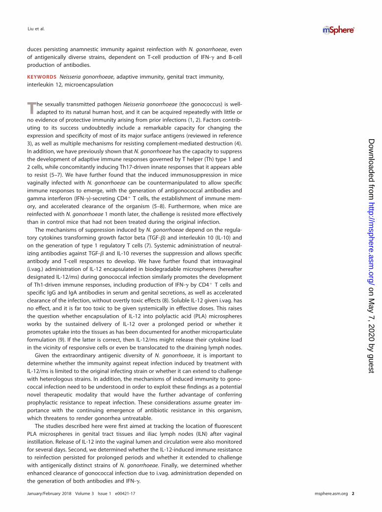
RESULTSMicroencapsulated IL-12 generates sustained release of IL-12 in the vagina.
Female BALB/c mice (six mice in each group) were given one dose of 1.0 �g IL-12encapsulated in PLA microspheres (IL-12/ms) or in soluble form intravaginally. To avoidremoval of the administered dose, serum samples and vaginal wash fluid samples werecollected from separate groups of mice on days 1 to 7, and IL-12 was measured by anenzyme-linked immunosorbent assay (ELISA). After a single dose of IL-12/ms, highconcentrations of IL-12 were maintained at slowly decreasing levels in vaginal washfluid samples for up to 4 days (Fig. 1). IL-12 was also detected in the sera of these mice,but the concentrations were low, �40 pg/ml (Fig. 1). The instillation of soluble IL-12yielded substantially lower recovery of IL-12 in vaginal wash fluid samples, and IL-12was barely detected in serum (Fig. 1).
We further investigated the location of PLA microspheres in genital tract tissues andpossibly in the draining ILN after vaginal instillation. A similar dose of PLA microspheresloaded with fluorescein isothiocyanate-labeled bovine serum albumin (FITC-BSA/ms)was instilled intravaginally, and tissues were collected from mice sacrificed 4 or 24 hlater. Tissues were also stained for CD11b and CD11c to identify presumptive antigen-presenting cells (dendritic cells and macrophages) or for CD4 and CD8 to identify Tcells. Confocal fluorescence microscopy showed that FITC-BSA/ms were largely retainedin the lumen of the vagina at 4 h (Fig. 2), and very few were seen within the tissue.CD11b� and CD11c� cells were observed in the tissue (Fig. 2B), but CD4� and CD8�
cells were rarely found. No particles were observed in the ILN. FITC-BSA/ms were notreliably detected in tissues obtained at 24 h. These results suggest that IL-12/msachieve their therapeutic effect by the sustained release of IL-12 at the epithelial surfaceor within the genital tract tissue, rather than through uptake by antigen-presentingcells and subsequent translocation to the draining lymph nodes.
Antibodies induced by i.vag. IL-12/ms treatment target both homologous andheterologous strains of N. gonorrhoeae. Our previous studies demonstrated thatIL-12/ms treatment during gonococcal infection induced the production of vaginal andserum IgG antibodies, as well as vaginal IgA antibodies against the homologousinfecting strain of N. gonorrhoeae (8). Considering gonococcal surface antigenic vari-ability, we determined whether the induced antibodies could also target heterologousstrains of N. gonorrhoeae. We therefore measured serum and vaginal wash antibodiesby ELISAs using plates with the wells coated with homologous (FA1090) or heterolo-gous (MS11 and FA19) gonococcal strains or with E. coli or nontypeable Haemophilusinfluenzae (NTHI). The results showed that vaginal and serum IgA and IgG antibodies to
Day 1
Day 2
Day 3
Day 4
Day 5
Day 6
Day 7
0
50
100
150
200
250
IL-12/ms: vaginal washIL-12/ms: serum
Soluble IL-12: serumSoluble IL-12: vaginal wash
[IL-1
2] (p
g/m
l)
FIG 1 Recovery of IL-12 in serum and vaginal wash fluid samples after i.vag. administration ofmicroencapsulated IL-12 (IL-12/ms) or soluble IL-12. Serum and vaginal wash fluid samples werecollected from separate groups of mice on days 1 to 7 after instillation of IL-12/ms or soluble IL-12 (1 �gof IL-12 per mouse) and assayed for IL-12 by ELISA. Values are means (bars) plus standard errors of themeans (SEM) (error bars) (n � 6 samples).
IL-12 Induces Immunity to N. gonorrhoeae
January/February 2018 Volume 3 Issue 1 e00421-17 msphere.asm.org 3
on May 7, 2020 by guest
http://msphere.asm
.org/D
ownloaded from

N. gonorrhoeae were detected to similar extents against both homologous and heter-ologous strains (Fig. 3). In contrast, antibodies were not detected above backgroundlevels against E. coli or NTHI (Fig. 3).
Antibodies were also analyzed by Western blotting to determine the patterns ofspecificity for different gonococcal antigens in homologous and heterologous strains,using outer membrane vesicles (OMV) which contain most of the surface antigensexpressed by N. gonorrhoeae. The protein profiles of N. gonorrhoeae FA1090, MS11, andFA19 OMV revealed by sodium dodecyl sulfate-polyacrylamide gel electrophoresis(SDS-PAGE) displayed some quantitative and qualitative differences probably reflectinglevels of expression and sequence variation (Fig. 4A). Western blot analyses of serumsamples from FA1090-infected mice treated with IL-12/ms against FA1090, MS11, orFA19 OMV separated by SDS-PAGE showed IgG antibodies reactive with bands migrat-ing at ~35 to 80 kDa, with partially similar reactivity against bands present in OMV fromall three strains (Fig. 4B). The IgG band at ~35 kDa probably corresponds to porin, as anantiporin antibody (H5) reacted with a band of similar mobility (Fig. 4B). Other bandswere visible at approximately 30, 40, 45, 60, and 70 kDa. Further work will be necessaryto identify the nature of the antigens detected. However, these results indicated thatIL-12/ms treatment of infected mice induced antibodies that recognized antigens inhomologous and heterologous strains of N. gonorrhoeae.
FIG 2 Confocal fluorescence micrographs of vaginal tissue excised from mice 4 h after i.vag. adminis-tration of FITC-BSA/ms. (A) FITC-BSA/ms (green) were located in the lumen (white arrows), but no CD4�
(red) or CD8� (blue) cells were observed. Bar � 500 �m. (B) Higher magnification showing FITC-BSA/ms(green) located in the lumen (white arrows) and CD11b� (blue) and CD11c� (red) cells in the tissue. Bar �100 �m.
IgA IgG0
5
10
15
FA1090 + blank ms: FA1090Control (blank ms alone)
FA1090 + IL12/ms: FA1090FA1090 + IL12/ms: MS11FA1090 + IL12/ms: FA19FA1090 + IL12/ms: E. coliFA1090 + IL12/ms: NTHI
#
***
***
*
A
Vag
inal
was
h sp
ecifi
c A
b re
lativ
e to
con
trol
IgA IgG05
101520253035
FA1090 + blank ms: FA1090Control (blank ms alone)
FA1090 + IL12/ms: FA1090FA1090 + IL12/ms: MS11FA1090 + IL12/ms: FA19FA1090 + IL12/ms: E. coliFA1090 + IL12/ms: NTHI
#***
***
*
B
Ser
um s
peci
fic A
b re
lativ
e to
con
trol
FIG 3 Treatment of mice infected with N. gonorrhoeae (FA1090) with IL-12/ms-induced vaginal (A) andserum (B) IgA and IgG antibodies (Ab) detected against homologous (FA1090) and heterologous (MS11 andFA19) strains of N. gonorrhoeae, but not against E. coli or NTHI. Values are means plus SEM (error bars) (n �5 samples). Values that are significantly different by ANOVA from the values for control samples fromuninfected mice treated with blank ms alone are indicated as follows: *, P � 0.01; #, P � 0.05.
Liu et al.
January/February 2018 Volume 3 Issue 1 e00421-17 msphere.asm.org 4
on May 7, 2020 by guest
http://msphere.asm
.org/D
ownloaded from

Intravaginal IL-12/ms treatment during primary N. gonorrhoeae infection in-duces resistance to reinfection with heterologous strains of N. gonorrhoeae. We
have previously demonstrated that IL-12/ms treatment significantly accelerates theclearance of genital gonococcal infection and induces immune memory and resistanceto reinfection with the homologous strain of N. gonorrhoeae (8). Given the extraordinaryantigenic diversity of N. gonorrhoeae, we assessed whether IL-12/ms treatment duringprimary N. gonorrhoeae infection would result in similar resistance to subsequentinfection with other strains as reinfection with the same strain. Groups of eight miceinfected with N. gonorrhoeae FA1090 were treated with IL-12/ms or blank microspheres(blank ms), and after the infection had run its course, the mice were treated withceftriaxone to ensure complete elimination of the gonococci. One month later, themice were inoculated with N. gonorrhoeae of either strain FA1090 or MS11 without anyfurther treatment, and the course of infection was monitored by vaginal swabbing andplating. The results showed that IL-12/ms treatment during FA1090 infection resultedin similar resistance to reinfection with either FA1090 or MS11 (Fig. 5A). After clearanceof the secondary infection, vaginal and serum antibodies were elevated to similar levelsagainst both strains FA1090 and MS11 (Fig. 5B and C). When ILN cells harvested attermination were assayed by flow cytometry for cytokine production by CD4� T cells,and IFN-� was similarly enhanced in IL-12/ms-treated mice reinfected with eitherFA1090 or MS11. Consistent with our previous findings, IL-4 production was notenhanced by IL-12/ms treatment during primary infection, and IL-17 production wasenhanced in all (re)infected mice regardless of IL-12/ms treatment (Fig. 5D).
In a reciprocal manner, treatment with IL-12/ms during primary infection withN. gonorrhoeae MS11 induced resistance to rechallenge with strain FA1090 (see Fig. S1in the supplemental material).
N. gonorrhoeae strains FA1090 and MS11 both possess porin of the same major type(PorB.1B). Therefore, to determine whether the major porin type is important to theeffect of IL-12/ms treatment on resistance to reinfection, further experiments wereperformed with strain FA19 (PorB.1A). IL-12/ms treatment during primary infection withstrain FA1090 induced resistance to rechallenge with strain FA19 (Fig. S2A). Serum andvaginal IgG and IgA antibody responses assayed after clearance showed cross-reactivityagainst FA19 (Fig. S2B and C), and IFN-� CD4 T cells from ILN was elevated (Fig. S2D).Further treatment and reinfection studies using different combinations of N. gonor-rhoeae strains FA1090, MS11, and FA19 were performed, and similar cross-protectionswere observed in all cases.
FIG 4 SDS-PAGE of gonococcal OMV preparations and Western blot analysis of serum samples frommice infected with N. gonorrhoeae FA1090 against antigens in OMV separated by SDS-PAGE. (A)SDS-polyacrylamide gel stained with Coomassie blue. Lane 1, molecular mass markers (in kilodaltons)(shown to the left of the gel); lane 2, OMV from N. gonorrhoeae FA1090; lane 3, OMV from N. gonorrhoeaeMS11; lane 4, OMV from N. gonorrhoeae FA19. (B) Western blots developed for IgG antibodies. Lane 1,molecular mass markers (in kilodaltons) (shown to the left of the gel); lanes 2 to 4, serum from a mouseinfected with N. gonorrhoeae FA1090 and treated with IL-12/ms, tested against OMV from FA1090 (lane2), MS11 (lane 3), or FA19 (lane 4); lane 5, serum from a mouse infected with N. gonorrhoeae FA1090 andtreated with blank ms, tested against OMV from FA1090; lane 6, H5 antibody (anti-porin PIB3) testedagainst OMV from FA1090.
IL-12 Induces Immunity to N. gonorrhoeae
January/February 2018 Volume 3 Issue 1 e00421-17 msphere.asm.org 5
on May 7, 2020 by guest
http://msphere.asm
.org/D
ownloaded from

Mice treated with IL-12/ms during primary infection with N. gonorrhoeae FA1090were also resistant to challenge with minimally passaged clinical isolates GC68 (aPorB.1B strain) and GC69 (PorB.1A) (Fig. S3).
Duration of the prophylactic effect against reinfection. To determine the dura-tion of resistance to reinfection induced by treatment of gonococcal infection withIL-12/ms, groups of mice (eight mice in each group) were infected with N. gonorrhoeaeFA1090 and treated with IL-12/ms or blank microspheres. After the infection had beencleared (as determined by vaginal swabbing and plating), the mice were treated withceftriaxone to ensure complete elimination of the gonococci. Two, 4, or 6 months later,the mice were inoculated with N. gonorrhoeae MS11 without any further treatment, andthe course of infection was monitored by vaginal swabbing and plating. The resultsshowed that the effect of treatment with IL-12/ms persisted for at least 6 months afterthe primary infection had been cleared (Fig. 6). Although there was a noticeable declinein the clearance times in control mice that had been treated with blank ms during theprimary infection (median clearance times of 12 days, 10 days, and 7.5 days after 2, 4,and 6 months, respectively), by comparison, mice that had been treated with IL-12/msduring the primary infection cleared the second infection significantly faster (medianclearance times of 7 days, 6 days, and 5 days after 2, 4, and 6 months, respectively) thanthe corresponding control mice did (Fig. 6).
Roles of IFN-� and antibody in accelerated clearance of gonococcal infectiondue to IL-12/ms treatment. IL-12/ms treatment of mice infected with N. gonorrhoeaeinduces the production of antibodies specific for gonococcal antigens and the gener-ation of Th1-governed responses as shown by the production of IFN-� by CD4� T cells.To investigate whether either or both of these aspects of IL-12-enhanced immune
FIG 5 Treatment of mice with IL-12/ms during primary infection with N. gonorrhoeae FA1090 induces resistance to reinfection with homologous(FA1090) or heterologous (MS11) strains and induces immune responses to either strain. (A, left) Recovery (mean CFU � SEM; n � 8) ofN. gonorrhoeae on vaginal swabs after reinfection with strain FA1090 or MS11 as shown. Curves that are significantly different (P � 0.01 byANOVA) are indicated by an asterisk. (Right) Percentage of animals remaining infected at each time point (P � 0.001 and �0.005 by Kaplan-Meieranalysis), comparing treatment with IL-12/ms versus blank ms, for reinfection with FA1090 and MS11, respectively. (B) Vaginal IgA and IgGantibodies after clearance of secondary infection tested against strain FA1090 or MS11. (C) Serum IgA and IgG antibodies after clearance ofsecondary infection tested against FA1090 or MS11. (D) Production of IFN-�, IL-4, and IL-17 by ILN CD4� cells recovered after clearance ofsecondary infection. In panels B, C, and D, values for mice treated with IL-12/ms that are significantly different (P � 0.01 by Student’s t test) fromthe values for mice treated with blank ms are indicated by an asterisk.
Liu et al.
January/February 2018 Volume 3 Issue 1 e00421-17 msphere.asm.org 6
on May 7, 2020 by guest
http://msphere.asm
.org/D
ownloaded from

responsiveness to N. gonorrhoeae are required for the observed accelerated clearanceof infection, experiments were performed using mutant C57BL/6 mice deficient in IFN-�(IFN-�-KO [KO stands for knocked out]) or B cells (�MT). Groups of wild-type C57BL/6(control) mice and immunodeficient mice (eight mice in each group) were infected withN. gonorrhoeae FA1090 and treated with IL-12/ms or blank ms as described above. Thecourse of vaginal gonococcal infection was not altered in control (blank-ms-treated)immunodeficient mice relative to wild-type mice. All wild-type and immunodeficientmice started to reduce the recoverable gonococcal load from day 5 or 6 onwards andhad cleared the infection by days 9 to 16 (median, 10 to 15 days).
In contrast to wild-type mice, clearance of gonococcal infection was not acceleratedin IFN-�-KO mice treated with IL-12/ms compared to mice treated with blank ms(Fig. 7A). Likewise, �MT mice treated with IL-12/ms did not show accelerated clearanceof gonococcal infection (Fig. 7B). Thus, deficiency of either IFN-� or B cells abrogatedthe therapeutic effect of IL-12/ms in accelerating clearance of genital gonococcalinfection. The production of gonococcus-specific vaginal and serum IgA and IgGantibodies induced by IL-12/ms treatment in wild-type mice was diminished in IFN-�-KO mice (Fig. 7C and D), and as expected, there was no generation of IFN-� by the
FIG 6 Effect of treatment with IL-12/ms during primary infection with N. gonorrhoeae FA1090 againstreinfection with N. gonorrhoeae MS11 persists for 2 months (A), 4 months (B), or 6 months (C) after theclearance of primary infection. The left panels show recovery (mean CFU plus SEM; n � 8) of N. gonorrhoeaeon vaginal swabs after reinfection with MS11 at each interval. Curves for mice treated with IL-12/ms aresignificantly different (P � 0.01 by ANOVA) from the curves for mice treated with blank ms at each interval(indicated by the asterisk). The right panels show percentage of animals remaining infected at each timepoint (P � 0.001 by Kaplan-Meier analysis), comparing IL-12/ms with blank ms treatment at each interval.
IL-12 Induces Immunity to N. gonorrhoeae
January/February 2018 Volume 3 Issue 1 e00421-17 msphere.asm.org 7
on May 7, 2020 by guest
http://msphere.asm
.org/D
ownloaded from

ILN T cells from IL-12/ms-treated IFN-�-KO mice (not shown). Similarly, no antibodyresponse to IL-12/ms treatment during infection was detected in �MT mice (notshown). In contrast, the enhanced production of IFN-� by CD4� T cells isolated from theILN of �MT mice treated with IL-12/ms was not affected (Fig. 7E). Furthermore, as notedpreviously, there was no IL-4 response, and IL-17 responses induced by the gonococcalinfection itself remained unaltered (Fig. 7E). These findings indicate that acceleratedclearance induced by IL-12/ms treatment in mouse N. gonorrhoeae infection dependedon both IFN-� and antibody production by B cells.
DISCUSSION
We have previously shown that the adaptive immune response to genital gonococ-cal infection in mice is suppressed by mechanisms that involve the regulatory cytokines
FIG 7 Therapeutic effect of IL-12/ms treatment on gonococcal infection depends on both IFN-� and B cells. (A) Accelerated clearance ofN. gonorrhoeae by treatment with IL-12/ms is abrogated in IFN-�-deficient (IFN-�-KO) mice compared to wild-type mice. (Left and middle)Recovery (mean CFU � SEM; n � 8) of N. gonorrhoeae on vaginal swabs after infection with strain FA1090 in wild-type (left) and IFN-�-KO (middle)mice. Curves are significantly different (P � 0.01 by ANOVA) comparing treatment with IL-12/ms versus blank ms for wild-type (WT) mice. Curvesare not significantly different (NS) for IFN-�-KO mice. (Right) Percentage of mice remaining infected at each time point. Curves are significantlydifferent (P � 0.005 by Kaplan-Meier analysis) comparing treatment with IL-12/ms versus blank ms for wild-type mice. Curves are not significantlydifferent (NS) for IFN-�-KO mice. (B) Accelerated clearance of N. gonorrhoeae by treatment with IL-12/ms is abrogated in B-cell-deficient (�MT)mice compared to wild-type mice. (Left and middle) Recovery (mean CFU � SEM; n � 8) of N. gonorrhoeae on vaginal swabs after infection withFA1090 in wild-type (left) and �MT (middle) mice. Curves are significantly different (P � 0.01 by ANOVA) comparing treatment with IL-12/msversus blank ms for wild-type mice. Curves are not significantly different (NS) for �MT mice. (Right) Percentage of mice remaining infected at eachtime point. Curves are significantly different (P � 0.002 by Kaplan-Meier analysis) comparing treatment with IL-12/ms versus blank ms forwild-type mice. Curves are not significantly different (NS) for �MT mice. (C and D) Vaginal (C) and serum (D) antibody responses induced byIL-12/ms treatment are diminished in IFN-�-deficient (IFN-�-KO) compared to wild-type mice. Values that are significantly different (by Student’st test) for mice treated with IL-12/ms from values for mice treated with blank ms are indicated as follows: *, P � 0.01; #, P � 0.05. (E) IFN-�responses induced by treatment with IL-12/ms are maintained in B-cell-deficient (�MT) mice. Values that are significantly different (P � 0.01 byStudent’s t test) comparing treatment with IL-12/ms versus blank ms in both wild-type and �MT mice are indicated by an asterisk.
Liu et al.
January/February 2018 Volume 3 Issue 1 e00421-17 msphere.asm.org 8
on May 7, 2020 by guest
http://msphere.asm
.org/D
ownloaded from

TGF-� and IL-10 and the generation of type 1 regulatory T cells (5–7) but that thisimmunosuppression can be reversed by administering neutralizing antibodies to TGF-�and IL-10 (6, 7) or by the i.vag. delivery of microencapsulated IL-12 (8). IL-12/mstreatment of mice infected with N. gonorrhoeae allows the emergence of Th1-drivenspecific immune responses, including the production of antigonococcal antibodies,secretion of IFN-� by CD4� T cells, establishment of recallable immune memory,accelerated clearance of the existing infection, and resistance to reinfection by thesame strain of N. gonorrhoeae 1 month later (8). The experiments reported heredemonstrate that the effect of i.vag.-administered IL-12/ms on immunity to gonococcalinfection persisted for at least 6 months, that it extended to antigenically different,heterologous strains of N. gonorrhoeae, and that it depended upon both IFN-� and Bcells.
IL-12 is a key cytokine for driving Th1-dependent adaptive immune responses (10),and it counteracts the suppressive effects of regulatory T cells (Tregs) and cytokinesTGF-� and IL-10 (11). Indeed, IL-12 was proposed for the treatment of certain cancersin which Tregs and TGF-� play a role in suppressing the activity of tumor-associatedCD4� and CD8� T cells, but the systemic administration of soluble IL-12 in effectivedoses is dangerously toxic (12). To avoid this problem, microencapsulation of IL-12 inbiodegradable microspheres was developed to deliver sustained but low therapeuticconcentrations of the cytokine in the vicinity of tumors (13). We found that thisformulation of IL-12 delivered i.vag. in N. gonorrhoeae-infected mice was effective inreversing the gonococcus-induced immunosuppression (8). IL-12 was released into thevaginal lumen after i.vag. instillation of IL-12/ms over the course of ~4 days, consistentwith the in vitro release of IL-12 from IL-12/ms suspended in phosphate-buffered saline(PBS) over the same time period (unpublished observations). In contrast, the same doseof soluble IL-12 delivered i.vag. dissipated rapidly, and we previously showed that it hadno measurable effect on the clearance of gonococcal infection or the development ofan immune response (8). Only low concentrations of IL-12 (�40 pg/ml) were detectedin the circulation system after i.vag. administration of IL-12/ms, and no overt ill effectswere observed in mice treated with this dose (1 �g) of IL-12/ms, which we previouslyshowed to be sufficient for the desired therapeutic effect in dose-response experiments(8). It has been reported that protein-coated nanoparticles (20 to 40 nm) can be takenup by epithelial cells in the female genital tract and induce immune responses to thecoupled protein (9). However, we found that our preparations of polylactic acidmicrospheres (0.1 to 5 �m) remained largely within the genital tract lumen. Thus, itappears that our formulation of IL-12/ms achieves its effect by the sustained release ofIL-12 at the mucosal surface and tissues of the genital tract, rather than by translocationto its draining lymph nodes.
Immune resistance to reinfection induced by IL-12/ms treatment during primaryinfection persisted for at least 6 months. It is not practically possible to extend theinterval before reinfection for longer than 6 months, as mice become increasinglyresistant to gonococcal infection as they age. This effect can be seen in Fig. 6 where thecontrol mice treated with blank ms during primary infection cleared the challengereinfection faster as the interval increased from 2 months to 4 months to 6 months.Nevertheless, the effect of prior IL-12/ms treatment in accelerating clearance of thesecondary infection was still seen after each interval. Assay of the antibodies present inserum and vaginal wash fluid samples after clearance of the secondary infection at6 months indicated that recallable immune memory was established, because we havenoted that antibody levels tend to decline over several months in the absence ofrestimulation. IL-12-induced IFN-� expression by CD4� T cells in ILN also declines over2 to 4 months after primary infection, but the finding that ILN CD4� T cells collectedfrom mice reinfected 6 months after the primary infection expressed IFN-� corroboratesthe establishment and recall of memory within the Th1 cell population.
The importance of both antibodies and IFN-� for the observed development ofimmune resistance to gonococcal infection induced by treatment with IL-12/ms wasdemonstrated by the use of immunodeficient mice lacking either B cells (�MT) or IFN-�.
IL-12 Induces Immunity to N. gonorrhoeae
January/February 2018 Volume 3 Issue 1 e00421-17 msphere.asm.org 9
on May 7, 2020 by guest
http://msphere.asm
.org/D
ownloaded from
The effect of treatment of gonococcal infection with IL-12/ms was abrogated in both ofthese mutants. IFN-� production was maintained in �MT mice, but this was inadequateto accelerate clearance; thus, we infer that antibody production by B cells is critical forimmune clearance of N. gonorrhoeae. However, IFN-�-deficient mice also had dimin-ished antibody responses despite IL-12 treatment during the infection. This suggeststhat IFN-� is required for antibody responses to N. gonorrhoeae, but it is also possiblethat IFN-� contributes in other ways to defense against N. gonorrhoeae. For example, itis known that IFN-� activates phagocytes, including both neutrophils and macro-phages, and upregulates the expression of receptors for IgG, especially the high-affinityFc gamma receptor I (Fc�R-I) (14, 15). Whether other IFN-�-induced cell-mediatedimmune mechanisms, such as cytotoxic activity, can contribute to immune defenseagainst N. gonorrhoeae seems unlikely, as it is largely an extracellular infection, but thispossibility cannot be formally ruled out. Although Th2-driven immune responses areimportant for certain types of antibody formation, notably the IgG1 subclass in mice,production of the Th2-driving cytokine IL-4 was not seen and moreover was notexpected for responses induced by IL-12, which drives Th1 responses. In contrast, IFN-�drives the development in mice of IgG2a antibodies, which are the most effectiveisotype of murine IgG in complement activation (16) and in opsonization (17). Thus, itis plausible to hypothesize that IL-12 treatment of gonococcal infection leads to thegeneration of antibodies that would be most effective in promoting complement-mediated and phagocytic defense against N. gonorrhoeae. However, other mechanismscannot be ruled out. While IgG occurs in murine genital secretions, its concentration isabout 10-fold lower than that of IgA in vaginal wash fluid samples (18). Both IgA andIgG antibodies were elevated in vaginal wash fluid samples by treatment of theinfection with IL-12/ms, but the relative proportions of these two classes of specificantigonococcal antibodies cannot be determined from the assays used without appro-priate calibration standards, which are not available.
N. gonorrhoeae is well-known for its extraordinary antigenic variability, involving theexpression and composition of many of its known surface antigens, including porin(19), lipo-oligosaccharide (20), opacity (Opa) proteins (21), and pilus protein (22). All ofthese proteins as well as other proteins have been proposed as vaccine antigens (23).However, none has yet been developed as a practicable vaccine. It was thereforesurprising to find that immunity to N. gonorrhoeae induced by the treatment ofinfection with IL-12/ms resulted in prophylactic resistance to subsequent reinfection,not only with the same strain as used in the primary infection but also against otherheterologous strains known to express different antigens. The full extent of thiscross-protection is not known at present, as it would require considerable effort todetermine this experimentally even with relatively few isolates from disparate sources.However, we note that the cross-protective effect appears to be independent of themajor porin type (PorB.1A versus PorB.1B), and likely also of Opa protein type, as theseprotein types differ between the strains tested (24–26). In addition, we observedcross-protection against reinfection with minimally passaged clinical isolates as well asbetween the widely used but still virulent “laboratory” strains FA1090, MS11, and FA19.Initial examination of antigens recognized by antibodies in sera from IL-12/ms-treated,infected mice reveals several cross-reactive antigens present in the different strains.Identification of the important antigens involved in cross-protective immunity willrequire extensive further studies using proteomic technology, with serum and vaginalwash fluid samples from multiple animals as well as numerous gonococcal isolates.
It is well-known that human gonorrhea can be acquired repeatedly, with apparentlyno generation of protective immunity to reinfection, or even of measurable immuneresponses that reliably correlate with infection or predict resistance to reinfection (1, 3).It has therefore been impossible to define a state of protective immunity againstgonorrhea in humans, and consequently, the determinants or even correlates ofimmune protection against N. gonorrhoeae in humans remain unknown. This situationis also reflected in mice, which are the only available species in which genital gono-coccal infection has been established for the study of immune responses to this
Liu et al.
January/February 2018 Volume 3 Issue 1 e00421-17 msphere.asm.org 10
on May 7, 2020 by guest
http://msphere.asm
.org/D
ownloaded from

otherwise exclusively human infection (27–31). Thus, while N. gonorrhoeae is eliminatedtypically within 2 to 3 weeks by mechanisms that remain uncertain but may simplyreflect the fact that N. gonorrhoeae is not adapted to survive in mice, there is nomeasurable antibody response, and animals that have recovered from the infection canbe reinfected even by the same strain with the same profile of clearance, indicating noinducible protective immunity arising from the prior infection. We have repeatedlyfound that genital gonococcal infection in mice induces the generation of Th17responses as shown by the production of IL-17 by CD4� T cells, regardless of anytreatment (8, 30; this study). IL-17 (and its “sister” Th17 cytokine, IL-22) are known toelicit innate defense mechanisms, including the secretion of antimicrobial proteins byepithelial cells, and the recruitment of phagocytes, especially neutrophils, which are thecharacteristic feature of the purulent discharge associated with clinical gonorrhea.However, there is accumulating evidence that N. gonorrhoeae can survive withinneutrophils and resist antimicrobial proteins such as defensins (32, 33). Thus, we havehypothesized that N. gonorrhoeae proactively induces Th17-driven innate responsesthat it can at least partially resist and that it concomitantly suppresses Th1- andTh2-driven adaptive immune responses that would eliminate it (31). Our findingsdemonstrate that treatment with IL-12/ms reverses the suppression of adaptive immu-nity by inducing Th1-driven responses, including the production of IFN-� and specificantigonococcal antibodies, without compromising Th17 responses. This affords a newapproach to therapeutic treatment of gonorrhea that has the additional advantage ofinducing prophylactic resistance to reinfection.
It has recently been reported that subjects in New Zealand who received themeningococcal outer membrane vesicle vaccine (MeNZB) showed reduced risk ofinfection with gonorrhea over the ensuing few years (34). Our studies of gonococcalimmunity in mice parallel these important findings in revealing that a state of immuneprotection against N. gonorrhoeae can be developed. The finding that therapeutictreatment of gonococcal infection with IL-12/ms has prophylactic potential againstreinfection implies that IL-12/ms serves as an immunomodulatory adjuvant and ineffect turns the existing, nonimmunogenic infection into a live vaccine. We haveexploited this observation to construct an experimental nonliving vaccine consisting ofgonococcal OMV plus IL-12/ms administered i.vag. (35). Both modalities of IL-12/msintervention generate essentially similar responses in terms of Th1-driven IFN-� andantibody production, establishment of immune memory, and immune defense againstgonococcal infection (35; this study). As such, results of the two studies are mutuallysupportive, and moreover, they afford the means to investigate in further detail themechanisms responsible for immune defense against N. gonorrhoeae.
MATERIALS AND METHODSMice. Female wild-type BALB/c mice were purchased from Jackson Laboratories (Bar Harbor, ME) and
used for the experiments unless otherwise specified. Female wild-type C57BL/6 mice and immunodefi-cient strains B6.129S7-Ifngtm1Ts/J (gamma interferon [IFN-�] deficient), B6.129S2-Ighmtm1Cgn/J (B celldeficient; also known as �MT) were also obtained from Jackson Laboratories. Mice were maintained ina biosafety level 2 (BSL2) suite in the Laboratory Animal Facility at the University at Buffalo, which is fullyaccredited by AAALAC. All animal protocols were approved by the Institutional Animal Care and UseCommittee of the University at Buffalo.
Bacteria. Neisseria gonorrhoeae strain FA1090 was provided by Ann Jerse (Uniformed ServicesUniversity of the Health Sciences, Bethesda, MD), strain MS11 was provided by Daniel Stein (Universityof Maryland), and strains FA19, GC68, and GC69 were provided by Marcia Hobbs (University of NorthCarolina at Chapel Hill). Strains GC68 and GC69 were derived from minimally passaged clinical isolates9087 and 0336, respectively, and transformed with the streptomycin resistance rpsL gene (35). Escherichia
coli K-12 was provided by Terry Connell (University at Buffalo). Nontypeable Haemophilus influenzae(NTHI) strain 6P24H1 was provided by Timothy Murphy (University at Buffalo). N. gonorrhoeae wascultured on GC agar supplemented with hemoglobin and IsoVitaleX (BD Diagnostic Systems, FranklinLakes, NJ) at 37°C in air with 5% CO2, and the resultant growth was checked for colony morphologyconsistent with Opa protein and pilus expression. NTHI was cultured on GC agar supplemented withhemoglobin only. E. coli was cultured on brain heart infusion (BHI) agar. Bacterial cell density wasdetermined by measuring optical density at 600 nm and referring to a previously determined calibrationcurve.
IL-12 Induces Immunity to N. gonorrhoeae
January/February 2018 Volume 3 Issue 1 e00421-17 msphere.asm.org 11
on May 7, 2020 by guest
http://msphere.asm
.org/D
ownloaded from

IL-12 microspheres. Murine interleukin 12 (IL-12) (Wyeth, Philadelphia, PA) was encapsulated intopolylactic acid microspheres (IL-12/ms) using the phase inversion nanoencapsulation (PIN) technology aspreviously described (36) except that bovine serum albumin (BSA) was replaced by sucrose (0.1%, wt/wt).Control (blank) microspheres (blank ms) were prepared in the same way but without IL-12.
For studies on tissue uptake, PIN microspheres were prepared using bovine serum albumin labeledwith fluorescein isothiocyanate (FITC).
Histology for uptake of microspheres. Vaginal tissue and iliac lymph nodes (ILN) were harvestedfrom euthanized mice 4 or 24 h after intravaginal (i.vag.) instillation of microspheres loaded withFITC-labeled bovine serum albumin (FITC-BSA/ms), embedded in Tissue-Plus optimal cutting tempera-ture (OCT) compound (Fisher Health Care, Houston, TX, USA), and snap-frozen in liquid nitrogen. Serialcryosections (25 �m) were prepared at �21°C with a Cryostar NX70 cryostat (Thermo Scientific,Kalamazoo, MI). Cryosections were kept at room temperature for at least 24 h prior to staining.Fluorochrome-conjugated antibodies were applied sequentially (37, 38) in the following order: T cellswere first stained with CD4 antibody labeled with phycoerythrin (PE) (eBioscience, San Diego, CA), andCD8� labeled with Alexa Fluor 647 (BD Pharmingen), followed by the macrophage marker CD11b labeledwith Alexa Fluor 633 (eBioscience) or the dendritic cell (DC) marker CD11c labeled with Alexa Fluor 633(BD Pharmingen). Antibodies were diluted with phosphate-buffered saline (PBS) (pH 7.4) containing 2%fetal calf serum; antibodies were diluted 1:5 for CD4 and CD8� and 1:10 for CD11b and CD11c andincubated at 37°C for 40 min. Images were captured using a Leica SP5 confocal laser scanningmicroscope (Leica, Wetzlar, Germany) and processed using Fiji software (39). Panels containing confocalimages were generated using Adobe Photoshop version 13.0 x32. The images were uniformly brightenedby 20% for clarity and marked using the drawing tools to highlight the results and to provide orientationof the tissues. No other image manipulations were performed.
Vaginal infection and treatment schedule. Female mice between 7 and 9 weeks old were preparedfor infection by subcutaneous (s.c.) injection on days �2, 0, and 2 of 0.5 mg Premarin (Pfizer, Philadelphia,PA), commenced on an antibiotic regime of vancomycin and streptomycin injected s.c., plus trim-ethoprim sulfate in the drinking water, and infected i.vag. on day 0 by instillation of 5 � 106 CFU offreshly grown N. gonorrhoeae strain FA1090 as described previously (8, 27). Mice were then treated i.vag.with IL-12/ms (containing 1 �g IL-12) in 40 �l PBS given on day 0 (approximately 6 h after infection) anddays 2 and 4 (8). Control groups were treated with blank ms alone.
After the primary infection had run its course and been cleared, as determined by vaginal swabbingand plating (see below), mice were treated with ceftriaxone (300 �g, intraperitoneal) to ensure completeelimination of the infection. One to six months later, mice were prepared for reinfection by treatmentwith Premarin and antibiotics and challenged i.vag. with 5 � 106 CFU of N. gonorrhoeae FA1090, MS11,FA19, GC68, or GC69 as described in Results (8). Vaginal swabs were collected daily, quantitativelydiluted, and plated on GC agar supplemented with hemoglobin, IsoVitaleX, and selective growthinhibitors (vancomycin, streptomycin, nisin, colistin, and trimethoprim). The limit of detection was100 CFU recovered per mouse. An individual who was blinded to experimental treatments countedgonococcal recovery, and all experiments were repeated two or three times for verification. Resultsshown are from one experiment. As reported previously (8, 35), a high degree of reproducibility wasobtained in replicate experiments, and data from replicate experiments are available on request.
Assay of serum and vaginal antibodies. Vaginal wash fluid and serum samples were collected frommice at the indicated time points. IgA and IgG antibodies were measured by an enzyme-linkedimmunosorbent assay (ELISA) of plates containing wells coated with whole gonococci and using alkalinephosphatase-conjugated goat anti-mouse IgA or IgG reagents (Southern Biotech, Birmingham, AL) andp-nitrophenyl phosphate substrate as described previously (5). H5 mouse monoclonal antibody (specificfor N. gonorrhoeae porin serovar PIB3; obtained from Marcia Hobbs) was used to establish standardcurves for IgG antibody assays. The total IgA and IgG concentrations in vaginal wash fluid samples wereassayed by ELISA of wells coated with anti-IgA or anti-IgG (Southern Biotech). Antibody data wereexpressed relative (fold increase) to the antibody levels detected in control samples (from sham-infectedmice) assayed simultaneously.
Cytokine production. Cells isolated from ILN were assayed for cytokine production by intracellularstaining and flow cytometry as described previously (8), using antibodies to mouse CD4, IFN-�, IL-4, andIL-17A conjugated with FITC, PE, or allophycocyanin (eBioscience).
Sodium dodecyl sulfate-polyacrylamide gel electrophoresis (SDS-PAGE) and Western blotting.Gonococcal outer membrane vesicles (OMV) were prepared by shearing in lithium acetate buffer asdescribed previously (35). OMV preparations were boiled for 5 min in SDS loading buffer containing2-mercaptoethanol. Protein quantification was done with the RC DC protein assay kit (Bio-Rad, Hercules,CA). From each sample, 10 �g of protein was electrophoresed on 10% Mini-PROTEAN TGX precast gels(Bio-Rad). A prestained protein ladder (PageRuler; Thermo Fisher Scientific) was used as a molecular massmarker. Replicate gels were either stained with Coomassie blue or transferred to nitrocellulose mem-branes using the electrophoresis transfer system (Bio-Rad). Membranes were blocked in PBS containing3% skim milk overnight at 4°C and incubated for 2 h with serum samples diluted 1:200 in PBS plus 3%skim milk. Specific antibodies bound to N. gonorrhoeae OMV preparations were detected with horse-radish peroxidase-conjugated goat anti-mouse IgG or anti-mouse IgA (Santa Cruz Biotechnology, PasoRobles, CA) diluted 1:4,000 in PBS plus 3% skim milk. The Pierce (Rockford, IL) detection kit was used forchemiluminescence detection, and images were collected with a ChemiDoc MP imaging system(Bio-Rad).
Statistical analysis. Data on recovery of N. gonorrhoeae (CFU) on vaginal swabs were analyzed bytwo-way analysis of variance (ANOVA) for repeated measures with Fisher’s protected least significant
Liu et al.
January/February 2018 Volume 3 Issue 1 e00421-17 msphere.asm.org 12
on May 7, 2020 by guest
http://msphere.asm
.org/D
ownloaded from
difference test. Clearance of infection, defined as the first of 3 consecutive days of zero recovery ofN. gonorrhoeae, was analyzed by Kaplan-Meier analysis with log rank test. Immune response data wereanalyzed by Student’s t (unpaired, two-tailed) for comparing two groups or by ANOVA with Bonferronitest for multiple comparisons. A P value of �0.05 was considered statistically significant. Data werecompiled and analyzed using Microsoft Excel and Prism 5 (GraphPad Software, San Diego, CA).
SUPPLEMENTAL MATERIALSupplemental material for this article may be found at https://doi.org/10.1128/
mSphere.00421-17.FIG S1, PDF file, 0.5 MB.FIG S2, PDF file, 1.9 MB.FIG S3, PDF file, 1 MB.
ACKNOWLEDGMENTSWe thank Ann E. Jerse (Uniformed Services University of the Health Sciences) for
valuable consultation and helpful advice during the course of this work, Marcia M.Hobbs (University of North Carolina at Chapel Hill) for providing clinical isolates ofN. gonorrhoeae transformed to streptomycin resistance, and the staff of the LaboratoryAnimal Facility, University at Buffalo, for expert care of the mice.
This study was supported by U.S. National Institutes of Health grant R44-AI104067(to Y.L.), and University at Albany and Wadsworth Center, New York State Departmentof Health start-up funds (to M.D.J.).
Y.L. and M.W.R. conceived and designed the animal experiments, analyzed andinterpreted the data, and wrote the manuscript with input from J.P., M.D.J., andN.K.E. Y.L., J.P., and L.A.H. performed the animal experiments and immunological assays,H.C.G. and M.D.J. performed the histological studies on tissue uptake with input fromN.K.E., and N.K.E. conceived and developed the cytokine microencapsulation process.All authors read and approved the final manuscript.
N.K.E. has ownership interest in TherapyX, Inc., which is developing sustained-release microparticulate adjuvants for inflammatory disease therapy. Y.L., J.P., and L.A.H.are salaried employees of TherapyX, Inc. M.W.R. serves as a paid consultant for Thera-pyX, Inc. The other authors declare that they have no conflict of interest.
REFERENCES1. Russell MW, Hook EW. 2009. Gonorrhea, p 963–981. In Barrett ADT,
Stanberry LR (ed), Vaccines for biodefense and emerging and neglecteddiseases. Academic Press, London, United Kingdom.
2. Zhu W, Chen CJ, Thomas CE, Anderson JE, Jerse AE, Sparling PF. 2011.Vaccines for gonorrhea: can we rise to the challenge? Front Microbiol2:124. https://doi.org/10.3389/fmicb.2011.00124.
3. Jerse AE, Bash MC, Russell MW. 2014. Vaccines against gonorrhea:current status and future challenges. Vaccine 32:1579 –1587. https://doi.org/10.1016/j.vaccine.2013.08.067.
4. Lewis LA, Burrowes E, Rice PA, Ram S. 2010. Interactions of Neisseria withcomplement, p 123–144. In Genco CA, Wetzler L (ed), Neisseria: molec-ular mechanisms of pathogenesis. Caister Academic Press, Norfolk,United Kingdom.
5. Liu Y, Russell MW. 2011. Diversion of the immune response to Neisseriagonorrhoeae from Th17 to Th1/Th2 by treatment with anti-TGF-� anti-body generates immunological memory and protective immunity. mBio2:e00095-11. https://doi.org/10.1128/mBio.00095-11.
6. Liu Y, Islam EA, Jarvis GA, Gray-Owen SD, Russell MW. 2012. Neisseriagonorrhoeae selectively suppresses the development of Th1 and Th2 cells,and enhances Th17 cell responses, through TGF-�-dependent mechanisms.Mucosal Immunol 5:320–331. https://doi.org/10.1038/mi.2012.12.
7. Liu Y, Liu W, Russell MW. 2014. Suppression of host adaptive immuneresponses by Neisseria gonorrhoeae: role of interleukin 10 and type 1regulatory T cells. Mucosal Immunol 7:165–176. https://doi.org/10.1038/mi.2013.36.
8. Liu Y, Egilmez NK, Russell MW. 2013. Enhancement of adaptive immunityto Neisseria gonorrhoeae by local intravaginal administration of micro-encapsulated IL-12. J Infect Dis 208:1821–1829. https://doi.org/10.1093/infdis/jit354.
9. Howe SE, Konjufca VH. 2014. Protein-coated nanoparticles are internal-ized by the epithelial cells of the female reproductive tract and inducesystemic and mucosal immune responses. PLoS One 9:e114601. https://doi.org/10.1371/journal.pone.0114601.
10. Trinchieri G. 1995. Interleukin-12: a proinflammatory cytokine with im-munoregulatory functions that bridge innate resistance and antigen-specific adaptive immunity. Annu Rev Immunol 13:251–276. https://doi.org/10.1146/annurev.iy.13.040195.001343.
11. Egilmez NK. 2011. Interleukin-12: effector mechanisms and homeostaticcounterregulation, p 3–11. In Manjili MH (ed), Cytokines: mechanisms,functions and abnormalities. Nova Science Publishers, Hauppauge, NY.
12. Cohen J. 1995. IL-12 deaths: explanation and a puzzle. Science 270:908.https://doi.org/10.1126/science.270.5238.908a.
13. Egilmez NK, Kilinc MO, Gu T, Conway TF. 2007. Controlled-release particu-late cytokine adjuvants for cancer therapy. Endocr Metab Immune DisordDrug Targets 7:266–270. https://doi.org/10.2174/187153007782794335.
14. Hoffmeyer F, Witte K, Schmidt RE. 1997. The high-affinity Fc�RI on PMN:regulation of expression and signal transduction. Immunology 92:544 –552. https://doi.org/10.1046/j.1365-2567.1997.00381.x.
15. Sivo J, Politis AD, Vogel SN. 1993. Differential effects of interferon-� andglucocorticoids on Fc�R gene expression in murine macrophages. JLeukoc Biol 54:451– 457.
16. Seino J, Eveleigh P, Warnaar S, van Haarlem LJ, van Es LA, Daha MR. 1993.Activation of human complement by mouse and mouse/human chime-ric monoclonal antibodies. Clin Exp Immunol 94:291–296. https://doi.org/10.1111/j.1365-2249.1993.tb03446.x.
17. Nimmerjahn F, Ravetch JV. 2005. Divergent immunoglobulin G subclassactivity through selective Fc receptor binding. Science 310:1510 –1512.https://doi.org/10.1126/science.1118948.
IL-12 Induces Immunity to N. gonorrhoeae
January/February 2018 Volume 3 Issue 1 e00421-17 msphere.asm.org 13
on May 7, 2020 by guest
http://msphere.asm
.org/D
ownloaded from

18. Wu H-Y, Abdu S, Stinson D, Russell MW. 2000. Generation of femalegenital tract antibody responses by local or central (common) mucosalimmunization. Infect Immun 68:5539 –5545. https://doi.org/10.1128/IAI.68.10.5539-5545.2000.
19. McKnew DL, Lynn F, Zenilman JM, Bash MC. 2003. Porin variation amongclinical isolates of Neisseria gonorrhoeae over a 10-year period, as deter-mined by Por variable region typing. J Infect Dis 187:1213–1222. https://doi.org/10.1086/374563.
20. Apicella MA, Shero M, Jarvis GA, Griffiss JM, Mandrell RE, Schneider H.1987. Phenotypic variation in epitope expression of the Neisseria gon-orrhoeae lipooligosaccharide. Infect Immun 55:1755–1761.
21. Stern A, Brown M, Nickel P, Meyer TF. 1986. Opacity genes in Neisseriagonorrhoeae: control of phase and antigenic variation. Cell 47:61–71.https://doi.org/10.1016/0092-8674(86)90366-1.
22. Cahoon LA, Seifert HS. 2011. Focusing homologous recombination: pilinantigenic variation in the pathogenic Neisseria. Mol Microbiol 81:1136 –1143. https://doi.org/10.1111/j.1365-2958.2011.07773.x.
23. Rice PA, Shafer WM, Ram S, Jerse AE. 2017. Neisseria gonorrhoeae: drugresistance, mouse models, and vaccine development. Annu Rev Microbiol71:665–686. https://doi.org/10.1146/annurev-micro-090816-093530.
24. Elkins C, Barkley KB, Carbonetti NH, Coimbre AJ, Sparling PF. 1994.Immunobiology of purified recombinant outer membrane porin proteinI of Neisseria gonorrhoeae. Mol Microbiol 14:1059 –1075. https://doi.org/10.1111/j.1365-2958.1994.tb01339.x.
25. Hobbs MM, Sparling PF, Cohen MS, Shafer WM, Deal CD, Jerse AE. 2011.Experimental gonococcal infection in male volunteers: cumulative ex-perience with Neisseria gonorrhoeae strains FA1090 and MS11mkC. FrontMicrobiol 2:123. https://doi.org/10.3389/fmicb.2011.00123.
26. Cole JG, Jerse AE. 2009. Functional characterization of antibodies againstNeisseria gonorrhoeae opacity protein loops. PLoS One 4:e8108. https://doi.org/10.1371/journal.pone.0008108.
27. Jerse AE. 1999. Experimental gonococcal genital tract infection andopacity protein expression in estradiol-treated mice. Infect Immun 67:5699 –5708.
28. Song W, Condron S, Mocca BT, Veit SJ, Hill D, Abbas A, Jerse AE. 2008.Local and humoral immune responses against primary and repeat Neis-seria gonorrhoeae genital tract infections of 17�-estradiol-treated mice.Vaccine 26:5741–5751. https://doi.org/10.1016/j.vaccine.2008.08.020.
29. Packiam M, Veit SJ, Anderson DJ, Ingalls RR, Jerse AE. 2010. Mousestrain-dependent differences in susceptibility to Neisseria gonorrhoeae
infection and induction of innate immune responses. Infect Immun78:433– 440. https://doi.org/10.1128/IAI.00711-09.
30. Feinen B, Jerse AE, Gaffen SL, Russell MW. 2010. Critical role of Th17responses in a murine model of Neisseria gonorrhoeae genital infection.Mucosal Immunol 3:312–321. https://doi.org/10.1038/mi.2009.139.
31. Liu Y, Feinen B, Russell MW. 2011. New concepts in immunity to Neisseriagonorrhoeae: innate responses and suppression of adaptive immunityfavor the pathogen, not the host. Front Microbiol 2:52. https://doi.org/10.3389/fmicb.2011.00052.
32. Johnson MB, Criss AK. 2011. Resistance of Neisseria gonorrhoeae toneutrophils. Front Microbiol 2:77. https://doi.org/10.3389/fmicb.2011.00077.
33. Jerse AE, Sharma ND, Simms AN, Crow ET, Snyder LA, Shafer WM. 2003.A gonococcal efflux pump system enhances bacterial survival in afemale mouse model of genital tract infection. Infect Immun 71:5576 –5582. https://doi.org/10.1128/IAI.71.10.5576-5582.2003.
34. Petousis-Harris H, Paynter J, Morgan J, Saxton P, McArdle B, Goodyear-Smith F, Black S. 2017. Effectiveness of a group B outer membranevesicle meningococcal vaccine against gonorrhoea in New Zealand: aretrospective case-control study. Lancet 390:1603–1610. https://doi.org/10.1016/S0140-6736(17)31449-6.
35. Liu Y, Hammer LA, Liu W, Hobbs MM, Zielke RA, Sikora AE, Jerse AE,Egilmez NK, Russell MW. 2017. Experimental vaccine induces Th1-drivenimmune responses and resistance to Neisseria gonorrhoeae infection ina murine model. Mucosal Immunol 10:1594 –1608. https://doi.org/10.1038/mi.2017.11.
36. Egilmez NK, Jong YS, Mathiowitz E, Bankert RB. 2003. Tumor vaccinationwith cytokine-encapsulated microspheres. Methods Mol Med 75:687–696.
37. De Jesus M, Ahlawat S, Mantis NJ. 2013. Isolating and immunostaininglymphocytes and dendritic cells from murine Peyer’s patches. J Vis Exp17:e50167. https://doi.org/10.3791/50167.
38. Gu T, De Jesus M, Gallagher HC, Burris TP, Egilmez NK. 2017. Oral IL-10suppresses colon carcinogenesis via elimination of pathogenic CD4� Tcells and induction of antitumor CD8� T-cell activity. Oncoimmunology6:e1319027. https://doi.org/10.1080/2162402X.2017.1319027.
39. Schindelin J, Arganda-Carreras I, Frise E, Kaynig V, Longair M, Pietzsch T,Preibisch S, Rueden C, Saalfeld S, Schmid B, Tinevez JY, White DJ,Hartenstein V, Eliceiri K, Tomancak P, Cardona A. 2012. Fiji: an open-source platform for biological image analysis. Nat Methods 9:676 – 682.https://doi.org/10.1038/nmeth.2019.
Liu et al.
January/February 2018 Volume 3 Issue 1 e00421-17 msphere.asm.org 14
on May 7, 2020 by guest
http://msphere.asm
.org/D
ownloaded from


![Repeat Dose Rabbit Vaginal Tolerance/Toxicity Study of ... · rabbits received intravaginal instillation of the maximum feasible volume (1.0 ml) of BR-DIM-VC (0% [placebo], 2%, 4%,](https://img.dokumen.tips/doc/110x75/5eb466583be0b636ba4fd17f/repeat-dose-rabbit-vaginal-tolerancetoxicity-study-of-rabbits-received-intravaginal.jpg)