Embed Size (px)
Citation preview
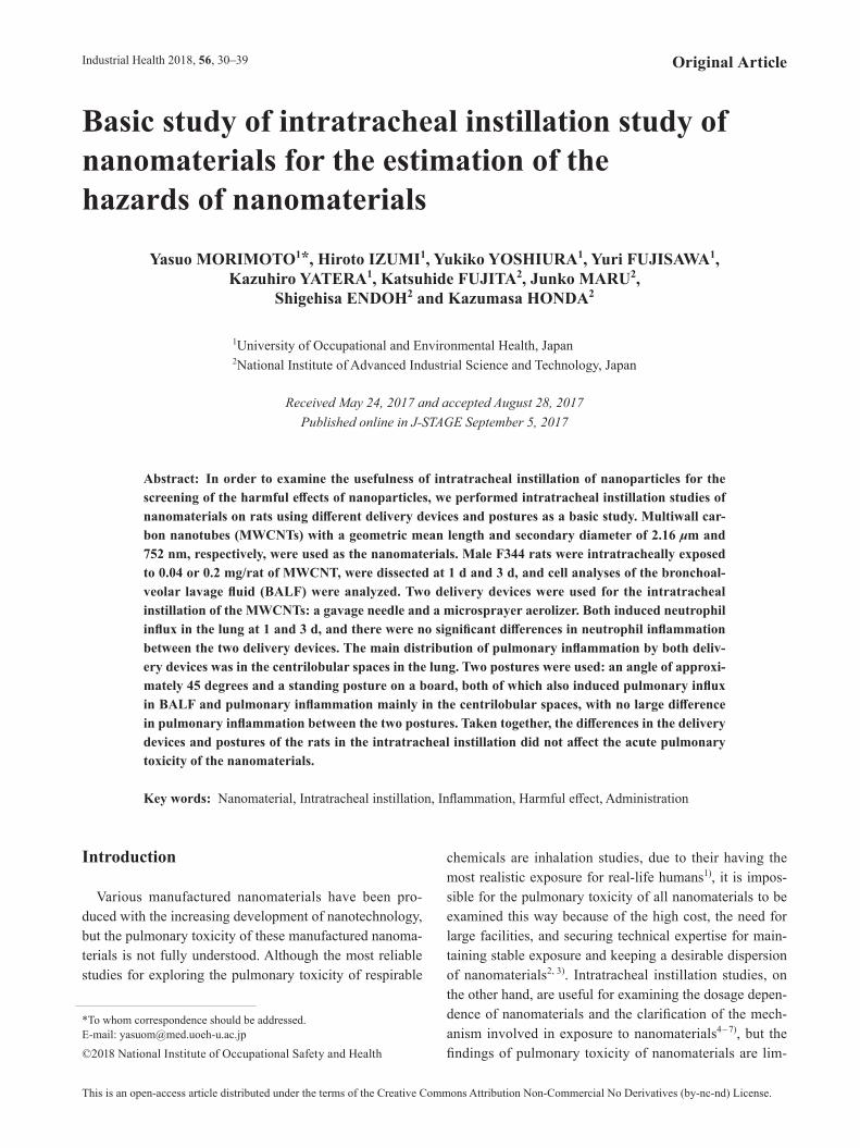
Y MORIMOTO et al.30
Industrial Health 2018, 56, 30–39
*To whom correspondence should be addressed.E-mail: [email protected]
©2018 National Institute of Occupational Safety and Health
Industrial Health 2018, 56, 30–39 Original Article
This is an open-access article distributed under the terms of the Creative Commons Attribution Non-Commercial No Derivatives (by-nc-nd) License.
Introduction
Various manufactured nanomaterials have been pro-duced with the increasing development of nanotechnology, but the pulmonary toxicity of these manufactured nanoma-terials is not fully understood. Although the most reliable studies for exploring the pulmonary toxicity of respirable
Basic study of intratracheal instillation study of nanomaterials for the estimation of the hazards of nanomaterials
Yasuo MORIMOTO1*, Hiroto IZUMI1, Yukiko YOSHIURA1, Yuri FUJISAWA1, Kazuhiro YATERA1, Katsuhide FUJITA2, Junko MARU2,
Shigehisa ENDOH2 and Kazumasa HONDA2
1University of Occupational and Environmental Health, Japan2National Institute of Advanced Industrial Science and Technology, Japan
Received May 24, 2017 and accepted August 28, 2017 Published online in J-STAGE September 5, 2017
Abstract: In order to examine the usefulness of intratracheal instillation of nanoparticles for the screening of the harmful effects of nanoparticles, we performed intratracheal instillation studies of nanomaterials on rats using different delivery devices and postures as a basic study. Multiwall car-bon nanotubes (MWCNTs) with a geometric mean length and secondary diameter of 2.16 μm and 752 nm, respectively, were used as the nanomaterials. Male F344 rats were intratracheally exposed to 0.04 or 0.2 mg/rat of MWCNT, were dissected at 1 d and 3 d, and cell analyses of the bronchoal-veolar lavage fluid (BALF) were analyzed. Two delivery devices were used for the intratracheal instillation of the MWCNTs: a gavage needle and a microsprayer aerolizer. Both induced neutrophil influx in the lung at 1 and 3 d, and there were no significant differences in neutrophil inflammation between the two delivery devices. The main distribution of pulmonary inflammation by both deliv-ery devices was in the centrilobular spaces in the lung. Two postures were used: an angle of approxi-mately 45 degrees and a standing posture on a board, both of which also induced pulmonary influx in BALF and pulmonary inflammation mainly in the centrilobular spaces, with no large difference in pulmonary inflammation between the two postures. Taken together, the differences in the delivery devices and postures of the rats in the intratracheal instillation did not affect the acute pulmonary toxicity of the nanomaterials.
Key words: Nanomaterial,Intratrachealinstillation,Inflammation,Harmfuleffect,Administration
chemicals are inhalation studies, due to their having the most realistic exposure for real-life humans1), it is impos-sible for the pulmonary toxicity of all nanomaterials to be examined this way because of the high cost, the need for large facilities, and securing technical expertise for main-taining stable exposure and keeping a desirable dispersion of nanomaterials2, 3). Intratracheal instillation studies, on the other hand, are useful for examining the dosage depen-denceofnanomaterialsandtheclarificationofthemech-anism involved in exposure to nanomaterials4 – 7), but the findingsofpulmonarytoxicityofnanomaterialsare lim-
BASIC STUDY FOLLOWING INTRATRACHEAL INSTILLATION 31
ited. Intratracheal instillation studies have garnered atten-tion in the research of the pulmonary toxicity of nanomate-rials because of their relatively inexpensive cost and their requirement of relatively simple equipment8).
Comparing inhalation and intratracheal instillation stud-ies for the pulmonary response induced by nanomateri-als, acutepulmonary inflammationdue to aboluseffectis observed following intratracheal instillation 3, 4), unlike inhalation studies. We conducted inhalation and intratra-cheal instillation studies of nanomaterials with high and low toxicity, and found that the pattern of pulmonary rank-ingofnanomaterialsisthesamewhenpulmonaryinflam-mation in not only the acute phase but also in the chronic phase are examined3, 9, 10). There are some other studies in whichtherankingofpulmonaryinflammationbynanoma-terials is the same in inhalation studies and intratracheal instillation studies4, 8, 11), suggesting that intratracheal instillation studies may be useful for ranking the harmful effectsofnanomaterials.
There are not yet enough studies about the methodology of intratracheal instillation. Although there is a report that the results of an interlaboratory evaluation of rodent pul-monary responses to intratracheally exposed nanomaterials were similar, a detailed methodology of the intratracheal instillation studies was not shown12). Even if there are dif-ferent results between laboratories, it cannot be denied that thedifferencesmightberelatedtoadifferenceinthebasictechniques of intratracheal instillation. Therefore, in order to examine whether or not pulmonary toxicity following intratrachealinstillationofnanomaterialsisareflectionofthetechniqueused,weexamineddifferencesinpulmonaryinflammationresultingfromdifferentdeliverydevicesanddifferentpositionsofanimalsinintratrachealinstillations.
Materials and Methods
Preparation of samples of multiwall carbon nanotube (MWCNT) suspensions
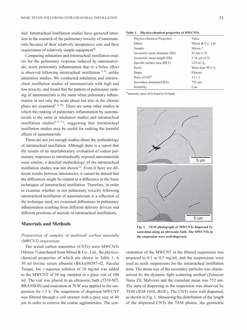
The tested carbon nanotubes (CNTs) were MWCNTs (Mitsui-7) purchased from Mitsui & Co., Ltd., the physico-chemical properties of which are shown in Table 1. A 50 ml bovine serum albumin (BSA)(08587-42, Nacalai Tesque, Inc ) aqueous solution of 10 mg/ml was added to the MWCNT of 50 mg sampled in a glass vial of 100 ml. The vial was placed in an ultrasonic bath (5510-MT, BRANSON) and sonication at 70 W was applied to the sus-pension for 1.5 h. The suspension of dispersed MWCNT wasfilteredthroughacellstrainerwithaporesizeof40μm in order to remove the coarse agglomerates. The con-
centrationoftheMWCNTinthefilteredsuspensionwasprepared to 0.1 or 0.5 mg/ml, and the suspensions were used as stock suspensions for the intratracheal instillation tests.Themeansizeofthesecondaryparticleswascharac-terizedbythedynamiclightscatteringmethod(ZetasizerNanoZS,Malvern)andthecumulantmeanwas752nm.The state of dispersing in the suspension was observed by TEM (JEM-1010, JEOL). The CNTs were well dispersed, as shown in Fig. 1. Measuring the distribution of the length of the dispersed CNTs the TEM photos, the geometric
Table 1. Physico-chemical properties of MWCNTs
Physico-chemical Properties ValueMaker Mitsui & Co., LtdSample Mitsui-7Geometric mean diameter (SD) 52 nm (1.5)Geometric mean length (SD) 2.16 μm (4.2)Specificsurfacearea(BET) 23.0 m2/gPurity More than 99.6 %Shape FibrousRatio of GD* 13 ± 3Secondary diameter(DLS) 752 nmSolubility Low
*Intensity ratio of G-band to D-band
Fig. 1. TEM photograph of MWCNTs dispersed by sonication using an ultrasonic bath. The MWCNTs in the suspension were well-dispersed.
Y MORIMOTO et al.32
Industrial Health 2018, 56, 30–39
mean and the geometric SD were 2.16 μm and 4.2, respec-tively. The geometric mean diameter and the geometric SD were 52 nm and 1.5, respectively.
AnimalsMale Fischer 344 rats (10 wk old) were purchased from
Charles River Laboratories International, Inc. (Japan). The animals were kept in the Laboratory Animal Research Center of the University of Occupational and Environmen-tal Health for two wk with access to free-feeding of com-mercial diet and water. All procedures and animal handling were done in accordance with the guidelines described in
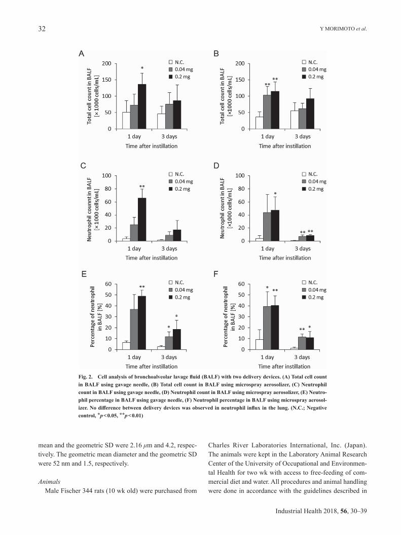
Fig. 2. Cell analysis of bronchoalveolar lavage fluid (BALF) with two delivery devices. (A) Total cell count in BALF using gavage needle, (B) Total cell count in BALF using microspray aerosolizer, (C) Neutrophil count in BALF using gavage needle, (D) Neutrophil count in BALF using microspray aerosolizer, (E) Neutro-phil percentage in BALF using gavage needle, (F) Neutrophil percentage in BALF using microspray aerosol-izer. No difference between delivery devices was observed in neutrophil influx in the lung. (N.C.; Negative control, *p<0.05, **p<0.01)
BASIC STUDY FOLLOWING INTRATRACHEAL INSTILLATION 33
the Japanese Guide for the Care and Use of Laboratory Animals as approved by the Animal Care and Use Com-mittee, University of Occupational and Environmental Health, Japan.
Intratracheal instillation of nanomaterialThe MWCNTs were suspended in 0.4 ml distilled
water, and 0.04 mg (0.16 mg/kg) or 0.2 mg (0.8 mg/kg) of MWCNTs was administered to rats (12 wk old) by a single intratracheal instillation. Negative control groups received distilled water including 10% BSA. Three meth-odsofintratrachealinstillationwereusedafteranesthetiza-tionbyisofluraneinhalation:1)Ratsmaintainedastand-ing posture on a board and were exposed to MWCNTs by a gavage needle inserted into the tracheal lumen; 2) Rats were kept in a supine posture angled approximately 45 degrees to a board, and were exposed to MWCNTs by a microspray aerosolizer inserted into the tracheal lumen;and 3) Rats maintained a standing posture on a board and were exposed toMWCNTsby amicrospray aerosolizerinserted into the tracheal lumen.
Animals (5 rats in each group) were dissected at 1 d and 3 d after the instillation.
There were 5 rats each in the control, low dose, and high dose groups at each time course. The right lungs were inflatedwithtotal20mlphysiologicalsalineunderapres-sure of 20 cm water, and BALF was collected and divided into two to three times. Between 15 and 18 ml of BALF was collected in collection tubes by free fall. The histo-pathological evaluation was performed with the left lung inflatedandfixedby10%formalinsolutionat25cmH2O pressure.
Analysis of inflammatory cells in BALF with cytospinFrom10to13mlofBALFfromthefirstsubgroupswas
centrifuged at 400 g at 4°C for 15 min. The supernatant was transferred to a new tube and used for measuring the cytokines in the BALF. The pellets were washed by sus-pensionwithpolymorphonuclearleukocyte(PMN)Buffer(137.9 mM NaCl, 2.7 mM KCl, 8.2 mM Na2HPO4, 1.5 mM KH2PO4, 5.6 mM C6H12O6) and centrifuged at 400 g at 4°C for 15 min. After the supernatant was removed, the pelletswereresuspendedwith1mlofPMNBuffer.Thecell number in the BALF was counted by Celltac (Nihon Kohden Corp., Tokyo, Japan), and cells were splashed on aslideglassusingcytospin.AfterthecellswerefixedandstainedwithDiff-Quik(SYSMEXCorp.,Hyogo,Japan),the number of neutrophils were counted by microscopic observation.
Parameter in BALFThe concentration of Rat cytokine-induced neutrophil
chemoattractant (CINC) -1 in the BALF was measured by ELISA kits, #RCN100 (R&D Systems, Minneapolis, MN). The concentration of total protein was measured using Pierce TM 660 nm Protein Assay Reagent (22660, Thermo FisherScientificK.K.Yokohama,Japan)bythecolorimet-ric determination method. The concentration of rat albu-minwasmeasuredbyAlbuminRatELISAQuantitationSet (E110-125) and ELISA Starter Accessory Package Kit Ⅰ(E101)(BethylLaboratories,Inc.Montgomery,TX).
HistopathologyThe lung tissue,whichwas inflated and fixedwith a
10% formalin solution under a pressure of 25 cm water, wasembeddedinparaffin,and4μm-thick sections were cut from the lobe, then stained with hematoxylin and eosin.
Statistical analysisAnalysis of variance (ANOVA) and Dunnett’s test were
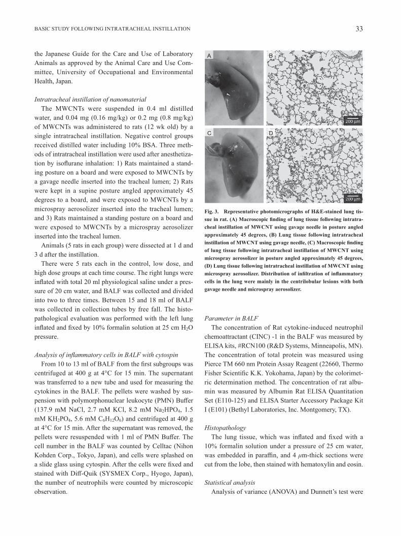
Fig. 3. Representative photomicrographs of H&E-stained lung tis-sue in rat. (A) Macroscopic finding of lung tissue following intratra-cheal instillation of MWCNT using gavage needle in posture angled approximately 45 degrees, (B) Lung tissue following intratracheal instillation of MWCNT using gavage needle, (C) Macroscopic finding of lung tissue following intratracheal instillation of MWCNT using microspray aerosolizer in posture angled approximately 45 degrees, (D) Lung tissue following intratracheal instillation of MWCNT using microspray aerosolizer. Distribution of infiltration of inflammatory cells in the lung were mainly in the centrilobular lesions with both gavage needle and microspray aerosolizer.

Y MORIMOTO et al.34
Industrial Health 2018, 56, 30–39
appliedwhereappropriate todetermineindividualdiffer-ences using a computer statistical package (SPSS, SPSS Inc., Chicago, IL, U.S.A.).
Results
1) Comparison of pulmonary inflammation and delivery devices
Figure 2 shows the total cell count, and the neutrophil count and percentage of neutrophils in BALF in the two
approachesofgavageneedleandmicrosprayeraerosolizer.These three parameters in BALF using both approaches increased somewhat in the 0.04 mg and 0.2 mg/rat at 1 d and3dpostexposure.Therewerenopersistentdifferencesin these data between the two approaches during the obser-vation periods. The level of these parameters at 1 d was higher than that at 3 d, but considering that MWCNTs have a high biopersistence, these phenomenon may be due to the addition of the bolus shot.Bothapproachesshowinfiltrationofinflammatorycells
Fig. 4. Concentration of parameter in BALF with two delivery devices. (A) Concentration of albumin in BALF using gavage needle, (B) Concentration of albumin in BALF using micro-spray aerosolizer, (C) Concentration of protein in BALF using gavage needle, (D) Concentra-tion of protein in BALF using microspray aerosolizer, (E) Concentration of CINC-1 in BALF using gavage needle, (F) Concentration of CINC-1 in BALF using microspray aerosolizer. No difference was observed between delivery devices for these factors. (N.C.; Negative control, *p<0.05, **p<0.01)
BASIC STUDY FOLLOWING INTRATRACHEAL INSTILLATION 35
such as macrophages and neutrophils in the centrilobular spaces, which are neighboring alveolus lesions around the peripheral respiratory tract (Fig. 3). No pathological fea-turessuchasseverefibrosis,emphysematouschanges,orgranulomatous changes were observed.
Figure 4 shows the albumin, protein, and CINC-1 con-centrations in BALF by the two approaches. Albumin, pro-tein, and CINC-1 are makers of alveolar-capillary perme-ability, lung injury and chemokine for neutrophil. Albumin and protein in the BALF was dose-dependently higher in
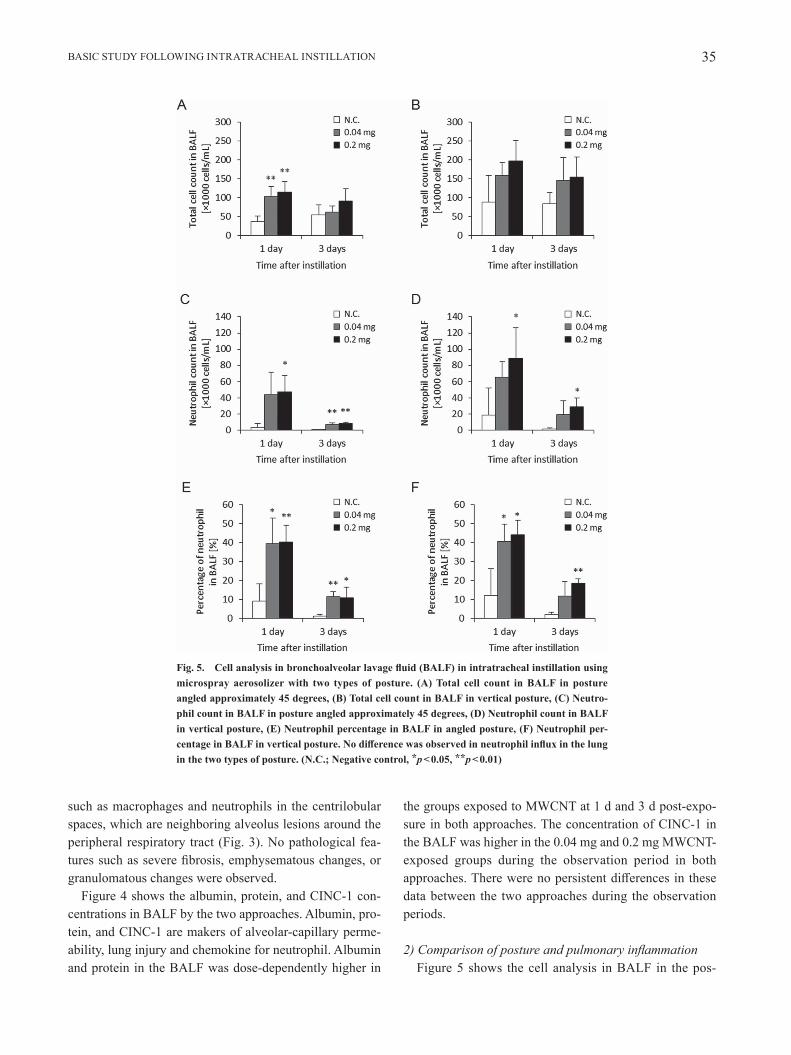
Fig. 5. Cell analysis in bronchoalveolar lavage fluid (BALF) in intratracheal instillation using microspray aerosolizer with two types of posture. (A) Total cell count in BALF in posture angled approximately 45 degrees, (B) Total cell count in BALF in vertical posture, (C) Neutro-phil count in BALF in posture angled approximately 45 degrees, (D) Neutrophil count in BALF in vertical posture, (E) Neutrophil percentage in BALF in angled posture, (F) Neutrophil per-centage in BALF in vertical posture. No difference was observed in neutrophil influx in the lung in the two types of posture. (N.C.; Negative control, *p<0.05, **p<0.01)
the groups exposed to MWCNT at 1 d and 3 d post-expo-sure in both approaches. The concentration of CINC-1 in the BALF was higher in the 0.04 mg and 0.2 mg MWCNT-exposed groups during the observation period in both approaches.Therewerenopersistentdifferencesinthesedata between the two approaches during the observation periods.
2) Comparison of posture and pulmonary inflammationFigure 5 shows the cell analysis in BALF in the pos-
Y MORIMOTO et al.36
Industrial Health 2018, 56, 30–39
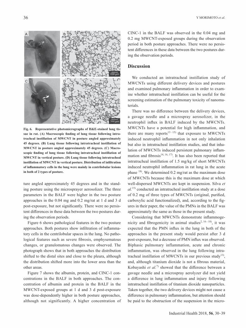
Fig. 6. Representative photomicrographs of H&E-stained lung tis-sue in rat. (A) Macroscopic finding of lung tissue following intra-tracheal instillation of MWCNT in posture angled approximately 45 degrees. (B) Lung tissue following intratracheal instillation of MWCNT in posture angled approximately 45 degrees. (C) Macro-scopic finding of lung tissue following intratracheal instillation of MWCNT in vertical posture. (D) Lung tissue following intratracheal instillation of MWCNT in vertical posture. Distribution of infiltration of inflammatory cells in the lung were mainly in centrilobular lesions in both of 2 types of posture.
ture angled approximately 45 degrees and in the stand-ingpostureusingthemicrosprayeraerosolizer.Thethreeparameters in the BALF were higher in the two posture approaches in the 0.04 mg and 0.2 mg/rat at 1 d and 3 d post-exposure,butnotsignificantly.Therewerenopersis-tentdifferencesinthesedatabetweenthetwoposturesdur-ing the observation periods.
Figure 6 shows pathological features in the two posture approaches.Bothposturesshowinfiltrationofinflamma-tory cells in the centrilobular spaces in the lung. No patho-logical features such as severefibrosis, emphysematouschanges, or granulomatous changes were observed. The photograph shows that in both approaches the distribution shifted to the distal sites and close to the pleura, although the distribution shifted more into the lower area than the other areas.
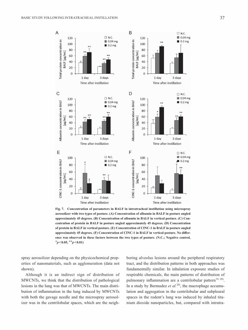
Figure 7 shows the albumin, protein, and CINC-1 con-centrations in the BALF in both approaches. The con-centration of albumin and protein in the BALF in the MWCNT-exposed groups at 1 d and 3 d post-exposure was dose-dependently higher in both posture approaches, although not significantly. A higher concentration of
CINC-1 in the BALF was observed in the 0.04 mg and 0.2 mg MWCNT-exposed groups during the observation period in both posture approaches. There were no persis-tentdifferencesinthesedatabetweenthetwoposturesdur-ing the observation periods.
Discussion
We conducted an intratracheal instillation study of MWCNTsusing different delivery devices and posturesandexaminedpulmonaryinflammationinordertoexam-ine whether intratracheal instillation can be useful for the screening estimation of the pulmonary toxicity of nanoma-terials.Therewasnodifferencebetweenthedeliverydevices,
a gavage needle and a microspray aerosolizer, in theneutrophil influx in BALF induced by the MWCNTs.MWCNTs have a potential for high inflammation, andthere are many reports13 – 15) that exposure to MWCNTs induced neutrophil inflammation in not only inhalationbut also in intratracheal instillation studies, and that inha-lationofMWCNTsinducedpersistentpulmonaryinflam-mationandfibrosis14, 16, 17). It has also been reported that intratracheal instillation of 1.5 mg/kg of short MWCNTs inducedneutrophil inflammation in rat lung in theacutephase 18). We determined 0.2 mg/rat as the maximum dose of MWCNTs because this is the maximum dose at which well-dispersed MWCNTs are kept in suspension. Silva et al.15) conducted an intratracheal instillation study at a dose of0.2mgofthreetypesofMWCNTs(original,purified,carboxylicacidfunctionalized),and,accordingtothefig-ures in their paper, the value of the PMNs in the BALF was approximately the same as those in the present study.Considering thatMWCNTs demonstrate inflammoge-
nicity and fibrogenicity in animal studies2, 16 – 18), it was expected that thePMN influx in the lung inbothof theapproaches in the present study would persist after 3 d post-exposure,butadecreaseofPMNinfluxwasobserved.Biphasic pulmonary inflammation, acute and chronic inflammation,wasobserved in the lung following intra-tracheal instillation of MWCNTs in our previous study13), and,although titaniumdioxide isnotafibrousmaterial,Kobayashi et al.7) showed that thedifferencebetween agavageneedle and amicrospray aerolyzer didnot yielda difference in lung inflammation and injury followingintratracheal instillation of titanium dioxide nanoparticles. Taken together, the two delivery devices might not cause a differenceinpulmonaryinflammation,butattentionshouldbe paid to the obstruction of the suspension in the micro-
BASIC STUDY FOLLOWING INTRATRACHEAL INSTILLATION 37
Fig. 7. Concentration of parameters in BALF in intratracheal instillation using microspray aerosolizer with two types of posture. (A) Concentration of albumin in BALF in posture angled approximately 45 degrees. (B) Concentration of albumin in BALF in vertical posture. (C) Con-centration of protein in BALF in posture angled approximately 45 degrees. (D) Concentration of protein in BALF in vertical posture. (E) Concentration of CINC-1 in BALF in posture angled approximately 45 degrees. (F) Concentration of CINC-1 in BALF in vertical posture. No differ-ence was observed in these factors between the two types of posture. (N.C.; Negative control, *p<0.05, **p<0.01)
sprayaerosolizerdependingonthephysicochemicalprop-erties of nanomaterials, such as agglomeration (data not shown).
Although it is an indirect sign of distribution of MWCNTs, we think that the distribution of pathological lesions in the lung was that of MWCNTs. The main distri-butionofinflammationinthelunginducedbyMWCNTswith both the gavage needle and the microspray aerosol-izerwasinthecentrilobularspaces,whicharetheneigh-
boring alveolus lesions around the peripheral respiratory tract, and the distribution patterns in both approaches was fundamentally similar. In inhalation exposure studies of respirable chemicals, the main patterns of distribution of pulmonary inflammationareacentrilobularpattern19, 20). InastudybyBermudezet al.20), the macrophage accumu-lation and aggregation in the centrilobular and subpleural spaces in the rodent’s lung was induced by inhaled tita-nium dioxide nanoparticles, but, compared with intratra-
Y MORIMOTO et al.38
Industrial Health 2018, 56, 30–39
cheal instillation, some inflammatory lesions followinginhalation exposure were present deep in the alveolar wall andthesubpleuralspace.Therearenostudiesontheeffectof inhaled nanoparticles on human lung, but there are some studies of exposure to welding fumes that include metal oxide nanoparticles21, 22). The distribution of opacity in the computed tomography image in patients with welding lung was compatible with that of centrilobular nodules, mean-ing that the deposition of nanoparticles from the welding fumes may be located in the centrilobular area21, 22).Althoughtherewaslittledifferenceinpathologicaldis-
tributions between the gavage needle and the microspray aerosolizer,thedistributionofinflammationinthegavageneedle type shifted slightly to the proximal site, and no differencewasobservedbetweenlobesinthedistributionofinflammation.Withthemicrosprayaerosolizer,ontheother hand, the distribution shifted to the distal sites and closetothepleura,althoughthedistributionofinflamma-tory changes shifted more into the lower lobes than the other lobes. We speculated that the strong pressure of the microsprayaerosolizermayhaveinducedthelobulardif-ferenceininflammatorychanges.Fujitaet al.18) reported that MWCNTs following intratracheal instillation by a microsprayaerosolizerpenetratedintothepleura,andXuet al.23) reported that MWCNTs were observed in the pleu-ral cavity in the acute phase following intratracheal instil-lationofMWCNTswithamicrosprayaerosolizer.
Intratracheal instillation of MWCNTs in the posture angled approximately 45 degrees and in the standing pos-tureinducedneutrophilinfluxinBALF,butthedifferenceinposturedidnotchangethedegreeofneutrophil influx.Themaindistributionofinflammationinthelunginducedby intratracheal instillation of MWCNTs in the 45 degree angled posture and in the standing posture was in the cen-trilobularspaces,buttherewasnodifferenceinpulmonaryinflammationintherat lunginthepathologicalfindings.Takentogether,adifferenceofpostureatthislevelcanbeignored when we estimate the pulmonary toxicity of nano-materials. Hasegawa-Baba et al.24) reported that the distri-bution of india ink in lung in the 45 degree angled posture were more dispersable than that in the standing posture in intratrachealinstillationstudies.Thereareadifferentten-dency of distribution of materials between our and their studies,suggesting that thedifference inviscosity liquidbetween india ink and MWCNT suspensions may connect withdifferenceofdistributioninthelung.Theremaybedifferencebetween lobes in theamount
of deposited nanomaterials using either posture for the intratracheal instillation of nanomaterials. Costa et al.25)
performed an intratracheal instillation of oil combustion particles and found greater amounts of deposition in the inferior lobe than in the superior lobe. Although we did not measure the deposition of MWCNTs in the present study, we can speculate this tendency for downward deposition from the photography of the lung after using the micro-sprayaerolizer.Costaet al.25) also performed an inhalation study of oil combustion particles and found a predomi-nance of downward deposition. These results indicate that thedifferenceinposturecanbeignored.
In summary, we performed an intratracheal instilla-tion study ofMWCNTs for rat using different cannulasandposturesasabasicstudy.Thedifference indeliverydevices(gavageneedleandmicrosprayeraerosolizer)didnot result in a change of level and distribution of pulmo-naryinflammationinrat lung,andneitherdidthediffer-ence in posture. Taken together, intratracheal instillation studies can assess the acute pulmonary toxicity of nanoma-terials within this condition.
Acknowledgment
This study is based on results obtained from a project commissioned by the New Energy and Industrial Technol-ogyDevelopmentOrganization(NEDO),Japan
References
1) Oberdörster G, Castranova V, Asgharian B, Sayre P (2015) Inhalation Exposure to Carbon Nanotubes (CNT) and Car-bon Nanofibers (CNF): Methodology and Dosimetry. JToxicol Environ Health B Crit Rev 18, 121–212.
2) MorimotoY,HorieM,KobayashiN,ShinoharaN,ShimadaM (2013) Inhalation toxicity assessment of carbon-based nanoparticles. Acc Chem Res 46, 770–81.
3) MorimotoY,IzumiH,YoshiuraY,TomonagaT,LeeBW,OkadaT,OyabuT,MyojoT,KawaiK,YateraK,ShimadaM,KuboM,YamamotoK,KitajimaS,KurodaE,HorieM,Kawaguchi K, Sasaki T (2016) Comparison of pulmonary inflammatoryresponsesfollowingintratrachealinstillationand inhalation of nanoparticles. Nanotoxicology 10, 607 –18.
4) Baisch BL, Corson NM, Wade-Mercer P, Gelein R, Kennell AJ, Oberdörster G, Elder A (2014) Equivalent titanium dioxide nanoparticle deposition by intratracheal instillation andwholebodyinhalation:theeffectofdoserateonacuterespiratorytractinflammation.PartFibreToxicol11, 5.
5) Prodan AM, Ciobanu CS, Popa CL, Iconaru SL, Predoi D (2014) Toxicity evaluation following intratracheal instilla-tion of iron oxide in a silica matrix in rats. BioMed Res Int 2014, 134260.
6) YoshiuraY, IzumiH,OyabuT,HashibaM,KambaraT,

BASIC STUDY FOLLOWING INTRATRACHEAL INSTILLATION 39
MizuguchiY,LeeBW,OkadaT,TomonagaT,MyojoT,YamamotoK,KitajimaS,HorieM,KurodaE,MorimotoY(2015) Pulmonary toxicity of well-dispersed titanium diox-ide nanoparticles following intratracheal instillation. J Nanopart Res 17, 241.
7) KobayashiT,OshimaY,TsubokuraY,HashizumeN,AjimiS, Kayashima T, Nakai M, Sasaki T, Kawaguchi K, ImatanakaN(2016)Effectsofdosevolumeanddeliverydevice on bronchoalveolar lavage parameters of intratrache-allyadministerednano-sizedTiO2 in rats.RegulToxicolPharmacol 81, 233–41.
8) MorimotoY,IzumiH,YoshiuraY,FujishimaK,YateraK,YamamotoK(2016)UsefulnessofIntratrachealInstillationStudies for Estimating Nanoparticle-Induced Pulmonary Toxicity. Int J Mol Sci 17, 165.
9) MorimotoY,IzumiH,YoshiuraY,TomonagaT,OyabuT,Myojo T, Kawai K, Yatera K, Shimada M, Kubo M,YamamotoK,KitajimaS,KurodaE,KawaguchiK,SasakiT(2016)EvaluationofPulmonaryToxicityofZincOxideNanoparticles Following Inhalation and Intratracheal Instil-lation. Int J Mol Sci 17, 1241.
10) OyabuT,MorimotoY,HirohashiM,HorieM,KambaraT,LeeBW,HashibaM,MizuguchiY,MyojoT,Kuroda E(2013) Dose-dependent pulmonary response of well-dispersed titanium dioxide nanoparticles following intratra-cheal instillation. J Nanopart Res 15, 1600.
11) MorimotoY, IzumiH,YoshiuraY, FujisawaY, FujitaK(2017)Significanceofintratrachealinstillationtestsforthescreeningofharmfuleffectsofnanomaterials.JUOEH39, 123–32.
12) Bonner JC, Silva RM, Taylor AJ, Brown JM, Hilderbrand SC, Castranova V, Porter D, Elder A, Oberdörster G, Harkema JR, Bramble LA, Kavanagh TJ, Botta D, Nel A, Pinkerton KE (2013) Interlaboratory evaluation of rodent pulmonary responses to engineered nanomaterials: the NIEHS Nano GO Consortium. Environ Health Perspect 121, 676–82.
13) MorimotoY,HirohashiM,OgamiA,OyabuT,MyojoT,TodorokiM,YamamotoM,HashibaM,MizuguchiY,LeeBW, Kuroda E, Shimada M,WangWN, Yamamoto K,FujitaK, Endoh S,UchidaK,KobayashiN,MizunoK,InadaM,TaoH,NakazatoT,NakanishiJ,TanakaI(2012)Pulmonary toxicity of well-dispersed multi-wall carbon nanotubes following inhalation and intratracheal instilla-tion. Nanotoxicology 6, 587–99.
14) Shvedova AA, Kisin E, Murray AR, Johnson VJ, Gorelik O, Arepalli S, Hubbs AF, Mercer RR, Keohavong P, Sussman N, Jin J,Yin J,StoneS,ChenBT,DeyeG,MaynardA,Castranova V, Baron PA, Kagan VE (2008) Inhalation vs. aspiration of single-walled carbon nanotubes in C57BL/6
mice:inflammation,fibrosis,oxidativestress,andmutagen-esis. Am J Physiol Lung Cell Mol Physiol 295, L552–65.
15) SilvaRM,DoudrickK,FranziLM,TeeSyC,AndersonDS,WuZ,MitraS,VuV,DutrowG,EvansJE,WesterhoffP,Van Winkle LS, Raabe OG, Pinkerton KE (2014) Instilla-tion versus inhalation of multiwalled carbon nanotubes: exposure-relatedhealtheffects,clearance,and the roleofparticle characteristics. ACS Nano 8, 8911–31.
16) Pauluhn J (2010) Subchronic 13-week inhalation exposure of rats tomultiwalled carbonnanotubes: toxic effects aredeterminedbydensityofagglomeratestructures,notfibrillar structures. Toxicol Sci 113, 226–42.
17) Ma-HockL,TreumannS,StraussV,BrillS,LuiziF,MertlerM,WienchK,GamerAO,vanRavenzwaayB,LandsiedelR (2009) Inhalation toxicity of multiwall carbon nanotubes in rats exposed for 3 months. Toxicol Sci 112, 468–81.
18) Fujita K, Fukuda M, Endoh S, Maru J, Kato H, Nakamura A, Shinohara N, Uchino K, Honda K (2016) Pulmonary and pleuralinflammationafterintratrachealinstillationofshortsingle-walled and multi-walled carbon nanotubes. Toxicol Lett 257, 23–37.
19) BermudezE,MangumJB,AsgharianB,WongBA,ReverdyEE,JanszenDB,HextPM,WarheitDB,EverittJI(2002)Long-term pulmonary responses of three laboratory rodent species to subchronic inhalation of pigmentary titanium dioxide particles. Toxicol Sci 70, 86–97.
20) BermudezE,MangumJB,WongBA,AsgharianB,HextPM, Warheit DB, Everitt JI (2004) Pulmonary responses of mice, rats, and hamsters to subchronic inhalation of ultra-finetitaniumdioxideparticles.ToxicolSci77, 347–57.
21) Akira M (1995) Uncommon pneumoconioses: CT and pathologicfindings.Radiology197, 403–9.
22) Chong S, Lee KS, Chung MJ, Han J, Kwon OJ, Kim TS (2006) Pneumoconiosis: comparison of imaging and patho-logicfindings.Radiographics26, 59–77.
23) XuJ,FutakuchiM,ShimizuH,AlexanderDB,YanagiharaK,FukamachiK,SuzuiM,KannoJ,HiroseA,OgataA,SakamotoY,NakaeD,OmoriT,TsudaH (2012)Multi-walled carbon nanotubes translocate into the pleural cavity and induce visceral mesothelial proliferation in rats. Cancer Sci 103, 2045–50.
24) Hasegawa-Baba Y, Kubota H, Takata A, Miyagawa M(2014) Intratracheal instillation methods and the distribu-tion of administered material in the lung of the rat. J Toxicol Pathol 27, 197–204.
25) Costa DL, Lehmann JR, Winsett D, Richards J, Ledbetter AD, Dreher KL (2006) Comparative pulmonary toxicologi-cal assessment of oil combustion particles following inhala-tion or instillation exposure. Toxicol Sci 91, 237–46.