Embed Size (px)
Citation preview

Intratracheal Seal Disc: A Novel Tracheostoma Closure Device
Karen J Christiansen RN, Niels Moeslund, Henrik Lauridsen PhD, Louise Devantier MD,Marianne C Rohde PhD, Benedict Kjærgaard MD, and Michael Pedersen PhD
BACKGROUND: Tracheostomy decannulation is accompanied by several clinical concerns due toair leakage. In this study, we introduced a novel tracheostoma closure device that facilitates the useof noninvasive ventilation, improvement of pulmonary function, and vocalization in the newlydecannulated patient. The biosafety and feasibility of the device were evaluated in an animal model.METHODS: Five Danish Landrace pigs were subjected to tracheostomy followed by decannulationand insertion of the tracheostoma closure device. Correct placement of the device was ensured byflexible tracheoscopy. The device consisted of an intratracheal silicone seal disc fixated by a cordthrough the stoma to an external part. At day 14, computed tomography (CT) was performed before thedevice was extracted. With the pulling of a cord, the disc unraveled into a thin thread and was extractedthrough the stoma. At day 21, CT was repeated before euthanasia. The trachea and epidermis wereexcised en bloc for histopathological evaluation. RESULTS: Insertion and correct placement of the discwas unproblematic in all animals. CT at day 14 confirmed a clear airway, appropriate placement of thedisc, and full closure of the tracheostoma. Extraction was successful in one animal but complicated inthe remaining animals. There was histological evidence of healing after the foreign body placement.CONCLUSIONS: The study demonstrated that the tracheostoma closure device is feasible and biosafein a porcine animal model, but the design and quality of the materials need to be improved beforeclinical trials. Key words: tracheostomy; decannulation; computed tomography; histology; animal model;device. [Respir Care 2017;62(7):970–977. © 2017 Daedalus Enterprises]
Introduction
Tracheotomy is a common procedure, and the numberof patients undergoing temporary tracheostomy is increas-
ing due to aging populations and comorbidity.1-3 Aninternational survey reported that 24% of mechanicallyventilated patients in intensive care units were venti-lated through a tracheostomy tube.4 Tracheostomy de-cannulation is required once the underlying indicationfor the tracheostomy has resolved.5-9 Removal of thetracheostomy tube leaves an artificial hole in the pa-tient’s airway, which is covered with occlusive dressingor gauze bandage. Patients are instructed to apply pres-sure over the dressing with their fingers when talking orcoughing to reduce air leakage.
Ms Christiansen, Mr Moeslund, Dr Lauridsen, Dr Devantier, and DrPedersen are affiliated with the Comparative Medicine Laboratory, De-partment of Clinical Medicine, and Dr Rohde is affiliated with the De-partment of Forensic Medicine, Aarhus University, Aarhus, Denmark.Ms Christiansen is also affiliated with the Department of Cardiology,Aarhus University Hospital, Aarhus, Denmark. Dr Devantier is affiliatedwith the Department of Otorhinolaryngology, Head and Neck Surgery,Aarhus University Hospital, Aarhus, Denmark. Dr Rohde is affiliatedwith the Department of Forensic Medicine, Aarhus University, Aarhus,Denmark. Dr Kjærgaard is affiliated with the Department of ClinicalMedicine, Aarhus University, Aarhus, Denmark and the Department ofThoracic Surgery, Aalborg University Hospital, Aalborg, Denmark.
Ms Christiansen and Mr Moeslund are co-first authors.
This work was supported by the A.P. Møller og Hustru Chastine Mc-Kinney Møllers Fond til almene Formaal. Ms Christiansen has disclosedan international patent application, number PCT/EP2015/064892. Theother authors have disclosed no conflicts of interest.
Supplementary material related to this paper is available at http://www.rcjournal.com.
Correspondence: Karen J Christiansen RN, Comparative Medicine Lab-oratory, Department of Clinical Medicine, Aarhus University, PalleJuul-Jensens Boulevard 99, 8200 Aarhus N, Denmark. E-mail:[email protected].
DOI: 10.4187/respcare.05301
970 RESPIRATORY CARE • JULY 2017 VOL 62 NO 7

Air leakage through the tracheostomy compromises vo-calization, throat clearing, deep sighs, and cough compe-tence.10,11 Tracheostomy dressing tends to loosen due toair leakage, and frequent changes are indicated when ex-cessive amounts of secretion pool from the tracheostoma.Secretions and the continuous flow of air may delay andeven hinder proper closure of the tracheostoma. In somepatients delayed closure of the tracheostoma induces de-velopment of a tracheocutaneous fistula requiring surgicalclosure. We believe that intratracheal sealing of a trache-ostomy will optimize lung function and promote woundhealing in the newly decannulated tracheostomy patient.
This study evaluated the feasibility of intratracheal seal-ing using a novel device, and the purposes were: (1) toevaluate whether an intratracheal closure device could beinserted via a tracheostomy tube; (2) to evaluate any acuteadverse effects due to the foreign material in the trachea;(3) to investigate the tension mechanism, design, and ma-terial; and (4) to evaluate its impact on trachea and pre-tracheal tissue.
Methods
Study Design
Five Danish Landrace pigs (�60 kg) were used in thisstudy. The animals underwent treatment with the sealingdevice for 2 weeks and were subsequently observed for 1week (Fig. 1).
Device Design
The device was fabricated in 2 parts: an intratrachealclosing device consisting of a silicone seal disc and anextratracheal fastening device consisting of an outer ten-sion and fastening housing. The silicone disc had a diam-eter of 26.5 mm and was cut in a spiral pattern to provokea spiral line of weakness for extraction. The silicone com-position of the disc permitted folding on insertion throughthe tracheostomy tube, and subsequent unfolding on entryinto the intratracheal space. Retraction of the device andtracheostomy tube formed a secure internal seal. Tensionwas maintained between the internal disc and the externalcover via a spring tethering system. This allowed secureplacement while allowing removal in the event of emer-gency. A peel thread was attached to the outer end of the
spiral and passed through the center to enable peeling ofthe disc to a cord. A fastening thread was attached atseveral points within the disc in a pattern not interferingwith the peeling (Fig. 2). The thread was connected to aspring in the outer housing for sufficient and continuoustension.
The outer housing consisted of 2 parts: a plate withtethering points for the peeling and the tension threads anda tension spring and a hole lined with polytetrafluoroeth-ylene to minimize friction where the threads passed throughthe hole. A lid covered the internal housing parts (Fig. 3).
A spring tethering system was used for safety and com-fort to ensure that the seal remained in position duringbody movement or during slight elevation of the housingpart for veterinary wound inspection and care. The springwas adjusted so that the force exerted by the seal discwould not exceed 25 mm Hg.12
The lengths of the threads in the outer housing wasadjusted to allow the housing to be lifted approximately2 cm off the skin without tensioning the peeling thread.The seal disc was designed for extraction through the tra-cheostoma canal if the housing was subjected to extensiveforce.
Animal Handling
The experimental procedures were in accordance withnational Danish legislation on the care and use of labora-tory animals. The study was approved by the Danish An-imal Experiment Inspectorate (approval 2014-15-0201-00265).
We used a porcine animal model because pigs’ trachealdimensions are comparable with those of adult humans.
QUICK LOOK
Current knowledge
Tracheostomy decannulation is an important step inrehabilitating the critically ill patient toward indepen-dent breathing. After decannulation, wound healing isimpaired by a variable flow of air and by secretionsuntil the canal has closed. Decannulation is associatedwith a risk of respiratory failure.
What this paper contributes to our knowledge
Treatment with an intratracheal seal disc was feasible inan animal model. The device was inserted through apercutaneous dilatational tracheostomy and fixated by aflexible tensioning system. All animals tolerated thedevice showing no signs of airway obstruction and thetracheas were intact at the end of the study.
Fig. 1. Timing of study. CT � computed tomography.
INTRATRACHEAL SEAL DISC IN A PORCINE MODEL
RESPIRATORY CARE • JULY 2017 VOL 62 NO 7 971
The animals were anesthetized with repeated intramuscu-lar injections of Zoletil Vet 50, a mixture of ketamine(6.25 mg/mL), tiletamine (6.25 mg/mL), benzodiazepine(6.25 mg/mL), synthetic opioid (butorphanol) (1.25 mg/mL),and xylazin (6.5 mg/mL). After the invasive procedure,the animal received an intramuscular injection of flu-nixin (50 mg/mL, 2.5 mL) and buprenorphin (0.3 mg/mL,3 mL) at 8-h intervals for 3 d.
The trachea was intubated with a cuffed 8-mm internaldiameter tube, and the lungs were ventilated mechanicallyusing a ventilator (S5 Advance, Datex-Ohmeda S5, Hel-sinki, Finland) with an 8–10 mL/kg tidal volume and aPEEP of 5 cm H2O. The frequency and tidal volume werecontinuously adjusted to maintain PaCO2
between 33 and42 mm Hg. The FIO2
was set to 60%. Before the invasiveprocedure, a long-acting antibiotic, Curamox (1 g), wasadministered intramuscularly. A percutaneous tracheos-tomy introducer set (Ciaglia Blue Rhino tracheostomy in-
troducer set, Cook Medical, Bloomington, Indiana) wasused, and an 8-mm inner diameter tracheostomy tube withan outer diameter of 11.9 mm (Portex Blue Line Ultra,Smith Medical, Kent, United Kingdom) was introducedinto the trachea under bronchoscopy guidance (aScope 3,AMBU, Copenhagen, Denmark). The oral tube was with-drawn to a level just beneath the vocal cords, and venti-lation was performed using the tracheostomy tube when-ever indicated. After 15 min, the tracheostomy tube wasused to insert the intratracheal seal disc into the trachea,the tracheostomy tube was removed, and the cord attachedto the disc was connected to the external device with anautomatic tensioner. The extratracheal fastening device wassutured to the skin, and the neck was dressed with circularself-adhesive bandages applied without tension. The posi-tion of the internal disc was controlled by tracheoscopy(see supplemental video at www.rcjournal.com). Thetranslaryngeal tracheal tube was removed after weaningfrom anesthesia, and the pigs were transferred to a stablefacility, where veterinary care and observation were man-aged by trained animal technicians. Feeding troughs werechanged to flat bowls to minimize mechanical force to-ward the front neck.
After 2 weeks, the animal was reanesthetized to a levelinducing sleep with preserved ventilation. Computed to-mography (CT) (time point: seal in place) of trachea wasperformed to determine the position of the disc. The discwas then peeled out (0 d post-extraction). The pig wasobserved for another week in the animal facility. Next, theanimal was reanesthetized, followed by another CT exam-ination (7 d post-extraction). Finally, the animal was sac-rificed, and histological specimens were obtained for anal-ysis.
CT
CT was employed to qualitatively evaluate device place-ment and to quantify any tracheal stenosis or dilation dur-ing and after treatment using a clinical system (SiemensSomatom Definition, Siemens Medical Solutions, Forch-heim, Germany) with the following parameters: field-of-view from snout to diaphragm, 0.6 � 0.6 � 0.6 mm3 voxelsize; 120 kVp; 200 mA. This resulted in an acquisitiontime of 90 s. The built-in (Syngo CT 2012B software)B45s convolution kernel was used to reconstruct the CTimages. The subsequent analysis was performed with theAmira software (FEI, Hillsboro, Oregon).
Histology
Areas around the tracheostomy and the seal were re-moved postmortem en bloc and formalin-fixed immedi-ately after removal, and multiple cross sections in the areaof the tracheostomy were made with a 5-mm interval,
Fig. 2. The silicone seal with a predetermined line of weakness.Two different threads are attached for tensioning and unravelingfunction, respectively.
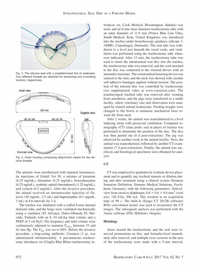
Fig. 3. Outer housing comprising attachment means for the sili-cone threads.
INTRATRACHEAL SEAL DISC IN A PORCINE MODEL
972 RESPIRATORY CARE • JULY 2017 VOL 62 NO 7
allowing inspection for inflammation, necrosis, and for-eign bodies. For microscopic evaluation, cross sectionswere sampled from the superficial, middle, and profoundareas (slice thickness 3–3.5 �m). The following stainswere used in the microscopic evaluation of the tissue:hematoxylin and eosin, routine stain for tissue and cellevaluation; elastic van Gieson for elastic fibers; and Mas-son’s trichrome, a 3-color staining suited for distinguish-ing cells from the surrounding connective tissue. Further-more, Perl’s’ Prussian blue was used for ferric iron depositsin tissue.
Results
Animal Behavior and Device Management
Insertion and intratracheal placement of the seal throughthe 8-mm inner diameter tube was successful. The animalsshowed no signs of airway obstruction during the 3 weeks
of observation; in one animal, 2 episodes of transitorycough were reported. The animals showed no signs of painor disability assessed from normal behavior and appetite.The tension between the seal and fastening device wassufficient to keep the seal in place. In one animal, thesealing disc was completely extracted during the removalprocedure, and in 4 animals, the disc broke during thepeeling procedure and left small pieces of silicone in thepretracheal tissue (see the supplementary materials at http://www.rcjournal.com). One disc could not be extractedthrough the tracheostoma canal and was removed endo-scopically.
CT
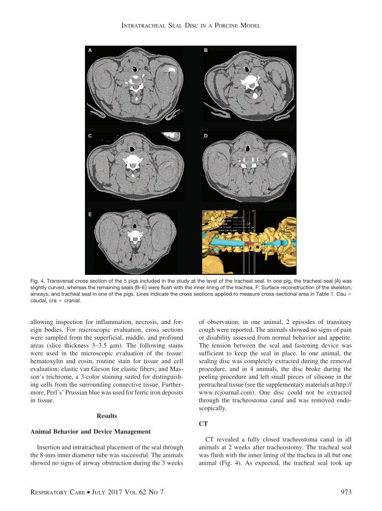
CT revealed a fully closed tracheostoma canal in allanimals at 2 weeks after tracheostomy. The tracheal sealwas flush with the inner lining of the trachea in all but oneanimal (Fig. 4). As expected, the tracheal seal took up
Fig. 4. Transversal cross section of the 5 pigs included in the study at the level of the tracheal seal. In one pig, the tracheal seal (A) wasslightly curved, whereas the remaining seals (B–E) were flush with the inner lining of the trachea. F: Surface reconstruction of the skeleton,airways, and tracheal seal in one of the pigs. Lines indicate the cross sections applied to measure cross-sectional area in Table 1. Cau �caudal, cra � cranial.
INTRATRACHEAL SEAL DISC IN A PORCINE MODEL
RESPIRATORY CARE • JULY 2017 VOL 62 NO 7 973
some luminal space in the trachea, which reduced the tra-cheal lumen. However, no signs of stenosis or dilation oftrachea were observed (Table 1 and see the supplementaryfigure at http://www.rcjournal.com).
Histology
After removal of the intact seal, abnormal macroscopicfindings were subtle and almost absent (Fig. 5). Micros-copy revealed evidence of healing after foreign body place-ment, including inflammatory cells, giant cells, and fibro-blasts. The affected area was around 1–2 mm in diameter
in the case with complete removal where the seal did notbreak off (Fig. 6). The seal broke during extraction in 4 ofthe pigs. These cases showed abundant signs of inflam-mation and acute inflammation with neutrophil granulo-cytes, presumably caused by reactions secondarily to the
Table 1. Cross-Sectional Area of Tracheal Lumen at 5 Points Associated With the Tracheal Seal
Pig Number Time PointMid-Seal,
CSA (mm2)Cra, CSA (mm2)
3 mm Cra,CSA (mm2)
Cau, CSA (mm2)3 mm Cau,CSA (mm2)
1 Seal in place 84.1 135.8 148.8 83.4 107.11 0 dpe 99.0 178.0 175.3 100.7 140.41 7 dpe 152.7 212.7 221.8 174.9 174.32 Seal in place 82.1 126.2 141.2 140.0 167.42 0 dpe 114.2 112.5 109.3 152.9 170.62 7 dpe 196.3 186.9 181.8 192.4 193.03 Seal in place 114.9 148.9 149.5 135.0 186.13 0 dpe NA NA NA NA NA3 7 dpe 172.2 129.3 100.5 192.9 196.64 Seal in place 122.4 114.8 133.2 180.9 191.84 0 dpe NA NA NA NA NA4 7 dpe 205.5 206.4 208.6 209.5 208.65 Seal in place 107.3 198.2 204.8 99.8 142.75 0 dpe NA NA NA NA NA5 7 dpe 193.9 229.0 242.8 184.0 180.2Mean � SD Seal in place 102.2 � 16.3 144.8 � 29.0 155.5 � 25.3 127.8 � 34.0 159.0 � 31.1Mean � SD 0 dpe 106.6 � 7.6 145.2 � 32.8 142.3 � 33.0 126.8 � 26.1 155.5 � 15.1Mean � SD 7 dpe 184.1 � 19.1 192.9 � 34.5 191.1 � 49.4 190.7 � 11.5 190.5 � 12.2
CSA � cross-sectional areadpe � days post-extractionCra � cranial end of seal3 mm Cra � 3 mm cranial to sealCau � caudal end of seal3 mm Cau � 3 mm caudal to sealNA � not applicable
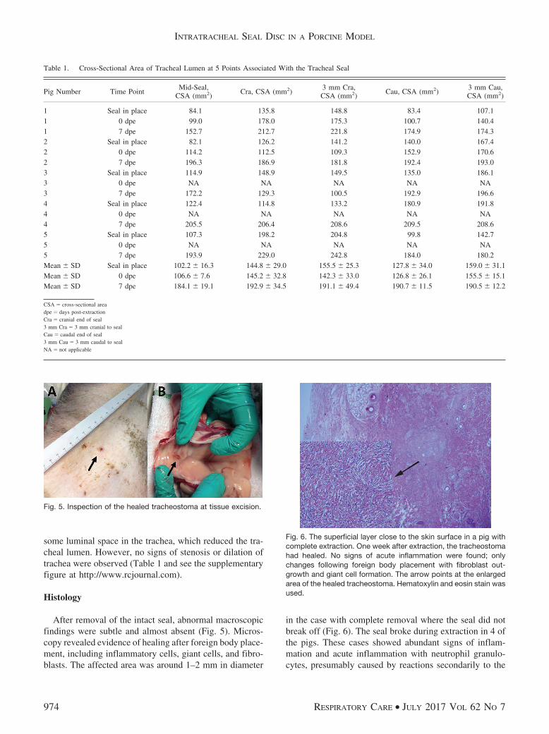
Fig. 5. Inspection of the healed tracheostoma at tissue excision.
Fig. 6. The superficial layer close to the skin surface in a pig withcomplete extraction. One week after extraction, the tracheostomahad healed. No signs of acute inflammation were found; onlychanges following foreign body placement with fibroblast out-growth and giant cell formation. The arrow points at the enlargedarea of the healed tracheostoma. Hematoxylin and eosin stain wasused.
INTRATRACHEAL SEAL DISC IN A PORCINE MODEL
974 RESPIRATORY CARE • JULY 2017 VOL 62 NO 7
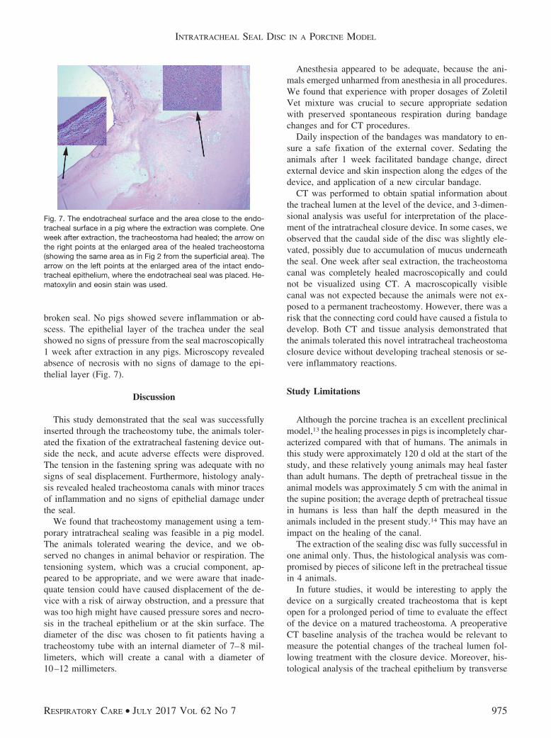
broken seal. No pigs showed severe inflammation or ab-scess. The epithelial layer of the trachea under the sealshowed no signs of pressure from the seal macroscopically1 week after extraction in any pigs. Microscopy revealedabsence of necrosis with no signs of damage to the epi-thelial layer (Fig. 7).
Discussion
This study demonstrated that the seal was successfullyinserted through the tracheostomy tube, the animals toler-ated the fixation of the extratracheal fastening device out-side the neck, and acute adverse effects were disproved.The tension in the fastening spring was adequate with nosigns of seal displacement. Furthermore, histology analy-sis revealed healed tracheostoma canals with minor tracesof inflammation and no signs of epithelial damage underthe seal.
We found that tracheostomy management using a tem-porary intratracheal sealing was feasible in a pig model.The animals tolerated wearing the device, and we ob-served no changes in animal behavior or respiration. Thetensioning system, which was a crucial component, ap-peared to be appropriate, and we were aware that inade-quate tension could have caused displacement of the de-vice with a risk of airway obstruction, and a pressure thatwas too high might have caused pressure sores and necro-sis in the tracheal epithelium or at the skin surface. Thediameter of the disc was chosen to fit patients having atracheostomy tube with an internal diameter of 7–8 mil-limeters, which will create a canal with a diameter of10–12 millimeters.
Anesthesia appeared to be adequate, because the ani-mals emerged unharmed from anesthesia in all procedures.We found that experience with proper dosages of ZoletilVet mixture was crucial to secure appropriate sedationwith preserved spontaneous respiration during bandagechanges and for CT procedures.
Daily inspection of the bandages was mandatory to en-sure a safe fixation of the external cover. Sedating theanimals after 1 week facilitated bandage change, directexternal device and skin inspection along the edges of thedevice, and application of a new circular bandage.
CT was performed to obtain spatial information aboutthe tracheal lumen at the level of the device, and 3-dimen-sional analysis was useful for interpretation of the place-ment of the intratracheal closure device. In some cases, weobserved that the caudal side of the disc was slightly ele-vated, possibly due to accumulation of mucus underneaththe seal. One week after seal extraction, the tracheostomacanal was completely healed macroscopically and couldnot be visualized using CT. A macroscopically visiblecanal was not expected because the animals were not ex-posed to a permanent tracheostomy. However, there was arisk that the connecting cord could have caused a fistula todevelop. Both CT and tissue analysis demonstrated thatthe animals tolerated this novel intratracheal tracheostomaclosure device without developing tracheal stenosis or se-vere inflammatory reactions.
Study Limitations
Although the porcine trachea is an excellent preclinicalmodel,13 the healing processes in pigs is incompletely char-acterized compared with that of humans. The animals inthis study were approximately 120 d old at the start of thestudy, and these relatively young animals may heal fasterthan adult humans. The depth of pretracheal tissue in theanimal models was approximately 5 cm with the animal inthe supine position; the average depth of pretracheal tissuein humans is less than half the depth measured in theanimals included in the present study.14 This may have animpact on the healing of the canal.
The extraction of the sealing disc was fully successful inone animal only. Thus, the histological analysis was com-promised by pieces of silicone left in the pretracheal tissuein 4 animals.
In future studies, it would be interesting to apply thedevice on a surgically created tracheostoma that is keptopen for a prolonged period of time to evaluate the effectof the device on a matured tracheostoma. A preoperativeCT baseline analysis of the trachea would be relevant tomeasure the potential changes of the tracheal lumen fol-lowing treatment with the closure device. Moreover, his-tological analysis of the tracheal epithelium by transverse
Fig. 7. The endotracheal surface and the area close to the endo-tracheal surface in a pig where the extraction was complete. Oneweek after extraction, the tracheostoma had healed; the arrow onthe right points at the enlarged area of the healed tracheostoma(showing the same area as in Fig 2 from the superficial area). Thearrow on the left points at the enlarged area of the intact endo-tracheal epithelium, where the endotracheal seal was placed. He-matoxylin and eosin stain was used.
INTRATRACHEAL SEAL DISC IN A PORCINE MODEL
RESPIRATORY CARE • JULY 2017 VOL 62 NO 7 975
section would be relevant to examine in detail any surfacedamage caused by the seal.
Clinical Perspectives
A speaking valve is one of the options for oral com-munication for tracheostomized patients, in whom thespeaking valve is placed onto the tracheostomy tube.Before decannulation, the patient should also tolerate adecannulation cap on the tracheostomy tube. Placementof a speaking valve provides many benefits to the pa-tient. First, it allows the patient to create positive end-expiratory pressures; second, it redirects air through theupper airway passing the vocal cords and thereby allowsthe patient to speak; and third, it enables the clinician toconfirm the patency of the patient’s upper airway.9,15 Atdecannulation, the speaking valve or the cap is removedalong with the tube, and the above-mentioned benefitsdisappear because air will leak via the open tracheos-toma. After decannulation, the tracheostoma is coveredby an outer dressing. In clinical practice, the dressingsare not airtight unless the patient applies pressure overthe dressing using his or her fingers. When the trache-ostoma is temporarily closed, vocalization is facilitatedby the air deflecting through the upper airway and vocalcords, and in parallel, cough efficacy is enhanced. Dueto general clinical fragility, some patients are unable toapply manual pressure over the dressing when needed.We expect that the use of an intratracheal sealing devicewill address the challenges related to air leakage throughthe open tracheostoma.
Noninvasive ventilation (NIV) is increasingly used intreatment of respiratory failure. Several trials have shownthat the use of NIV advances extubation in the endo-tracheally intubated patient with prolonged weaning.Application of NIV immediately after extubation is ef-fective in avoiding respiratory failure after extubation,reduces duration of intubation and complication rates,and improves survival.16,17 More extensive use of NIVcan free up ventilators and other ICU equipment forpatients with respiratory insufficiency whose survivaldepends exclusively on invasive ventilation.18 The lit-erature is intensely concerned about respiratory fragilityin the phase of tracheostomy tube removal. In the newlydecannulated patient, NIV treatment is likely to failbecause the system requires an airtight circuit betweenthe mechanical ventilator and the lungs. Patients with anintratracheal sealing of the tracheostomy may benefitfrom NIV and intermittent CPAP on equal terms withendotracheally intubated patients.
Formation of granulation tissue is acknowledged as animportant late complication of tracheostomy. It remainsuncertain how granulation tissue will affect the use of aclosure device and vice versa.19-21 Future studies will show
the extent of clinical indications for application of a clo-sure device at decannulation; we expect that the indica-tions will address patients with long-term tracheostomy.Heffner22 wrote, “The patients run a marathon toward re-covery, and we should not neglect the last mile, which isof equal importance as the first 25, if they are to cross thefinish line.”
Conclusions
This study demonstrated the feasibility of a novel de-vice for intratracheal sealing of a tracheostoma. We dem-onstrated that an intratracheal seal disc could be insertedthrough a tracheostomy tube and that the fastening deviceplaced on the neck was tolerated in an animal model. Wealso demonstrated that the device could be worn with noadverse effect on breathing patterns. Weaknesses in thedesign of the existing prototype were revealed during theextraction procedure, which contributed with knowledgethat may assist further development of the product. Aftertreatment with the closure device, we found that the tra-cheostomy canal had healed. Macroscopically, we con-firmed that the healed wound were flush with the ventralwall of the trachea. Tracheal stenosis and dilation weredisproved by CT. However, additional studies are neededto evaluate whether the closure device is beneficial for thehealing of tracheostoma canals.
ACKNOWLEDGMENTS
We thank Professor Jens Christian Djurhuus (Department of ClinicalMedicine, Aarhus University) for scientific supervision in studydesign.
REFERENCES
1. Santus P, Gramegna A, Radovanovic D, Raccanelli R, Valenti V,Rabbiosi D, et al. A systematic review on tracheostomy decannula-tion: a proposal of a quantitative semiquantitative clinical score.BMC Pulm Med 2014;14:201.
2. Budweiser S, Baur T, Jorres RA, Kollert F, Pfeifer M, Heinemann F.Predictors of successful decannulation using a tracheostomy retainerin patients with prolonged weaning and persisting respiratory failure.Respiration 2012;84(6):469-476.
3. Pandian V, Miller CR, Schiavi AJ, Yarmus L, Contractor A, HautER, et al. Utilization of a standardized tracheostomy capping anddecannulation protocol to improve patient safety. Laryngoscope 2014;124(8):1794-1800.
4. Esteban A, Anzueto A, Alıa I, Gordo F, Apezteguıa C, Palizas F, etal. How is mechanical ventilation employed in the intensive careunit? An international utilization review. Am J Respir Crit Care Med2000;161(5):1450-1458.
5. Freeman S. Care of adult patients with a temporary tracheostomy.Nurs Stand 2011;26(2):49-56.
6. Paul F. Tracheostomy care and management in general wards andcommunity settings: literature review. Nurs Crit Care 2010;15(2):76-85.
INTRATRACHEAL SEAL DISC IN A PORCINE MODEL
976 RESPIRATORY CARE • JULY 2017 VOL 62 NO 7
7. Chadda K, Louis B, Benaissa L, Annane D, Gajdos P, Raphael JC,et al. Physiological effects of decannulation in tracheostomized pa-tients. Intensive Care Med 2002;28(12):1761-1767.
8. Martinez GH, Fernandez R, Casado MS, Cuena R, Lopez-Reina P,Zamora S, Luzon E. Tracheostomy tube in place at intensive careunit discharge is associated with increased ward mortality. RespirCare 2009;54(12):1644-1652.
9. O’Connor HH, White AC. Tracheostomy decannulation. Respir Care2010;55(8):1076-1081.
10. McCool FD. Global physiology and pathophysiology of cough: ACCPevidence-based clinical practice guidelines. Chest 2006;129(1 Suppl):48S–53S.
11. Christopher KL. Tracheostomy decannulation. Respir Care 2005;50(4):538-541.
12. Hess DR, Altobelli NP. Tracheostomy tubes. Respir Care 2014;59(6):956-971; discussion 971-953.
13. Hoffman B, Martin M, Brown BN, Bonassar LJ, Cheetham J. Bio-mechanical and biochemical characterization of porcine tracheal car-tilage. Laryngoscope 2016;126(10):E325–E331
14. Mallick A, Bodenham A, Elliot S, Oram J. An investigation into thelength of standard tracheostomy tubes in critical care patients. An-aesthesia 2008;63(3):302-306.
15. Mitchell RB, Hussey HM, Setzen G, Jacobs IN, Nussenbaum B,Dawson C, et al. Clinical consensus statement: tracheostomy care.Otolaryngol Head Neck Surg 2013;148(1):6-20.
16. Ferrer M, Sellares J, Torres A. Noninvasive ventilation in with-drawal from mechanical ventilation. Semin Respir Crit Care Med2014;35(4):507-518.
17. Burns KE, Adhikari NK, Keenan SP, Meade M. Use of non-invasiveventilation to wean critically ill adults off invasive ventilation: meta-analysis and systematic review. BMJ 2009;338:b1574.
18. Kopic J, Paradzik MT. Expanding the use of noninvasive ventilationduring an epidemic. Disaster Med Public Health Prep 2014;8(4):310-314.
19. Epstein SK. Late complications of tracheostomy. Respir Care 2005;50(4):542-549.
20. Bhatia G, Abraham V, Louis L. Tracheal granulation as a cause ofunrecognized airway narrowing. J Anaesthesiol Clin Pharmacol 2012;28(2):235-238.
21. Ledl C, Mertl-Roetzer M. Tracheal and tracheostomal hypergranu-lation and related stenosis in long-term cannulated patients: does thetracheostomy procedure make a difference? Ann Otol Rhinol Laryn-gol 2009;118(12):876-880.
22. Heffner JE. Tracheostomy decannulation: marathons and finish lines.Crit Care 2008;12(2):128.
INTRATRACHEAL SEAL DISC IN A PORCINE MODEL
RESPIRATORY CARE • JULY 2017 VOL 62 NO 7 977