Embed Size (px)
Citation preview

THE SURGICAL TREATMENT OF PRIMARY
CARCINOMA OF THE LUNG Report of an Unusual Case
T . E. JONES, M . D . , JOHN R . PAXTON, M . D . , and
H . SCOTT VAN ORDSTRAND, M . D .
Studies during recent years have shown that primary carcinoma of the lungs actually is occurring more frequently. The importance of early diagnosis, followed by early treatment, cannot be stressed too greatly. Surgery is the treatment of choice. The management of the patient with primary carcinoma of the lung entails close cooperation between the internist, the bronchoscopist, and the surgeon.
Incidence: Necropsy studies have revealed the lung to be a far more common site for the development of a primary malignant growth than has been appreciated previously. In a group of 7,685 consecutive necropsies done at the Cleveland City Hospital, and reported by Koletsky1, the lung was the second most frequent site of origin of primary malignancies, being exceeded only by the stomach. It has been shown that approximately 10 per cent of all primary cancers originate in the lungs. Simons2 showed that the high incidence of lung cancer today is due to a relative and an absolute increase in frequency. Dublin showed that approximately 150,000 persons die from all types of cancer in the United States each year. This would indicate an average death rate of approximately 15,000 people each year from primary cancer of the lungs in this country.
In a five month period (February 1 to July 1, 1940) thirteen proved cases of primary carcinoma of the lungs were seen at the Cleveland Clinic out of 347 patients presenting respiratory symptoms. As over half of these 347 patients were below the considered cancer age limit, this would suggest a relatively high clinical incidence of primary neoplasms of the lung.
Pathology: It has been shown that between 75 and 90 per cent of all primary carcinomas of the lungs are bronchogenic, the remainder being peripheral. The general consensus of opinion is that all primary carci-nomas of the lungs arise in the basal layer of the bronchial epithelium, regardless of whether the site of origin is in the main stem bronchi, or in the terminal bronchioles3. The degree of differentiation of this parent cell determines whether or not the tumor will be a squamous cell carci-noma, an adenocarcinoma, or an undifferentiated round cell, spindle cell, or oat cell type of carcinoma.
265
require permission. on January 29, 2022. For personal use only. All other useswww.ccjm.orgDownloaded from
T. E. JONES. JOHN R. PAXTON ANi) H. SCOTT VAN ORDSTRAND
Symptomatology: The symptoms depend upon the location of the tumor and the degree of involvement. There is no one classical symptom complex in carcinoma of the lungs. In many of the patients the clinical picture simulates a lung abscess, there being bouts of fever with produc-tive cough. The majority of patients eventually complain of pain in the chest, which, to some degree, is a characteristic feature. Cough com-monly is an early symptom, as the majority of tumors are centrally located and produce early bronchial irritation. The sepsis in many cases is due to the obstructed area of suppurative pneumonitis. Hemoptysis, caused by ulceration of the neoplasm, is a frequent symptom in the later stages.
Diagnosis: The physical signs depend entirely upon the location of the tumor and the degree of obstruction of the lung tissue. There is no typical roentgen configuration in carcinoma of the lungs. Any paren-chymal shadow in the adult lung which cannot be diagnosed otherwise should be considered a carcinoma until disproved. With few exceptions, the shadow seen on the roentgenogram in a case of carcinoma of the lungs is not of the tumor itself but of the area of secondary obstructive pneu-monitis. In only a few cases can the tumor be visualized. Roentgeno-graphically, carcinoma of the lung has been shown to simulate almost all other disease processes. The ulcerating form commonly is confused with tuberculosis and lung abscess.
Bronchoscopic examination is the most important single aid in the diagnosis of primary carcinoma of the lungs. Studies have shown that in over 75 per cent of the cases the tumors are so situated in the major bronchi that they can be visualized, and biopsies obtained through the bronchoscope. Bronchoscopy is the best means of making the diagnosis early, as this has been accomplished in a considerable number of patients even before roentgen changes were apparent.
Aspiration lung biopsy has been condemned by some workers, but in a small percentage of cases in which peripheral tumors cannot be visual-ized bronchoscopically, this procedure is warranted, particularly when it is felt that exploratory thoracotomy would otherwise be necessary. We have seen a few cases of solitary, large, metastatic tumors in which this procedure has prevented needless surgery.
Exploratory thoracotomy is justified in two groups of patients: (1) those in whom the diagnosis has been established but in whom the opera-bility is questionable, and (2) those in whom primary bronchogenic carcinoma is strongly suspected, but not proved pathologically, and where it is thought wise to proceed without further delay.
Treatment: At the present time, the only known forms of treatment are irradiation, fulguration, and surgical excision. All forms of irradia-
266
require permission. on January 29, 2022. For personal use only. All other useswww.ccjm.orgDownloaded from

SURGICAL TREATMENT OF PRIMARY CARCINOMA OF THE LUNG
tion to the present time have been failures as curative agents. Graham4
and his associates have been unable to find any record of a single verified five year cure by this method of treatment. This undoubtedly is due to the high radioresistence in the majority oi carcinomas of the lung. Portmann5 feels that the decrease in the lung shadow which often occurs following irradiation is due to improvement in the obstructed pneu-monitis, rather than to an actual decrease in the size of the tumor itself. The lack of clinical improvement from irradiation probably can be explained by the central location of the majority of the tumors.
Intrabronchial insertion of radium for lesions in a stem bronchus has not been eminently helpful and, of course, is practicable only in a very small percentage of cases. Likewise, fulguration has a very limited field, because of the few patients having carcinomatous polyps within reach of the cautery through the bronchoscope.
Since the above procedures are limited in their applicability and will not cure the patient, surgery is the only treatment in the majority of cases. Carcinoma of the lung can be cured by radical surgery if no metastasis or extension to adjacent structure has occurred. Pneu-monectomy is the procedure of choice, although a few cases have been reported as cured by lobectomy. One of us (T. E. J.) has a nine year proved cure of bronchogenic carcinoma following lobectomy.
Exploratory thoracotomy is indicated in any case of proved primary carcinoma of the lung without direct clinical evidence of metastasis and also in any case of suspected lung neoplasm. This is true even in spite of roentgen evidence of mediastinal extension, as occasionally the glands will be inflammatory secondary to lung suppuration and not to metastasis. Exploration also is justified even in the presence of a complete atelectasis of the involved lung which may be caused by edema secondary to the bronchial neoplasm or to a pedunculated polyp with malignant degenera-tion, as will be shown in the case reported.
Pneumonectomy may be indicated even though direct extension to the chest wall has occurred over a small area, or this area of chest wall may be excised. However, this decreases the possibility of a cure as metastasis probably will have occurred through the intercostal lymphatics.
The contraindications to exploratory thoracotomy are (1) obvious clinical metastasis, (2) a neoplasm located at the carina or tracheal wall instead of a main stem bronchus. The presence of Horner's syndrome is usually considered a contraindication, as it signifies mediastinal in-volvement of the sympathetic chain.
Operations were performed on nine of the thirteen cases in this group of cases. The other four were clinically inoperable or refused surgical intervention. The following table summarizes the operated cases:
267
require permission. on January 29, 2022. For personal use only. All other useswww.ccjm.orgDownloaded from

T. E. JONES. JOHN R. PAXTON ANi) H. SCOTT VAN ORDSTRAND
TABLE 1
Sex Age
Cough Fever Chest pain Horner's syndrome Tumor suspected by roentgen examination Ulcerating type of carcinoma by roentgen examination Side of lesion
Diagnosis confirmed by bronchoscopic biopsy Aspiration needle biopsy Exploratory thoracotomy Pneumonectomy
.8 men, 1 woman Youngest 41, oldest 68,
average 52 years 7 cases 6 cases 7 cases 2 cases 4 cases 2 cases Right, 5 cases,
left, 4 cases 6 cases 2 cases 1 case 7 cases
The surgical mortality in this group of cases to date is two out of nine. Although this small group of cases chosen over a short period of time does not warrant many conclusions, the following points are of clinical interest: (1) The youngest patient was forty-one years of age and the oldest sixty-eight, the average age being fifty-two years. (2) Seven of the nine cases had early symptoms of cough; two had never coughed. Six had bouts of fever. Seven had pain in the chest. The two cases with Horner's syndrome were found at exploration to be inoperable for removability of the tumor. (3) Carcinoma of the lung was suspected roentgenographically in only four of the nine cases. The other five were more typical of other entities, such as tuberculosis, bronchiectasis, and so-called "unresolved pneumonia." In two patients the ulceration re-sembled a tuberculous cavity or lung abscess on the films. (4) The diagnosis was confirmed by bronchoscopic biopsy in six cases, by aspira-tion needle biopsy in two, and by exploratory thoracotomy in the one remaining case. (5) In seven of the nine cases, pneumonectomy was carried out. One case in this series was most unusual and merits a detailed report as follows:
CASE REPORT
A forty-one year old, married, white woman was admitted to the Clinic on March 27, 1940, complaining of a cough which had been present for two years.
Present History: The patient stated that the present condition started with an attack of influenza in December, 1938. About two weeks later she was taken to the hospital and pus was aspirated from the left chest. She was told that she had tuberculosis and she was placed in a sanatorium for two months. During this time repeated sputum examinations failed to show tubercule bacilli. She stated that the opinions of the doctors as to whether or not she had tuberculosis were divided. Since that time she had continued to cough.
The past history was irrelevant.
268
require permission. on January 29, 2022. For personal use only. All other useswww.ccjm.orgDownloaded from

SURGICAL TREATMENT OF PRIMARY CARCINOMA OF THE LUNG
A . B .
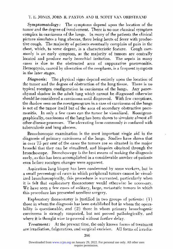
FIGURE 1 : A . Roentgenogram taken on admission, showing complete atelectasis of the left lung. B. Postoperative roentgenogram showing a moderate degree of empyema (pneumonec tomy sixteen days prev ious ly ) .
Physical Examination: The temperature was 98.8° F., the pulse 106. and the blood pressure 108 systolic, 75 diastolic. The percussion note over the entire left chest was flat. The breath sounds were vesicular above the seventh dorsal vertebra, but were absent below this level. No rales were heard in the chest.
The physical diagnosis was atelectasis of the left lower lobe, etiology a ques-tionable bronchial neoplasm.
The laboratory examinations were negative except for a mild secondary anemia. A blood count revealed 3,500,000 red cells with 65 per cent hemoglobin. Roentgen examination showed atelectasis of the left lung (Fig. 1 A ) . Broncho-scopy was performed on April 18, 1940. Protruding into the left main stem bronchus from the region of the upper lobe bronchus was a mass of friable tissue obstructing the lumen. Biopsy was taken and was reported to be squamous cell carcinoma.
Left pneumonectomy was performed on April 24, 1940, and a completely atelectatic lung was found diffusely adherent to the lateral chest wall, pulling the heart and mediastinum into the left chest. An intercostal drainage tube was in-serted into the bottom of the pleural space because of contamination. The post-operative course was uneventful until the sixteenth day when a temperature of 101° F. developed. Roentgen examination of the chest showed a fluid level above the intercostal tube (Fig. I B ) . Thoracentesis was done several times, and finally a rib resection was done on May twentieth, twenty-six days after pneu-monectomy, revealing a small localized empyema about the tube. Convalescence was uneventful and she was discharged on the thirty-fifth postoperative day.
Pathological Examination: In the left main stem bronchus (Fig. 2 A and B) just proximal to the major branches, was a pedunculated polypoid tumor I x 2 cm. protruding up the main stem bronchus. Advanced bronchiectasis of the entire lung was present, secondary to the polypoid obstruction. Microscopic examina-tion revealed a carcinoma simplex originating in the bronchial mucosa. Exami-nation of three mediastinal lymph nodes showed no evidence of metastasis.
269
require permission. on January 29, 2022. For personal use only. All other useswww.ccjm.orgDownloaded from

t . e . j o n e s . j o h n r . p a x t o n a n i ) h . s c o t t v a n o r d s t r a n d
L E F T LUNG
T R A C H E A
BRONCHIAL POLYP!
A. 1?.
FIGURE 2 : A. Drawing from surgical specimen, showing a pedunculated polyp arising at the point of division of the bronchi of the left upper and lower lobe. This polyp extended centrally into the left main stem bronchus, occluding the drainage of the entire lung. B. Photograph of an anteroposterior cross section of the left lung demonstrating the advanced bronchiectasis secondary to the obstructing polyp.
This case presents early carcinomatous degeneration of a benign bronchial polyp. The location of the polyp rendered useless all hope of treatment by bronchoscopic approach.
SUMMARY
There apparently is both a relative and an absolute increase in the incidence of primary carcinoma of the lung. Our hope of recognition of early cases rests on close cooperation between the internist and the bronchoscopist, as well as the surgeon.
With our present day knowledge, the only hope of cure is by surgery, either in the form of lobectomy or pneumonectomy.
A group of cases is discussed with reference to surgical approach, with a report of an unusual case. 1. Koletsky, Simon: Primary carcinoma of the lung; clinical and pathological study of 100
cases, Arch. Int. Med., 62 :636-651, (October) 1938. 2. Simons, E. J . : Primary Carcinoma of the Lung, Chicago, Year Book Publishers, Inc., 1937. 3. Dublin, L. I . : Statistics on morbidity and mortality from cancer in the United Slates, Am.
J. Cancer, 29 :736-742 , (April) 1937. 4. Graham, E. A. , Singer, J. I., and Ballon, II. C. : Surgical Diseases of the Chest, Phila-
delphia, Lea and Febiger, 1935. 5. Portmann, U. V . : The role of roentgen therapy in the treatment of bronchiogenic
carcinoma, Cleveland Clin. 0»art . , 7 :119 -122 , (April) 1940.
270
require permission. on January 29, 2022. For personal use only. All other useswww.ccjm.orgDownloaded from


![Inflammation and cancer: How hot is the link? · carcinoma [30], colon carcinoma, lung carcinoma, squamous cell carcinoma, pancreatic cancer [31,32], ovarian carcinoma biochemical](https://img.dokumen.tips/doc/110x75/5fcdd6c81c76a34db570e7e6/iniammation-and-cancer-how-hot-is-the-link-carcinoma-30-colon-carcinoma.jpg)