Embed Size (px)
Citation preview

Bronchogenic CarcinomaBronchogenic Carcinoma(Lung Cancer)(Lung Cancer)
Respiratory department

DefinitionDefinition
Bronchogenic carcinoma refers
to the malignant tumor which g
rows in the bronchus. Originat
ing from mucus or gland of br
onchus.

Incidence and mortalityIncidence and mortality
Bronchogenic carcinoma has increased re
markable in incidence and mortality during
half of the century and has become the mo
st frequent visceral malignant diseases of m
en.The mortality of lung cancer hold the fir
st place among all kinds carcinomas.

EtiologyEtiology
The cause of lung cancer is unknown.It is believed
that there are following related factors.
1. Excessive cigarette smoking:Smoking
index(Brinkman Index) is equal to cigarettes per
day smoking time(years).
Passive smoking is also a carcinogen factor.

EtiologyEtiology
2.Atmospheric pollution.It was found that c
arcinogenic factor is benzpyrene .
3.Occupational factors.
4Radioactivity in the atmosphere .
5.Diets and Nutrition.
6.Chronic irritation.
7.Genetic factors.
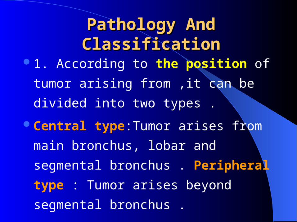
Pathology And ClassificationPathology And Classification
1. According to the position of tumor arising
from ,it can be divided into two types .
Central type:Tumor arises from main
bronchus, lobar and segmental bronchus .
Peripheral type : Tumor arises beyond
segmental bronchus .
Pathology And Pathology And ClassificationClassification
2.According to cytology,it is convenient to classify into four kinds of types.
(1).Squamous cell carcinoma.(2).Small cell anaplastic carcinoma.(3).Large cell anaplastic carcinoma.(4).Adenocarcinoma(including alveolar cel
l carcinoma).
Pathology And Pathology And ClassificationClassification
According to the different principles
of management,it is divided into two
types.
SCLC:small cell lung carcinoma.
NSCLC:non small cell lung carcinoma.
Clinical featuresClinical features
There are no symptoms of early lung cancer in some patients.
Symptoms caused by lung cancer are non-specific:perhaps an audible wheeze or a slight cough,symptoms of infection (fever ,purulent sputum) , of obstruction (wheezing,dyspnea), or ulceration of bronchial mucosa (hemoptysis).
Clinical featuresClinical features
1.Respiratory symptoms. (1).Cough: (2).Hemoptysis: (3).Dyspnea.:
(4).Wheeze or stridor:
(5).Chest pain :
(6).Fever:
Clinical featuresClinical features
2.Symptoms caused by the near
organs or tissue involved by tumor.
(1).Dysphagia.
(2).Hoarseness.
(3).Pleural effusion due to invasion of the
pleura.
Clinical FeaturesClinical Features
(4).Horner’s syndrome.It is caused by inva
ding the cervical sympathetic ganglia on the
involved side the pupil is small ptosis of the
up eyelids,retraction of the eyeball and no s
weat of the face.
(5)Cardiac effusion
Clinical feturesClinical fetures
(6).Superior vena caval syndrome. Due to obstruction of the superior vena caval,the patient may have noticed that his collar is tight, the neck is enlarged and the jugular vein and the veins of anterior chest wall are distension and edema of the face.
3.Symptoms caused by metastasis.liver, skeleton,brain, supra clavicle lymph nodes.
Clinical feturesClinical fetures
4.Paraneoplastic syndrome.Because tumor cell can secrete ectopic hormone, antigen or enzyme the patients with Lung Cancer sometimes may have some paraneoplastic syndrome Including:
(1) Collagen tissue disorder such as finger clubbing , hypertrophic pulmonray osteoarthropathy 。
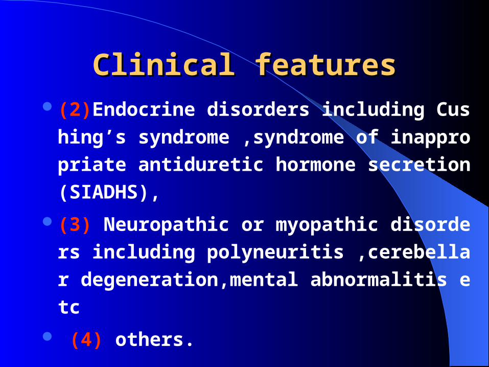
Clinical featuresClinical features(2)Endocrine disorders including Cushing’s
syndrome ,syndrome of inappropriate antid
uretic hormone secretion(SIADHS),
(3) Neuropathic or myopathic disorders incl
uding polyneuritis ,cerebellar degeneration,
mental abnormalitis etc
(4) others.

Radiographic FindingsRadiographic Findings
The appearance on the x-ray film depends on the position ,size and stage of the tumor 1.Peripheral type :It may be various such as infiltrative or nodular, lobulated or umbilicus sign,liner protrusions from the shadow into the surrounding lung, cavitation which is often eccentric irregular in the inner wall owing to the necrosis of the neoplasm.
Radiographic FindingsRadiographic Findings
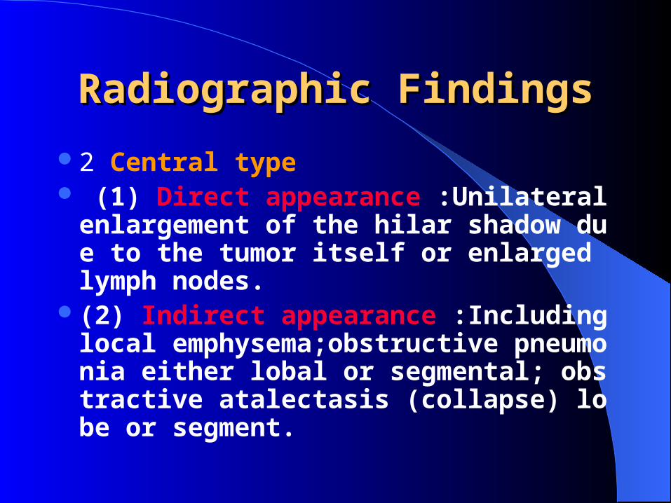
2 Central type (1) Direct appearance :Unilateral enlarg
ement of the hilar shadow due to the tumor itself or enlarged lymph nodes.
(2) Indirect appearance :Including local emphysema;obstructive pneumonia either lobal or segmental; obstractive atalectasis (collapse) lobe or segment.
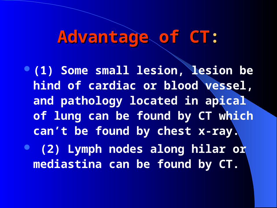
Advantage of CTAdvantage of CT::
(1) Some small lesion, lesion behind of cardiac or blood vessel,and pathology located in apical of lung can be found by CT which can’t be found by chest x-ray.
(2) Lymph nodes along hilar or mediastina can be found by CT.
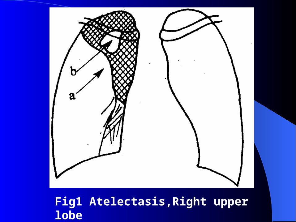
Fig1 Atelectasis,Right upper lobe
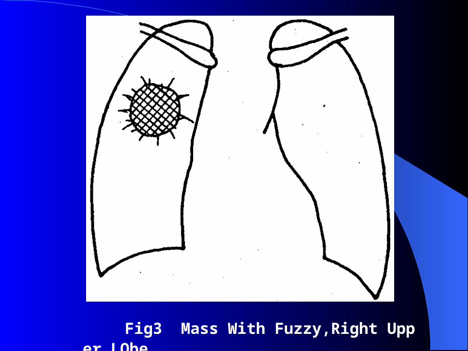
Fig3 Mass With Fuzzy,Right Upper LObe

Fig4 Mass In right Lobe,Lateral portion
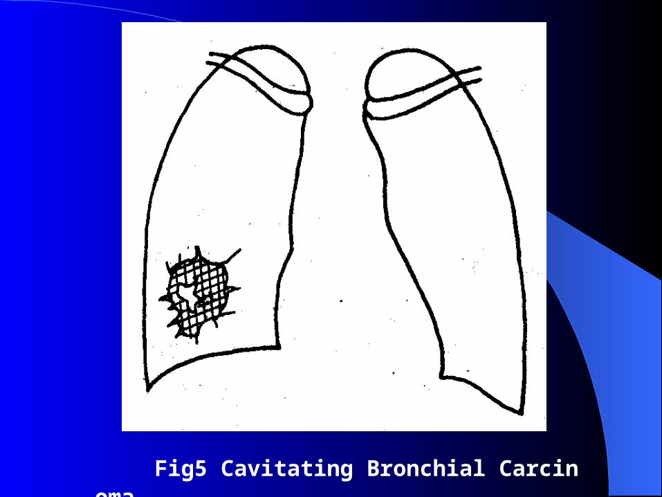
Fig5 Cavitating Bronchial Carcinoma
Examination of sputumExamination of sputum
Cytologic examination of bronchial secretio
ns(or sputum)may reveal exfoliated maligna
nt cells recognizable to the pathologist who
is specially trained for such work.The sputu
m must to be fresh, send on time, repeat(4-6
times)..
BronchoscopeBronchoscope
Bronchoscope may verify the existence of tumor , of Central type, and cytologic diagnosis of lung cancer should be obtained though FBC
.Blind biopsy may be help to the diagnosis of the tumor beyond the range of bronchoscope vision
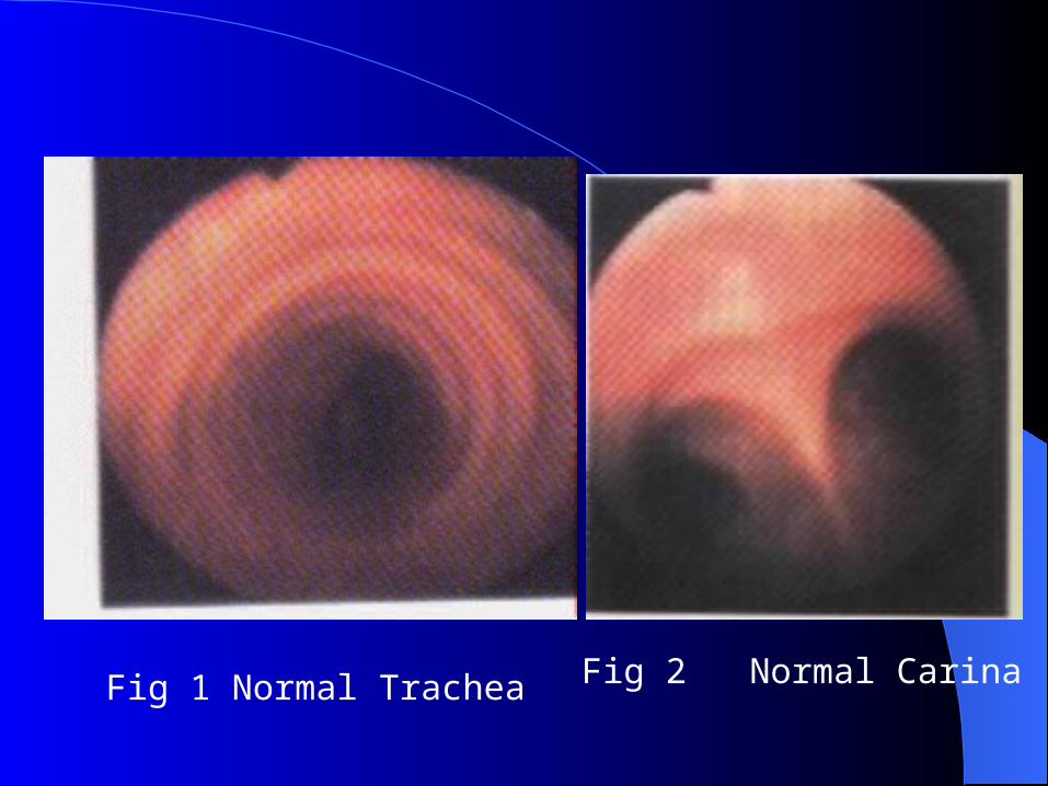
Fig 1 Normal Trachea Fig 2 Normal Carina
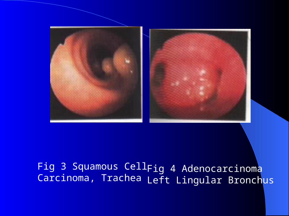
Fig 3 Squamous Cell Carcinoma, Trachea
Fig 4 AdenocarcinomaLeft Lingular Bronchus
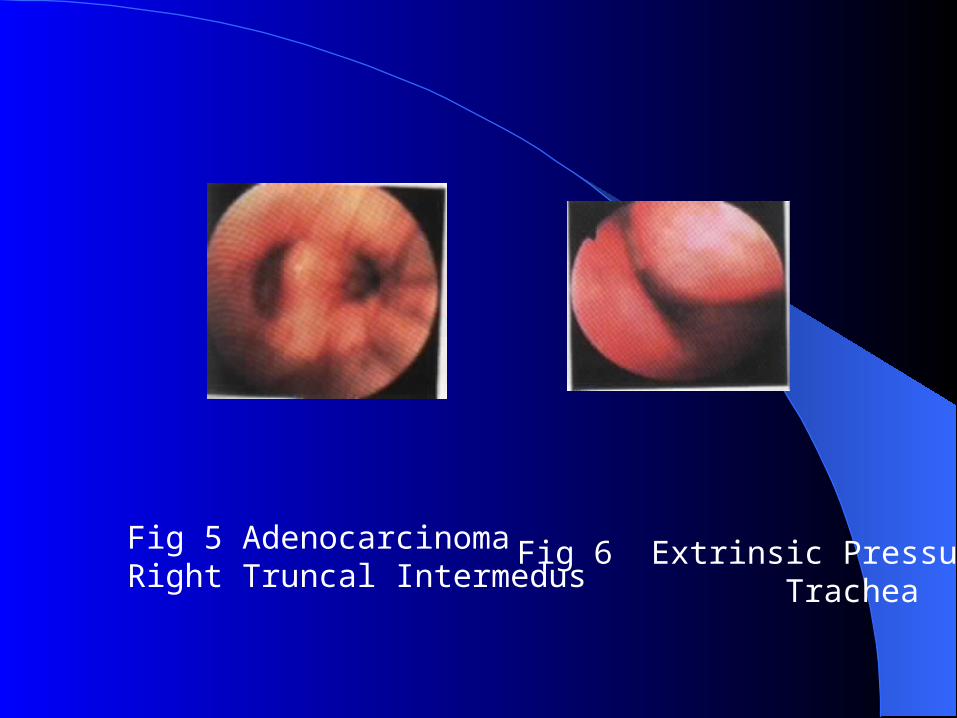
Fig 5 AdenocarcinomaRight Truncal Intermedus
Fig 6 Extrinsic Pressure Trachea
Lung BiopsyLung Biopsy
1.Biopsy with fiberoptic bronchoscope; 2.Transthoracic neddle biopsy with CT directed or B type ultrasonic;
3.Biopsy with thoracoscopy ;4.Biopsy with medistinoscopy;5.Exploratory thoracotomy.
DiagnosisDiagnosis
1.Symptom -free: General investigation of high risk group (male,morn than 40 years old,cigarette consumption 20/per day). Taking a x-ray film and examining sputum for cancer cell every half year
Early stage of the bronchogenic carcinoma Refers to the tumor is still located at the bronchus ,no invade the hilar lymph nodes,pleura as well as dista
nt metastases,its diameter is often <3cm.
DiagnosisDiagnosis
Diagnosis procedure:
1. X-ray film(-) and sputum for cytology (-)
FBC(-) follow up once a month /year.
2. X-ray film(+) and sputum for cytology (+)
FBC to identify the cancer cell type CT , MRI
therapy.

DiagnosisDiagnosis
Diagnosis procedure:
3. X-ray film(-) and sputum for cytology (+)
ruling out the tumor of upper respiratory tract
first FBC.
4 X-ray film(+) and sputum for cytology (-)
FBC(-) lung biopsy.
Differential diagnosisDifferential diagnosis
1.Solitary nodule: Tuberculoma, Benign Tu
mor
2.Cavitation:Lung Abscess, Tuberculosis,
3. Enlargement of hilar shadow: Hamarto
ma
4.Others: Pleural Effusion,Widening Of Me
diatinal.
TreatmentTreatment 1.Rresection by operation ; 2.Radiotherapy ; 3.Chemotherapy; 4.Immunotherapy ; 5.Traditional Chinese medicine therapy etc. The therapeutic principle of lung cancer is compr
ehensive: rescect the tumor as far as possible then combine with other treatments ; other treatments first then operation depending on the cytologic type, position,size and stage of the tumor.
TreatmentTreatment
SCLC: Ⅰ Chemotherapy , operation. Ⅱ Chemotherapy,radiotherapy.NSCLC: Ⅰ Operation. Ⅱ Most :operation→chemotherapy Small parts: radiotherapy.
TreatmentTreatment
Ⅲ: Operation + chemotherapy;
radiotherapy +chemotherapy.
Ⅳ: chemotherapy+ radiotherapy(relieve
some symptoms,such as pain, dyspnea, o
bstruction etc).

![Transbronchial Needle Aspiration Staging of Bronchogenic ...downloads.hindawi.com/journals/dte/1996/237680.pdfChest, 80,48-50. [18] Transbronchialneedle bronchogenic carcinoma, In:](https://img.dokumen.tips/doc/110x75/5fef28f6c0cad34ae7313439/transbronchial-needle-aspiration-staging-of-bronchogenic-chest-8048-50-18.jpg)