Embed Size (px)
Citation preview

Retrospective Theses and Dissertations Iowa State University Capstones, Theses andDissertations
1990
The structure and function of conjunctiva-associated lymphoid tissue in chickens and turkeysAndrew Stephen FixIowa State University
Follow this and additional works at: https://lib.dr.iastate.edu/rtd
Part of the Animal Sciences Commons, and the Veterinary Medicine Commons
This Dissertation is brought to you for free and open access by the Iowa State University Capstones, Theses and Dissertations at Iowa State UniversityDigital Repository. It has been accepted for inclusion in Retrospective Theses and Dissertations by an authorized administrator of Iowa State UniversityDigital Repository. For more information, please contact [email protected].
Recommended CitationFix, Andrew Stephen, "The structure and function of conjunctiva-associated lymphoid tissue in chickens and turkeys " (1990).Retrospective Theses and Dissertations. 9496.https://lib.dr.iastate.edu/rtd/9496

INFORMATION TO USERS
The most advanced technology has been used to photograph and reproduce this manuscript from the microfilm master. UMI films the text directly from the original or copy submitted. Thus, some thesis and dissertation copies are in typewriter face, while others may be from any type of computer printer.
Hie quality of this reproduction is dependent upon the quality of the copy submitted. Broken or indistinct print, colored or poor quality illustrations and photographs, print bleedthrough, substandard margins, and improper alignment can adversely affect reproduction.
In the unlikely event that the author did not send UMI a complete manuscript and there are missing pages, these will be noted. Also, if unauthorized copyright material had to be removed, a note will indicate the deletion.
Oversize materials (e.g., maps, drawings, charts) are reproduced by sectioning the original, beginning at the upper left-hand corner and continuing from left to right in equal sections with small overlaps. Each original is also photographed in one exposure and is included in reduced form at the back of the book.
Photographs included in the original manuscript have been reproduced xerographically in this copy. Higher quality 6" x 9" black and white photographic prints are available for any photographs or illustrations appearing in this copy for an additional charge. Contact UMI directly to order.
University Microfilms International A Bell & Howell Information Company
300 North Zeeb Road, Ann Arbor, fvll 48106-1346 USA 313/761-4700 800/521-0600

Order Number 0110408
The structure and function of coi^unctiva-associated lymphoid tissue in chickens and turkeys
Fix, Andrew Stephen, Ph.D.
Iowa State University, 1990
U M I SOON.ZeebRd.
AAor, MI 48106


NOTE TO USERS
THE ORIGINAL DOCUMENT RECEIVED BY U.M.I. CONTAINED PAGES WITH PHOTOGRAPHS WHICH MAY NOT REPRODUCE PROPERLY.
THIS REPRODUCTION IS THE BEST AVAILABLE COPY.


The structure and function of
conjunctiva-associated lymphoid tissue
In chickens and turkeys
by
Andrew Stephen Fix
A Dissertation Submitted to the
Graduate Faculty In Partial Fulfillment of the
Requirements for the Degree of
DOCTOR OF PHILOSOPHY
Maj or: Veterinary Pathology
Approved:
In Cha Maior Work
For the MajeTr Department
For the Gftâdù/^e College
Iowa State University Ames, Iowa
1990
Copyright © Andrew Stephen Fix, 1990. All rights reserved.
Signature was redacted for privacy.
Signature was redacted for privacy.
Signature was redacted for privacy.

ii
TABLE OF CONTENTS
Page
GENERAL INTRODUCTION 1
Objectives of the dissertation 2
Explanation of dissertation format 3
LITERATURE REVIEW 4
Mucosal immunity and mucosal lymphoid tissues 4
Avian immunology 12
Avian paraocular anatomy and immunity 17
PART I. CONJUNCTIVA-ASSOCIATED LYMPHOID TISSUE (CALT) 22 IN NORMAL AND BORDETELLA AVIUM-INFECTED TURKEYS
ABSTRACT 24
INTRODUCTION 25
MATERIALS AND METHODS 27
RESULTS 29
Gross appearance of CALT 29
Light microscopy 30
Scanning electron microscopy 31
Transmission electron microscopy 32
DISCUSSION 44
PART II. MORPHOLOGIC CHARACTERIZATION OF CONJUNCTIVA- 48 ASSOCIATED LYMPHOID TISSUE (CALT) IN CHICKENS
ABSTRACT 50
INTRODUCTION 51
MATERIALS AND METHODS 53

ill
RESULTS 55
Eyelid structure in 1-day-old chickens 55
Lymphoid component of CALT 56
Epithelium of CALT 57
Vessels of CALT 58
DISCUSSION 70
PART III. PARTICLE UPTAKE BY CONJUNCTIVA-ASSOCIATED 74 LYMPHOID TISSUE (CALT) IN TURKEYS
ABSTRACT 76
INTRODUCTION 77
MATERIALS AND METHODS 79
RESULTS 82
DISCUSSION 89
PART IV. QUANTIFICATION OF PARTICLE UPTAKE BY 93 CONJUNCTIVA-ASSOCIATED LYMPHOID TISSUE (CALT) IN CHICKENS
ABSTRACT 95
INTRODUCTION 96
MATERIALS AND METHODS 98
RESULTS 103
DISCUSSION 107
GENERAL SUMMARY
LITERATURE CITED
ACKNOWLEDGMENTS
110
114
135

1
GENERAL INTRODUCTION
Avian Immunology has advanced considerably since Gllck
Initially suggested an Immunologic role for the bursa of
Fabriclus in 1956.^^ Since that time, many aspects of the
avian immune system have been researched and clarified. These
Include immunoglobulin types,histocompatibility
antigens,^®® cellular cooperativlty,usf 134 lymphocyte surface
i m m u n o g l o b u l i n i s o t y p e s , a n d l y m p h o i d t i s s u e s . O n e
of the most extensively studied avian lymphoid tissues is the
Harder Ian gland .14,163,199,200 This retrobulbar gland has been
shown to contain IgA-secretlng plasma cells and to have an
Important role in local ocular immunity.^,41,180 Application
of antigen to the conjunctival space leads, to an increase in
the number of plasma cells in the Harderian gland as well as
the appearance of specific antibody in tears.
However, in spite of the marked interest in the Harderian
gland, the site for initial lymphocyte contact with antigen
has not been established. The only attempt at determining
particulate material uptake by the Harderian gland failed to
document transport across the glandular epithelium.
The mucosal immune system is important for defense
against potential mucosal pathogens. This is accomplished
primarily through the production of secretory igA,22,186,189
which arises relatively late in phylogeny and ontogeny.®^
Mucosal lymphoid tissues are central to the function of the
mucosal immune system. These tissues, which exist along many
epithelial surfaces, are Important in the uptake of antigen
a n d i n t h e g e n e r a t i o n o f a n i m m u n e r e s p o n s e . M a n y
specialized structural features of mucosal lymphoid tissues
have been reported, including a flattened superficial
lymphoeplthelium and intraepithelial lymphocytes.
Particularly well-studied in this group are gut-associated
lymphoid tissue (GALT) and bronchus-associated lymphoid tissue

2
(BALT) .20,24,30 Although most of these studies have been
restricted to mammals, some characterization of GALT and BALT
has been done in avian species.More recently, similar
lymphoid tissue has been described in the conjunctiva of
rabbits and guinea pigs.^^'^^'^® This conjunctiva-associated
lymphoid tissue, or CALT, has not been described or studied in
avian species.
Many experiments using microorganisms or particulate
tracer materials have documented the capacity for uptake of
particles by mammalian GALT and BALT.106,140,141,183 Here,
uptake usually occurs by specialized mucosal lymphoepithelium
through selective pinocytosis at the cell surface. The only
study of experimental uptake by CALT has been done in guinea
pigs.^^® In that report, uptake was demonstrated but it was
not selective for the CALT lymphoepithelium.
The poultry industry relies heavily on vaccination for
the prevention and control of many potentially devastating
infectious diseases. Economic constraints have dictated the
delivery of many of these vaccines to large numbers of birds
in water or by aerosolization. Several vaccines are also
delivered by eyedrop inoculation. These vaccine exposure
methods make respiratory, gastrointestinal, and ocular
lymphoid tissues important in the successful development of an
immune response. Logically, any lymphoid tissue present
within the conjunctiva that experiences vaccine contact could
also be important in mucosal immunity.
Objectives of the dissertation
The objectives of these studies in turkeys and chickens
were (1) to describe the structure of CALT, (2) to
characterize the development of CALT during the post-hatching
period, (3) to examine the response of CALT to local antigenic
stimulation, (4) to demonstrate and characterize the uptake of

3
particulate tracers by CALT, and (5) to quantitate tracer
uptake by CALT.
Explanation of dissertation format
This dissertation is presented in the alternate thesis
format. It contains four manuscripts presented in the style
of the journal. Veterinary Pathology. The manuscripts are
preceded by a general introduction and literature review and
are followed by a general summary and the literature cited.
All citations for all parts are located in the literature
cited section. The Ph.D. candidate, Andrew S. Fix, was the
principal investigator for each study.

4
LITERATURE REVIEW
Mucosal membranes are epithelial surfaces that line many
regions of the body, such as the gastrointestinal and
respiratory tracts. The location of these epithelial surfaces
places them in direct contact with the external environment.
Accordingly, the ability to develop a protective immune
response toward potentially pathogenic microorganisms along
these epithelial surfaces is important. Mucosal lymphoid
tissues are located along these surfaces specifically for this
purpose. In these tissues, antigen contact and subsequent
antigen uptake are important early steps in the mucosal immune
response. This review addresses the pertinent literature in
three areas: an initial discussion of avian and mammalian
mucosal lymphoid tissues, a consideration of avian immunology,
and a final discussion focused on avian paraocular immunity.
Mucosal immunity and mucosal lymphoid tissues
Mucosal lymphoid tissues, which are present along many
mucosal surfaces of mammals and birds, have been studied and
characterized in a number of species. In the past several
decades, considerable interest has developed in the structural
and functional significance of these mucosa-associated
lymphoid tissues, or MALT.^^ In spite of their location in
many different sites, MALT are morphologically and
functionally similar. Major components of this system exist
in the gastrointestinal and respiratory tracts. In addition,
similar tissues have been described along several other
mucosal surfaces. In the first part of this brief review, a
discussion of the general structural features of MALT will be
followed by a consideration of the immunologic function of
these tissues.

5
Gut-associated lymphoid tissues, or 6ALT, are present
throughout the gastrointestinal tract in mammals and birds.
6ALT represents a substantial part of the total lymphoid
tissue present in the body. GALT includes lymphoid tissues in
the oral cavity, pharynx, small intestine, cecum, colon, bursa
of Fabricius, and rectum.In many species, aggregates of
submucosal lymphoid tissue encircle the entrance to the
digestive tract in the form of pharyngeal tonsils.In pigs,
palatine tonsils are exceptionally prominent in the soft
palate and provide a relatively large surface area.^°^
Particularly well-studied are the small intestinal Peyer's
patches, which, in humans, increase in size and number from
early fetal life to puberty. 3? » 57,144 The number of these
patches in the small intestine, as well as the density of
lymphoid follicles within a given patch, is highly variable
between species.In ruminants, both jejunal and ileal
Peyer's patches exist. Like the bursa of Fabricius, ileal
patches involute at maturity and have a primary function in
B-cell generation; jejunal patches remain throughout life and
function as secondary reactive lymphoid tissues.98,99,ise-isa
Peyer's patches are also well-characterized in pigs®^'®^'^®® and
chickens.Lymphoglandular complexes, which are submucosal
epithelial diverticula associated with lymphoid tissue below
the muscularis mucosa, have been characterized in the large
intestine of cattle, 109»no pigs,^^^ dogs,^^ and humans.
Lastly, the appendix of rabbits and humans contains abundant
lymphoid tissue and is an important component of GALT in those
species,In rabbits, the appendix has surface recesses
that delineate lymphoid nodules and which harbor luminal
bacteria.
Bronchus-associated lymphoid tissue, or HALT, is another
important mucosal lymphoid tissue. Originally, HALT was
identified along the bronchial epithelium in rabbits, guinea
pigs, rats, mice, dogs, pigs, chickens, and humans.

6
Subsequent studies have examined BALT in rats,rabbits,
cattle,^ and turkeys.Mammalian BALT is located below
bronchial epithelium and contains a narrow neck of lymphocytes
connecting subepithelial lymphocytes to deeper
follicles.In contrast, avian BALT forms prominent
subepithelial lymphoid nodules that elevate the epithelium and
extend well into the lumen of pulmonary bronchi.2?»
Several other mucosal surfaces have also been found to
contain regional lymphoid tissue, including reproductive,
nasopharyngeal, and conjunctival. Lymphoid tissues resembling
other MALT exist in the lateral walls of the mouse
nasopharynx,in salivary gland ducts,and in the human
fallopian tube and uterus.The latter tissue apparently
undergoes cyclical shedding during the menstrual cycle. More
pertinent to this review are descriptions of conjunctiva-
associated lymphoid tissue, or CALT.
The original descriptions of CALT were in laboratory
rabbits and guinea pigs.^^'^^ These reports describe randomly
scattered to discretely clustered macroscopic lymphoid
nodules, with a diameter of 0.2 mm to 0.8 mm, in palpebral
conjunctiva. In the rabbit, these nodules are common near the
nasolacrimal duct opening, average 40 per lower eyelid and 19
per upper eyelid, and are not evident before 4 weeks of age.
Microscopically, CALT in rabbits consists of dense lymphoid
infiltrates without germinal centers and plasma cells. In
addition, lymphocytes heavily infiltrate the overlying
epithelium and fill large adjacent lymphatic channels. Later
study of CALT in rabbits supported the original findings but
noted the presence of germinal centers and a lack of goblet
cells in the overlying CALT epithelium.Based on structural
similarities, these studies suggested that CALT is a component
of the mucosal immune system. Limited work has been
done with CALT since these original descriptions ̂85,178
CALT has not been described in avian species.
7
Mucosal lymphoid tissues exhibit remarkably similar
structure, despite their diverse anatomic locations among
different species. Specific divisions of these tissues
Include the epithelium, the lymphoid component, and the
vasculature. Since the most complete morphologic descriptions
of MALT have been generated through studies of 6ALT and BALT,
these tissues provide the basis for our understanding of
mucosal lymphoid tissue organization.
Perhaps the most unique division of MALT Is the surface
epithelium, commonly referred to as follicle-associated
epithelium or lymphoeplthellum. This epithelium Is usually
devoid of goblet cells, and It Is heavily Infiltrated by
Intraepithelial lymphocytes primarily of the
suppressor/cytotoxlc phenotype.21,24 Lymphoeplthellal cells
typically have short microvilli, few lysosomes, flattened
apical cytoplasm, abundant apical vesicles, and reduced
affinity for ruthenium red.^^^ Originally, cells composing the
epithelium over human Peyer's patches were termed "M" cells,
based on their lack of microvilli and the presence of unique
surface mlcrofolds.^^^ These cells encase lymphocytes under a
thin apical cytoplasm and are specialized for the uptake and
transport of luminal material. 144,202 addition to the
general features of lymphoeplthellal cells listed above, M
cells have Increased cholesterol content, stain poorly for
alkaline phosphatase but intensely for esterase, and develop
directly from undifferentiated crypt epithelial
cells.143 ̂ cells have been identified in the GALT
of chickens, rabbits, pigs, calves, mice, rats, guinea pigs,
monkeys, hamsters, and dogs.®®'^®^ Similar cells exist in
other mucosal lymphoid tissues including BALT^° and CALT.^®
A second major division of MALT, the lymphoid component,
contains cells similar in identity and organization to
nonmucosal lymphoid tissues. Below the lymphoeplthellum are
solitary or clustered lymphoid nodules composed of

8
lymphocytes, macrophages, reticular stromal cells, and
germinal centers. Germinal centers contain expanding clones
of B-cells generally committed to IgA production.They are
common throughout 6ALT, but are infrequent in SALT, except in
chickens.20,30,147 in lymph nodes, paracortical areas are
populated by T cells. The greatest number of T-helper cells
are thought to be in the paracortex, whereas more T-suppressor
cells exist primarily within the lymphoepithelium.^^'^^
Macrophages are also distributed throughout the cellular areas
of mucosal lymphoid tissues. These macrophages are important
for the presentation of antigen and are positive for major
histocompatibility class II antigens.Recently, mucosal
mast cells, which differ in several respects from their
connective tissue counterparts, have been examined for their
possible role in mucosal immunity.
A third important division of MALT is the vasculature,
specifically the high endothelial venules, or HEV. These
specialized vessels are adapted for the homing and
r e c i r c u l a t i o n o f l y m p h o c y t e s i n t o l y m p h o i d t i s s u e s . S u c h
vessels are not confined to MALT, but also exist in other
lymphoid tissues including lymph nodes and spleen. HEV, also
called cuboidal endothelial venules or post-capillary venules,
have plump, round to oval endothelial cells that closely pack
the circumference of the vessel.A complete phylogenetic
examination of vertebrate lymphoid tissue described HEV in the
lymph node, appendix, Peyer's patch, and tonsil of mammals,
but only in the spleen of birds, reptiles, amphibians, and
fish.More recently, similar vessels have been identified
in BALT of rats,^®^ guinea pigs,^®^ and turkeys.HEV also
exist in chicken GALT,^^ although no direct analogy to
mammalian HEV was made. In the original reports of CALT in
rabbits and guinea pigs, no specific description of HEV was
made.^^'^^'^®

9
Any discussion of mucosal lymphoid tissues is incomplete
without addressing potential functions in mucosal immunity.
Evidence indicates that the function of MALT is rather
uniform: to protect mucosal surfaces through antigen uptake
and the production of secretory IgA. Although a number of
excellent papers have reviewed this subject at
length, 21-23,29,87 pertinent aspects considered here will
include antigen uptake, secretory IgA production, and
lymphocyte homing.
Antigen uptake by mucosal lymphoid tissues is a
prerequisite for the successful generation of a mucosal immune
response. Studies evaluating antigen uptake by MALT have
concentrated on GALT, primarily using particulate (carbon,
latex beads) and macromolecular (ferritin, horseradish
perodoxidase) tracer materials. Uptake occurs primarily by
pinocytosis along the lymphoepithelial apical surface.
Histologically, carbon has been demonstrated crossing the
lymphoepithelium of the rabbit appendix and the mouse Peyer's
patch, with subsequent localization in mesenteric lymph
nodes.®®'Similar results have been shown using latex
beads in rats, mice, and dogs.^°®'^°^'^®® Latex bead uptake in
Peyer's patches occurs with equal frequency in conventional
and germfree mice.^°^ The ultrastructural details of uptake
across M cells have been elucidated using horseradish
peroxidase in the mouse Peyer's patch.In these
studies, tracer material was preferentially taken up within 5
minutes by H cell apical pits, transported via cytoplasmic
vesicles, and released into intercellular spaces between M
cells and their enfolded lymphocytes. Tracer transport in
GALT is not strictly unidirectional, since horseradish
peroxidase administered intravenously also localized in the
lymphoepithelium of mouse Peyer's patch, rabbit appendix, and
chicken bursa of Fabricius.^® Although fewer studies have
assessed uptake in BALT, several tracers (carbon, latex beads,

10
and horseradish peroxidase) have been shown to be taken up by
BALT lymphoepithellum. 183,192 ^he only report evaluating
uptake in CALT failed to demonstrate selective tracer
transport by conjunctival lymphoepithellum in guinea pigs.^^^
A considerable portion of humoral defense along mucosal
surfaces occurs through the action of secretory IgA. This
immunoglobulin has an alpha heavy chain and is frequently
dimerized by the binding of the J chain peptide. The dimeric
configuration enhances mucosal transport.Delivery of
secretory IgA along mucosal surfaces requires several specific
steps. Antigen-presenting cells must process and present
antigenic fragments to immature B cells.In mucosal
lymphoid tissues, B immunoblasts are largely committed to IgA
production.58,91 Evidence indicates that, at least in GALT,
the preferential secretion of IgA is mediated by specific T
cell subsets which evoke an isotype switch in B
immunoblasts.18 similar isotype switching may also occur in
guinea pig CALT.85 once sensitized to antigen, IgA-committed
precursors leave the lymphoid tissue of origin and localize to
mucosal surfaces as IgA-producing plasma cells. This
localization is dependent on a number of variables, including
chemotactic factors, cell receptors, organ characteristics,
macrophage distribution, and vascular features.!®'®^ Delivery
of IgA across the epithelium requires binding of polymeric IgA
to secretory component, and subsequent transport through the
interior of the epithelial cell. 160,175,187 secretory
component, a polypeptide molecule containing five
immunoglobulin-like domains, binds to IgA and is subsequently
cleaved from the mucosal transport receptor located base-
laterally on the epithelial cell.189 secretory IgA is
then released onto the adjacent mucosal surface. Secretory
component has been identified in several epithelial types,
including rabbit and human lacrimal glands.^'^8,77

11
Selective localization of circulating lymphocytes to
various mucosal lymphoid tissues is an important aspect of
mucosal immunity. This subject has been extensively
reviewed. 19,45,89,204 Lymphocyte recirculation patterns depend
on the tissue of origin of the lymphoblast. The idea that
lymphoblasts home to their tissue of origin has produced a
conceptual separation of mucosal and nonmucosal immunity.
Experimentally, lymphocytes derived from 6ALT home to the
gastrointestinal tract wall or Peyer's patches, whereas cells
derived from peripheral lymph nodes return to those nodes.
Using frozen sections of lymph nodes in an In vitro binding
assay, several studies have clarified the role of high
endothelial venules (HEV) as binding sites for the entry of
l y m p h o c y t e s i n t o l y m p h n o d e s . C i r c u l a t i n g
lymphocytes, which normally have a relatively smooth surface,
develop microvilli as they pass through the HEV of lymph
nodes.The association between circulating lymphocytes and
HEV is mediated by lymphocyte surface adhesion molecules that
recognize specific receptors on HEV.^°* After binding,
lymphocytes migrate through the endothelium and enter the
surrounding lymphoid tissue. It is likely that different
adhesion molecules dictate the homing of lymphocytes to
different tissues.Homing can be specifically confined to
certain mucosal lymphoid tissues. For example, lymphocytes
derived from bronchial lymph nodes localize more to the lungs
than to the gut.However, homing is not always predictable
or completely tissue specific. In mice, %-thymidine-labelled
lymphocytes from the mesenteric lymph node preferentially
repopulate multiple mucosal sites, including the
gastrointestinal tract, uterus, and mammary gland.In BALT
of rats and guinea pigs, HEV specificity more closely
resembles the HEV of mesenteric nodes than Peyer's patches.
There is also evidence that lymphocyte recirculation accounts
for the immunological connection between the gastrointestinal

12
tract and the mammary gland.However, the significance of
this connection apparently varies with species.88» 90,167
Avian immunology
Research in avian immunology has provided considerable
insight into understanding the overall function of the immune
system. Recently, many excellent reviews of this subject have
been published in textbooks®^and in journals. 56,79,8i, 184
Although much of the avian immune system resembles that of
mammals, some unique features exist, including the bursa of
Fabricius, the Harderian gland, and the lack of peripheral
lymph nodes. As in mammals, avian lymphoid tissues can be
divided into primary (central) and secondary (peripheral),
each with a special function. These tissues contain the cells
that provide the humoral and cell-mediated immunity for
immunologic protection in birds. Avian primary lymphoid
organs include the thymus and bursa of Fabricius, which
facilitate the maturation and clonal expansion of T and B
cells, respectively. Primary lymphoid tissues are also
referred to as lymphoepithelial, since their lymphocytes
develop in close association with an epithelial component.
Secondary lymphoid tissues have an important stromal
component, and are also called lymphoreticular. They are more
diffuse than the primary tissues and are important in direct
response to antigen.
The avian thymus, the maturation center for T cells,
develops from pharyngeal pouch epithelium. It is divided into
12 to 14 distinct lobes, each containing lobules with an outer
cortex and an inner medulla. A vascular barrier to antigen,
the blood-thymic barrier, exists in the cortex but not in the
medulla. Cell types present in the thymus include
lymphocytes, epithelial cells, myoid cells, macrophages, and
endothelial cells of HEV. Unlike the mammalian thymus, the

13
avian thymus also has a secondary immunologic function.
Germinal centers exist in the medulla, where the vascular
barrier is incomplete. Shortly after hatching, B cells
migrate into this region and plasma cells are evident there by
4 weeks of age.*'^°®
The bursa of Fabricius, central to the generation of B
cells, develops from the dorsal epithelium of the primitive
cloaca.Developing epithelial cells form infoldings and
plicae which are colonized by lymphoid cells that subsequently
define cortical and medullary areas.^ Proliferation of these
colonizing cells produces bursal follicles, which total
between 8,000 and 12,000 per bursa.The epithelium over
these follicles develops the classic features of a
l y m p h o e p i t h e l i u m s p e c i a l i z e d f o r a n t i g e n u p t a k e . H o w e v e r ,
the interfollicular epithelium lacks this specialization.
Tracer particle solutions placed in contact with bursal
lymphoepithelium are transported by pinocytosis. After
uptake, tracers localize in bursal follicles and are
distributed to other regions of the bursa by phagocytic
cells. 15,129,130,174 Lixg the avian thymus, the bursa also has
a secondary lymphoid function. Uptake by the bursal
lymphoepithelium is important in this role. After local
immunization, antibody-producing cells have been observed
inside the bursa.T cells, identified histochemically by
acid alpha-naphthyl acetate staining, are also present in
specific regions of the bursa.In chickens, the bursa
decreases in weight from 10 to 16 weeks of age, with
involution virtually complete by 24 weeks of age.^^^
Secondary lymphoid tissues in birds, including the
spleen, atypical lymph nodes, and mucosal lymphoid tissues,
are not as well studied as in mammals. The spleen is similar
to the mammalian counterpart, except for the presence of
ellipsoidal sheaths of reticular cells around penicillary
arteries.When challenged with intravenous antigen, B and T

14
cell areas in the spleen cooperate and produce germinal
c e n t e r s i n t h e p e r i a r t e r i a l l y m p h o i d t i s s u e a r e a s . T h e
generation of splenic germinal centers is maximal at 4 to 5
weeks of age, which corresponds to peak bursal development.
Organized peripheral lymph nodes are not characteristic
features of avian secondary lymphoid tissues. Instead» birds
form regional concentrations of lymphoid tissue locally in
organs, tissues, and lymphatic vessels.However, structures
resembling lymph nodes have been described. Chickens have
lymphoid accumulations along the posterior tibio-popliteal and
lower femoral veins.These femoral lymph nodules have
afferent and efferent lymphatics, peripheral germinal centers,
and an intricate internal sinus system. Although barely
detectable in normal birds, these structures become
hyperplastic after local antigen delivery.Ducks have
similar paired nodes in the lumbar region that contain
macrophages and vessels resembling HEV.^°^ In addition, the
pineal gland in chickens also contains lymphoid tissue and
antibody-producing plasma cells.
Mucosal lymphoid tissues, primarily 6ALT and SALT,
comprise another group of secondary lymphoid tissues in birds.
Peyer's patches, the cecal tonsil, and Meckel's diverticulum
occur along the gastrointestinal tract. In chickens, the
number of Peyer's patches increases until 16 weeks of age,
then decreases until a single residual patch remains at the
ileocecal junction.^® Structurally, avian Peyer's patches
resemble those in mammals.^® Chickens also possess cecal
tonsils in the proximal region of each cecum.Like the
mammalian palatine tonsil, these structures have epithelial
crypts that are continually exposed to luminal contents.
Equal numbers of B and T cells exist in cecal tonsils in
chickens at 5 weeks of age; however, B cells predominate in
older birds.^ Meckel's diverticulum, the embryologie yolk sac
remnant that separates the jejunum from the ileum, is

15
Infiltrated with lymphoid cells by 2 weeks of age.^^^ Like the
Harderian gland, this area contains large numbers of plasma
cells and remains functional until at least 20 weeks of age.^^^
Ducks have a rather unique form of GALT. Annular bands of
lymphoid tissue completely encircle the intestinal wall at
regular intervals on either side of Meckel's diverticulum.
These bands are only rudimentary at hatching, but develop
prominent follicles with geirminal centers by maturity.
Although not mentioned in recent reviews of avian
immunology ̂ ̂ 7,82,149 was characterized in chickens almost
two decades ago.^^'^^ In those studies, lymphoid nodules with
germinal centers formed finger-like projections into the
bronchial lumen. According to the authors, chickens had more
lymphoid tissue in the trachea than any other species examined
(rabbit, guinea pig, rat, mouse, dog, pig, human). Their use
of the term "trachea" is somewhat perplexing, since by
definition, BALT is confined to the pulmonary bronchus.
Subsequent and more complete descriptions of BALT have been
reported in turkeys.In this species, BALT nodules
protrude into the primary intrapulmonary bronchus and are
specifically associated with longitudinal mucosal folds and
the openings of secondary bronchi.This study also
demonstrated a progressive increase in the number and size of
BALT nodules with increasing age of turkeys. In another study
that involved turkeys infected with Bordetella avium. BALT
nodules were more numerous and more widely distributed than in
normal birds.In contrast to mammals,specific
experiments documenting tracer uptake have not been done in
avian BALT. Although the ultrastructural features of turkey
BALT lymphoepithe1ium does not suggest specialization for
antigen uptake, epithelial discontinuities induced by
infiltrating lymphocytes may allow antigens to pass from the
lumen to underlying lymphoid elements.

16
Several studies have localized antibody-producing cells
in avian mucosal lymphoid tissues by immunohistochemistry. In
the chicken gastrointestinal tract, IgA-positive cells have
been identified in the duodenum and cecal tonsil, but not in
Peyer's patches.This has been verified by some reports,
but not by others.1° The latter study found primarily
IgG-positive cells along the chicken gastrointestinal tract.
In turkey intestine, IgG-positive cells are sparse and
IgA-positive cells equal those positive for IgM.ss
Antibiotics reduce the number of immunoglobulin-bearing cells
in the gastrointestinal tract of both chickens and
turkeys.Along chicken bronchial mucosa, IgG- and
IgM-positive cells occur most commonly, with fewer
IgA-positive cells evident.
Humoral and cellular aspects of avian immunology are well
characterized. Three major immunoglobulins occur in avian
serum, a 7S immunoglobulin analogous to mammalian IgG, IgM,
and an IgA-like immunoglobulin.Serum IgG and IgM are
present in highest concentration, with IgA representing less
than 4% of total serum immunoglobulins. Only 20% of serum
IgA is monomeric, with the remainder existing as dimers or
multimers.In chickens, IgA predominates in bile and
intestinal secretions,but not in saliva, tears, or
seminal plasma.1°* In contrast, IgG predominates in tracheal
secretions.*8 The transport of biliary IgA has been
extensively studied in chickens, and a molecule similar to
mammalian secretory component is likely involved.31' 152,196
the chicken egg, IgG is concentrated in the yolk and IgM and
IgA are confined principally to the white.Similar serum
and secretory immunoglobulin profiles exist in turkeys, with
IgG and IgM detectable in many secretions.polymeric IgA
occurs in the bile and intestinal secretions of turkeys,m
with some IgA also detectable in saliva, tears, and trachea.

17
Although IgM and IgA are present in the turkey egg, IgG is the
predominant immunoglobulin found.**
Several aspects of cellular immunity have been studied in
birds and many functional types of T cells have been
identified. Chickens have T helper and T suppressor cells
that have similar functions to their mammalian
counterparts.32,79,115 ^ suppressor cells are bursal dependent,
since bursectomized chickens demonstrate reduced suppressor
activity.in contrast, bursectomy does not reduce cytotoxic
T cell function.** Delayed-type hypersensitivity reactions
occur in chicks exposed to tuberculin, diphtheria toxoid, and
human gamma-globulin.®^ Contact hypersensitivity reactions to
topically administered low molecular weight compounds are
modulated by B cells.Other cellular aspects of avian
immunology that have been explored include homograft and
allograft rejection, graft-versus-host responses, and the
major histocompatibility complex.®®'®^
Avian paraocular anatomy and immunity
The general structural features of the tissues and organs
surrounding the avian eye are presented in several recent
textbooks of avian anatomy.69,94,95,132 However, one of the
earliest and most thorough accounts was in the English sparrow
in 1918 by Slonaker. Birds have a closed, bony orbit that
contains the eyeball, paraocular glands, extraocular muscles,
nerves, blood vessels, fat, and connective tissue. A
nicitating membrane, or third eyelid, exists in the rostral
angle of the conjunctival space. For lubrication and cleaning
of the cornea, this thin fibrous membrane is rapidly swept
across the cornea by a tendon that connects to a specific
retrobulbar muscle, the pyramidalis. Paraocular glands
present in birds include the lacrimal gland and the Harderian
gland. The lacrimal gland, which contributes to tear

18
production, is located at the lateral orbital rim and drains
into the conjunctiva by several small ducts. The Harderian
gland is a retrobulbar, tubulo-alveolar gland that also
contributes to tear secretions. It empties into the
conjunctival space behind the nictitating membrane through a
single duct. To cover and protect the cornea, birds have
upper and lower eyelids like mammals. However, detailed
descriptions of these structures are lacking in avian anatomy
texts and journals.
Considerable scientific literature exists delineating the
role of the Harderian gland in avian paraocular immunity.
Although the existence of the gland was known for centuries,
in 1968 Bang and Bang described "invasive lymphoid
infiltrates" along the lacrimal gland, the Harderian gland,
the ducts of those glands, and the duct system of the lateral
nasal gland.These workers emphasized that infiltrates
along the ducts were primarily small lymphocyte nodules with
germinal centers. In contrast, the Harderian gland was
infiltrated with large populations of plasma cells.
Subsequent studies have verified and extended the original
findings in chickens ,97,163,199,200 ^nd have added descriptions
o f t h e H a r d e r i a n g l a n d i n t u r k e y s ^ ^ ' ^ ^ ^ a n d d u c k s . I n
addition, plasma cells exist in the Harderian gland of many
wild bird species,and have also been found in germfree
chickens.Some reports have emphasized the large number of
Russell bodies in Harderian gland plasma cells.i**
Morphologically, the Harderian gland has the following
features: a compound tubulo-acinar architecture with lobules,
columnar epithelium, fibrovascular interstitial septae, and a
thin connective tissue capsule.Histochemically, the
merocrine secretions are mostly sulfated
mucopolysaccharides.Four types of epithelial cells have
been characterized ultrastructurally.Several lobule types
exist in the Harderian gland, and the relative plasma cell

19
density varies between types.Plasma cell infiltration is
interstitial, bursal dependent,124,179 ^^d increases with
age.^^° In chickens, early infiltration occurs between 2 and 4
weeks of age, primarily with cells positive for IgM.^
However, IgG- and IgA-positive cells predominate from weeks 4
to 9, with mostly IgA-positive cells present thereafter.s, 180
In contrast, others have found primarily IgM- and IgG-positive
cells in the Harderian gland of adult chickens.The gland
also contains macrophages, lymphocytes, myoepithelial cells,
and heterophils. 14,163,169 Macrophages, which are structurally
heterogeneous, exist in both intraepithelial and subepithelial
sites and sometimes are associated with dense homogenous
intercellular material.i** Ultrastructurally, pre-plasma cells
or "plasmablasts" form desmosome-like junctional complexes
with each other and with nearby macrophages.169
A number of respiratory diseases exist in birds that have
considerable negative economic impact on the poultry industry.
These include Newcastle disease, infectious bronchitis, avian
pox, infectious laryngotracheitis, and turkey bordetellosis.
Many important aspects of resistance to these respiratory
pathogens have recently been reviewed.In an effort to
improve health and increase productivity, the poultry industry
has developed many vaccines to help control these diseases. A
significant number of these vaccinations require delivery on a
flock basis, since individual bird handling is cost
prohibitive. Therefore, administration is usually by water or
aerosol. Protocols for the administration of these vaccines
vary widely in the industry. Since vaccination success
depends on epithelial surface contact, mucosal lymphoid
tissues are undoubtedly important in the generation of
protective immunity. For several decades, the avian Harderian
gland has been regarded as central to the production of local
immunity, especially in response to eyedrop or aerosol
vaccination.

20
Several studies have assessed antibody production by
Harderian gland plasma cells. Specific homologous antibody
was detected in saline extracts of Harderian gland after
conjunctival inoculation with sheep red blood cells, Newcastle
disease virus, infectious bronchitis virus, and Mycoplasma
aallisepticum.However, no antibody was detectable in the
Harderian gland after parenteral inoculation with these
agents. Similar results have been demonstrated by others with
conjunctival delivery of Newcastle disease virus^^^'^^^ and
bovine serum albumin.Using immunofluorescence, this latter
report also identified plasma cells with antibody specificity
for bovine serum albumin. Studies using infectious agents
have indicated that during inflammation, some circulating
antibody enters Harderian gland secretions by transudation.%
Structural changes in the avian Harderian gland after
conjunctival delivery of infectious agents have been examined
in chickens. Eyedrop delivery of live Newcastle disease virus
and live infectious bronchitis virus produced a significant
increase in Harderian gland plasma cells; however, aerosol or
intravenéous inoculation produced only a slight increase.
These studies also documented acute inflammatory changes in
affected Harderian glands. After inoculation with infectious
bronchitis virus, lymphoid follicles formed in the Harderian
gland in 14 to 21 days. An opposite effect occurred after
conjunctival inoculation of infectious bursal disease virus in
3-week-old broilers, since Harderian gland plasma cells became
necrotic for 5 to 14 days.®^ However, plasma cell numbers did
eventually return to normal. Similar inoculation in 1-day-old
broilers prevented the normal post-hatching infiltration of
plasma cells into the Harderian gland.Comparable effects
by infectious bursal disease virus have been substantiated by
others.
In spite of the strong interest in the Harderian gland as
a paraocular lymphoid organ, few attempts to document antigen

21
uptake by this gland have been made. After conjunctival
delivery, carbon and colloidal gold tracers were found In the
Harderlan gland secretory duct.Although these tracers were
trapped within glandular secretions, no evidence of direct
epithelial contact or transport were reported. The uptake of
tracer particles or antigen has not been documented anywhere
along paraocular surfaces or the upper respiratory tract of
birds.

22
PART I. CONJUNCTIVA-ASSOCIATED LYMPHOID TISSUE (CALT)
IN NORMAL AND BORDETELLA MIÏÏM-INFECTED TURKEYS

23
CONJUNCTIVA-ASSOCIATED LYMPHOID TISSUE (CALT)
IN NORMAL AND BORDETELLA M112U-INFECTED TURKEYS
A. S. Fix and L. H. Arp
Published in Veterinary Pathology 26: 222-230, 1989
From the Department of Veterinary Pathology, Iowa state
University, Ames, Iowa 50010

24
ABSTRACT
Conjunctiva-associated lymphoid tissue (CALT) was
characterized in normal and Bordetella avium-infected turkey
poults during the first 5 weeks of life. At 1, 5, 12, 19, 25 ,
and 33 days post-hatching (DPH), upper and lower eyelids were
examined by gross, histologic, and electron microscopic
techniques. CALT was confined to the proximal part of the
lower eyelid near the conjunctival fornix; it appeared by 5
DPH as individual lymphoid nodules and as dense masses by 19
DPH. In the upper eyelid, CALT was present only as isolated
nodules. Histologically, CALT was composed of dense
lymphocyte infiltrates within subepithelial connective tissue,
intraepithelial lymphocytes, and flattened lymphoid-associated
epithelium that lacked goblet cells. Germinal centers were in
CALT by 19 DPH. By scanning electron microscopy, epithelial
cells over lymphoid areas were flat and had short, irregular
microvilli; non-lymphoid areas were covered by cells with
tall, regular microvilli. Transmission electron microscopy
revealed that with increasing age of birds, the epithelium
over conjunctival lymphoid infiltrates became progressively
flattened and infiltrated by lymphocytes. Some blood vessels
in CALT had high endothelial cells; lymphocytes were in the
lumen and between or beneath endothelial cells. In
avium-infected poults, CALT was increased, developed
earlier, and contained more germinal centers than in normal
poults. We conclude that CALT of turkeys closely resembles
other mucosal lymphoid tissues and may serve as a site for
local antigen uptake.

25
INTRODUCTION
Mucosal lymphoid tissues contain cells which allow
antigen uptake and processing with subsequent generation of an
antibody response, primarily in the form of secretory IgA.^^
Gut-associated lymphoid tissue (GALT)16,24,146 ^nd
bronchus-associated lymphoid tissue (SALT)20,190 have been
described in both birds and mammals. The Harderian gland,
which contains plasma cells^^° and secretory IgA, ^ has also
been described in birds.Conjunctiva-associated
lymphoid tissue (CALT) is morphologically similar to GALT and
BALT, but descriptions are limited to rabbits and guinea
pigs.^^'^^'^^ CALT contains multiple nodules of lymphocytes
beneath the palpebral conjunctiva of the lower eyelid.
Unlike CALT, the avian Harderian gland has little opportunity
for antigen contact since it is confined to the orbit and is
not exposed to the conjunctival surface. It is possible that
CALT functions as a site for initial antigen contact with
subsequent distribution of antibody-producing cells to mucosal
surfaces and secretory glands.
CALT might be expected to undergo some degree of
hyperplasia if subjected to a local antigenic challenge, such
as that which accompanies an infection. Bordetella avium is
the cause of turkey bordetellosis (coryza), a highly
contagious upper respiratory tract disease of young turkeys
characterized by oculonasal discharge, sneezing, and
conjunctivitis.Turkeys infected with Bli. avium have
hyperplastic peribronchial lymphocytic nodules (BALT),
probably in response to sustained antigenic stimulation
associated with infection.Similarly, the conjunctivitis
associated with avium infection^^^ might provide antigenic
exposure to lymphoid tissue present within the conjunctiva.
The specific objectives of the present study were to describe
anatomical features of the turkey eyelid pertinent to CALT,
26
characterize the development of CALT in normal turkeys, and to
compare the development of CALT in normal and &L avium-
infected turkey poults.

27
MATERIALS AND METHODS
Seventy-seven 1-day-old Nicholas strain Broad-Breasted
White turkeys were obtained commercially (Midwest Turkey
Hatchery Inc., Dike, Iowa) and divided Into two groups. All
turkeys were provided accessory heat, turkey starter, and
water ad libitum. Turkeys In one group (n = 35) were
Inoculated In each eye and each nostril with 50 (il of a
Bordetella avium strain 75 stock culture (6.8 x 10^
colony-forming unlts/ml) as previously described.The other
group (n = 42) remained as nonlnoculated, normal turkeys and
were housed separately from the Inoculated group. At specific
Intervals post-hatching (5, 12, 19, 25, and 33 days), seven
turkeys from each group were euthanatized after blood
collection (1 ml) and the upper and lower eyelids removed with
the conjunctiva Intact. A group of seven nonlnoculated,
normal birds received similar treatment at 1 day of age. For
all turkeys at all intervals, serologic response to ̂ avium
was determined with a mlcrotlter agglutination test as
previously described.Eyelids were characterized
morphologically by gross, histologic, scanning electron
microscopic, and transmission electron microscopic techniques.
For gross observation, a procedure developed for the
specific staining of gastrointestinal Beyer's patches was
used.57 Briefly, eyelids were fixed in 3% acetic acid for 24
hours, washed in water, stained with 0.5% aqueous methylene
blue, and washed again in water until sufficient stain was
removed from non-lymphoid areas to maximize contrast between
these areas and conjunctiva-associated lymphoid tissue.
Eyelids were then placed on a glass plate and studied by
transillumination.
For histologic examination, eyelids were pinned on cork
to prevent distortion of the conjunctival epithelium and to
best approximate in vivo orientation. Following fixation in

28
10% neutral buffered formalin, eyelids were trimmed in
cross-section perpendicular to the free lid margin, processed
by routine paraffin technique, and sectioned at 5 pm.
Sections were stained with hematoxylin and eosin (HE),
periodic acid-Schiff, and Masson's trichrome. Selected
eyelids were embedded in glycol methacrylate (Bio Rad,
Richmond, California), sectioned at 2 ixm, and stained with
0.1% aqueous toluidine blue or HE.
Tissues for scanning electron microscopy were fixed in
ice-cold (4 C) 3% glutaraldehyde and 8% tannic acid in 0.05 M
Sorensen's phosphate buffer, pH 7.3. To enhance conductivity,
specimens were further processed in tannic acid and osmium
tetroxide as previously described. Tissues were rinsed in
deionized water, dehydrated in ethanol and Freon 113, critical
point dried from carbon dioxide, mounted on aluminum stubs,
sputter-coated with gold-palladium for 2.5 minutes, and
examined with a Cambridge Stereoscan 200 scanning electron
microscope.
For transmission electron microscopy, tissues were fixed
in 3% glutaraldehyde in 0.05 M Sorensen's phosphate buffer, pH
7.3, and post-fixed in 1% osmium tetroxide in the same buffer.
After dehydration in alcohol and embedding in epoxy resin
(EMbed 812, Electron Microscopy Sciences, Fort Washington,
Pennsylvania), semi-thin sections were examined by light
microscopy. Ultrathln sections were cut and stained with 2%
methanolIc uranyl acetate and Reynolds' lead citrate and
examined with a Hitachi HS-9 transmission electron microscope.

29
RESULTS
All Bordetella avium-infected turkeys developed clinical
signs typical of bordetellosis including sneezing, oculonasal
discharge, and conjunctivitis. The noninoculated group
remained clinically normal throughout the post-hatching
period. In the inoculated group, serologic response to
Et avium infection was detected by the microtiter
agglutination test at 12 days post-hatching (DPH) and peaked
by 25 DPH. Normal, noninoculated birds consistently had no
titers. Although both upper and lower eyelids were examined
in this study, only an occasional isolated lymphoid nodule was
present within the conjunctiva of upper eyelids. Subsequent
descriptions refer only to conjunctiva-associated lymphoid
tissue (CALT) within the lower eyelid (ventral palpebra).
Gross appearance of CALT
For orientation, the conjunctival surface from the
ventral palpebra of a normal turkey poult (19 DPH) is
presented in diagrammatic form (Fig. 1). CALT is evident as
dense nodules within proximal semilunar longitudinal folds and
fissures along the proximal portion of the palpebral
conjunctiva, close to the conjunctival fornix. CALT is more
concentrated along the nasal (rostral) margin of the eyelid
than along the temporal margin. Distal linear folds and
fissures, located immediately below the palpebral rim, do not
contain lymphoid tissue.
In normal turkeys, the gross appearance and development
of CALT followed a distinct temporal pattern in all birds
examined. Although not evident at 1 DPH, CALT was seen by 5
DPH as individual nodules along proximal longitudinal folds
and fissures. These nodules coalesced into aggregates by 12
DPH and formed dense masses by 19 DPH. By 33 DPH,

30
considerable lymphoid tissue was present beneath the
conjunctival epithelium (Fig. 2a}.
In fij. avium-Infected turkeys, the development of CALT
closely resembled the pattern in normal birds. However, by 12
DPH, relatively more lymphoid tissue was evident beneath the
conjunctiva of infected birds. The increase in lymphoid
tissue continued to be detectable at 19 and 25 DPH and was
pronounced by 33 DPH (Fig. 2b).
Light ffijçrçgçgpy
CALT was consistently localized to longitudinal folds and
fissures in the proximal part of the lower eyelid, close to
the the conjunctival fornix (Fig. 3). No lymphoid tissue was
present along the more distally located epithelial plateau or
the longitudinal folds and fissures next to the palpebral rim.
In normal turkeys, the developmental progression of CALT
in the post-hatching period had a consistent histologic
pattern in all birds examined. At 1 DPH, proximal folds and
fissures contained tall columnar to cuboidal epithelial cells,
numerous goblet cells, and only occasional individual
lymphocytes within subepithelial connective tissue. By 5 DPH,
lymphocytes were numerous within the subepithelial tissue of
conjunctival folds, and occasional intraepithelial lymphocytes
(lEL) were evident (Fig. 4). Distinct lymphoid nodules that
distended and disrupted folds and fissures were seen by 12 DPH
and were prominent by 19 DPH (Fig. 5). These nodules were
covered by a thin, flattened epithelium that contained
numerous lEL (Fig. 6). Germinal centers were detected within
lymphoid nodules by 19 DPH and were prominent by 33 DPH.
These centers were characterized by large blast cells, mitotic
figures, pyknotic nuclear debris within macrophages, and
peripheral collagenous and reticular fibers. Multiple
germinal centers per nodule were seen occasionally.

31
Bordetella avium-Infected turkeys had histologic features
of CALT development similar to normal turkeys, but lymphoid
tissue was more prominent at each post-hatching interval
examined. At 5 DPH, in contrast to normal turkeys (Fig. 4),
early epithelial attenuation was detectable over lymphoid
areas in avium-infected birds (Fig. 7). By 19 DPH, several
infected individuals had extremely prominent, dome-shaped
nodules containing multiple germinal centers. Infected birds
generally had larger and more prominent germinal centers than
did normal birds.
Scanning electron microscopy
In normal turkeys, differences between conjunctival
epithelial cells over lymphoid and non-lymphoid areas were
detected by scanning electron microscopy. In non-lymphoid
areas, the region containing proximal longitudinal folds and
fissures consisted of uniform, parallel, cleft-like fissures
separated by folds of epithelium. Cells at the surface of
folds had tall, regular microvilli, but occasional cells had
long, prominent microvilli (Fig. 8). In lymphoid areas,
proximal folds and fissures were distended and disrupted as
infiltrating lymphocytes formed nodules and large aggregates.
Epithelial cells between adjacent lymphoid nodules were
densely compressed while those over lymphoid nodules had a
flattened and stretched appearance (Fig. 9). Host of the
epithelial cells between adjacent lymphoid nodules had uniform
microvilli, but occasional cells had surfaces with more
stellate projections (Fig. 10). In comparison, epithelial
cells over lymphoid areas had short, regular to irregular
microvilli that incompletely covered many cells (Fig. 11).
In avium-infected turkeys, epithelial cells over both
lymphoid and non-lymphoid areas were similar to epithelial
cells in normal turkeys. However, distention and disruption

32
of proximal folds and fissures by lymphoid nodules were more
extreme in the infected birds and occurred earlier.
Trangffiiggjpn glgçtrgn miçgggçgPY
During the post-hatching period in all turkeys, the
conjunctival epithelium over lymphoid nodules became
progressively flattened and infiltrated with lymphocytes. At
1 DPH, epithelial cells in proximal folds and fissures were
uniform, had tall, regular microvilli and terminal junctions,
and contained occasional cytoplasmic vacuoles (Fig. 12).
Basal cells were more electron dense than superficial cells,
contained intermediate filaments, and had a continuous,
uninterrupted basement membrane. By 5 DPH, numerous solitary
lEL were evident within the epithelium (Fig. 13). These lEL,
which frequently breached the basement membrane, had
cytoplasmic extensions that projected between and separated
basal epithelial cells. Superficial epithelial cells were
slightly flattened over lEL and had prominent terminal
junctions. Microvilli were tall and regular. At 12 and 19
DPH epithelial flattening and lEL infiltration were more
pronounced. Flattened superficial cells covered lEL and had
shorter microvilli than at 5 DPH. Solitary lEL were common,
but a few small lEL clusters were seen. By 25 DPH,
superficial epithelial cells were extremely flattened, had
infrequent short, irregular microvilli, and covered large
aggregates of lEL (Fig. 14). Basal epithelial cells were
compressed between lEL and contained prominent intermediate
filaments. Lymphocytes typically formed distinct
intraepithelial aggregates, and solitary lEL were less common
than at 12 and 19 DPH. Epithelial characteristics at 33 DPH
were similar to 25 DPH.
Conjunctival blood vessels in lymphoid areas, but not in
non-lymphoid areas, developed high endothelium and close

33
membrane association with intraluminal lymphocytes in all
turkeys examined during the post-hatching period.
Subepithelial capillaries and venules at 1 DPH contained thin
endothelial cells and erythrocytes (Fig. 12). At 5 DPH,
endothelial cells in venules were thicker, and intraluminal
lymphocytes had cytoplasmic projections which contacted the
endothelial surface. Tall, plump endothelial cells which
projected into the venule lumen were evident by 12 DPH
(Fig. 15). Lymphocytes in these venules had features similar
to those in vessels at 5 DPH but were also seen beneath
endothelial cells and within the vessel wall itself. Venules
in lymphoid areas at 25 DPH were larger, had continuous high
endothelium, and contained lymphocytes between and beneath
adjacent endothelial cells. These characteristics were unique
to lymphoid-associated venules and were not seen in other
conjunctival venules where lymphoid nodules were lacking.
Significant differences in the ultrastructural features
of CALT could not be detected between normal and fij. avium-
infected turkeys.

Conjunctival surface of the lower eyelid, 19-day-old
normal turkey poult. Conjunctiva-associated
lymphoid tissue associated with proximal semilunar
longitudinal folds and fissures. Distal linear
longitudinal folds and fissures and smooth
epithelial plateau lack lymphoid tissue
Conjunctiva-associated lymphoid tissue within lower
palpebral conjunctiva, 33-day-old normal (a) and
Bordetella avium-infected (b) turkeys poults.
Increased lymphoid tissue in the infected bird (b).
Acetic acid fixation, aqueous methylene blue
Bar = 3 mm
Proximal half of lower eyelid, 25-day-old Bordetella
avium-infected turkey poult. Conjunctiva-associated
lymphoid tissue distends and disrupts proximal
longitudinal folds and fissures (arrows) near fornix
(1). Smooth epithelial plateau (2) lacks lymphoid
tissue. Note germinal centers within lymphoid
nodule (3). HE. Bar = 1 mm

Distal linear longitudinal folds and fisaurea
Palpebral Nasal
palpebral commissure
Temporal palpebral
commissure,
Proximal lemNunar tongNudkMl
WdemndMsaums

Fig. 4: Conjunctival fold, 5-day-old normal turkey poult.
Note subepithelial lymphocytes, occasional
intraepithelial lymphocytes, and limited epithelial
attenuation. HE. Bar = 20 pm
Fig. 5: Lymphoid nodules distend and disrupt conjunctival
folds, 19-day-old normal turkey poult. Compare tall
columnar epithelial cells in non-lymphoid areas
(long arrow) to flattened cells over lymphoid
nodules (short arrow). Glycol methacrylate,
toluidine blue. Bar = 100 pm
Fig. 6: Higher magnification of flattened epithelium over
lymphoid nodules from Fig. 5. Intraepithelial
lymphocyte clusters (arrowheads) between epithelial
cells and above basal lamina. Glycol methacrylate,
toluidine blue. Bar = 15 pm
Fig. 7: Conjunctival fold, 5-day-old Bordetella avium-
infected turkey poult with numerous subepithelial
lymphocytes, epithelial attenuation, and
intraepithelial lymphocytes. Compare to normal
bird. Fig. 4. HE. Bar = 50 f*m


Conjunctival epithelium from a non-lymphoid area
within the lower eyelid, 5-day-old normal turkey
poult. Microvilli are tall and regular (1) or long
and prominent (2). Bar = 2 pm
Conjunctiva-associated lymphoid epithelium,
19-day-old normal turkey poult. Compressed cells
(1) located between nodules covered by flattened
cells (2). Bar = 50 pm
Epithelial cells between lymphoid nodules from (1)
in Fig. 9. Microvilli uniform, but occasional cells
have stellate projections (arrow). Bar = 2 pm
Epithelial cells over lymphoid nodules from (2) in
Fig. 9. Microvilli are short, irregular, and
Incompletely cover some cells. Bar = 5 nm


Fig. 12: Conjunctival epithelium, 1-day-old normal turkey
poult. Note uniform, tall microvilli, cytoplasmic
vacuoles, terminal junctions, and intermediate
filaments. Erythrocyte within subepithelial
capillary below basal lamina (1). Bar = 2 pm
Fig. 13: Conjunctival epithelium, 5-day-old normal turkey
poult. Intraepithelial lymphocytes (1) and
prominent terminal junctions (arrowheads).
Bar = 2 nm


Fig. 14; Conjunctival epithelium, 25-day-old normal turkey
poult. Flattened epithelial cell with short,
irregular microvilli over intraepithelial lymphocyte
aggregate. Note junctions with adjacent cell
(arrow). Bar = 2 pm
Fig. 15; Lymphoid-associated venule, 12-day-old normal turkey
poult. Lymphocytes (1) in lumen (2) associated with
high endothelium by cytoplasmic processes
(arrowheads). Note three subendothelial
lymphocytes. Bar = 2 fm


44
DISCUSSION
Based on location, epithelial structure, vascular
features, and cellular composition, conjunctiva-associated
lymphoid tissue (CALT) of turkeys is probably important in
mucosal immunity. The conjunctival location may provide
exposure to environmental antigens, including microorganisms,
which are routinely encountered by the bird. These antigens
could gain access to responsive lymphoid cells by uptake
through specialized conjunctival epithelial cells. The
vessels containing high endothelium and associated lymphocytes
may facilitate localization and entry of appropriate
lymphocytes into the region. Although the lymphoid cell
population in CALT has not been specifically characterized in
turkeys, both B and T cell components are likely. Using
mitogenic stimulation and immunofluorescence in rabbits, CALT
has been shown to contain both T and B cells, with a large
proportion of IgA-committed B cells.These IgA precursor
cells may undergo expansion within the blast cell areas of
rabbit CALT described in previous studies.A similar
event may occur within the germinal centers found in the
turkey CALT of the present study. This is particularly likely
since the antigenically stimulated Bordetella avium-infected
birds had more germinal centers within conjunctival lymphoid
tissue. After differentiation into mature IgA-producing
plasma cells, localization of these cells to various ocular
surfaces is likely to provide the delivery of secretory IgA to
the nearby epithelium.^®
Similarities and differences exist between features of
CALT in rabbits^^'^^'^® and CALT of the turkeys in the present
report. In both species, lymphoid nodules are not evident at
birth or hatching, appear more common in the lower palpebra
than the upper palpebra, are confined primarily to the
proximal conjunctival region, and vary in distribution from

45
single, scattered nodules to dense aggregates.
Histologically, CALT in both species contains intraepithelial
lymphocytes but lacks goblet cells and plasma cells.
However, the time of appearance of CALT differs. Lymphoid
tissue is evident in turkey conjunctiva by 5 days
post-hatching (DPH) but does not appear in rabbits until
sometime after 4 weeks of age.^^ Rabbits also have more
nodules in the upper conjunctiva than turkeys.Lymphoid-
associated epithelial cells in rabbit CALT have abundant, long
microvilli and microplicae,*? but in turkeys these cells have
sparse, short, and irregular microvilli and lack microplicae.
CALT in rabbits lacks the prominent germinal centers,
significant epithelial attenuation, high endothelial venule
(HEV)-like blood vessels, and specific association with
conjunctival epithelial folds and fissures^^#47,78 that are
characteristic in turkeys. In addition, rabbit CALT contains
lymphocyte-packed lymphatic channels peripheral to lymphoid
nodules that are not seen in turkeys.
Epithelial and vascular features of CALT in turkeys are
similar to previously characterized components of the mucosa-
associated lymphoid system. The close association of lymphoid
nodules with a corresponding epithelium is typical in avian
and mammalian descriptions of gut-associated lymphoid
tissue^^'146,149 bronchus-associated lymphoid
tissue.In these reports, the lymphoid-associated
epithelium is characterized by cell attenuation, fewer
microvilli on epithelial cells, loss of goblet cells, and
infiltration by lymphocytes. Several terms are used in the
literature for this specialized epithelium, including
follicle-associated epithelium and lymphoepithelium.The
lymphoepithelium allows selective sampling of local antigens
through M cells,!** and facilitates presentation of those
a n t i g e n s t o n e a r b y c e l l s o f t h e i m m u n e s y s t e m . T h e
venules in turkey CALT closely resemble specialized HEV of

46
other mucosa-assoclated lymphoid tissues. HEV are
characterized by cuboidal endothelium, intravascular and
intramural lymphocytes, and specific cell recognition
mechanisms that determine migration pathways for recirculating
lymphocytes.176,204 endothelial cell-lymphocyte
association in HEV occurs through cytoplasmic projections and
is probably a receptor-mediated event.
The existence of plasma cells and the associated
production of secretory IgA is well documented in the
p a r a o c u l a r g l a n d s o f r a b b i t s , ? ? h u m a n s , a n d
birds. 14,82,149,199 These reports describe pure populations of
interstitial plasma cells in the mammalian lacrimal gland and
the avian Harderian gland, both of which are major
contributors to tear production through ducts that empty into
the conjunctival space. Secretory IgA is
immunohistochemically detectable within glandular acini and
plasma cells in these glands.Additionally, secretory
component, the transport molecule responsible for
transepithelial delivery of secretory IgA, is also detectable
in both human and rabbit lacrimal gland epithelium. T'??
Although these reports collectively provide strong evidence
for the existence of protective immune mechanisms at the
ocular surface, the initial site of antigen contact is still
undetermined.
CALT in rabbits has been proposed to function in early
antigen contact with the subsequent generation of
IgA-committed blast cells that recirculate and localize to
specific mucosal surfaces.13,47,78 Based on the morphologic
features of turkey CALT presented in this study, the strong
resemblance of this tissue to its counterpart in rabbits, and
the documented existence of IgA in the lacrimal secretions of
turkeys,we propose that CALT in turkeys functions as an
initial site of antigen uptake. Processing of antigen and
presentation to immature lymphocytes might cause these cells

47
to differentiate into IgA-producing plasma cells that home to
mucosal surfaces and secrete IgA for transport across local
epithelial surfaces. Although these cells may home to any
mucosal surface, the large population of plasma cells in the
Harderian gland of turkeys^^^'^^° suggests that this gland is
important for antibody delivery into ocular secretions. Since
these secretions also contact the upper respiratory passages
via the naso-lacrimal duct, antibody from the Harderian gland
can potentially contact a very large mucosal surface. Initial
antigenic exposure via CALT, with subsequent localization of
plasma cells to the Harderian gland or nearby epithelial
tissues, may provide a homing-loop mechanism specific for the
protection of ocular and upper respiratory surfaces in
turkeys. If this proves to be true, protection against
significant upper respiratory diseases may be achieved or
enhanced through the use of eyedrop and aerosolized vaccines.

48
PART II. MORPHOLOGIC CHARACTERIZATION OF CONJUNCTIVA-
ASSOCIATED LYMPHOID TISSUE (CALT) IN CHICKENS

49
MORPHOLOGIC CHARACTERIZATION OF CONJUNCTIVA-ASSOCIATED
LYMPHOID TISSUE (CALT) IN CHICKENS
A. S. Fix and L. H. Arp
Accepted for publication in the
American Journal of Veterinary Research
From the Department of Veterinary Pathology, College of
Veterinary Medicine, Iowa State University, Ames, Iowa 50011.
50
ABSTRACT
Conjunctiva-associated lymphoid tissue (CALT) in the
eyelids of chickens was studied by gross, histologic, and
electron microscopic techniques. Structural features were
characterized at 1 day of age and at 1, 2, 3, 4, 6, 8, 12, and
16 weeks post-hatching (WPH). Beginning at 1 WPH, prominent
lymphoid nodules containing a heterogenous population of
lymphocytes, lymphoblasts, and macrophages were first observed
within conjunctival folds and fissures of the lower eyelid.
Nodules contained germinal centers by 2 WPH and plasma cells
by 4 WPH. The epithelium associated with these nodules was
flattened, had short, irregular microvilli, contained
intraepithelial lymphocytes (lEL), and lacked goblet cells.
High endothelial venules were located at the base of lymphoid
nodules and contained lymphocytes within and below the
cuboidal endothelium. In the upper eyelid, CALT was
morphologically similar to lymphoid tissue in the lower
eyelid, but nodules were smaller and more random, lacked an
association with epithelial folds and fissures, and were
clustered around the opening of the naso-lacrimal duct. By 12
WPH, CALT was characterized by the presence of basal germinal
centers outlined by collagenous stroma, suprafollicular plasma
cells, columnar epithelium with goblet cells, and fewer lEL.
Based on these features, CALT in chickens has morphologic
characteristics similar to other components of the mucosal
immune system and therefore may have a role in mucosal
immunity.

51
INTRODUCTION
Conjunctiva-associated lymphoid tissue (CALT),
recently described in rabbits,guinea pigs,*? and
turkeys,71 has been proposed to function as a component of the
mucosal immune system. 13# 47,71,78 mucosal immune system is
important for immunologic protection along mucosal surfaces.
Unique features of this system include antigen uptake through
specialized epithelium^^'i*®'^®^ and transepithelial delivery of
secretory immunoglobulin.In mammals, gut-associated
lymphoid tissue (GALT)^*'^*® and bronchus-associated lymphoid
tissue have received the most study; however,
similar tissue has also been identified in salivary ducts,
endometrium,122 and the nasopharynx.Although GALT and BALT
have been identified in birds ̂ ,27,82, lie, 149 the avian
Harderian gland has also received considerable attention
regarding its role in mucosal immunity.5,14,82,149
The chicken Harderian gland is a tubulo-acinar secretory
gland which contains a significant plasma cell population by
several weeks of age.^^l'i®® The infiltrating plasma cells are
located in the glandular interstitium, have a bursa-dependent
development,4,179 secrete IgA.® The retrobulbar location
of the Harderian gland provides only limited access to thé
conjunctival mucosa. Secretions empty into the conjunctival
space via a duct; subsequent drainage into the upper
respiratory tract occurs through the naso-lacrimal duct.^*
Eyedrop delivery of antigen produces an increase in the
numbers of plasma cells in the Harderian gland,specific
antibody appearance in its secretions,and minor development
of lymphoid follicles.The site of antigen uptake that
leads to plasma cell generation and localization in the
Harderian gland is currently unknown.
Since the control of several economically important
chicken diseases is currently attempted through eyedrop.

52
aerosol, and oral vaccination, mucosal immunity is likely
important in the success of these nonparenteral vaccination
strategies.Many of these vaccines may induce
protective immunity through contact with mucosal lymphoid
tissues. Since vaccine delivery by eyedrop, aerosol, and oral
exposure also provides conjunctival contact, lymphoid tissue
within the conjunctiva may be important in antigen uptake.
The purpose of this study was to describe anatomical features
of chicken eyelids pertinent to CALT, characterize the gross,
histologic, and ultrastructural features of CALT in chickens,
and address the possible role of this tissue in mucosal
immunity.

53
MATERIALS AND METHODS
One-day-old broiler chickens (n = 108) were obtained from
a local hatchery (Hoover Hatchery, Rudd, Iowa) after debeaking
and subcutaneous administration of a killed Marek's disease
virus vaccine in the dorsal cervical region. Chickens were
housed in a well-ventilated room in an indoor laboratory
animal facility concurrently housing no other chickens. After
2 weeks in temperature-controlled brooders, chickens were
moved to wire cages for 2 additional weeks, then placed on
concrete flooring with sawdust litter for the remainder of the
study. Free access to fresh water and feed was provided at
all times.
At 1 day and 1, 2, 3, 4, 6, 8, 12, and 16 weeks post-
hatching (WPH), 12 chickens were euthanatized by intravenous
administration of sodium pentobarbital. Upper and lower
eyelids were removed immediately with special care to keep the
naso-lacrimal duct opening and conjunctiva intact. Upper and
lower eyelids from each bird were maintained as a set, and a
group of sets from 4 birds was fixed in either 3% acetic acid,
10% neutral buffered formalin, or 3% glutaraldehyde in 0.05 M
Sorensen's phosphate buffer, pH 7.3.
For gross examination of CALT, eyelids fixed in acetic
acid were prepared as previously described.s? Briefly,
eyelids were fixed for 24 hours in 3% acetic acid, stained in
0.5% aqueous methylene blue for 1 minute, and then washed in
cold water for 2 to 3 hours to remove stain from non-lymphoid
areas. Washing maximized contrast between lymphoid and
non-lymphoid tissues. Eyelids were placed on glass and
stained lymphoid tissue was visualized by transillumination
with a dissecting microscope.
For histologic evaluation, cross sections of
formalin-fixed eyelids were trimmed perpendicularly to the
free lid margin, processed routinely by paraffin technique.

54
and sectioned at 4 /Lun. Eyelids were then stained with
hematoxylin and eosln (HE), periodic acld-Schlff, or Masson's
trlchrome.
Eyelids fixed in glutaraldehyde were post-fixed in 1%
osmium tetroxide, sequentially dehydrated in ethanols, and
embedded in epoxy resin. Thin sections were cut with an
ultramicrotome, stained with 2% methanolic uranyl acetate and
Reynolds' lead citrate as previously described,and examined
with a Hitachi HS-9 transmission electron microscope.

55
RESULTS
All chickens remained healthy and free of clinical
disease throughout the study period. By gross and histologic
examination, CALT was not detected in the upper or lower
eyelids of any 1-day-old chickens.
Evelid structure in 1-dav-old chickens
The lower eyelid conjunctiva in 1-day-old chickens had
longitudinal epithelial folds and fissures running closely
parallel to the conjunctival fornix (Fig. 1).
Macroscopically, these folds and fissures extended from the
nasal palpebral commissure toward the temporal palpebral
commissure in the proximal part of the conjunctiva. In these
areas, tall columnar epithelial cells and goblet cells covered
a central fibrovascular connective tissue stroma (Fig. 2).
This supporting stroma contained scattered subepithelial
lymphocytes and prominent capillaries. Ultrastructurally,
electron dense basal epithelial cells along a continuous
basement membrane were located below more superficial, less
electron dense columnar cells (Fig. 3). Epithelial cells had
intercellular junctions, intermediate filaments, clear
vesicles, and prominent apical microvilli. Capillaries
appeared immediately below the basement membrane.
In contrast to the lower eyelid, the upper eyelid in
one-day-old chickens had two major components: an outer
feathered portion lined by a stratified squamous conjunctival
epithelium, and an inner fibrous portion that originated from
the conjunctival side of the outer portion (Fig. 4). This
inner portion extended toward the eyeball and had a columnar
epithelium that formed a series of small, regular ridges
parallel to the free lid margin. The proximal folds and

56
fissures characteristic of the lower eyelid were lacking in
the upper eyelid.
Lymphoid comppnent <?t ÇALT
Lymphoid tissue was not found in the eyelids of 1-day-old
chickens. However, in subsequent weeks, lower eyelid folds
contained increasingly prominent lymphoid nodules (Fig. 5).
At 1 WPH, isolated clusters of lymphocytes formed small
subepithelial nodules randomly along the parallel folds in the
lower eyelid proximal conjunctiva. By 2 WPH, larger lymphoid
nodules obliterated adjacent fissures and formed confluent
areas of lymphoid infiltration (Fig. 6). These large lymphoid
nodules contained a mixed population of lymphocytes,
lymphoblasts, and macrophages and greatly expanded individual
proximal conjunctival folds. Germinal centers in these
nodules were numerous, contained lymphoblasts in mitosis, and
occurred in both superficial and deep CALT regions (Fig. 7).
By 12 WPH, germinal centers were deeper than at earlier weeks
and were isolated from surrounding lymphoid tissue by a
prominent circumferential collagenous stroma. Occasional
solitary plasma cells were subepithelial at 3 WPH; by 4 WPH,
there were numerous small clusters of subepithelial plasma
cells. At 16 WPH, plasma cells and mature lymphocytes
populated suprafollicular areas but interfollicular areas
contained a more heterogenous population of lymphocytes,
lymphoblasts, and macrophages. Occasional intraepithelial
plasma cells were between basal epithelial cells after 4 WPH.
In some chickens after 6 WPH, diffuse intraepithelial
lymphocytes (lEL), subepithelial lymphocytes, and epithelial
goblet cell metaplasia were variably present along the
normally lymphocyte-free pseudostratified conjunctiva from the
extreme proximal edge of CALT to the conjunctival fornix.

57
In the upper eyelid conjunctiva, macroscopic CALT nodules
were considerably fewer than in the lower eyelid and were
characteristically clustered around the opening of the
naso-lacrimal duct (Fig. 8). Histologically, several small
lymphoid aggregates were below the columnar epithelial ridges
of the inner fibrous flap at 2 WPH. Comparable nodules were
not evident below the stratified epithelium of the outer
feathered portion until 4 WPH. Nodules in both areas were
more prominent at 6 WPH, contained occasional germinal
centers, but remained solitary. At 12 WPH, nodules were
confluent in some areas and diffuse subepithelial lymphoid
infiltrates were occasionally seen. However, upper eyelid
CALT never reached the degree of development seen in the lower
eyelid. Goblet cell metaplasia was occasionally found in
conjunctival epithelium of the upper eyelid.
Epithelium of CALT
In areas of CALT, a flattened epithelium covered lymphoid
nodules and contained numerous lEL (Fig. 7). From 1 to 4 WPH
in lower eyelids, epithelial cell flattening, lymphocyte
infiltration, and lack of goblet cells occurred concurrently
and progressively with subepithelial lymphoid nodule
development. Generally, epithelial cells at the top of
conjunctival folds were flatter than cells along adjacent
fissures. Extreme infiltration by lEL, concurrent with
maximal development of CALT at 3 to 6 WPH, made histologic
delineation of a distinct epithelium difficult.
Ultrastructurally, epithelial distortion and disruption was
evident as lEL and plasma cells infiltrated the epithelium
across a discontinuous basement membrane (Fig. 9). Flattened
epithelial cells had short, irregular microvilli, distinct
linear bundles of intermediate filaments, apical vesicles, and
prominent intercellular junctions (Fig. 10}. Basal epithelial

58
cells were more electron dense than superficial epithelial
cells. The epithelial basement membrane was frequently
interrupted by cytoplasmic processes of lymphocytes. Although
initially solitary, lEL were in small clusters by 2 WPH. lEL
interdigitated with disrupted and distorted processes of
flattened epithelium, but lacked intercellular junctions
(Fig. 10). By 12 WPH, epithelial cells in some segments were
again taller, basal cells had a more distinct basement
membrane, and lEL were less numerous. In addition, goblet
cells previously absent within the flattened epithelium were
present once again along conjunctival folds.
The developmental features of CALT epithelium within the
upper eyelid were similar to those within the lower eyelid.
However, since upper eyelid CALT was not as prominent,
epithelial flattening and lEL infiltration were not as
extensive.
vessels of CALT
Characteristic blood vessels observed in CALT by
transmission electron microscopy included subepithelial
capillaries and high endothelial venules (HEV). In 1-day-old
chickens, capillaries examined by transmission electron
microscopy immediately below the epithelium in proximal lower
eyelid folds had long, thin endothelial processes (Fig. 11).
These capillaries also frequently contained intraluminal
lymphocytes. As CALT developed, the endothelial processes of
these capillaries became more attenuated, with shorter
processes persisting in chickens after 6 WPH. In addition to
these subepithelial capillaries, HEV developed concurrently
with CALT (Fig. 12). The location of these venules was
restricted to the base of lymphoid nodules. HEV frequently
contained lymphocytes that formed close membrane contacts with
endothelium by cytoplasmic projections. Lymphocytes were

59
located between and immediately below adjacent endothelial
cells, crossed the HEV wall, and formed dense clusters around
HEV. In contrast, lymphocytes were not seen in any of these
locations in the superficial subepithelial capillaries
described above. HEV of similar structure and location were
identified histologically below lymphoid nodules in the upper
eyelid.

Cross section of the lower eyelid, l-day-old
chicken. F = proximal conjunctival fornix,
M = distal free lid margin. Proximal conjunctival
folds and fissures where lymphoid tissue develops
(arrow). HE. Bar = 1 mm
Proximal conjunctival folds and fissures, l-day-old
chicken (area indicated by arrow in Fig. 1). Tall
columnar epithelial cells and goblet cells along a
fibrovascular stroma. HE. Bar = 50 pm
Electron photomicrograph of epithelium in proximal
folds and fissures, l-day-old chicken (area
indicated by arrow in Fig. 1). Electron dense basal
cells along a continuous basement membrane (arrows).
Bar = 5 pm

M

Cross section of the upper eyelid, 1-day-old
chicken. O = outer feathered portion, I = inner
fibrous portion. Regular epithelial ridges along
inner portion (arrow). HE. Bar - 1 nun
Gross photogragh of the lower eyelid, 3-week-old
chicken. Note linear association of dark lymphoid
nodules with faint proximal conjunctival epithelial
folds and fissures. Acetic acid fixation, aqueous
methylene blue. Bar = 3 mm
Proximal conjunctival folds and fissures, lower
eyelid, 2-week-old chicken. Note expansion of
conjunctival folds by lymphoid nodules; compare to
Fig. 2. HE. Bar = 100 fm

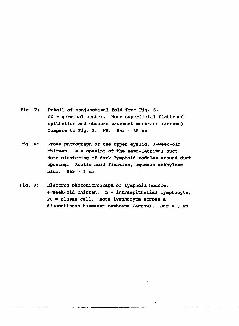
Detail of conjunctival fold from Fig, 6.
GC = germinal center. Note superficial flattened
epithelium and obscure basement membrane (arrows).
Compare to Fig. 2. HE. Bar = 25 pm
Gross photograph of the upper eyelid, 3-week-old
chicken. N = opening of the naso-lacrimal duct.
Note clustering of dark lymphoid nodules around duct
opening. Acetic acid fixation, aqueous methylene
blue. Bar = 3 mm
Electron photomicrograph of lymphoid nodule,
4-week-old chicken. L = intraepithelial lymphocyte,
PC = plasma cell. Note lymphocyte across a
discontinous basement membrane (arrow). Bar = 3 pm


Fig. 10: Electron photomicrograph of epithelium over a
lymphoid nodule, 2-week-old chicken.
I = intermediate filaments, L = intraepithelial
lymphocyte. Note irregular microvilli, prominent
intercellular junctions between epithelial cells
(arrows), and apical vesicles (arrowheads).
Bar = 1 im


Fig. 11: Electron photomicrograph of a subepithelial
capillary, 1-day-old chicken. E = endothelial cell,
L = lymphocyte. Note long endothelial processes
contacting lymphocyte. Bar = 2 #m
Fig. 12: Electron photomicrograph of a high endothelial
venule, 4-week-old chicken. E = endothelial cell,
L = lymphocyte, R = red blood cell in venule lumen.
Note thickness of endothelium compared to Fig. 11.
Bar = 3 pm


70
DISCUSSION
CALT In chickens has morphologic characteristics which
are similar to other mucosa-associated lymphoid tissues. 6ALT
and BALT both consist of heterogenous nodules of lymphocytes
and macrophages within a loose stroma, an overlying attenuated
epithelium composed of cells with short microvilli, epithelial
infiltration by lymphocytes, and relatively few goblet
cells.These tissues typically contain specialized
M-cells which allow the uptake of antigen across the
epithelial surfaice.^®'^^®'^®^ In addition to characteristic
lymphoid nodules and epithelium, mucosal lymphoid tissues
contain HEV which are specialized for regulating the migration
o f l y m p h o c y t e s i n t o s p e c i f i c l y m p h o i d t i s s u e s . S i n c e
CALT in chickens has many of the above characteristics, it
likely has a function in mucosal immunity.
The location and general structure of CALT in chickens
are similar to CALT in turkeys.?! In both species, CALT is
present most prominently in longitudinal folds and fissures of
the lower eyelid, appears shortly after hatching, and has
similar epithelial and vascular histomorphologic features.?!
Although CALT has not been described in turkeys over 33 days
of age,71 older turkeys also have features of CALT similar to
the older chickens in the present report (A. S. Fix, personal
observation).
Differences exist between CALT in chickens and in
turkeys. The most apparent differences include more prominent
lymphoid nodules in the chicken upper eyelid and the
clustering of these nodules around the opening of the
naso-lacrimal duct, since this duct drains ocular secretions,
antigens and microorganisms in these secretions would make
contact with lymphoid tissue around the duct opening. Mucosal
lymphoid tissue in this location may optimize the chance for
antigen contact and uptake. Other characteristics of chicken

71
CALT not found in turkey CALT^^ include diffuse lymphoid
infiltrates along the proximal conjunctiva near the lower
eyelid fornix, a prominent plasma cell component, epithelial
goblet cell metaplasia in both upper and lower eyelids, and
superficial subepithelial capillaries with long endothelial
processes. In chickens, CALT development was prominent at an
earlier age than in turkeys.?!
CALT in chickens is less similar to CALT in rabbits and
in guinea pigs than to CALT in turkeys. In contrast to the
relatively early development of CALT in chickens, CALT in
rabbits does not appear until after 4 weeks of age.l^
Additionally, chicken CALT has prominent germinal centers, an
attenuated epithelium, numerous plasma cells, and HEV. None
of these features are described in CALT of rabbits or of
guinea pigs.^^'^^ Rabbit CALT has prominent lymphocyte-packed
lymphatic channels not seen in the chickens of the present
study.However, the greater number of upper eyelid
conjunctival lymphoid nodules in our chickens more closely
resembles the distribution of CALT in rabbits than in
turkeys.
The characteristic morphologic features of CALT
development in chickens over the 16 weeks of this study may
suggest a change in function. After hatching, conjunctival
lymphoid infiltration and epithelial modification occurred
gradually until development peaked at 3 to 6 WPH. However,
after 12 WPH, the epithelium was once again columnar with
goblet cells, had a more prominent basement membrane, and
contained less numerous lEL. In addition, CALT germinal
centers were more basal in nodules and were surrounded by a
collagenous circumferential stroma. Also concurrent with CALT
development was the progressive increase in plasma cells
within and below the epithelium. These morphologic
differences in CALT between younger and older chickens suggest
the early ability of CALT to sample and react to antigens in

72
the conjunctival space, with a later emphasis on the delivery
of antibody to the conjunctival surface from superficial
plasma cells.
Considerable evidence exists supporting a role for the
chicken HarderIan gland in local paraocular immunity. Maximal
infiltration by plasma cells into this gland occurs by about 6
weeks of age. By Immunofluorescence, most of these cells
stain for IgM in chickens under 4 weeks of age, IgG- and
IgA-posltlve cells occur with equal frequency until 11 weeks
of age, and IgA-posltlve cells predominate in older chickens.^
Several reports have described lymphoid follicle formation and
specific antibodies in HarderIan gland secretions after
eyedrop delivery of antigens such as bovine serum albumin,
sheep red blood cells, Newcastle disease virus, and infectious
bronchitis virus.153,18i However, detailed understanding
of the Immune response to these antigens and Harderian gland
infiltration with plasma cells is currently lacking.
CALT in chickens may serve as a site for antigen uptake
and processing, followed by localization of antibody-producing
plasma cells to the Harderian gland. A similar role has
recently been proposed for CALT in rabbits, guinea pigs, and
turkeys.If this is also true for chickens, some of
the Increased appearance of plasma cells and specific antibody
in the Harderian gland after eyedrop delivery of antigen may
be explained.Based on this hypothesis, antigen
introduced along the conjunctival epithelium would contact the
subepithelial lymphoid population after crossing the
specialized epithelium in CALT by a currently unknown
mechanism. Subsequent to the generation of an Immune
response, committed B-cells would mature into
antibody-producing plasma cells, localize within the Harderian
gland, and deliver antibody into Harderian secretions. CALT
would then provide the affector function in local paraocular

73
immunity, with the effector function provided by the plasma
cell-rich Harderian gland.
This study has characterized structural features of CALT
in chickens and documented a similarity to many mucosal
lymphoid tissues in other species. CALT therefore has a
probable role in mucosal immunity. Based on previous
documentation of plasma cells in and antibody production by
the chicken Harderian gland, CALT may serve as a site of
antigen uptake within the conjunctiva. If this proves to be
true, a better understanding of the interaction between
nonparenteral vaccines and CALT may enhance the control of
economically significant respiratory diseases in chickens.

74
PART III. PARTICLE UPTAKE BY CONJUNCTIVA-ASSOCIATED LYMPHOID
TISSUE (CALT) IN TURKEYS

75
PARTICLE UPTAKE BY
CONJUNCTIVA-ASSOCIATED LYMPHOID TISSUE (CALT)
IN TURKEYS
A. S. Fix and L. H. Arp
Accepted for publication in Avian Diseases
From the Department of Veterinary Pathology, Iowa State
University, Ames, Iowa 50011

76
ABSTRACT
Uptake of tracer particles was assessed in lower eyelid
conjunctiva-associated lymphoid tissue (CALT) of 3-week-old
turkeys. Tracer particle suspensions (carbon, iron oxide, or
three sizes of latex beads - 0.81 fm, 1.7 /im, and 2.9 urn) were
placed into the experimentally sealed conjunctival space.
After 5, 15, or 30 minutes, eyelids were removed and CALT was
examined by light microscopy. Uptake was confirmed for all
tracers and occurred within the lymphoepithelium of CALT. The
uptake of latex beads was not as frequent as for carbon and
iron. Tracers were increasingly evident in lymphoepithelium
and subepithelial macrophage clusters as contact time
increased. These findings provide additional evidence that
CALT is capable of antigen uptake and may play a role in
paraocular and upper respiratory immunity in turkeys.

77
INTRODUCTION
Conjunctiva-associated lymphoid tissue (CALT) is a
recently described entity in turkeys that has many of the
structural features of mucosa-associated lymphoid tissues.?!
CALT is associated with distinct folds and fissures in the
proximal region of the lower-eyelid conjunctiva. Although not
present at hatching, CALT develops into prominent lymphoid
nodules by 3 weeks of age. Morphologic features include dense
lymphoid infiltrates below a flattened lymphoepithelium,
intraepithelial lymphocytes, lack of goblet cells, and high
endothelial venules.?! CALT in turkeys resembles that
described in rabbits and guinea pigs^^'^^'^^ and undergoes
hyperplasia in response to local Bordetella avium infection.
The location and structural similarity of CALT with other
mucosal lymphoid tissues, including gut-associated lymphoid
tissue (6ALT) and bronchus-associated lymphoid tissue
(BALT),^^ has led to the proposal that CALT is a functional
component of the mucosal immune system.13,47,71,78
Mucosal lymphoid tissues are important in the defense of
mucosal surfaces through the uptake of microbial antigens and
the generation of a protective antibody response, primarily in
the form of secretory IgA.^^ These tissues contain a
specialized epithelium that is adapted for the transepithelial
uptake of antigens and microorganisms.^®^ A unique cell type,
the M-cell, performs this function in the human
gastrointestinal tract.similar epithelial types are
described in other tissues and are referred to as a follicle-
associated epithelium or lymphoepithelium.Many studies
have documented the uptake of tracer particles (carbon, latex
beads, horseradish peroxidase) by the lymphoepithelium of
mucosal lymphoid tissues.183,198 Additional studies
have demonstrated that the uptake of viruses,bacteria,
and protozoa^*! also occurs.
78
Vaccination via mucosal exposure is critical for
p r o t e c t i v e i m m u n i t y a g a i n s t m a n y i n f e c t i o u s d i s e a s e s . T h e
avian Harderian gland, a tubulo-alveolar gland containing
plasma cells and secretory IgA, has been recognized as having
a major role in local ocular immunity.However, this
gland lacks many structural features of mucosal lymphoid
tissues, including a distinct lymphoepithelium, prominent
intraepithelial lymphocytes, and high endothelial venules. In
addition, although carbon tracer enters the duct of the '
Harderian gland after experimental eyedrop delivery, uptake
across the epithelium was not detected.The present study
was designed to determine if tracer particle uptake occurs in
CALT of turkeys and if uptake is influenced by tracer type and
time of exposure.

79
MATERIALS AND METHODS
Fifty 1-day-old Nicholas strain broad-breasted white
turkeys were obtained from a commercial hatchery (Midwest
Turkey Hatchery, Dike, Iowa) and housed in an isolated room
away from other animals within a well-ventilated indoor
laboratory animal facility. Turkeys were kept in temperature-
controlled brooders and were given starter ration and water ad
libitum. No vaccinations were administered at the hatchery or
during the experiment.
Tracer particle suspensions (carbon, iron, latex beads)
were prepared to assess uptake by conjunctiva-associated
lymphoid tissue (CALT) in the lower eyelid. For carbon, 1 ml
of black India Ink (Faber-Castell Corp., Newark, New Jersey)
was diluted in 20 ml phosphate-buffered saline (PBS) and
centrifuged at 5000 x g for 30 minutes. After the supernatant
was discarded, the pellet was resuspended in 10 ml PBS. For
iron, 100 mg of black iron oxide particles (0.5 urn diameter,
Polysciences, Inc., Warrington, Pennsylvania) was suspended in
10 ml PBS. For latex beads, three different diameters were
selected (0.8 fm, 1.7 im, and 2.9 pm). One ml of stock
suspension from each size (Polystyrene Microspheres, 2.5%
solids-latex, Polysciences, Inc.) was centrifuged at 3000 x g
for 10 minutes, and, after the supernatant was discarded, the
pellet was resuspended in 10 ml PBS. All tracer suspensions
were warmed to approximately 38 C before use.
An experimental technique was developed to control tracer
particle suspension contact with CALT. At 3 weeks of age,
turkeys were anesthetized with sodium pentobarbital, and
eyelid margins were sealed bilaterally with adhesive (Duro
Superglue-5, Woodhill Permatex, Cleveland, Ohio). Each naso
lacrimal duct was ligated with a single subcutaneous suture
anterior to the nasal palpebral commissure to seal the
conjunctival space. Preliminary light microscopic evaluation

80
of eyelids prepared by this technique ensured that the
conjunctival space was closed and CALT was not morphologically
damaged. Tracer suspensions were injected by syringe with a
22-gauge needle through the upper eyelid into the sealed
conjunctival space of the right eye. Sufficient tracer
suspension was injected to ensure adequate contact with CALT
(1 ml). Each tracer suspension was injected into the
conjunctival space of nine turkeys. For negative controls,
PBS without tracer was injected into the left sealed
conjunctival space of each bird. For positive controls and to
assess the microscopic appearance of tracer material in liver
and spleen, five additional turkeys received 0.5 ml of each
tracer suspension intravenously.
At 5, 15, and 30 minutes postinjection (PI), three
turkeys from each tracer group were sacrificed with an
intravenous overdose of sodium pentobarbital. Lower eyelids
were removed and fixed in 10% neutral buffered formalin.
After 24 hours, the middle of each eyelid was trimmed in
cross-section perpendicular to the eyelid margin. Positive
control turkeys receiving intravenous tracer were sacrificed
in a similar manner at 5 minutes PI. Liver and spleen were
harvested immediately and fixed in 10% formalin. All tissues
exposed to carbon and iron were processed by routine paraffin
technique, sectioned at 5 fin, and stained with hematoxylin and
eosin (HE) for light microscopy or fast green (FG) for
photomicrography.Tissues exposed to latex beads were
embedded in plastic (glycol methacrylate, 6MA Kit, Polaron,
Cambridge, Massachusetts), sectioned at 2 urn, and stained with
0.1% aqueous toluidine blue (TB) as described.
A minimum of 10 sections from each block were examined by
light microscopy. Observations were recorded with particular
attention to tracer location within CALT at the specific
intervals PI. Microscopic assessment of uptake was restricted

81
to the CALT lynphoepithellvun and nonlynphold epithelium in
proximal conjunctival folds and fissures of the lower eyelid.

82
RESULTS
All tracer particles had a characteristic histologic
appearance in positive control tissues (liver and spleen). In
liver, tracers were located in Kupffer cells along hepatic
sinusoids. Similarly, splenic tracer particles were confined
to phagocytic cells associated with sheathed arteries
(Schweiger-Seidel sheaths). Carbon particles were numerous,
appeared as uniform 1 to 2 pm light-brown granules, and
occasionally formed large clumps filling cell cytoplasm
(Fig. 1). Iron particles were black and irregular, with a
tendency to clump into aggregates of 5 /xm diameter or more.
Fast green (FG) stain provided better contrast for tracer
visualization than hematoxylin and eosin (HE). However, FG
provided poor cellular detail of the tissue. In plastic
sections stained with toluidine blue (TB), latex beads
appeared as small, round to oval, clear cytoplasmic spaces.
By adjusting the microscope condenser to enhance contrast,
latex beads were distinctly refractile and easily identified.
Generally, the number of beads within a given cell was
inversely proportional to the size of the bead. Cellular
detail in TB-stained plastic sections was superior to those
embedded in paraffin. No tracer material was observed in any
eyelid section from saline-injected negative controls.
Carbon, iron oxide, and latex beads were located
primarily within or below the conjunctiva-associated lymphoid
tissue (CALT) lymphoepithe1ium, although occasionally these
tracers were also seen in the more typical nonlymphoid
epithelium of the conjunctiva. The appearance of carbon and
iron oxide particles in CALT was similar to positive control
liver and spleen sections, although some larger aggregates of
iron oxide (greater than 6.0 (m) were displaced artifactually
by sectioning. This was verified by evaluation of additional
serial sections. By 30 minutes post-injection (PI), increased

83
numbers of heterophils were observed near the conjunctival
epithelium in a few sections.
At 5 minutes PI, carbon and iron oxide particles were
within the CALT lymphoepithelium in approximately one-half of
the sections examined. These tracers appeared as small (2 to
4 /Ltm) granules (Fig. 2). In a few areas, carbon and iron
oxide particles were concentrated in small clumps (5 to 8 fim)
immediately below the lymphoepithelium. By 15 minutes PI,
larger and more distinct clumps (10 to 15 (xm) were typical
(Fig. 3). These areas were located further below the
lymphoepithelium than at 5 minutes and suggested localization
in subepithelial macrophages. At 30 minutes PI, tracer
particles were more dispersed within CALT and were in deeper
sites than at previous times (Fig. 4). The distinct clumping
observed earlier was not as evident. Occasional tracer-laden
phagocytic cells (macrophages and heterophils) were seen above
the lymphoepithelial surface or free in the conjunctival
space. Small, thin cytoplasmic strands extended between these
cells and the underlying lymphoepithelium.
Localization of all three sizes of latex beads in the
CALT lymphoepithelium was less common than for carbon and
iron. Generally, 0.81 nm beads were located superficially,
near the top of lymphoepithelial folds. However, the 1.7 ̂ m
and 2.9 urn beads were located in the lymphoepithelium near the
base of conjunctival fissures between adjacent folds. The
small 0.81 /xm latex beads were seen in dense clusters below
the superficial lymphoepithelium only at 30 minutes PI (Fig.
5), although occasional solitary beads were also observed. A
solitary 1.7 fin bead was seen in the lymphoepithelium of one
section at 5 minutes PI. Other sections contained solitary
and loose clusters of beads within and below the
lymphoepithelium at 30 minutes (Fig. 6). The 2.9 pm beads
were solitary and within lymphoepithelial cells at 5 minutes
PI. At 30 minutes PI, several of these large beads were also

84
in loose clusters below the lymphoepithelium (Fig. 7). The
location of the 1.7 fm and 2.9 urn beads in the conjunctival
fissure lymphoepithelium was in contrast to the superficial
lymphoepithelial location of carbon, iron oxide, and 0.81 im
beads.

Figures 1 through 4. Uptake of carbon and iron oxide in
3-week-old turkeys
Fig. 1: Liver, positive control, intravenous carbon. Note
clustering of carbon in Kupffer cells lining
sinusoidal endothelium. F6. Bar = 10 pm
Fig. 2: Conjunctiva-associated lymphoid tissue, top of
lymphoepithelial fold, carbon, 5 minutes PI. Tracer
is primarily intraepithelial (arrow). F6.
Bar = 10 fim
Fig. 3: Conjunctiva-associated lymphoid tissue, top of
lymphoepithelial fold, carbon, 15 minutes PI.
Tracer is clustered in subepithelial macrophages
(arrow). FG. Bar = 10 pm
Fig. 4: Conjunctiva-associated lymphoid tissue, top of
lymphoepithelial fold, iron oxide, 30 minutes PI.
Tracer is deeper in lymphoid tissue and more
dispersed than at earlier times (arrow). FG.
Bar = 10 (im


Figures 5 through 7. Uptake of latex beads in 3-week-old
turkeys
Fig. 5: Conjunctiva-associated lymphoid tissue, top of
lymphoepithelial fold, 0.81 jum latex beads,
30 minutes PI. Note clustering of numerous beads in
a subepithelial macrophage (arrow). Solitary beads
are also present. TB. Bar = 10 fm
Fig. 6: Conjunctiva-associated lymphoid tissue, lateral
fissure, 1.7 nm latex beads, 30 minutes PI.
Intraepithelial and subepithelial beads are solitary
and in loose clusters (arrow). TB. Bar - 10 nm
Fig. 7; Conjunctiva-associated lymphoid tissue, lateral
fissure, 2.9 pm latex beads, 30 minutes PI. Beads
are primarily solitary (arrows) and in
intraepithelial and subepithelial sites. TB.
Bar = 10 /im


89
DISCUSSION
The experimental tracer particle studies evaluated in the
present report support our earlier proposal that conjunctiva-
associated lymphoid tissue (CALT) in turkeys has a role in
mucosal immunity.We have shown that the location of
carbon, iron oxide, and latex bead tracers is restricted to
the lymphoepithellum of CALT. Tracer localization within the
lymphoepithellum of a mucosal lymphoid tissue is consistent
with uptake of that tracer and structural modifications within
CALT probably correlate with this function. The overall
location in proximal conjunctival folds, the characteristic
flattened lymphoepithellum, and intraepithelial lymphocytes
all may facilitate contact between material in the
conjunctival space and lymphoid cells. Tracer uptake has been
documented in other mucosal immune system tissues, including
gut-associated lymphoid tissue (GALT)®^'^®^'^®® and bronchus-
associated lymphoid tissue (SALT).183,192 interestingly,
although the structure of CALT in rabbits and guinea pigs
resembles CALT in turkeys, 13,47,71,78 tracer uptake is
apparently not selective for the lymphoepithelial cells of
guinea pig CALT.^^® In that study, experimental conjunctival
delivery of horseradish peroxidase produced uptake in both
lymphoid and nonlymphold epithelial areas. This is in direct
conflict with the present study, in which uptake was
restricted to the lymphoepithellum of CALT.
Differences in the frequency and site of uptake in our
study are likely a function of tracer size and composition.
Trapping between conjunctival folds may have facilitated
contact between the larger beads and the epithelium. The size
of the larger beads may have limited phagocytosis of multiple
beads by one macrophage. Because surface adsorption is
apparently a prerequisite for uptake,latex may not produce
the same surface interaction between particle and epithelial

90
cell that occurs with carbon and iron. Uptake is also
apparently influenced by the relative activity of nearby
lymphoid tissue,surface charge,and tracer
concentration.
The precise cellular mechanism of uptake could not be
determined with the histological observations in the present
study. Although tracers appeared to be intraepithelial, the
abundant intraepithelial lymphocyte population in CALT made
the determination of exact cellular location difficult. An
"intraepithelial" location was not necessarily synonymous with
actual localization in the cytoplasm of an epithelial cell.
Tracer particles could be in epithelial cell cytoplasmic
vacuoles, within the processes of intraepithelial phagocytic
cells, or between adjacent epithelial cells. Endocytosis and
vesicle transport are the most common way that particles are
taken up by GALT,^^° BALT,^®^ and the bursa of Fabricius.^®
Transepithelial endocytosis could have occurred in our
turkeys, since several of the larger beads appeared to be
directly in epithelial cell cytoplasm. However, localization
is also possible within macrophage pseudopods extended between
epithelial cells, as described in Giardia muris uptake in
mouse Peyer's patch epithelium.Localization between
adjacent epithelial cells would require disruption of
epithelial cell tight junctions. This is less likely but has
been observed with surgical manipulation in the guinea pig
small intestine.It is also possible that different tracers
used in the present study were taken up by different
mechanisms. Ultrastructural studies are required to
definitively determine how tracer particles cross the
lymphoepithelium of CALT in turkeys.
The detection of tracer-laden macrophages deeper in CALT
after longer exposure time suggests active movement and
migration of these cells within tissue. An interesting
finding in the present study was the presence of tracer-laden

91
phagocytic cells on the lymphoepithelial surface and within
the conjunctival space. Similar findings were reported when
Vibrio cholerae was used to detect microbial uptake in rabbit
Peyer's patches.The movement and migration of phagocytic
cells after uptake may have significance for antigen
presentation and the generation of an immune response.
The experimental procedure developed in the present study
was designed to maximize the opportunity for tracer uptake to
occur, not to simulate conditions likely to be encountered in
the field. The confirmation and characterization of uptake in
CALT was our primary objective before attempting more applied
experimentation. Admittedly, several aspects of the
procedure, including anesthesia, surgical trauma, tracer
delivery method, and the restriction of normal ocular
movements, may have influenced the outcome. However, we
believe that the uptake of biological materials via CALT is
quite possible and may occur during natural infection of
paraocular tissues and administration of eyedrop and
aerosolized vaccines.
The uptake of tracer particles by CALT in the present
study provides additional evidence that CALT is a component of
the mucosal immune system and strengthens the specific role
proposed for CALT in mucosal immunity of turkeys.?! In the
turkey Harderian gland, secretory IgA and plasma cells are
present,and the importance of this gland in avian
paraocular immunity is recognized.In chickens,
conjunctival delivery of antigen produces an increase in
Harderian gland plasma cells and specific antibody in ocular
secretions., 153,lei suggest that under field conditions,
uptake of environmental microorganisms and vaccines through
CALT results in the development of an immune response. This
uptake may be responsible for a portion of the antibody
production by the Harderian gland that contributes to
protective immunity. The improved development of vaccines and

92
vaccination protocols specifically targeting CALT may enhance
our attempt to control infectious disease in turkeys.
93
PART IV. QUANTIFICATION OF PARTICLE UPTAKE BY
CONJUNCTIVA-ASSOCIATED LYMPHOID TISSUE (CALT)
IN CHICKENS
94
QUANTIFICATION OF PARTICLE UPTAKE BY
CONJUNCTIVA-ASSOCIATED LYMPHOID TISSUE (CALT)
IN CHICKENS
A. S. Fix and L. H. Arp
Accepted for publication in Avian Diseases
From the Department of Veterinary Pathology, Iowa State
University, Ames, Iowa 50011

95
ABSTRACT
Tracer particle uptake by conjunctiva-associated lymphoid
tissue (CALT) was quantified in the lower eyelids of l, 3, 5,
and 7-week-old broiler chickens. CALT was measured
histologically by computerized image analysis in all birds and
in additional 1-day-old and 16-week-old chickens not subjected
to uptake assessment. Suspensions of carbon or iron oxide
were placed in contact with CALT for 5, 15, or 30 minutes
(contact time). After eyelid removal, tracer uptake was
scored by light microscopy, CALT was measured, and a
mathematically-derived uptake index was evaluated
statistically. At each age examined, computer-generated
measurements showed a significant increase in the proportion
of CALT lymphoepithelium within proximal eyelids. Tracer
uptake was confined to CALT lymphoepithelium at all ages and
at all contact times. Uptake increased significantly between
3 and 5 weeks of age and between 5 and 15 minutes of tracer
contact. Based on these uptake data for CALT in chickens, a
minimal age-specific maturity is suggested which may influence
function in mucosal immunity.

96
INTRODUCTION
Conjunctiva-associated lymphoid tissue (CALT) is located
primarily in the lower eyelid in turkeys,?! chickens,7%
rabbits,and guinea pigs.*? In chickens and turkeys, CALT
develops after hatching in association with distinct folds and
fissures in the proximal conjunctiva.71,72 cALT develops,
these conjunctival folds become progressively expanded by
lymphoid nodules and develop a flattened epithelium containing
lymphocytes and lacking goblet cells. The epithelium
associated with these lymphoid follicles resembles specialized
cells important for antigen uptake in other mucosal lymphoid
tissues and is termed a follicle-associated epithelium or
lymphoepithelium (LE)During the post-hatching period, LE
appears concurrently, with the development of lymphoid nodules
in proximal conjunctival folds and the original nonlymphoid
epithelium (nonLE) gradually diminishes. CALT in chickens and
turkeys7i'72 morphologically resembles other mucosal lymphoid
tissues^®'^*'70 which are important for the immunologic
protection of mucosal surfaces.
Tracer particle uptake is associated with mucosal
lymphoid tissue LE and has been demonstrated experimentally
using a wide variety of materials. 183,202 However, limited
work has been done with CALT. The uptake of horseradish
peroxidase has been documented in guinea pig conjunctiva, but
no selectivity for CALT LE was found.In contrast, we
recently characterized the selective uptake of carbon, iron
oxide, and latex beads by CALT LE in 3-week-old turkeys.73 in
our study, tracers were intraepithelial and clustered in
subepithelial macrophages within 5 minutes of contact.
However, as in most other previous assessments of uptake by
mucosal lymphoid tissues, we made no attempt at quantification
or in determining age effects on uptake. The objectives of
the present study were to use computer-assisted measurements

97
to assess tracer uptake by CALT in chickens and to
statistically analyze effects due to age, tracer contact time,
and tracer particle type.

98
MATERIALS AND METHODS
Eighty-two l-day-old broiler chickens were obtained from
a local hatchery (Hoover Hatchery, Rudd, Iowa) after debeaking
and subcutaneous administration of a killed Marek's disease
virus vaccine in the dorsal cervical region. Chickens were
housed in a well-ventilated room in an indoor laboratory
animal facility concurrently housing no other chickens. After
2 weeks in temperature-controlled brooders, chickens were
moved to wire cages for 3 weeks and then placed on concrete
flooring with sawdust litter until the end of the experiment.
Free access to fresh water and feed was provided at all times.
Tracer particle uptake was quantitatively assessed using
two tracers (carbon and iron) in chickens of four ages (1, 3,
5, and 7 weeks) at three contact times (5, 15, and
30 minutes). Three chickens were used for each treatment
combination of age, contact time, and tracer type. Computer-
generated morphologic measurement of lower eyelid CALT nonLE
and LE was performed in all of the above chickens and in
additional l-day-old (n = 5) and 16-week-old (n = 5) chickens.
Tracer particle suspensions of carbon and iron oxide were
prepared to assess uptake by CALT in the lower eyelid. For
carbon, 1 ml of black India Ink (Faber-Castell Corp., Newark,
New Jersey) was diluted in 20 ml phosphate buffered saline
(PBS, pH 7.2) and centrifuged at 5,000 x g for 30 minutes.
After discarding the supernatant, the pellet was resuspended
in 10 ml PBS. For iron, 100 mg of black iron oxide particles
(0.5 nm diameter, Polysciences, Inc., Warrington,
Pennsylvania) was suspended in 10 ml PBS. Tracer solutions
were warmed to approximately 38 C prior to use.
At 1, 3, 5, and 7 weeks of age, 18 chickens were
anesthetized with sodium pentobarbital and eyelid margins were
sealed with adhesive bilaterally (Duro Superglue-5, Woodhill
Permatex, Cleveland, Ohio). Each naso-lacrimal duct was

99
llgated with a single subcutaneous suture anterior to the
nasal palpebral commissure to seal the conjunctival space.
One ml of each tracer suspension was injected by syringe
(22 gauge needle) through the right upper eyelid into the
sealed conjunctival space of 9 chickens in each age group.
Sufficient tracer suspension was injected to ensure adequate
contact with CALT. For controls, PBS without tracer was
injected into the left sealed conjunctival space of each bird.
Within each treatment combination of age and tracer type,
3 chickens were euthanatized with sodium pentobarbital after
5, 15, or 30 minutes of contact time. Lower eyelids were
removed and fixed in 10% neutral buffered formalin. After
24 hours, the middle of each eyelid was trimmed in cross-
section perpendicular to the eyelid margin. Eyelids were
processed by routine paraffin technique after graded alcohol
dehydration. Twenty-five serial sections were cut at 5 ixm
intervals from each block and divided into 5 consecutive
groups. Within each group, the first section was stained with
hematoxylin and eosin (HE), the second section was stained
with fast green (FG), and the remaining 3 sections were
discarded. Microscopic assessment of tracer particle uptake
was restricted to lymphoepithelium (LE) and
nonlymphoepitheliùm (nonLE) in proximal conjunctival folds and
fissures of the lower eyelid (Fig. 1).
A measurement (in mm) of the surface contour distance
along CALT nonLE and LE was determined in each section with a
computerized image analysis system (Zeiss SEN-IPS Image
Analysis System, Zeiss-Kontron;IBAS Version 1.3, Thornwood,
New York). Using the HE sections described above,
conjunctival folds and fissures containing CALT were imaged
with computer digitalization under 2.0 x magnification (Fig.
1). The eyelid image was discriminated in shades of gray and
the surface contour was traced. This tracing converted the
eyelid surface contour to a single line representing the

100
linear surface distance (Fig. 2). By simultaneously comparing
the computer-generated line with the actual tissue section
(performed by A. S. Fix), regions of LE and nonLE were
differentiated and the single line tracing was cut
appropriately (Fig. 2, dotted lines). The computer then
generated a numerical surface contour distance in mm for nonLE
and LE in each section. To assess trends in CALT development
with age, the surface contour distance in mm was converted to
percent nonLE and LE in each section. Preliminary studies
comparing the first and last sections from a given block
revealed no significant differences in surface contour
distance. Therefore, only the middle HE section from each
block was measured by computer.
All F6 sections were examined by light microscopy, and
tracer particle uptake was scored numerically (performed by A.
S. Fix) for each site of uptake as follows: 1 = individual
tracer particle, 2 = small cluster of less than 10 particles,
3 = large cluster of greater than 10 particles (Fig. 3).
Tracer particles had to clearly be within or below the
superficial epithelium in order to be counted. Uptake was
scored separately for nonLE and LE within each eyelid.
An uptake index was generated for both nonLE and LE in
each section examined by dividing the uptake score by the
respective surface contour distance in mm. This adjusted the
numerical uptake score for the variability in CALT between
sections. Uptake indices were analyzed by analysis of
variance (ANOVA) using the general linear models procedure
(PROC GLM) with SAS software (Statistical Analysis System
Institute, Inc., Gary, North Carolina). Data were considered
in a factorial experimental design with 24 treatments (4 ages
X 3 contact times x 2 tracers). To assess morphologic changes
in CALT with age, additional ANOVA was performed on computer-
generated surface distance data.

Proximal conjunctival folds and fissures,
conjunctiva-associated lymphoid tissue, lower
eyelid, 3-week-old chicken. Note distention of left
two folds by lymphoid nodules. HE. Bar = 1 mm
Computer-generated tracing of surface contour from
Fig. 1. Dotted lines Indicate demarcation of
lymphoeplthellum (LE) from nonlymphoeplthellum
(nonLE) bordering conjunctiva-associated lymphoid
tissue (CALT)
Subepithelial clusters of carbon tracer below the
lymphoeplthellum of conjunctiva-associated lymphoid
tissue, 3-week-old chicken. Uptake was scored based
on particle density at each site of uptake:
individual particles = 1 (not shown); small clusters
of <10 particles = 2 (arrowhead); larger clusters of
>10 particles = 3 (arrow). FG. Bar = 10 fxm

•hawtAî
%
3

103
RESULTS
A significant increase in the lynphoepithelium (LE)
surface contour distance existed in proximal conjunctival
folds between all ages examined (P > 0.05). Significant
differences were detected for measurements in mm and
percentage LE (Fig. 4). Increases in percentage LE occurred
by 3 weeks of age and over 80% of proximal conjunctival
epithelium was LE by 7 weeks of age. Chickens 16 weeks of age
had 100% LE in proximal conjunctival folds.
Carbon and iron tracers were observed primarily within
and below the LE of CALT along proximal conjunctival folds in
fast green (F6) sections (Fig. 3). Tracer particles were
intraepithelial as well as in subepithelial clusters, probably
within macrophages. The localization of carbon particles was
more consistent than for iron particles, since some surface-
adherent iron was displaced into deeper tissue by sectioning.
The consistency of tracer localization in CALT was verified by
examination of adjacent hematoxylin and eosin (HE) and FG
sections.
The uptake index for LE was significantly greater than
for nonlymphoepithelium (nonLE) when averaged over all
treatment groups (P = 0.01). Within LE, significant
differences were detected in uptake indices for age, contact
time, and tracer type (Table 1). The mean uptake index
increased significantly between 3 and 5 weeks of age and 5 and
15 minutes of contact time. Carbon produced a significantly
higher mean uptake index in LE than iron. When iron tracer
was excluded from statistical analyses and only carbon data
were assessed, the differences detected for age and time were
similar as described above and had a higher level of
significance.

Fig. 4: Percentage of conjunctival folds and fissures occupied by
chicken conjunctiva-associated lymphoid tissue (CALT)
lymphoepithelium (LE) and nonlymphoepithelium (nonLE) at
specific weeks post-hatching. LE = black; nonLE = light
gray; age 0=1 day post-hatching. Percentages represent
computer-generated measurements of LE and nonLE surface
contour distance in histologic cross section

Percentage
- * - r o o ) - ^ c n o > ^ o o c o o o o o o o o o o o o o
f
> <g w (D
cn (0
a>
SOT

106
Table 1. Effects of age, contact time, and tracer type on
particle uptake by conjunctiva-associated lymphoid
tissue in chickens
Treatment Groupé Uptake Index®
Age 1 week 1.3*
3 weeks 1.6®
5 weeks 6.1%
7 weeks 6.2%
Contact time 5 minutes 2.3®
15 minutes 4.6%
30 minutes 4.6%
Tracer carbon 5.3®
iron 2.3%
^Statistical comparisons valid only within each treatment
group.
^Mean uptake index. Values within each treatment group
followed by different letters are significantly different
(P < 0.05). Standard error of the mean for each treatment
group: age = 0.29, contact time = 0.26, tracer = 0.21.

107
DISCUSSION
This Study confirmed that conjunctiva-associated lymphoid
tissue (CALT) in chickens undergoes an age-dependent increase
in size and that the uptake of particulate material within the
conjunctival space occurs through CALT. Uptake occurs
primarily through lymphoepithelium (LE), increases between 3
and 5 weeks of age, and is quite rapid. These data support
the idea that CALT is a functional component of the mucosal
immune system and that through the uptake of antigen, CALT may
have a role in paraocular and upper respiratory immunity of
chickens.
The qualitative characteristics of carbon and iron uptake
in these chickens closely resembles the uptake of the same
tracers in turkeys.In both species, post-uptake
localization occurred in the folds of CALT in both
intraepithelial and subepithelial sites. Although multiple
ages were not evaluated in the turkeys of our previous study,
uptake was extremely rapid and increased with contact time.?^
The similarity in the results of these two avian studies
suggests common functional features of CALT in birds.
However, a similar study assessing the uptake of horseradish
peroxidase by CALT in guinea pigs failed to demonstrate
selective localization in LE.^^^ The significance of this
species difference is currently unknown.
The quantitative, age-dependent increase in the size of
LE detected by our computer measurements is consistent with
previous qualitative observations of CALT in chickens?^ and
turkeys.71 In both species, lower eyelid CALT is not present
at hatching but is prominent and contains germinal centers by
2 to 3 weeks of age.^^'^^ Similarly, CALT in rabbits is not
evident until after 4 weeks of age.^^ The structure of CALT
in rabbits, turkeys, and chickens^^'^^'^^ is comparable to
other mucosal lymphoid tissues.^* Therefore, the location and

108
accessibility of CALT makes it a valuable model for study of
postnatal development of mucosal lymphoid tissues.
The morphologic change from proximal conjunctival
nonlymphoepithelium (nonLE) to LE correlates with the
increased capacity for particle uptake detected in older
chickens. As chickens age, the nonLE, which is composed of
tall columnar epithelial cells and goblet cells, changes to a
more flattened LE lacking goblet cells but containing
intraepithelial lymphocytes.^^ This epithelial change
probably promotes the capacity for particle uptake, which
occurs through specialized epithelial cells in mucosal
lymphoid tissues.Most studies assessing uptake in mucosal
lymphoid tissues have examined post-neonatal animals, usually
of one Our approach was unique in evaluating
and correlating the simultaneous structural and functional
development of CALT from hatching to 7 weeks of age. Although
the only significant morphologic change in chicken CALT
between 3 and 6 weeks of age is the appearance of plasma
cells,our data suggest that uptake increases during this
period. Therefore, animal age should be strongly considered
when planning and interpreting uptake studies involving
mucosal lymphoid tissues.
Tracer localization within or below the CALT LE in the
present study was remarkably rapid, suggesting that minimal
contact time is necessary to trigger uptake. As with carbon
in the bursa of Fabricius^^° and horseradish peroxidase in
GALT,^^° we found uptake occurring within 5 minutes of contact
time. If similarly fast uptake occurs under field conditions,
then a mechanism exists for rapid sampling of potentially
antigenic materials located within the conjunctival space.
This characteristic is highly advantageous if vaccination
strategies are to be developed for specific immunization
through CALT.

109
Although we have demonstrated that uptake of tracer
particles occurs through CALT in chickens experimentally,
direct evidence that this occurs with antigens delivered under
field conditions is lacking. However, CALT does undergo
lymphoid hyperplasia in turkeys infected with Bordetella
avium.as has been described for BALT in similarly infected
turkeys.In addition, evidence suggests that eyedrop
delivery of antigen results in increased plasma cell
infiltration into and specific antibody response by the
chicken Harderian gland. 41,61,153,18i therefore consider it
likely that CALT functions in antigen uptake and has a role in
the mucosal immune system of chickens. In addition, CALT may
have an age-specific maturity which correlates with this
uptake function.

110
GENERAL SUMMARY
These studies have described the gross, histologic, and
ultrastructural features of conjunctiva-associated lymphoid
tissue (CALT) in turkeys and chickens. In addition, the
uptake of particulate tracer material by CALT has been
documented in both species. This work represents the first
descriptive and experimental assessment of CALT in birds.
Overall, CALT in turkeys and chickens is quite similar in
location and structure to that described in rabbits and guinea
pigs.^^'^^'^^ However, studies concerning the function of CALT
in mammals have been limited.In contrast to the present
avian studies, functional evaluation of CALT in mammals failed
to document selective tracer uptake by CALT
lymphoepithelium.17*
CALT in turkeys and chickens has many features found in
mucosal lymphoid tissues. In these avian studies, CALT was
absent at hatching and progressively developed during the
post-hatching period. Within one week, lymphoid nodules were
evident Within the proximal conjunctival longitudinal folds
and fissures in the lower eyelid of both species. By three
weeks of age, lymphoid nodules were quite prominent and
occupied a large portion of the folds and fissures.
Computerized image analysis documented the statistical
significance of these age-related changes in chicken CALT.
Many epithelial and vascular structural features found in
mucosal lymphoid tissues^^'^^®'^®^'^®^ were evident. The
superficial epithelial cells that formed the lymphoepithelium
were flattened, had prominent intercellular junctions,
contained apical vesicles, and had short, irregular
microvilli. Within the lymphoepithelium, intraepithelial
lymphocytes, a lack of goblet cells, and a discontinuous
epithelial basement membrane were also characteristic.
Vessels associated with CALT, particularly those at the base

Ill
of lymphoid nodules, had high endothelial cells and adherent
intraluminal lymphocytes. Germinal centers and lymphoid
hyperplasia associated with Bordetella avium infection in
turkeys indicated the reactivity of CALT to local antigenic
stimulation. Collectively, similarities between avian CALT
and other mucosal lymphoid tissues strongly suggest a role for
CALT in mucosal immunity.
The uptake of tracer particles documented by these
studies further supports CALT as a component of the mucosal
immune system. Mucosal lymphoid tissues have the unique
ability to sample mucosal surfaces by the transepithelial
uptake of particulate material.in the present studies,
carbon, iron oxide, and latex beads were selectively taken up
across the CALT lymphoepithelium in turkeys. Clustering
within subepithelial macrophages was characteristic after
uptake. Tracer uptake also increased with contact time and
was more extensive in older birds. In addition, the site of
tracer uptake varied with particle size, occurring
superficially along lymphoepithelial folds for smaller
particles but deeper within fissures for larger particles.
These qualitative features of tracer uptake in turkeys were
substantiated by computerized image analysis and quantitative
studies in chickens. A statistically significant increase in
uptake was demonstrated between 5 and 15 minutes of tracer
contact time and between 3 and 5 weeks of age.
The results of these studies offer a unique hypothesis
concerning paraocular immunity in birds, particularly when
interpreted in light of the preexisting literature on the
avian HarderIan gland. Since CALT in turkeys and chickens has
structural features consistent with mucosal lymphoid tissues
and the uptake of tracer material has been documented
experimentally, a role in mucosal immunity is quite likely.
The avian Harderian gland contains predominantly plasma cells
and secretes IgA.®'®^'^®® These experiments Indicate that CALT

112
could provide an initial site of antigen uptake; subsequent
processing and presentation of antigen may lead to an antibody
response that provides mucosal protection. Localization of
plasma cells to the Harderian gland or to other local
epithelial tissues may provide a homing-loop mechanism
specific for protection against upper respiratory pathogens.
The extent to which CALT functions as a mucosal lymphoid
tissue under natural circumstances is entirely unknown. A
number of vaccines for important avian pathogens are
administered by mucosal routes. These include aerosolized
delivery for respiratory contact, oral delivery for intestinal
contact, and eyedrop delivery for ocular contact. Based on
simultaneous exposure of large numbers of birds, these
delivery methods could easily provide vaccine contact with
conjunctival lymphoid tissue. Specific studies examining the
role of CALT in immune responsiveness to these vaccines are
necessary.
Given the age-dependent development and maturation that
occurs in avian CALT, vaccination protocols used in the
poultry industry may require reassessment. For example, some
vaccination programs begin with eyedrop delivery of vaccine in
day-old chicks. Based on the present studies, which indicate
a complete lack of lymphoid tissue in the conjunctiva at one
day of age, the usefulness of this technique may be
questionable. Delay of vaccination until CALT is functionally
more mature would appear more appropriate. However, a
specific maturation time has not been determined and may be
influenced by numerous factors including species, breed,
strain, nutritional status, and local antigenic stimulation.
In addition, the role CALT may have in response to vaccines
may differ for killed and modified-live products.
Many important questions remain to be answered concerning
the role of CALT in avian mucosal immunity. Specific
mechanisms of transepithelial tracer uptake require assessment

113
at the electron microscopic level. The temporal appearance
and isotype specificity of plasma cells in CALT should be
evaluated. The localization of lymphocytes originating in
CALT and the potential homing of those lymphocytes to the
Harderian gland are completely unexplored. Information gained
from further research in these areas will enhance our
understanding of conjunctiva-associated lymphoid tissue in
birds and may improve our control of economically significant
avian diseases.

114
LITERATURE CITED
1. Ackerman 6A, Knouff RA: Lymphocytopoiesls in the bursa
of Fabricius. Am J Anat 104(163-178, 1959
2. Aitken ID, Parry SH: Local immunity in the respiratory
tract of the chicken. I. Transudation of circulating
antibody in normal and virus-infected birds. Immunology
31:33-37, 1976
3. Albini B, Wick 6: Immunoglobulin determinants on the
surface of chicken lymphoid cells. Int Arch Allergy
44:804-822, 1973
4. Albini B, Wick G: Delineation of B and T lymphoid cells
in the chicken. J Immunol 112:444-450, 1974
5. Albini B, Wick G, Rose E, Orlans E: Immunoglobulin
production in chicken Harderian glands. Int Arch Allergy
47:23-34, 1974
6. Allansmith MR: Defense of the ocular surface. Int
Ophthalmol Clin 19:93-109, 1978
7. Allansmith MR, Gillette TE: Secretory component in human
ocular tissues. Am J Ophthalmol 89:353-361, 1980
8. Allansmith MR, Kajiyama G, Abelson MB, Simon MS: Plasma
cell content of main and accessory lacrimal glands and
conjunctiva. Am J Ophthal 82:819-826, 1976
9. Anderson ML, Moore FF, Hyde DM, Dungworth DL: Bronchus
associated lymphoid tissue in the lungs of cattle:
relationship to age. Res Vet Sci 41:211-220, 1986
10. Arai N, Hashimoto Y, Kitagawa H, Kon Y, Kudo N:
Immunohistochemical study on the distribution of lymphoid
tissues in the upper alimentary and respiratory tracts of
chickens. Jpn J Vet Sci 50:183-192, 1988
11. Arp LH, Cheville NF: Tracheal lesions in young turkeys
infected with Bordetella avium. Am J Vet Res
45:2196-2200, 1984

115
12. Atkins AM, Schofield 6C: Lynphoglandular complexes in the
large intestine of the dog. J Anat 113:169-178, 1972
13. Axelrod AJ, Chandler JW: Morphologic characteristics of
conjunctival lymphoid tissue in the rabbit. In;
Immunology and Immunopathology of the Eye. Proceedings of
the 2nd International Symposium, ed. Silverstein AM and
O'Connor 6R, pp. 292-301. Masson and Cie, New York, NY,
1979
14. Bang B6, Bang FB: Localized lymphoid tissues and plasma
cells in paraocular and paranasal organ systems in
chickens. Am J Pathol 53*735-751, 1968
15. Beezhold DH, Sachs H6, Van Alten PJ: The development of
transport ability by embryonic follicle-associated
epithelium. J Reticuloendothel Soc 34:143-152, 1983
16. Befus AO, Johnston N, Leslie 6A, Bienenstock J:
Gut-associated lymphoid tissue in the chicken. I.
Morphology, ontogeny, and some functional characteristics
of Peyer's patches. J Immunol 125:2626-2632, 1980
17. Benedict AA, Berestecky JM: Special features of avian
immunoglobulins. In; Avian Immunology; Basis and
Practice, Vol. 1., ed. Toivanen A and Toivanen P., pp.
114-125. CRC Press, Inc, Boca Raton, Florida, 1987
18. Benson EB, strober W; Regulation of igA secretion by T
cell clones derived from the human gastrointestinal
tract. J Immunol 140:1874-1882, 1988
19. Bienenstock J: Review and discussion of homing of
lymphoid cells to mucosal membranes; the selective
localization of cells in mucosal tissues. In; Recent
Advances in Mucosal Immunity, ed. Strober W, Hanson LA,
and Sell KW, pp. 35-43. Raven Press, New York, 1982
20. Bienenstock J: Bronchus-associated lymphoid tissue. In:
Immunology of the Lung, ed. Bienenstock J, pp. 96-118.
McGraw-Hill, New York, NY, 1984

116
21. Blenenstock J: The mucosal immunologic network. Ann
Allergy 53:535-540, 1984
22. Blenenstock J , Befus AD: Mucosal Immunology. Immunology
41:249-270, 1980
23. Blenenstock J, Befus AD, NcDermott M: Mucosal Immunity.
In: The Mucosal Immune System; Current Topics In
Veterinary Medicine and Animal Science, Vol. 12, ed.
Bourne FJ, pp. 5-27. Martlnus Nljhoff, The Hague, 1981
24. Blenenstock J, Befus D: Gut and bronchus associated
lymphoid tissue. Am J Anat 170:437-445, 1984
25. Blenenstock J, Gauldle J, Perey DYE: Synthesis of IgG,
IgA, IgM by chicken tissues: Immunofluorescent and 14C
amino acid Incorporation studies. J Immunol
111:1112-1118, 1973
26. Blenenstock J, Johnson N: A morphologic study of rabbit
bronchial lymphoid aggregates and lymphoepithelium. Lab
Invest 35:343-348, 1976
27. Blenenstock J, Johnson N, Perey DYE: Bronchial lymphoid
tissue. I. Morphologic characteristics. Lab Invest
28:686-692, 1973
28. Blenenstock J, Johnson N, Perey DYE: Bronchial lymphoid
tissue. II. Functional characteristics. Lab Invest
28:693-698, 1973
29. Blenenstock J, McDermott M, Befus D, O'Neill M: A common
mucosal immunologic system involving the bronchus,
breast, and bowel. Adv Exp Med Biol 107:53-59, 1977
30. Blenenstock J, McDermott MR, Befus AD: The significance
of bronchus-associated lymphoid tissue. Bull Eur
Physlopathol Resplr 18:153-177, 1982
31. Blenenstock J, Perey DYE, Gauldle J, Underdown B: Chicken
IgA: physicochemical and Immunochemical characteristics.
J Immunol 110:524-533, 1973

117
32. Blaese RM, Weiden PL, Koski I, Dooley N: Infectious
agammaglobulinemia: transmission of immunodeficiency with
grafts of agammaglobulinemic cells. J Exp Ned
140:1097-1101, 1974
33. Bland PW, Britton DC: Morphological study of
antigen-sampling structures in the rat large intestine.
Infect Immun 43:693-699, 1984
34. Bockman DE, Boydston WR: Participation of follicle
associated epithelium (FAE), macrophages, and plasma
cells in the function of appendix. In: Scanning Electron
Microscopy, Part III, pp. 1341-1350. SEM Inc., Chicago,
IL, 1982
35. Bockman DE, Cooper MD: Fine structural analysis of
pinocytosis in lymphoid follicle-associated epithelium in
chick bursa and rabbit appendix. Fed Proc 30:511, 1971
36. Bockman DE, Cooper MD: Pinocytosis by epithelium
associated with lymphoid follicles in the bursa of
Fabricius, appendix, and Peyer's patches. An electron
microscopic study. Am J Anat 136:455-478, 1973
37. Bockman DE, Cooper MD: Early lymphoepithelial
relationships in human appendix. A combined light- and
electron-microscopic study. Gastroenterology
68:1160-1168, 1975
38. Bockman DE, Stevens W: Gut-associated lymphoepithelial
tissue: bidirectional transport of tracer by specialized
epithelial cells associated with lymphoid follicles. J
Reticuloendothel Soc 21:245-254, 1977
39. Brobby GW: On the Harderian gland of the domestic duck
fAnas platvrhvnchus^ - Morphological and histochemical
investigations. Z Zellforsch 133:223-230, 1972
40. Burns RB: Plasma cells in the avian Harderian gland and
the morphology of the gland in the rook. Can J Zool
53:1258-1269, 1975

118
41. Burns RB: Specific antibody production against a soluble
antigen in the Harderian gland of the domestic fowl. Clin
Exp Immunol 26:371-374, 1976
42. Burns RB: Possible route of antigen uptake by the
Harderian gland of the domestic fowl. Br Poult Sci
18:407-409, 1977
43. Burns RB, Maxwell MH: The structure of the Harderian and
lacrimal gland ducts of the turkey, fowl and duck. A
light microscope study. J Anat 128:285-292, 1979
44. Butcher EC, Scollay RG, Weissman IL: Lymphocyte adherence
to high endothelial venules: Characterization of a
modified in vitro assay, and examination of the binding
of syngeneic and allogeneic lymphocyte populations. J
Immunol 123:1996-2003, 1979
45. Butcher EC, Stevens SK, Reichert RA, Scollay RG, Weissman
IL: Lymphocyte-endothelial cell recognition in lymphocyte
migration and the segregation of mucosal and nonmucosal
immunity. In; Recent Advances in Mucosal Immunity, ed.
Strober W, Hanson LA, and Sell KW, pp. 3-23. Raven Press,
New York, NY, 1982
46. Bye WA, Allan CH, Trier JS: Structure, distribution, and
origin of M cells in Peyer's patches of mouse ileum.
Gastroenterology 86:789-801, 1984
47. Chandler JW, Axelrod AJ: Conjunctiva-associated lymphoid
tissue: A probable component of the mucosa-associated
lymphoid system. In: Immunologic Diseases of the Mucous
Membranes, ed. O'Connor RG, pp. 63-70. Masson and Cie,
New York, NY, 1980
48. Chhabra PC, Goel MC: Normal profile of immunoglobulins in
sera and tracheal washings of chickens. Res Vet Sci
29:148-152, 1980
49. Childers NK, Bruce MG, McGhee JR: Molecular mechanisms of
immunoglobulin A defense. Annu Rev Microbiol 43:503-536,
1989
119
50. Chin Y, Carey GD, Woodruff JJ: Lymphocyte recognition of
lymph node high endothelium. IV. Cell surface structures
mediating entry into lymph nodes. J Immunol
129:1911-1915, 1982
51. Chu RM, Clock RD, Ross RF: Gut-associated lymphoid
tissues of young swine with emphasis on dome epithelium
of aggregate lymph nodules (Peyer's patches) of the small
intestine. Am J Vet Res 4011720-1728, 1979
52. Chu RM, Glock RD, Ross RF, Cox DF: Lymphoid tissues of
the small intestine of swine from birth to one month of
age. Am J Vet Res 40:1713-1719, 1979
53. Cogburn LA, Glick B: Lymphopoiesis in the chicken pineal
gland. Am J Anat 162:131-142, 1981
54. Cogburn LA, Glick B: Functional lymphocytes in the
chicken pineal gland. J Immunol 130:2109-2112, 1983
55. Cook J, Nagi SA, Sahin N, Wagner G: Distribution of
immunoglobulin-bearing cells in the gut-associated
lymphoid tissues of the turkey: Effect of antibiotics.
Am J Vet Res 45:2189-2192, 1984
56. Cooper MD, Peterson RDA, South MA, Good RA: The functions
of the thymus system and the bursa system in the chicken.
J Exp Med 123:75-102, 1967
57. Comes JS: Number, size, and distribution of Peyer's
patches in the human small intestine. Part I. The
development of Peyer's patches. Gut 6:225-229, 1965
58. Craig SW, Cebra JJ: Peyer's patches: an enriched source
of precursors for IgA producing immunocytes in the
rabbit. J Exp Med 134:188-200, 1971
59. Czinn SJ, Lamm ME: Selective chemotaxis of subsets of B
lymphocytes from gut-associated lymphoid tissue and its
implications for the recruitment of mucosal plasma cells.
J Immunol 136:3607-3611, 1986

120
60. Oarbyshlre JH: Immunity and resistance in respiratory
tract diseases. In; Avian Immunology: Basis and Practice,
Vol. II, ed. Toivanen A and Toivanen P, pp. 130-141. CRC
Press, Inc., Boca Raton, FL, 1987
61. Davelaar F6, Kouwenhoven B: Changes in the Harderian
gland of the chicken following conjunctival and
intranasal infection with infectious bronchitis virus in
one- and 20-day-old chickens. Avian Pathol 5:39-50, 1976
62. Dohms JE, Lee KP, Rosenberger JK: Plasma cell changes in
the gland of Harder following infectious bursal disease
infection of the chicken. Avian Dis 25:683-695, 1981
63. Dohms JE, Lee KP, Rosenberger JK, Metz AL: Plasma cell
quantitation in the gland of Harder during infectious
bursal disease virus infection of 3-week-old broiler
chickens. Avian Dis 32:624-631, 1988
64. Dohms JE, Saif YM, Bacon WL: Metabolism and passive
transfer of immunoglobulins in the turkey hen. Am J Vet
Res 39:1472-1481, 1978
65. Dohms JE, Saif YM, Pitts JE: Isolation of turkey
immunoglobulin-A. Avian Dis 22:151-156, 1978
66. Droege W: The antigen-inexperienced thymic suppressor
cells: a class of lymphocytes in the young chicken thymus
that inhibits antibody production and cell-mediated
immune responses. J Immunol 6:279-287, 1976
67. Eerola E, Veromaa T, Toivanen P: Special features in the
structural organization of the avian lymphoid system. In:
Avian Immunology: Basis and Practice, Vol I. ed. Toivanen
A and Toivanen P, pp. 10-21. CRC Press, Inc., Boca Raton,
FL, 1987
68. Egberts HJA, Brinkhoff MGM, Mouwen JMVM, Van Dijk JE,
Koninkz JFJG: Biology and pathology of the intestinal
M-cell. A review. Tijdschr Diergeneeskd 110:1085-1088,
1985

121
69. Evans H: Organa Sensorla. In: Nomina Anatomica Avium, ed.
Baumel JJ, King AS, Lucas AN, and Evans H, pp. 505-521.
Academic Press, London, 1979
70. Fagerland JA, Arp LH: A morphologic study of
bronchus-associated lymphoid tissue in turkeys. Am J Anat
189(24-34, 1990
71. Fix AS, Arp LH: Conjunctiva-associated lymphoid tissue
(CALT) in normal and Bordetella avium-infected turkeys.
Vet Pathol 26:222-230, 1989
72. Fix AS, Arp LH: Morphologic characterization of
conjunctiva-associated lymphoid tissue (CALT) in
chickens. Am J Vet Res, accepted for publication, 1990
73. Fix AS, Arp LH: Particle uptake by conjunctiva-associated
lymphoid tissue (CALT) in turkeys. Avian Dis, accepted
for publication, 1990
74. Fournier M, Vai F, Derenne JP, Pariente R: Bronchial
lymphoepithelial nodules in the rat. Am Rev Respir Dis
116:685-694, 1977
75. Franklin RM: Secretory IgA production in the lacrimal
gland. In: Immunologic Diseases of the Mucous Membranes,
ed. O'Connor GR, pp. 13-19. Masson and Cie, New York, NY,
1980
76. Franklin RM, Kenyon KR, Tomasi TB, Jr: Immunohistologic
studies of human lacrimal gland: Localization of
immunoglobulins, secretory component and lactoferrin. J
Immunol 110:984-992, 1973
77. Franklin RM, Prendergast RA, Silverstein AM: Secretory
immune system of rabbit ocular adnexa. Invest Ophthalmol
Visual Sci 18:1093-1096, 1979
78. Franklin RM, Remus LE: Conjunctival-associated lymphoid
tissue: evidence for a role in the secretory immune
system. Invest Ophthalmol Visual Sci 25:181-187, 1984
79. Click B: The avian immune system. Avian Dis 23:282-289,
1979

122
80. Click B: The normal growth of the bursa of Fabricius in
chickens. Poult Sci 35:843-851, 1956
81. Click B: The bursa of Fabricius and immunoglobulin
synthesis. Int Rev Cytol 48*345-402, 1977
82. Click B: Immunophysiology. In: Avian Physiology, ed.
Sturkie PD, 4th ed., pp. 87-101. Springer-Verlag, New
York, NY, 1986
83. Click B, Holbrook KA, Olah I, Perkins WD, Stinson R: A
scanning electron microscope study of the caecal tonsil:
the identification of a bacterial attachment to the villi
of the caecal tonsil and the possible presence of
lymphatics in the caecal tonsil. Poult Sci 57:1408-1416,
1978
84. Click BT, Chang TS, Jaap RC: The bursa of Fabricius and
antibody production in the domestic fowl. Poult Sci
35:224-226, 1956
85. Haldar JP, Khatami M, Donnelly JJ, Rockey JH:
Experimental allergic conjunctivitis: production of
different isotypes of antibody by conjunctival-associated
lymphoid tissue in culture. Regional Immunology 1:92-99,
1988
86. Hanaoka M, Williams RM, Waksman BH: Appendix and IgH
antibody formation. III. Uptake and distribution of
soluble or alum-precipitated bovine gamma-globulin
injected into the rabbit appendix. Lab Invest 24:31-37,
1971
87. Hanson LA, Ahlstedt S, Andersson B: Mucosal immunity.
Annals New York Academy of Sciences 409:1-21, 1983
88. Harp JA, Moon HW: Lymphocyte localization in lymph nodes
of pubescent, prepartum, and postpartum sheep. Vet
Immunol Immunopathol 15:297-310, 1987
89. Husband AJ: Mucosal immune interactions in intestine,
respiratory tract and mammary gland. Prog Vet Microbiol
Immunol 1:25-57, 1985

123
90. Husband AJ: Perspectives in mucosal immunity: A ruminant
model. Vet Immunol Immunopathol 171357-365, 1987
91. Husband AJ, Gowans JL: The origin and antigen-dependent
distribution of IgA-containing cells in the intestine. J
Exp Med 148:1146-1160, 1978
92. Joel DD, Laissue JA, LeFevre ME: Distribution and fate of
ingested carbon particles in mice. J Reticuloendothel Soc
24:477-487, 1978
93. Joel DD, Sordat B, Hess MW, Cottier H: Uptake and
retention of particles from the intestine by Peyer's
patches in mice. Experientia 26:694, 1970
94. King AS, McLelland J: Special sense organs. In; Birds:
Their Structure and Function, 2nd ed., pp. 297-301.
Bailliere Tindall, London, 1984
95. King AS, McLelland J: Form and Function in Birds, Vol. 3,
pp. 332-335. Academic Press, New York, NY, 1985
96. Kirchner H, Blaese RM: Pokeweed mitogen-, concanavalin
A-, and phytohemagglutinin-induced development of
cytotoxic effector lymphocytes. J Exp Med 138:812-824,
1973
97. Kittner Z, Olah I, Toro I: Histology and ultrastructure
of the Harderian glands - accessory lacrimal gland - of
the chicken. Acta Biol Acad Sci Hung 29:29-41, 1978
98. Landsverk T: Is the ileo-caecal Peyer's patch in
ruminants a mammalian "bursa-eguivalent"? Acta Pathol
Microbiol Immunol Scand 92:77-79, 1984
99. Larson HJ, Landsverk T: Distribution of T and B
lymphocytes in jejunal and ileocaecal Péyer's patches of
lambs. Res Vet Sci 40:105-111, 1986
100. Lause DB, Bockman DE: Heterogeneity, position, and
functional capability of the macrophages in Peyer's
patches. Cell Tissue Res 218:557-566, 1981

124
101. Lawn AM, Rose ME: Presence of a complete endothelial
barrier between lymph node and lymphoid tissue in the
lumbar lymph nodes of the duck (ÂDâfi olatvrhvnchos). Res
Vet Sci 30:335-342, 1981
102. Lawrence EC, Arnaud-Battandier F, Koski IR, Oooley NJ,
Muchmore AV, Blaese RM: Tissue distribution of
immunoglobulin-secreting cells in normal and
IgA-deficient chickens. J Immunol 123:1767-1771, 1979
103. Lebacg-Verheyden A, Vaerman JP, Heremans JF:
Immunohistologic distribution of the chicken
immunoglobulins. J Immunol 109:652-654, 1972
104. Lebacg-Verheyden A, Vaerman JP, Heremans JF:
Quantification and distribution of chicken
immunoglobulins IgA, IgM and IgG in serum and secretions.
Immunology 27:683-692, 1974
105. Lefevre ME, Joel DD, Schidlovsky 6: Retention of ingested
latex particles in Peyer's patches of germfree and
conventional mice. Proc Soc Exp Biol Med 179:522-528,
1985
106. LeFevre ME, Olivo R, Vanderhoff JW, Joel DD: Accumulation
of latex in Peyer's patches and its subsequent appearance
in villi and mesenteric lymph nodes. Proc Soc Exp Biol
Med 159:298-302, 1978
107. LeFevre ME, Vanderhoff JW, Laissue JA, Joel DJ:
Accumulation of 2 micron latex particles in mouse Peyer's
patches during chronic latex feeding. Experientia
34:120-122, 1970
108. Ley DH, Yamamoto R, Bickford AA: The pathogenesis of
infectious bursal disease: serologic, histopathologic,
and clinical observations. Avian Dis 27:1060-1085, 1983
109. Liebler EM, Pohlenz JF, Cheville NF: Gut-associated
lymphoid tissue in the large intestine of calves. II.
Electron microscopy. Vet Pathol 25:509-515, 1988

125
110. Llebler EM, Pohlenz JF, Woode 6N: Gut-associated lymphoid
tissue in the large intestine of calves. I. Distribution
and histology. Vet Pathol 25:503-508, 1988
111. Lim OJ, Maheswaran SK: Purification and identification of
turkey inmunoglobulin-A. Avian Dis 21:675-696, 1977
112. Madara JL, Bye WA, Trier JS: Structural features of and
cholesterol distribution in M-cell membranes in guinea
pig, rat, and mouse Peyer's patches. Gastroenterology
87:1091-1103, 1984
113. Malkinson M, Small PA, Jr: Local immunity against
Newcastle disease virus in the newly hatched chicken's
respiratory tract. Infect Immun 16:587-591, 1977
114. Maxwell MH, Burns RB: The ultrastructure of the
epithelium of the ducts of the Harderian and lacrimal
glands of the turkey, fowl and duck. J Anat 128:445-459,
1979
115. McArthur WP, Gilmour DG, Thorbecke GJ: Immunocompetent
cells in the chicken. II. Synergism between thymus cells
and either bursa or bone marrow cells in the humoral
immune response to sheep erythrocytes. Cell Immunol
8:103-111, 1973
116. McCorkle FM, Stinson RS, Olah I, Glick B: The chicken's
femoral-lymph nodules: T and B cells and the immune
response. J Immunol 123:667-669, 1979
117. McDermott MR, Bienenstock J: Evidence for a common
mucosal immunologic system. I. Migration of B
immunoblasts into intestinal, respiratory, and genital
tissues. J Immunol 122:1892-1898, 1979
118. McGarry RC, Bourns TKR: Annular bands of lymphoid tissue
in the intestine of the mallard duck Anas Dlatvrhvnchos.
J Morphol 163:1-8, 1980
119. Mestecky J: The common mucosal immune system and current
strategies for induction of immune responses in external
secretions. J Clin Immunol 7:265-276, 1987

126
120. Miller JJ: Studies of the phylogeny and ontogeny of the
specialized lymphatic tissue venules. Lab Invest
21:484-490, 1969
121. Morfitt DC, Pohlenz JFL: Porcine colonic lymphoglandular
complex: distribution, structure, and epithelium. Am J
Anat 184*41-51, 1989
122. Morris H, Edwards J, Tiltman A, Emms M: Endometrial
lymphoid tissue: an immunological study. J Clin Pathol
38x644-652, 1985
123. Mostov KE, Friedlander M, Blobel 6: The receptor for
transepithelial transport of IgA and IgM contains
multiple immunoglobulin-like domains. Nature 308:37-43,
1984
124. Mueller AP, Sato K, Click B: The chicken lacrimal gland,
gland of Harder, caecal tonsil, and accessory spleens as
sources of antibody-producing cells. Cell Immunol
2:140-152, 1971
125. Nagura H, Asai J, Katsumata Y, Kojima K: Role of electric
surface charge of cell membrane in phagocytosis. Acta
Pathol Jpn 23:279-290, 1973
126. Nair PR, Schroeder HE: Duct-associated lymphoid tissue
(DALT) of minor salivary glands and mucosal immunity.
Immunology 57:171-180, 1986
127. Nakano T, Muto H: Structure and probable functional role
of the nasopharyngeal lymphoepithelial tissue of the
mouse. Okajimas Folia Anat Jpn 63:371-386, 1987
128. Nagi SA, Sahin N, Wagner 6, Williams J: Adverse effects
of antibiotics on the development of gut-associated
lymphoid tissues and the serum immunoglobulins in
chickens. Am J Vet Res 45:1425-1429, 1984
129. Naukkarinen A: Transport of colloidal carbon from the
lymphoid follicles to other tissue compartments in the
chicken bursa of Fabricius. J Reticuloendothel Soc
31:423-432, 1982

127
130. Naukkarlnen A , Sorvari TE: Cellular transport of
colloidal carbon in the follicle-associated epithelium of
the chicken bursa of Fabricius. J Reticuloendothel Soc
28:473-482, 1980
131. Naukkarinen A, Sorvari TE: Involution of the chicken
bursa of Fabricius: a light microscopic study with
special reference to transport of colloidal carbon in the
involuting bursa. J Leukocyte Biol 35:281-290, 1984
132. Nickel R, Schummer A, Seiferle E: Sensory organs. In:
Anatomy of the Domestic Birds, pp. 154-155. Verlag Paul
Parey, Berlin, 1977
133. O'Leary AD, Sweeney EC: Lymphoglandular complexes of the
normal colon: histochemistry and immunohistochemistry.
Irish J Med Sci 156:142-148, 1987
134. Odend'Hal S, Breazile JE: An Area of T cell localization
in the cloacal bursa of white leghorn chickens. Am J Vet
Res 41:255-258, 1980
135. Ogra PL, Fishaut M, Gallagher MR: Viral vaccination via
the mucosal routes. Rev Infect Dis 2:352-369, 1980
136. Olah I, Click B: The number and size of the follicular
epithelium (FE) and follicles in the bursa of Fabricius.
Poult Sci 57:1445-1450, 1978
137. Olah I, Click B: Splenic white pulp and associated
vascular channels in chicken spleen. Am J Anat
165:445-480, 1982
138. Olah I, Click B: Avian lymph node: light and electron
microscopic study. Anat Rec 205:287-299, 1983
139. Olah I, Click B, Taylor RL, Jr: Meckel's diverticulum.
II. A novel lymphoepithelial organ in the chicken. Anat
Rec 208:253-263, 1984
140. Owen RL: Sequential uptake of horseradish peroxidase by
lymphoid follicle epithelium of Beyer's patches in the
normal unobstructed mouse intestine: An ultrastructural
study. Castroenterology 72:440-451, 1977

128
141. Owen RL, Allen CL, Stevens DP: Phagocytosis of Giardia
muris by macrophages in Peyer's patch epithelium in mice.
Infect immun 23:591-601, 1981
142. Owen RL, Apple RT, Bhalla DK: Morphometric and
cytochemical analysis of lysosomes in rat Peyer's patch
follicle epithelium: their reduction in volume fraction
and acid phosphatase content in N cells compared to
adjacent enterocytes. Anat Rec 216*521-527, 1986
143. Owen RL, Bhalla DK: Cytochemical analysis of alkaline
phosphatase and esterase activities and of lectin-binding
and anionic sites in rat and mouse Peyer's patch M cells.
Am J Anat 168:199-212, 1983
144. Owen RL, Jones AL: Epithelial cell specialization within
human Peyer's patches: An ultrastructural study of
intestinal lymphoid follicles. Gastroenterology
66:189-203, 1974
145. Owen RL, Pierce NF, Apple RT, Cray Jr. WC: M cell
transport of Vibrio cholerae from the intestinal lumen
into Peyer's patches: a mechanism for antigen sampling
and for microbial transepithelial migration. J Infect Dis
6:1108-1118, 1986
146. Pappo J, Owen R: The lymphoid system and immunologic
defense of the digestive tract, in: Ultrastructure of the
Digestive Tract, ed. Motta PM, and Fugita H, pp. 181-199.
Martinus Nijhoff Publishing, Boston, MA, 1988
147. Parrot DMV: The gut-associated lymphoid tissues and
gastrointestinal immunity. In: Immunological Aspects of
the Liver and Gastrointestinal Tract, ed. Ferguson A and
MacSween RNM, pp. 1-32. University Park Press, Baltimore,
MD, 1976
148. Parry SH, Aitken ID: Local immunity in the respiratory
tract of the chicken. II. The secretory immune response
to Newcastle disease virus and the role of IgA. Vet
Microbiol 2:143-165, 1977

129
149. Payne LN, Powell PC: The lymphoid system. In: Physiology
and Biochemistry of the Domestic Fowl, ed. Freeman BM,
pp. 278-312. Academic Press, New York, NY, 1984
150. Pejkovski C, Davelaar F6, Kouwenhoven B:
Immunosuppressive effect of infectious bursal disease
virus in vaccination against infectious bronchitis. Avian
Pathol 8t95-106, 1979
151. Pierce KS, Morfitt DC: Simple, reliable detection of
latex microspheres in high quality tissue sections, stain
Technol 64x249-251, 1989
152. Porter P, Parry SH: Further characterization of IgA in
chicken serum and secretions with evidence of a possible
analogue of mammalian secretory component. Immunology
31:407-415, 1976
153. Powell JR, Aitken ID, Survashe BD: The response of the
Harderian gland of the flow to antigen given by the
ocular route. II. Antibody production. Avian Pathol
8:363-373, 1979
154. Racz P, Tenner-Racz K, Myrvic QN, Fainter LK: Functional
architecture of bronchial-associated lymphoid tissue and
lymphoepithelium in pulmonary cell-mediated reactions in
the rabbit. J Reticuloendothel Soc 22:59-83, 1977
155. Reynolds ES: The use of lead citrate at high pH as an
electron-opaque stain in electron microscopy. J Cell
Biology 17:208-212, 1963
156. Reynolds JD, Morris B: The evolution and involution of
Peyer's patches in fetal and postnatal sheep. Eur J
Immunol 13:627-635, 1983
157. Reynolds JD, Morris B: The effect of antigen on the
development of Peyer's patches in sheep. Eur J Immunol
14:1-6, 1984
158. Reynolds JD, Pabst R: The emigration of lymphocytes from
Peyer's patches in sheep. Eur J Immunol 14:7-13, 1984
130
159. Rhodes RS, Karnovsky MJ: Loss of macromolecular barrier
function associated with surgical trauma to the
intestine. Lab Invest 25*220-229, 1971
160. Rodriguez-Boulan E, Nelson WJ: Morphogenesis of the
polarized epithelial cell phenotype. Science 245:718-724,
1989
161. Rose ME, Orlans E, Buttress N: Immunoglobulin classes in
the hen's egg; their segregation in yolk and white. Eur J
Immunol 4:521-523, 1974
162. Rose ME, Orlans E, Payne WR, Hesketh P: The origin of IgA
in chicken bile: its rapid active transport from blood.
Eur J Immunol 11:561-564, 1981
163. Rothwell B, Wight PAL, Burns RB, MacKenzie 6M: The
Harderian glands of the domestic fowl. III.
Ultrastructure. J Anat 112:233-250, 1972
164. Sachs HG, Beezhold DH, Van Alten PJ: The effect of
cyclophosphamide on the structure and function of the
bursal epithelium. J Reticuloendothel Soc 26:1-9, 1979
165. Saif YN, Dohms JE: Isolation and characterization of
immunoglobulins G and M of the turkey. Avian Dis
20:79-95, 1976
166. Saif YM, Moorhead PD, Dearth RN, Jackwood DJ:
Observations on Alcaliaenes faecalis infection in
turkeys. Avian Dis 24:665-684, 1980
167. Salmon H: The intestinal and mammary immune system in
pigs. Vet Immunol Immunopathol 17:367-388, 1987
168. Sanders BG, Case WL: Chicken secretory immunoglobulin:
chemical and immunological characterization of chicken
IgA. Comp Biochem Physiol 568:273-278, 1977
169. Schramm U: Lymphoid cells in the Harderian gland of
birds. An electron microscopical study. Cell Tissue Res
205:85-94, 1980

131
170. Sheenan DC, Hrapchak BB: Theory and Practice of
Histotechnology, 481 pp. The C V Mosby Company, St Louis,
NO, 1980
171. Shimada K, Silverstein AM: Induction of booster antibody
formation without specific antigenic drive. Cell Immunol
18:484-488, 1975
172. Simmons DG: Turkey coryza. In: Disease of Poultry, ed.
Hofstad MS, 8th ed., pp. 251-256. Iowa State University
Press, Ames, lA, 1984
173. Slonaker JR: A physiological study of the anatomy of the
eye and its accessory parts of the English Sparrow
(Passer domesticusl. J Morph 31:351-459, 1918
174. Sorvari T, Sorvari R, Ruotsalainen P, Toivanen A,
Toivanen P: Uptake of environmental antigens by the bursa
of Fabricius. Nature 253:217-219, 1975
175. South MA, Cooper MD, Wollheim FA, Hong R, Good RA: The
IgA system. I. Studies on the transport and
immunochemistry of IgA in the saliva. J Exp Med
123:615-627, 1966
176. Stamper HB, Woodruff JJ: Lymphocyte homing into lymph
nodes: in vitro demonstration of the selective affinity
of recirculating lymphocytes for high-endothelial
venules. J Exp Med 144:828-833, 1976
177. Stevens SK, Weissman IL, Butcher EC: Differences in the
migration of B and T lymphocytes: Organ-selective
localization In vivo and the role of
lymphocyte-endothelial cell recognition. J Immunol
128:844-851, 1982
178. Stock EL, Sobut RA, Roth SI: The uptake of horseradish
peroxidase by the conjunctival epithelium of the
guinea-pig. Exp Eye Res 45:327-337, 1987
179. Sundick RS, Albini B, Wick 6: Chicken Harder's gland:
evidence for a relatively pure bursa-dependent lymphoid
cell population. Cell Immunol 7:332-335, 1973

132
180. Survashe BD, Aitken ID: Immunocompetent cells in avian
paraocular glands. Res Vet Sci 24t182-190, 1978
181. Survashe BD, Aitken ID, Powell JR: The response of the
Harderian gland of the fowl to antigen given by the
ocular route. I. Histological changes. Avian Pathol
8:77-93, 1979
182. Sweney LR, Shapiro BL: Rapid preparation of uncoated
biological specimens for scanning electron microscopy.
Stain Technol 52t221-227, 1977
183. Tenner-Racz K, Racz P, Myrvik QN, Ockers JR, Geister R:
Uptake and transport of horseradish peroxidase by a
lymphoepithelium of the bronchial-associated lymphoid
tissue in normal and bacillus Calmette-Guerin-immunized
and challenged rabbits. Lab Invest 41:106-115, 1979
184. Tizard I: Avian immune responses: a brief review. Avian
Dis 23:290-298, 1979
185. Toivanen A, Toivanen P: Histocompability requirements for
cellular cooperation in the chicken: generation of
germinal centers. J Immunol 118:431-436, 1977
186. Tomasi TB: Overview of the IgA system and its role in
mucosal defense. In; Immunologic Diseases of the Mucous
Membranes, ed. O'Connor GR, pp. 1-11. Nasson and Cie, New
York, NY, 1980
187. Tomasi TB, Tan EM, Soloman A, Prendergast RA:
Characteristics of an immune system common to certain
external secretions. J Exp Med 121:101-123, 1965
188. Torres-Medina A: Morphologic characteristics of the
epithelial surface of aggregated lymphoid follicles
(Peyer's patches) in the small intestine of newborn
gnotobiotic calves and pigs. Am J Vet Res 42:232-236,
1981
189. Underdown BJ, Schiff JM: Immunoglobulin A: Strategic
defense initiative at the mucosal surface. Ann Rev
Immunol 4:389-417, 1986

133
190. Van Alstlne W6, Arp LH: Effects of Bordetella avium
Infection on the pulmonary clearance of Escherichia coll
In turkeys. Am J Vet Res 481922-926, 1987
191. Van Alstlne W6, Arp LH: Histologic evaluation of lung and
bronchus-associated lymphoid tissue in young turkeys
infected with Bordetella avium. Am J Vet Res 49:835-839,
1988
192. Van Der Brugge-Gamelkoorn 6, Van De Ende M, Smlnia T:
Uptake of antigens and inert particles by bronchus
associated lymphoid tissue (BALT) epithelium in the rat.
Cell Biol Int Rep 91524, 1985
193. Van Der Brugge-Gamelkoorn GJ, Kraal G: The specificity of
the high endothelial venule in bronchus-associated
lymphoid tissue (BALT). J Immunol 134:3746-3750, 1985
194. Van Ewijk W, Brons NHC, Rozing J: Scanning electron
microscopy of homing and recirculating lymphocyte
populations. Cell Immunol 19:245-261, 1975
195. Waltenbaugh CR, Van Alten PJ: The production of antibody
by bursal lymphocytes. J Immunology 113:1079-1084, 1974
196. Watainabe H, Kobayashi K, Isayama Y: Peculiar secretory
IgA system identified in chickens II. Identification and
distribution of free secretory component and
immunoglobulins of IgA, IgM, and IgG in chicken external
secretions. J Immunol 115:998-1001, 1975
197. Watson AY, Brain JD: Uptake of iron oxide aerosols by
mouse airway epithelium. Lab Invest 40:450-459, 1979
198. Wells CL, Maddaus MA, Erlandsen SL, Simmons RL: Evidence
for the phagocytic transport of intestinal particles in
dogs and rats. Infect Immun 56:278-282, 1988
199. Wight PAL, Burns RB, Rothwell B, MacKenzie GM: The
Harderian gland of the domestic fowl. I. Histology, with
reference to the genesis of plasma cells and Russell
bodies. J Anat 110:307-315, 1971

134
200. Wight PAL, MacKenzie GM, Rothwell B, Burns RB: The
Harderlan glands of the domestic fowl. II.
Histochemistry. J Anat 110:323-333, 1971
201. Williams SH, Rowland AC: The palatine tonsils of the pig:
an afferent route to the lymphoid tissue. J Anat
113:131-137, 1972
202. Wolf JL, Bye WA: The membranous epithelial (M) cell and
the mucosal immune system. Ann Rev Med 35:95-112, 1984
203. Wolf JL, Rubin DH, Finberg R, Kauffman RS, Sharpe AH,
Trier JS, Fields BN: Intestinal M cells: a pathway for
entry of reovirus into the host. Science 212:471-572,
1981
204. Woodruff JJ, Clarke LM: Specific cell-adhesion mechanisms
determining migration pathways of recirculating
lymphocytes. Ann Rev Immunol 5:201-222, 1987
205. Woods R, Linna J: The transport of cells from the bursa
of Fabricius to the spleen and the thymus. Acta Pathol
Microbiol Scand 64:470-476, 1965

135
ACKNOWLEDGMENTS
Many people have contributed directly or indirectly to
the successful completion of this dissertation and to my
graduate education. I could not have reached this point in my
life without considerable help and support from many
individuals along the way.
Sincere appreciation and love go first of all to my
wonderful family. My wife, Judy, has endured the trials and
tribulations of my graduate training as a true friend with
limitless understanding and patience. I could not have
completed it without her love and companionship. Daughter
Katie, arriving during the final year when things were perhaps
their most complex, brought incredible joy and a welcome sense
of perspective to an otherwise difficult time. My parents,
Joe and Patty, provided innumerable opportunities and
experiences that created a stimulating environment of
curiosity and learning. Through their love and example, I
learned to expect the best from myself, to focus on and
complete tasks in front of me, and to communicate with those
around me. Since this dissertation is really the culmination
of lifelong learning and experience, I gratefully dedicate it
to my wonderful parents.
I wish to thank the faculty and staff of the Department
of Veterinary Pathology for creating a warm, friendly, and
intellectually stimulating atmosphere for academic study and
training. Everyone there contributed substantially to my
education and training. I specifically thank Dr. Dave
Morfitt, Jane Fagerland, Kay Pierce, and Dr. Mark Evans for
expert technical help and advice. I also thank the members of
my graduate committee, Drs. Arp, Kluge, Myers, Cheville, Roth,
and Griffith, for their guidance and support during my work.
Dr. Kluge was particularly helpful and encouraging by
assisting with research support and travel to meetings for

136
presentation of results. Special thanks go to my major
professor and mentor, Dr. Larry Arp, for his endless
enthusiasm, motivation, understanding, and scientific insight.
By his excellent example, I have learned many skills
concerning scientific experimentation, research, writing, and
teaching that will serve me well during a career in veterinary
pathology.
Finally, I thank the Lord for giving me the background,
talent, and determination necessary for completion of this
degree. He has given me much in life for which I am extremely
grateful. Without God's grace and love, none of this would
have been possible.

![National Library of Serbia...feronasa] bulbar conjunctiva [61 According to some references, pans of conjunctiva higher goblet cell density are Inferonasal bulbar conjunctiva, tarsal](https://img.dokumen.tips/doc/110x75/6084bbb33561423ad20313c4/national-library-of-feronasa-bulbar-conjunctiva-61-according-to-some-references.jpg)