Embed Size (px)
Citation preview
IntroductionStaphylococcus aureus (SA) is an opportunistic pathogenthat colonizes the skin (primarily the anterior nasalvestibule) of approximately 20% of the population with-out causing clinical symptoms (1, 2). If, however, the skinis damaged by trauma, inoculation by needles, or directimplantation of medical devices, SA can gain entry into
the host and colonize and infect a variety of tissues (3). SAcan cause lethal infections often associated with abscessformation such as endocarditis and pneumonia. Due toa developing resistance to multiple antibiotics, SA is rec-ognized worldwide as a major health threat (2, 4–7).
Adherence of SA to host tissues is mediated by a familyof adhesins termed MSCRAMMs (microbial surface com-ponents recognizing adhesive matrix molecules), andexpression of MSCRAMMs appears to be necessary forcolonization of different tissues (8). The SA Map (MHCclass II analog protein) protein, also referred to as EAPand p70, is a secreted protein that can bind to a variety ofECM components, including fibronectin, fibrinogen, vi-tronectin, bone sialoprotein, and thrombospondin(9–18). Map-deficient SA, however, is not impaired in itsadhesion to different ECM components, suggesting thatother adhesins can mediate SA adherence to the ECMand that Map may serve functions other than that of anadhesin (19, 20). Map contains six repeated domains of110 amino acids; each containing a 30–amino acid sub-domain with similarity to a sequence in the peptide-bind-ing groove of the MHC class II β chain of various mam-mals (21). This striking similarity between Map andMHC II molecules suggested the possibility that Mapmay posses the ability to affect T cell function.
Map has been demonstrated to induce immunoglob-ulin synthesis and the proliferation of blood mononu-clear cells in addition to shifting T cell responses in aTh2 direction (12, 13, 20, 21). The effects of Map on Tcell responses may play a critical role in SA survival since
The Journal of Clinical Investigation | November 2002 | Volume 110 | Number 10 1461
The Staphylococcus aureus Map protein is animmunomodulator that interferes with T cell–mediated responses
Lawrence Y. Lee,1 Yuko J. Miyamoto,2 Bradley W. McIntyre,2 Magnus Höök,1
Kirk W. McCrea,1 Damien McDevitt,1 and Eric L. Brown1
1The Center for Extracellular Matrix Biology, Texas A&M University System Health Science Center, Albert B. Alkek Institute of Biosciences and Technology, Houston, Texas, USA
2Department of Immunology, The University of Texas M.D. Anderson Cancer Center, Houston, Texas, USA
Staphylococcus aureus (SA) is an opportunistic pathogen that affects a variety of organ systems and isresponsible for many diseases worldwide. SA express an MHC class II analog protein (Map), whichmay potentiate SA survival by modulating host immunity. We tested this hypothesis in mice by gen-erating Map-deficient SA (Map–SA) and comparing disease outcome to wild-type Map+SA–infectedmice. Map–SA–infected mice presented with significantly reduced levels of arthritis, osteomyelitis, andabscess formation compared with control animals. Furthermore, Map–SA–infected nude mice devel-oped arthritis and osteomyelitis to a severity similar to Map+SA–infected controls, suggesting that Tcells can affect disease outcome following SA infection and Map may attenuate cellular immunityagainst SA. The capacity of Map to alter T cell function was tested more specifically in vitro and in vivousing native and recombinant forms of Map. T cells or mice treated with recombinant Map hadreduced T cell proliferative responses and a significantly reduced delayed-type hypersensitivityresponse to challenge antigen, respectively. These data suggest a role for Map as an immunomodula-tory protein that may play a role in persistent SA infections by affecting protective cellular immunity.
J. Clin. Invest. 110:1461–1471 (2002). doi:10.1172/JCI200216318.
Received for publication July 1, 2002, and accepted in revised formSeptember 23, 2002.
Address correspondence to: Eric L. Brown, The Center forExtracellular Matrix Biology, Texas A&M University HealthScience Center, Albert B. Alkek Institute of Biosciences andTechnology, 2121 West Holcombe Boulevard, Suite 603,Houston, Texas 77030-7552, USA. Phone: (713) 677-7572; Fax: (713) 677-7576; E-mail: [email protected] J. Miyamoto’s present address is: Department ofPharmacology, School of Medicine, University of North Carolina,Chapel Hill, North Carolina, USA.Kirk W. McCrea’s present address is: University of Michigan,School of Public Health, Ann Arbor, Michigan, USA.Damien McDevitt’s present address is: MicrobiologyDepartment, Glaxo-Smith-Kline Pharmaceuticals, Collegeville, Pennsylvania, USA.Conflict of interest: The authors have declared that no conflict ofinterest exists.Nonstandard abbreviations used: Staphylococcus aureus (SA);microbial surface components recognizing adhesive matrixmolecules (MSCRAMMs); delayed-type hypersensitivity (DTH);temperature sensitive origin of replication (ts ori); decorin-binding protein A (DbpA); Lennox broth (LB); binding buffer(BB); inactive B. burgdorferi (iBb); antigen-presenting cells (APCs);3-{4,5-Dimethylthizol-2-y}-2,5diphenyl-tetraxolium bromide(MTT); propidium iodide (PI); microbial immunomodulatorymolecule (MIM); open-reading frames (ORF).

the induction of Th1 (cellular immunity) responses dur-ing the course of SA infections have been associated withbacterial clearance in mice (22). The role of T cells in pro-tection against SA infections in humans is less well-defined. SA infections do not usually result in protectiveimmunity, and individuals can be subjected to persistentand repeated staphylococcal infections (23–26). WhileSA infections affecting the skin appear to be exacerbat-ed by strong Th1-type response, it is clear that cellularimmunity is critical in orchestrating the clearance of sys-temic SA infections and in preventing reinfection withthe same or similar pathogen(s) (27–31). The capacity ofMap to inhibit or shift the T cell response in a Th2 direc-tion may serve as an additional mechanism facilitatingSA survival and persistence.
Recently, Map has been described as an anti-inflam-matory agent with the capacity to interfere with theinnate host defense systems by preventing neutrophilrecruitment, primarily by interacting with ICAM-1 onendothelial cells (20). In the studies reported here, wedemonstrate that Map can also interfere with acquiredimmunity in a chronic murine SA-infection model (32).We demonstrated that BALB/c mice infected with SAgenetically manipulated to be deficient in Map (Map–SA)had significantly reduced levels of arthritis and abscessformation (heart and kidneys) following reinfection withwild-type SA (Map+SA) compared with mice infected andreinfected with Map+SA or mice receiving a single inocu-lum of Map+SA. Furthermore, when T cell–deficientnude mice (nu/nu) were used in SA infection studies,Map–SA was only marginally less effective in causingarthritis and osteomyelitis compared with Map+SA–infected nude mice. Conversely, infections of the geno-type control mice (nu/+) with Map+SA or Map–SA,respectively, resulted in disease presentation similar tothat observed in SA-infected BALB/c mice. Since Th1responses appear to be important in the control of SAinfections (22), we examined the ability of isolated nativeor recombinant Map to modulate the Th1-mediateddelayed-type hypersensitivity (DTH) response in miceand the effects of recombinant Map on the proliferationof a Th1 cell line in vitro. These data suggest that Map isa virulence factor that may play a role in SA persistenceand survival by altering T cell function in vivo; however,it is unclear if and how Map’s homology to MHC IIrelates to its T cell inhibitory properties.
MethodsMice. Specific pathogen-free (MTV–) BALB/c,C3H/Hen, nu/nu (nude), and nu/+ mice were pur-chased from Harlan Sprague Dawley (Indianapolis,Indiana, USA). The animals were maintained in facilities approved by the American Association for Accreditation of Laboratory Animal Care in accordance with current regulations and standards of the United States Department of Agriculture, Department of Health and Human Services, and NIH. All animal procedures were approved by the In-stitutional Animal Care and Use Committee. In the
experiments reported here we used female mice thatwere 8–10 weeks old at the start of each experiment.
Construction of a SA strain Newman Map deletion mutant.An allelic-replacement mutagenesis strategy was adopt-ed to replace the native map locus with a gene thatencodes drug resistance in SA strain Newman. The mapgene and its up- and downstream flanking sequenceswere cloned on two overlapping plasmids: pMap5 har-boring 1.9 kb of the map gene and 3.3 kb of downstreamsequence and pMap6 harboring 1 kb of the map geneand 1 kb of upstream sequence. These are pBluescriptSK(+) plasmids containing gene fragments from theoriginal lambda gt 11 library of SA strain FDA 574 (21).The 3.3-kb downstream sequence of map was PCRamplified using pMap5 as template and primers R1(NcoI/HincII), 5′-TGACCATGGCGTCGACATTTGTAGA-GATGT-3′ (2174–2202 bp) and F1 (T7 primer frompBluescript), 5′-AATACGACTCACTATAGG-3′. The PCRproduct was purified, digested with KpnI/NcoI and the1.7-kb digestion product was gel purified and ligatedinto pMap6, which had been digested with KpnI/NcoI toremove the map sequence. The resulting plasmid pMap7contained both flanking sequences and only 50nucleotides of the map gene.
Plasmid pRN6680 was digested with HincII/SmaI andthe tmn-TcR marker was blunt-end ligated into HincII-digested pMap7 to generate plasmid pMap8 (33). Toconstruct a shuttle plasmid that would replicate in SAbut had a temperature sensitive origin of replication (tsori), plasmid pMap8 was ligated with the SA ts ori frompE194. This ts ori is on a 3.7-kb PstI fragment that hadbeen cloned from pRN5101 into the PstI site in pBlue-script. The PstI sites of this ts ori were filled in and ligat-ed into the SmaI site of pMap8. The resulting plasmidwas called pMap9. Plasmid pMap9 was introduced intoSA strain RN4220 by electroporation and then platedon TSA containing 10 µg/ml tetracycline and incubat-ed at 30°C (34, 35). The temperature sensitivity of plas-mid pMap9 in RN4220 was confirmed. The growth ofa culture at 40°C in the presence of 10 µg/ml of tetra-cycline selected for plasmid pMap9 loss and isolation ofmutants where allelic-replacement had occurred. TheDmap:tmn mutation was then transferred from SAstrain RN4220 into SA strain Newman by phage 85transduction (36). The correct chromosomal mutationsin both strains RN4220 and Newman were confirmedby Southern hybridization analysis using gene probesfor both the map and tmn genes (data not shown). West-ern immunoblotting of proteins released from the cellsurface of Newman wild-type and Newman Dmap:tmncells with 1 M LiCl also confirmed that the mutantfailed to express the Map protein (data not shown).
Expression and purification of recombinant proteins. Arecombinant version of Map designated Map19,decorin-binding protein A (DbpA), Ace19 and Ace40,were expressed using the pQE vector (QIAGEN Inc.,Chatsworth, California, USA) in Escherichia coli(JM101) (Stratagene, La Jolla, California, USA) harbor-ing the corresponding plasmids (Table 1) (37–41). The
1462 The Journal of Clinical Investigation | November 2002 | Volume 110 | Number 10

resulting proteins contain an N-terminal His tag thatallowed for purification using metal ion-chelatingchromatography. Map19 contains residues 50–237encompassing the first and second MHC II analog sub-domains and is derived from strain FDA574 (21). Theamino acid sequence is 84% identical and 95% similarto that of Newman Map in this region (21). E. coli weregrown at 37°C in Lennox broth (LB) containing theappropriate antibiotics until they reached an A600 of 0.6(42). Isopropyl-β-D-thiogalactopyranoside (Life Tech-nologies Inc., Gaithersburg, Maryland, USA) was addedto a final concentration of 0.2 mM, and the cells wereincubated at 37°C for an additional 4 hours. Cells froma 1-l culture were harvested by centrifugation, resus-pended in 10 ml binding buffer (BB) (20 mM Tris HCl,0.5 M NaCl, 15 mM imidazole, pH 8.0), and lysed in aFrench pressure cell at 11,000 pounds/inch2 (41). Thelysate was centrifuged at 40,000 g for 15 minutes andthe supernatant filtered through a 0.45-µm filter. A 1-ml iminodiacetic acid Sepharose column (Sigma-Aldrich, St. Louis, Missouri, USA) was charged with 75mM NiCl2
.6H2O and equilibrated with BB. The filteredsupernatant was applied to the column and washedwith 10 volumes of BB, then 10 volumes of BB con-taining 60 mM imidazole. The bound proteins wereeluted with BB containing 200 mM imidazole, dialyzedagainst PBS containing 10 mM EDTA, then dialyzedagainst PBS (41). Protein concentrations were deter-mined by the bicinchoninic acid protein assay (PierceChemical Co., Rockford, Illinois, USA), and proteinswere stored at –20°C until use.
Quantitation of SA and intravenous injections. Map+SA andMap–SA (strain Newman) were grown overnight in LB(Difco Laboratories, Detroit, Michigan, USA) media at37°C with shaking and were used in all infection experi-ments. Five hundred microliters of this culture was usedto inoculate 50 ml of fresh LB in a 250-ml Erlenmeyerflask. The new cultures were grown as above until theoptical density reached 0.5 at 600 nm using a 1-cm quartzcuvette. Aliquots of each culture were quantified forcolony-forming units. The remainder of each culture waswashed three times in sterile PBS. The cultures, based onprior growth curve determinations, were diluted toapproximately 2 × 10⁷ CFU/ml. Mice were injected intra-venously with 107 SA in 0.5 ml PBS and monitored for up to 8 weeks. At the conclusion of the experiment, mice were sacrificed and the joints were examined histo-logically for osteomyelitis and arthritis as described
previously (43, 44). Briefly, histopathological examina-tion of hematoxylin- and eosin-stained formalin-fixedhind tibiotarsal joints were scored according to the levelof cellular infiltrate (primarily neutrophils) as follows: 0,no arthritis/osteomyelitis; 1, minimal or rare (<10% tis-sue involvement); 2, mild (10–20%); 3, frequent (20–50%);4, severe (>50%) (43, 44). All sections were examined with-out knowledge of the infection status of the mouse.
Quantification of SA in tissues. Mice (four mice/group)were infected with Map–SA or Map+SA as describedabove. Mice were sacrificed at various time pointsafter infection (0.5, 1, 2, and 4 days), and their blood,heart, kidneys, and tibiotarsal joints (devoid of skin)were collected. Blood was collected aseptically intosterile Vacutainer tubes containing sodium heparin(Becton Dickinson and Co., Franklin Lakes, New Jer-sey, USA) as described (43, 44). The heart and bothkidneys were collected aseptically and homogenizedunder sterile conditions using a mortar and pestle.Both hind tibiotarsal joints were homogenized usinga Brinkman PT 10/35 homogenizer under sterile con-ditions. All homogenized tissues were serially dilut-ed in sterile PBS and inoculated onto 5% sheep bloodagar plates to estimate the bacterial density for eachtissue. Plates were incubated at 37°C, and the num-ber of colonies were counted 24–48 hours later. No
The Journal of Clinical Investigation | November 2002 | Volume 110 | Number 10 1463
Table 1Recombinant proteins
Protein Relevant characteristics Reference
Map19 30 kDa; recombinant His tag protein contains the first and second MHC II analog domains (12–13, 20–21)from S. aureus strain FDA574, potential MSCRAMM, and anti-inflammatory agent
Ace40 40 kDA; recombinant His tag protein contains the minimal collagen-binding domain from E. faecalis, MSCRAMM (39–40)Ace19 19 kDa; recombinant His tag protein (40), Truncated form of Ace40 with no collagen-binding activity, MSCRAMMDbpA 20 kDa; recombinant His tag protein, Decorin-binding protein A from B. burdorferi, MSCRAMM (41)
Figure 1Mean weight loss from Map–SA- and Map+SA-infected mice. Micewere intravenously infected with either 107 Map–SA or Map+SA.Weights were taken before infection and every 7 days after infectionfor 4 weeks. Data are expressed as the mean ± SE of the mean of 26and 24 mice/group for Map–SA– and Map+SA–infected mice, respec-tively. *P < 0.05, Student t test.

bacterial colonies were detected in tissues harvestedand cultured from uninfected mice.
Extraction of Map from SA. Map+SA and Map–SA weregrown overnight as described above. Bacteria were pellet-ed by centrifugation and resuspended in 1 M LiCl (one-tenth of the original media volume). The suspension wasincubated at 42°C with shaking for 2 hours. The bacte-ria were pelleted and the supernatant containing nonco-valently attached surface proteins was removed andquantified for protein by determining the A280 using 1 MLiCl as a blank. Extracted proteins were diluted to 0.2mg/ml in PBS and passed through a 0.22-µm filter forsterilization prior to intraperitoneal injection (45).
DTH assay. Mice were immunized with 20 µg of DbpAin CFA (Sigma-Aldrich) (day 0) (44). Seven days afterimmunization, mice were challenged in the hind foot-pads with 2.5 µg DbpA (50 µl PBS) (44). At the time ofimmunization, days 2, 4, and 6 after immunization,mice were injected intraperitoneally with 100 µg ofnative Map extracted from Map+SA, supernatant fromMap–SA, or with various concentrations (1–500 µg) ofthe recombinant proteins Map19, Ace19, or Ace40 in500 µl of PBS (38, 39, 41, 46–48). The footpads weremeasured before challenge and 24 hours later, using aspring-loaded micrometer (Mitutoyo, Tokyo, Japan).Mice were anesthetized with Isoflurane during footpadmeasurements (49).
Adoptive T cell transfer. BALB/c mice (five mice/group)were immunized with DbpA and were treated with
recombinant Map19 or recombinant Ace40 asdescribed above. The day after the last Map19 or Ace40treatment, mice were sacrificed, and the spleens fromeach treatment group were enriched for T cells by pas-sage over nylon wool columns as described previously(50). Twenty-four hours after intraperitoneal injection
1464 The Journal of Clinical Investigation | November 2002 | Volume 110 | Number 10
Figure 2The number of bacteria isolated fromvarious tissues. CFUs from blood,heart, kidneys, and joints were deter-mined from mice infected with eitherMap–SA (open squares) or Map+SA(filled squares) at various times afterinfection. Data are expressed as themean CFU ± SE of the mean of 4mice/group at each time point. Therewas no statistical differences betweeninfection groups at any of the timepoints examined (Student t test). *Thebacterial density in the joints harvest-ed from Map–SA–infected mice 4 daysafter infection was 5,750 ± 2,453.
Figure 3Hematoxylin- and eosin-stained tissue sections from Map–SA– orMap+SA–infected mice. (a) Severe osteomyelitis (BALB/c mouseinfected with Map+SA). (b) No arthritis or osteomyelitis (BALB/cmouse infected with Map–SA). (c–d) Severe arthritis (nu/nu and nu/+mice infected with either Map–SA or Map+SA, respectively). (e–f) Kid-ney sections (BALB/c mice infected with Map–SA or Map+SA, respec-tively) and (g–h) heart sections (BALB/c mice infected with Map–SAor Map+SA, respectively).

of T cells (107 nylon wool–enriched T cells/mouse in500 µl of complete T lymphocyte media [CTL]), micewere challenged in the hind footpads with DbpA, andthe DTH response was assessed as described above.CTL medium: RPMI-1640 containing 2 mM L-gluta-mine, 100 U/ml penicillin, 100 µg/ml streptomycin, 50µg/ml gentamicin, 0.2 mM nonessential amino acids,11 µg/ml sodium pyruvate, 0.02 M N-2-hydrox-yethylpiperaxine-N′-2ethanesulfonic acid, and 5 × 10⁻⁵
N 2-mercaptoethanol plus 10% heat-inactivated FBS.In vitro proliferation of BAT2.2 T cells. The Borrelia
burgdorferi–specific mouse Th1 cell line BAT2.2 wasstimulated with whole, inactive B. burgdorferi (iBb) andsyngeneic (C3H/Hen) antigen-presenting cells (APCs),as described previously (43, 50). Briefly, 105 BAT2.2 Tcells were cultured in 96-well flat-bottom plates (Corn-ing-Costar Corp., Cambridge, Massachusetts, USA)along with 3 × 10⁵ mitomycin-treated APCs in CTLmedium and Borrelia (2 µg) in the presence of variousproteins. Each treatment group was done in triplicatein a final volume of 200 µl complete medium. Each pro-tein (10, 50, and 100 µg) was added to each well, and theT cells were allowed to proliferate for 24–48 hours at37°C. Four hours before the end of the proliferation
period, 20 µl/well of 3-{4,5-Dimethylthizol-2-y}-2,5diphenyl-tetraxolium bromide (MTT) (5 mg/ml;Sigma-Aldrich) was added to each well. After 4 hours ofincubation at 37°C, 100 µl of solubilization buffer (0.04N HCL in isopropanol) was added to each well andabsorbance measured at 590 nm. Data are expressed asmean plus or minus SE of the mean of triplicate wells.
Assessment of Map-induced apoptosis of BAT2.2 cells. BAT2.2T cells (106/well; 5 U IL-2/ml) were incubated in the pres-ence of Map19 or Ace19 in a final volume of 1.5 ml com-plete media and examined for apoptosis using an Apop-totic DNA Ladder Kit (Roche Molecular Biochemicals,Indianapolis, Indiana, USA), according to the manufac-turer’s instructions. Two hundred micrograms of eachprotein was used and apoptosis measured after a 24-hourincubation at 37°C. Extracted DNA was treated with 10µg/ml RNase (DNase free) for 1 hour at room tempera-ture before examination by agarose gel electrophoresis.
Flow cytometry. T cells isolated for adoptive transferexperiments were nylon wool–enriched as described pre-viously and examined by flow cytometric analysis to ver-ify purity (50). Cells were washed in PBS containing 3%FBS (FACS buffer) and stained with the followingmAb’s: FITC-conjugated anti-mouse CD8a (Ly2) and
The Journal of Clinical Investigation | November 2002 | Volume 110 | Number 10 1465
Table 2Histological examination of joints and abscess formation assessment in heart and kidneys harvested from Map–SA– and Map+SA–infected mice
Infecting strains Mean arthritis score Arthritis frequency(%) Mean osteomyelitis score Osteomyelitis frequency (%)
Map+A 1.93 ± 0.38B 13/14 (93%)C 2.86 ± 0.40D 12/14 (86%)E
Map–A 0.93 ± 0.28 8/16 (50%) 0.06 ± 0.06 1/16 (6%)
Abscess formation (tissue examined)Infecting strains Heart KidneysMap+A 8/14 (57%)F 21/28 (75%)F
Map–A 0/16 (0%) 1/32 (3%)
ABALB/c mice were infected intravenously with either 107 Map+SA or Map–SA strain Newman. Eight weeks later hind tibiotarsal joints were examined histo-logically for arthritis and hearts and kidneys were examined grossly and histologically for abscess formation. BP < 0.05 versus Map–/– 8-week group; Student ttest. CP < 0.05 versus Map–/– 8-week group; Fisher’s exact test. DP < 0.0001 versus Map–/– 8-week group; Student t test. EP < 0.0001 versus Map–/– 8-week group;Fisher’s exact test. FP < 0.0005 versus Map–/– 8-week group; Fisher’s exact test.
Table 3Histological examination of joints and abscess formation assessment in heart and kidneys harvested from Map–SA– and Map+SA–infectedmice reinfected with Map+SA
Infecting strains Mean arthritis score Arthritis frequency (%) Mean osteomyelitis score Osteomyelitis frequency (%)
Map–/Map+A,B 0.84 ± 0.18C 14/26 (54%)D 0.57 ± 0.23C 6/26E (23%)Map+/Map+A,B 1.65 ± 0.27 18/21 (86%) 1.95 ± 0.37 14/21 (66%)–/Map+A,B 2.06 ± 0.26 28/32 (88%) 1.48 ± 0.31 14/32 (44%)
Abscess formation (tissue examined)
Infecting strains Heart Kidneys
Map–/Map+A,B 12/26 (46%)F,G 13/52 (25%)H
Map+/Map+A,B 17/19 (89%) 33/38 (86%)–/Map+A,B 29/31(94%) 48/62 (77%)
ABALB/c mice were infected intravenously with either 107 Map+ or Map–SA strain Newman or left untreated. Four weeks after primary infection, mice from allgroups received 107 Map+SA intravenously. Four weeks later, the right hind-limb joint was harvested and examined histologically for arthritis and osteomyelitis.In addition, hearts and kidneys were examined grossly and histologically for abscess formation. BThe data are pooled observations from three separate exper-iments. CP < 0.005 versus Map+/Map+ group; Students t test. DP < 0.0001 versus –/Map+; Fisher’s exact test. EP < 0.0001 versus Map+/Map+ and –Map+ groups;Fisher’s exact test. FP < 0.005 versus Map+/Map+ group; Fisher’s exact test. GP < 0.0001 versus –/Map+; Fisher’s exact test versus –/Map+; Fisher’s exact test. HP < 0.0001 versus Map+/Map+ and –Map+ groups; Fisher’s exact test.

phycoerythrin-conjugated anti-mouse CD4 (L3T4)(PharMingen, San Diego, California, USA). The cellswere incubated with the directly conjugated Ab’s for 1hour at 4°C and then washed with cold FACS bufferprior to FACS analysis. Annexin V-FITC (PharMingen)and propidium iodide (PI) (Sigma-Aldrich) staining of106 BAT2.2 cells (5 U IL-2/ml) was performed 24 hoursafter incubation with either Map19 or DbpA (0.2 µg/µl)or left untreated in CTL media (2 ml), according to man-ufacturer’s instructions. Cells from all groups werewashed in cold FACS buffer and then analyzed on aCoulter EpicProfile flow cytometer (Coulter Corp.,Miami, Florida, USA).
ResultsExperimental SA infection. Mice challenged with a sub-lethal dose of virulent SA can develop severe arthritisand osteomyelitis in addition to abscesses in the heartand kidneys (32). To explore the role that Map mightplay in the pathogenesis of SA infections, BALB/c micewere infected with either 107 Map–SA or Map+SA strainNewman. During the initial stages of the infection, thegroups of animals inoculated with the two strains weresimilarly affected, as indicated by recorded weight loss-es (Figure 1). The mean weight loss recorded forMap–SA– and Map+SA–infected mice 7 days after infec-tion was –2.27 ± 0.52 and –2.58 ± 0.32 g, respectively;however, by day 21 after infection, the weight ofMap–SA–infected mice returned to postinfection levelscompared with Map+SA–infected mice, which were anaverage of 1.41 ± 0.37 g below preinfection weight read-ings (Figure 1). Furthermore, bacterial densities betweeninfection groups were indistinguishable during the earlystages of infection (Figure 2), suggesting that there wereno differences in the capacity of Map–SA to colonize tis-sues when compared with Map+SA colonization.
Eight weeks after infection, 12 of 14 (86%)Map+SA–infected mice developed osteomyelitis, but only1 of 16 (6%) Map–SA–infected mice was diagnosed witha bone infection (Figure 3, a and b, and Table 2). Arthri-tis frequency and severity were also significantly greaterin Map+SA–infected mice compared with Map–SA–infected counterparts (Table 2). Furthermore, 8 of 14(57%) Map+SA–infected mice developed heart abscessesand 21 of 28 (75%) kidneys examined were similarly
affected (Figure 3, f and h, and Table 2). Conversely, noneof the 16 Map–SA–infected mice developed heartabscesses and only 1 of 32 (3%) kidneys presented withan abscess (Figure 3, e and g, and Table 2). Taken togeth-er, these results demonstrated that the Map–SA strainwas significantly less virulent in our animal model whencompared with the parental Map+SA strain, suggestingthat Map may be a virulence factor in chronic SA infec-tions. Based on the sequence similarities with MHC classII molecules, we speculated that Map might act as animmunomodulator and that the observed differencesbetween the Map+SA and Map–SA strains were due tosuppressed host defenses in Map+SA–infected mice.
This hypothesis was tested in several experiments. First,mice were infected with either 107 Map+SA, Map–SA, orleft untreated. Four weeks later, mice from all groupswere reinfected with 107 Map+SA organisms. After anadditional 4 weeks, mice from all groups were sacrificedand examined as described above. The results from theseexperiments suggested that mice initially infected withMap–SA were partially protected against the subsequentMap+SA challenge infection. These mice presented with
1466 The Journal of Clinical Investigation | November 2002 | Volume 110 | Number 10
Figure 4Native Map-mediated inhibition of DTH. DbpA-immunized micewere treated with native Map on the day of immunization (day 0)and on days 2, 4, and 6 after immunization. On day 7, mice werechallenged with DbpA, and footpads were measured 0 and 24 hoursafter challenge. Mice treated with supernatant from Map+SA had asignificantly reduced DTH response compared with immunized andchallenged mice (*P < 0.0001; Student t test). Data are expressed asthe mean ± SE of five mice.
Table 4Histological examination of joints harvested from Map–SA– or Map+SA–infected nude miceA
Infecting strains Mean arthritis score Arthritis frequency(%) Mean osteomyelitis score Osteomyelitis frequency (%)
nu/+/Map+SA 3.00 ± 0.24B 16/16 (100%) 3.19 ± 0.36D,E 14/16 (88%)nu/+/Map–SA 1.78 ± 0.21 21/23 (91%) 0.34 ± 0.15 5/23 (22%)G
nu/nu/Map+SA 2.58 ± 0.25 12/12 (100%) 3.00 ± 0.32 12/12 (100%)nu/nu/Map–SA 2.50 ± 0.26C 21/24 (88%) 1.58 ± 0.20F 15/24 (63%)
AHsd nu/nu and nu/+ mice were infected intravenously with either 107 Map–SA or Map+SA strain Newman or left untreated. Four weeks after primary infection,mice from all groups received 107 Map+SA intravenously. Four weeks later, the right hind-limb joint was harvested and examined histologically for arthritis andosteomyelitis. The data are pooled observations from two separate experiments. BP < 0.005 versus nu/+/Map–SA; Student t test. CP < 0.05 versus nu/+ Map–SAgroup; Student t test. DP < 0.0001 versus nu/+ Map–SA group; Student t test. EP < 0.001 versus nu/nu Map–SA group; Student t test. FP < 0.001 versus nu/+Map–SA group; Student t test. GP < 0.05 versus all groups; Fisher’s exact test.

significantly fewer heart (46%) and kidney (25%) abscess-es following Map+SA challenge compared with morethan 75% abscess formation in hearts and kidneys fromMap+SA–infected and challenged mice or mice infectedwith a single dose of Map+SA (i.e., –/Map+) (Table 3).Arthritis and osteomyelitis incidence and severity werealso significantly reduced in Map–SA–infected mice chal-lenged with Map+SA compared with control mice (Table3). One possible explanation for these data is thatMap–SA–infected mice were capable of clearing the infec-tion since their cellular responses were not impaired byMap while concomitantly establishing a memoryresponse capable of clearing the second infection ofMap+SA. If Map functioned to impair T cell–mediatedimmunity, then mice initially infected with Map+SA neverfully cleared the infection and were likely not to have beenable to develop a memory response capable of recogniz-ing a second SA infection.
SA-infected nude mice. T cell–deficient nude (nu/nu)and genotype control (nu/+) mice were used to furtherassess the connection between SA virulence, Map, andT cell immunity. If Map is an immunomodulator withT cell inhibitory activities that facilitate SA survivaland persistence, then Map–SA–infected nu/nu miceshould present with similar disease as Map+SA–infect-ed nu/+ mice. Map–SA-infected nu/nu mice had signif-icantly higher arthritis and osteomyelitis scores com-pared with nu/+Map–SA–infected mice. Furthermore,the arthritis scores between nu/nu/Map–SA– andnu/+/Map+SA–infected mice were similar (Figure 3, cand d, and Table 4), even though nu/+/Map+SA–infect-ed mice presented with significantly higher osteomye-litis scores (Table 4). Abscess formation was observedin all Map+SA-infected mice but not in mice infectedwith Map–SA, regardless of genotype. These data
suggested that (a) the presence of Map on SA canaffect the severity of arthritis and osteomyelitisthrough a T cell–mediated mechanism, and (b) Mapplays a role in abscess formation through a T cell–inde-pendent mechanism(s).
Map-mediated inhibition of DTH. A direct in vivo assay forcellular immunity was used to assess the ability of Mapto interfere with DTH. DTH responses are initiated andmediated by Th1 CD4+ T cells in response to recall anti-gens, and these responses result in specific, measurableinflammation at the site of challenge. Mice immunizedwith a recombinant form of the B. burgdorferiMSCRAMM, DbpA, emulsified in CFA developed a sig-nificant DTH response to DbpA as measured by footpadswelling 7 days after immunization (Figure 4 and 5) (41,44). Mice treated with native Map (Map+SA super-natant), however, on the day of immunization (day 0)and days 2, 4, and 6 after immunization had a signifi-cantly reduced DTH response to DbpA challenge com-pared with untreated but immunized control mice andto mice treated with supernatants from Map–SA (Figure4). Similarly, mice treated with a recombinant form ofMap (Map19) had a significantly reduced DTH responsefollowing DbpA challenge compared with untreated butimmunized controls or the recombinant control proteinAce19-treated mice (Figure 5). Recombinant Map19inhibited the DTH response in both BALB/c andC3H/Hen mice following immunization and challenge,demonstrating that Map has inhibitory affects not spe-cific to particular mouse strains (Figure 5 and data notshown, respectively). The effects of Map19 were largelydose dependent with concentrations of 25 µg/mouseexhibiting significant inhibitory effects (Figure 6).
Adoptively transferred T cells from Map19-treated mice.Since adoptively transferred immune T cells conferimmunity in syngeneic recipient mice, a cell transfermodel was used to further dissect the cellular eventsunderlying Map-mediated inhibition of DTH. Mice
The Journal of Clinical Investigation | November 2002 | Volume 110 | Number 10 1467
Figure 5Recombinant Map19-mediated inhibition of DTH. DbpA-immu-nized mice were treated with recombinant Map19 on the day ofimmunization (day 0) and on days 2, 4, and 6 after immunization.On day 7, mice were challenged with DbpA and footpads were meas-ured 0 and 24 hours after challenge. Mice treated with recombinantMap19 had a significantly reduced DTH response compared withimmunized and challenged mice (*P < 0.0001; Student t test). Dataare expressed as the mean ± SE of five mice.
Figure 6Dose-dependent inhibition of DTH. DbpA-immunized mice weretreated with various doses of Map19 (1–500 µg) or Ace19 (500 µg),as described previously. On day 7, mice were challenged with DbpA,and the footpads measured 0 and 24 hours after challenge. Signifi-cant values are indicated by an asterisk (P < 0.05; Student t test).Data are expressed as the mean ± SE of five mice.
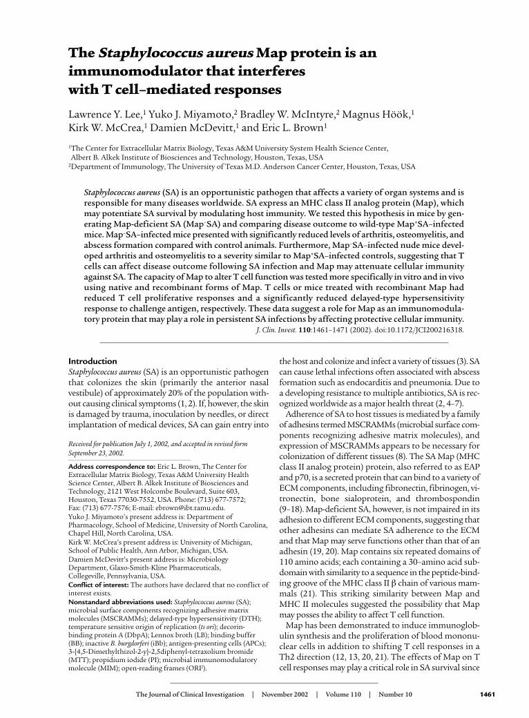
immunized with DbpA were either left untreated orinjected intraperitoneally with either Map19 or therecombinant control protein Ace40 on the day of immu-nization (day 0) and on days 2, 4, and 6 after immuniza-tion. On day 7, mice were sacrificed, and single cell sus-pensions from whole spleens were prepared andenriched for T cells by passage over nylon wool columns(50). Adoptive transfer of nylon wool–purified T cellsfrom Map19-treated mice did not elicit a DTH responseto DbpA in naive recipients compared with mice adop-tively transferred with enriched T cells from Ace40-treat-ed mice or untreated immune T cell control groups (Fig-ure 7). Flow cytometric analysis of nylon wool–collectedcells revealed a profile that was 46.83% ± 0.92% CD4+,31.63% ± 0.96% CD8+, 1.2% ± 0.26% CD4+CD8+, and20.4% ± 1.33% CD4–CD8–. These data are expressed asthe mean percentage of positive cells plus or minus SEfor the three groups examined.
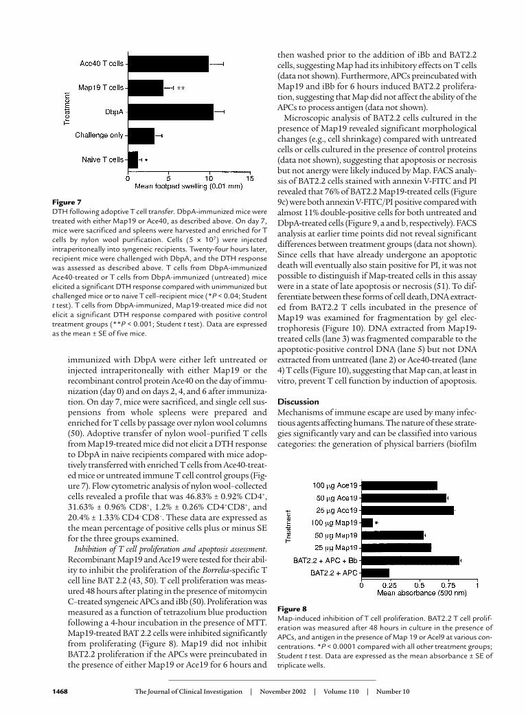
Inhibition of T cell proliferation and apoptosis assessment.Recombinant Map19 and Ace19 were tested for their abil-ity to inhibit the proliferation of the Borrelia-specific Tcell line BAT 2.2 (43, 50). T cell proliferation was meas-ured 48 hours after plating in the presence of mitomycinC–treated syngeneic APCs and iBb (50). Proliferation wasmeasured as a function of tetrazolium blue productionfollowing a 4-hour incubation in the presence of MTT.Map19-treated BAT 2.2 cells were inhibited significantlyfrom proliferating (Figure 8). Map19 did not inhibitBAT2.2 proliferation if the APCs were preincubated inthe presence of either Map19 or Ace19 for 6 hours and
then washed prior to the addition of iBb and BAT2.2cells, suggesting Map had its inhibitory effects on T cells(data not shown). Furthermore, APCs preincubated withMap19 and iBb for 6 hours induced BAT2.2 prolifera-tion, suggesting that Map did not affect the ability of theAPCs to process antigen (data not shown).
Microscopic analysis of BAT2.2 cells cultured in thepresence of Map19 revealed significant morphologicalchanges (e.g., cell shrinkage) compared with untreatedcells or cells cultured in the presence of control proteins(data not shown), suggesting that apoptosis or necrosisbut not anergy were likely induced by Map. FACS analy-sis of BAT2.2 cells stained with annexin V-FITC and PIrevealed that 76% of BAT2.2 Map19-treated cells (Figure9c) were both annexin V-FITC/PI positive compared withalmost 11% double-positive cells for both untreated andDbpA-treated cells (Figure 9, a and b, respectively). FACSanalysis at earlier time points did not reveal significantdifferences between treatment groups (data not shown).Since cells that have already undergone an apoptoticdeath will eventually also stain positive for PI, it was notpossible to distinguish if Map-treated cells in this assaywere in a state of late apoptosis or necrosis (51). To dif-ferentiate between these forms of cell death, DNA extract-ed from BAT2.2 T cells incubated in the presence ofMap19 was examined for fragmentation by gel elec-trophoresis (Figure 10). DNA extracted from Map19-treated cells (lane 3) was fragmented comparable to theapoptotic-positive control DNA (lane 5) but not DNAextracted from untreated (lane 2) or Ace40-treated (lane4) T cells (Figure 10), suggesting that Map can, at least invitro, prevent T cell function by induction of apoptosis.
DiscussionMechanisms of immune escape are used by many infec-tious agents affecting humans. The nature of these strate-gies significantly vary and can be classified into variouscategories: the generation of physical barriers (biofilm
1468 The Journal of Clinical Investigation | November 2002 | Volume 110 | Number 10
Figure 7DTH following adoptive T cell transfer. DbpA-immunized mice weretreated with either Map19 or Ace40, as described above. On day 7,mice were sacrificed and spleens were harvested and enriched for Tcells by nylon wool purification. Cells (5 × 10⁷) were injectedintraperitoneally into syngeneic recipients. Twenty-four hours later,recipient mice were challenged with DbpA, and the DTH responsewas assessed as described above. T cells from DbpA-immunizedAce40-treated or T cells from DbpA-immunized (untreated) miceelicited a significant DTH response compared with unimmunized butchallenged mice or to naive T cell–recipient mice (*P < 0.04; Studentt test). T cells from DbpA-immunized, Map19-treated mice did notelicit a significant DTH response compared with positive controltreatment groups (**P < 0.001; Student t test). Data are expressedas the mean ± SE of five mice.
Figure 8Map-induced inhibition of T cell proliferation. BAT2.2 T cell prolif-eration was measured after 48 hours in culture in the presence ofAPCs, and antigen in the presence of Map 19 or Acel9 at various con-centrations. *P < 0.0001 compared with all other treatment groups;Student t test. Data are expressed as the mean absorbance ± SE oftriplicate wells.

formation) that protect bacteria against immune attack(52, 53); the secretion of immunoregulatory cytokineanalogs or the induction of host cytokines that potenti-ate nonprotective immunity, resulting in pathogen sur-vival (54–58); antigenic variation, which can prevent thegeneration of a protective immune response (59–61);antigenic mimicry, which can result in either immunemodulation (e.g., MHC I analogs) or sequestration of thepathogen from the immune system (62, 63); toxin andprotease production, which can destroy different celltypes, including immune cells (2, 52); and finally, directmodulation of host cell functions, which can result in theinduction or inhibition of apoptosis, respectively,depending on the pathogen (64–69).
Reinfection of humans with SA is one of the hallmarksof diseases caused by this pathogen, and a difficulty inclearing these types of infections is not only a function ofdisseminated disease but of SA immune escape mecha-nisms (23–26). One possible reason for recurring infec-tions is that leukocytes from patients with chronic orrecurrent SA infections have impaired chemotactic,phagocytic, and bactericidal functions (25, 30, 31).Whether these defects in lymphocyte function were aresult of the bacterial infection or a preexisting conditionin these individuals is not known (25, 30, 31). Morespecifically, superantigens and protein A produced by SAduring an infection serve immune-evasion functions thatmay potentiate ongoing and recurrent infections (2).
Here we describe a third SA MIM (microbialimmunomodulatory molecule) with potential T cell–limiting properties. The first observation suggestingMap acted in this capacity stemmed from double infec-tion studies in which a primary infection with Map–SAconferred significant protection against reinfection withMap+SA. This contrasted significantly with the patholo-gies observed in mice receiving primary and secondaryMap+SA infections. One explanation for these data isthat T cell–mediated responses in Map+SA–infected micewere abrogated by the presence of Map. In contrast,Map–SA–infected mice developed cell-mediated immu-nity over the course of infection, resulting in bacterialclearance and in a memory response capable of control-ling a secondary Map+SA infection. That a primaryMap–SA infection conferred significant but incompleteprotection against Map+SA challenge suggested that thebalance or competition between an anamnestic responseand Map-mediated immunomodulation could be affect-ed by the challenge dose. Although these data suggested
that Map acted as an immunoregulatory molecule, wecould not exclude the following two possibilities: first, ifMap is a critical adhesin required during the infectionprocess, its removal may have resulted in an attenuatedSA strain that served to “vaccinate” mice against the sec-ond Map+SA infection. This is not a likely scenario sinceMap-deficient SA still efficiently adhere to individualECM molecules (19, 20). Furthermore, the mean weightloss between Map–SA– and Map+SA–infected mice wassimilar during the first weeks after infection, and thebacterial densities in various tissues were indistinguish-able between infection groups, suggesting that bacterialadhesion, a critical early step during infection, was notcompromised in Map–SA. This weight-loss pattern is sig-nificantly different from that recorded for mice infectedwith SA deficient in the collagen-binding protein CNA(CNA-SA). Weight loss differences in these animals, com-pared with CNA+SA–infected controls diverged from thestart of the infection process (E.L. Brown et al., unpub-lished observations). Second, a diminished disease statefollowing Map–SA infection may have been a result ofpolar effects caused by map inactivation following trans-formation resulting in the inactivation of other genesrequired for SA survival or virulence. Although a map-complemented Map–SA strain Newman is not yet avail-able, the significant effects of both isolated native Map
The Journal of Clinical Investigation | November 2002 | Volume 110 | Number 10 1469
Figure 9The effect of Map19 on annexin V/PI staining.BAT2.2 cells (106) (5 U IL-2/ml) were incubated inmedia alone (a) or in the presence of either 400 µgof either Ace40 (b) or Map19 (c) for 24 hours andanalyzed by FACS following incubations with annex-in V-FITC and PI.
Figure 10Map-induced apoptosis of BAT2.2 T cells. BAT2.2 cells (2 × 10⁶) (5U IL-2/ml) were incubated in media alone (lane 2) or in the presenceof either 100 µg Map19 (lane 3) or Ace40 (lane 4). DNA from U937cells were used as a positive control (lane 5). Lane 1, 100-bp ladder.
and of recombinant Map19 on cellular immunitystrongly suggested that the lack of virulence observed forMap–SA was a function of the bacteria’s inability toaffect host immunity and not a result of the map inacti-vation process. In addition, nu/nu mice infected withMap–SA presented with more severe arthritis comparedwith nu/+ mice infected with Map–SA. Interestingly,abscess formation was absent in nu/nu and nu/+ Map-SA–infected mice but not in Map+SA–infected miceof either genotype. These data suggested a potential rolefor T cells in controlling joint pathology but not in pro-tection against abscess formation. These experimentssomewhat contradict findings in a rat model that sug-gested that T cells are critical for abscess formation andwith recent work describing Map as an anti-inflamma-tory agent in a mouse peritonitis model (20, 70). How-ever, both of these studies examined acute SA infectionsin contrast to the assessment of chronic diseasedescribed here. Furthermore, the infection routes(intraperitoneal) and the parameters used to assess dis-ease were significantly different in both studies (20, 70).These differences make it difficult to reconcile the anti-inflammatory effects of Map described in a thioglycol-late-induced peritonitis model with the abscesses andsevere joint disease caused by a chronic SA infection (20).
Additional evidence suggesting that Map affected T cellfunction stemmed from experiments demonstrating thatMap inhibited DTH responses and induced T cell deathin vitro most likely by apoptosis. The effects of Map on Tcell function appeared only to affect mature T cells stim-ulated in the context of antigen and MHC II. It is inter-esting to speculate that this effect of Map on T cells is afunction of the homology between Map and MHC classII molecules since the proliferation of naive T cells fol-lowing incubation with concanavalin A or by Ab cross-linking of the T cell receptor was not inhibited by Map19(McIntyre and Miyamoto, unpublished observations).Interference of Map with the interactions between acti-vated T cells and MHC class II–bearing APCs may explainwhy Map only affected T cell proliferation in the contextof antigen and APCs. Furthermore, a human T cell linecultured in the presence of plate-bound fibronectin andαCD3 proliferated in the presence of Map19 (B.W. McIn-tyre and Y.J. Miyamoto, unpublished observations).These data suggested that activated T cells, but not naiveT cells, were susceptible to inhibition of proliferation byMap and that T cell lines induced to proliferate via “non-classical” pathways bypassed Map-mediated mechanismsthat resulted in the loss of T cell function.
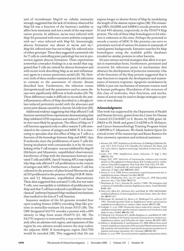
Sequence analysis of the SA genome revealed fouropen-reading frames (ORFs) encoding Map-like pro-teins in meticillin-resistant SA strain N315 (38). TheORF designated SA1751 encodes a protein with a 69%identity to Map from strain FDA574 (21, 38). TheSA1751 sequence is truncated by a stop codon immedi-ately after an adenine-rich region, and expansion of thisregion by one adenine would remove the stop codon;the adjacent MHC II homologous region (SA1750)would be encoded (38). This suggested that SA can
express longer or shorter forms of Map by modulatingthe length of the alanine repeat region (38). The remain-ing ORFs (SA2006 and SA0841) encode proteins with34 and 44% identity, respectively, to the FDA574 Mapprotein. The role of these Map homologues in SA infec-tion is unknown at this time. Perhaps the potential toencode a variety of MHC II–like proteins can serve topotentiate survival of various SA strains in mammals ofvaried genetic backgrounds. Extensive searches for Maphomologues using the available public databasesrevealed no similar proteins in other bacteria.
SA uses various survival strategies that allow it to per-sist in mammalian hosts. Furthermore, persistent andrecurrent SA infections point to mechanisms thatobstruct the development of protective immunity. Oneof the functions of the Map protein suggested that itmay function to impede the development and mainte-nance of memory responses. Apoptotic modulation byMIMs is just one mechanism of immune evasion usedby human pathogens. Elucidation of the structure ofthis class of molecules, their functions, and mecha-nisms of action may be used to design strategies to pre-vent or treat disease.
AcknowledgmentsThis work was supported by the Department of Healthand Human Services, grants from the Center for DiseaseControl (CCU618387) to E. Brown, by NIH grant AI-20624 to M. Höök and grant CA-62596 to B. McIntyre,and Cancer Immunobiology Training Program GrantCA09598 to Y. Miyamoto. We thank Andrew Spicer forcritical review of the manuscript and Karen Ramirez forflow cytometry operation and technical assistance.
1. Kissane, J.M. 1997. Staphylococcal infections. In Pathology of Infectious Dis-eases. Vol. I. D.H. Connor, F.W. Chandler, H.J. Schwartz, H.J. Manz, andE.E. Lack, editors. Appleton and Lange. Stamford, Connecticut, USA.805–816.
2. Lowy, F.D. 1998. Staphylococcus aureus infections. N. Engl. J. Med.339:520–532.
3. Rupp, M.E. 1997. Infections of intravascular catheters and vasculardevices. In The staphylococci in human disease. K.B. Crossley and G.L. Archer,editors. Churchill Liningstone. New York, New York, USA. 379–399.
4. Tacconelli, E., Tumbarello, M., and Cauda, R. 1998. Staphylococcus aureusinfections. N. Engl. J. Med. 339:2026–2027.
5. Petros, S., et al. 1998. Severe community acquired pneumonia due toStaphylococcus aureus. Intensive Care Med. 24:189.
6. Kim, H.S., Weilbaecher, D.G., Lie, J.T., and Titus, J.L. 1978. Myocardialabscesses. Am. J. Clin. Pathol. 70:18–23.
7. Hiramatsu, K., Cui, L., Kuroda, M., and Ito, T 2001. The emergence andevolution of methicillin-resistant Staphylococcus aureus. Trends Microbiol.9:486–493.
8. Patti, J.M., Allen, B.L., McGavin, M.J., and Höök, M. 1994. MSCRAMM-mediated adherence of microorganisms to host tissues. Annu. Rev. Micro-biol. 48:585–617.
9. Herrmann, M., Suchard, S.J., Boxer, L.A., Waldvogel, F.A., and Lew, P.D.1991. Thrombospondin binds to Staphylococcus aureus and promotesstaphylococcal adherence to surfaces. Infect. Immun. 59:279–288.
10. Fujigaki, Y., et al. 1998. Glomerular injury induced by cationic 70-kDstaphylococcal protein; specific immune response is not involved in earlyphase in rats. J. Pathol. 184:436–445.
11. Chhatwal, G.S., Preissner, K.T., Muller-Berghaus, G., and Blobel, H. 1987.Specific binding of the human S protein (vitronectin) to streptococci,Staphylococcus aureus, and Escherichia coli. Infect. Immun. 55:1878–1883.
12. Jahreis, A., Beckheinrich, P., and Haustein, U.F. 2000. Effects of two novelcationic staphylococcal proteins (NP-tase and p70)and enterotoxin B onIgE synthesis and interleukin-4 and interferon-gamma production inpatients with atopic dermatitis. Br. J. Dermatol. 142:680–687.
13. Jahreis, A., et al. 1995. Two novel cationic staphylococcal proteins induce
1470 The Journal of Clinical Investigation | November 2002 | Volume 110 | Number 10

IL-2 secretion, proliferation and immunoglobulin synthesis in periph-eral blood mononuclear cells (PBMC) of both healthy controls andpatients with common variable immunodeficiency (CVID). Clin. Exp.Immunol. 100:406–411.
14. Hudson, M.C., Ramp, W.K., and Frankenburg, K.P. 1999. Staphylococcusaureus adhesion to bone matrix and bone-associated biomaterials. FEMSMicrobiol. Lett. 173:279–284.
15. Lopes, J.D., dos Reis, M., and Brentani, R.R. 1985. Presence of lamininreceptors in Staphylococcus aureus. Science. 229:275–277.
16. O’Connell, D.P., et al. 1998. The fibrinogen-binding MSCRAMM(clumping factor) of Staphylococcus aureus has a Ca2+-dependent inhibito-ry site. J. Biol. Chem. 273:6821–6829.
17. Vercellotti, G.M., et al. 1985. Extracellular matrix proteins (fibronectin,laminin, and type IV collagen) bind and aggregate bacteria. Am. J. Pathol.120:13–21.
18. Wann, E.R., Gurusiddappa, S., and Höök, M. 2000. The fibronectin-binding MSCRAMM FnbpA of Staphylococcus aureus is a bifunctional pro-tein that also binds to fibrinogen. J. Biol. Chem. 275:13863–13871.
19. Kreikemeyer, B., McDevitt, D., and Podbielski, A. 2002. The role of theMap protein in S. aureus matrix protein and eukaryotic cell adherence.Int. J. Med. Microbiol. 292:283–295.
20. Chavakis, T., et al. 2002. Staphylococcus aureus extracellular adherence pro-tein serves as anti- inflammatory factor by inhibiting the recruitment ofhost leukocytes. Nat. Med. 8:687–693.
21. Jonsson, K., McDevitt, D., McGavin, M.H., Patti, J.M., and Höök, M.1995. Staphylococcus aureus expresses a major histocompatibility complexclass II analog. J. Biol. Chem. 270:21457–21460.
22. Guillen, C., et al. 2002. Enhanced Th1 response to Staphylococcusaureus infection in human lactoferrin-transgenic mice. J. Immunol.168:3950–3957.
23. Chang, H.R., et al. 2000. Use of pulsed-field gel electrophoresis in theanalysis of recurrent Staphylococcus aureus infections in patients on con-tinuous ambulatory peritoneal dialysis. Am. J. Nephrol. 20:463–467.
24. Hartstein, A.I., Mulligan, M.E., Morthland, V.H., and Kwok, R.Y. 1992.Recurrent Staphylococcus aureus bacteremia. J. Clin. Microbiol. 30:670–674.
25. Monteil, M., Hobbs, J., and Citron, K. 1987. Selective immunodeficien-cy affecting staphylococcal response. Lancet. 2:880–883.
26. Shayegani, M., De Courcy, S.J., Jr., and Mudd, S. 1973. Cell-mediatedimmunity in mice infected with S. aureus and elicited with specific bac-terial antigens. J. Reticuloendothel. Soc. 14:44–51.
27. Easmon, C.S., and Glynn, A.A. 1975. Cell-mediated immune responsesin Staphylococcus aureus infections in mice. Immunology. 29:75–85.
28. Ficker, L., Seal, D., and Wright, P. 1989. Staphylococcal infection and thelimbus: study of the cell-mediated immune response. Eye. 3:190–193.
29. Sarai, Y., et al. 1977. Immunological properties in staphylococcal toxicepidermal necrolysis. Dermatologica. 155:315–318.
30. Verbrugh, H.A., et al. 1980. Phagocytic and chemotactic function of poly-morphonuclear and mononuclear leucocytes in patients with recurrentstaphylococcal infections. Scand. J. Infect. Dis. 12:111–116.
31. Valmin, K., Hallberg, T., and Hedstrom, S.A. 1982. Recurrent Staphylo-coccal furunculosis: lymphocyte subsets and plasma immunoglobulins.Scand. J. Infect. Dis. 14:153–154.
32. Bremell, T., Lange, S., Yacoub, A., Ryden, C., and Tarkowski, A. 1991.Experimental Staphylococcus aureus arthritis in mice. Infect. Immun.59:2615–2623.
33. Sloane, R., et al. 1991. A toxic shock syndrome toxin mutant of Staphy-lococcus aureus isolated by allelic replacement lacks virulence in a rabbituterine model. FEMS Microbiol. Lett. 62:239–244.
34. Hussain, M., et al. 2002. Insertional inactivation of Eap in Staphylococcusaureus strain Newman confers reduced staphylococcal binding to fibrob-lasts. Infect. Immun. 70:2933–2940.
35. Kreiswirth, B.N., et al. 1983. The toxic shock syndrome exotoxin struc-tural gene is not detectably transmitted by a prophage. Nature.305:709–712.
36. Duthie, E.S., and Lorenz, L.L. 1952. Staphylococcal coagulase: mode ofaction and antigenicity. J. Gen. Microbiol. 6:95–107.
37. Ponnuraj, K., et al. 2002. Crystallization and preliminary x-ray crystal-lographic analysis of Ace: a collagen-binding MSCRAMM from Entero-coccus faecalis. Biochim. Biophys. Acta. 1596:173–176.
38. Kuroda, M., et al. 2001. Whole genome sequencing of meticillin-resist-ant Staphylococcus aureus. Lancet. 357:1225–1240.
39. Visai, L., et al. 2000. Monoclonal antibodies to CNA, a collagen-bindingmicrobial surface component recognizing adhesive matrix molecules,detach Staphylococcus aureus from a collagen substrate. J. Biol. Chem.275:39837–39845.
40. Rich, R.L., et al. 1999. Ace is a collagen-binding MSCRAMM from Ente-rococcus faecalis. J. Biol. Chem. 274:26939–26945.
41. Guo, B.P., Brown, E.L., Dorward, D.W., Rosenberg, L.C., and Höök, M.1998. Decorin-binding adhesins from Borrelia burgdorferi. Mol. Microbiol.30:711–723.
42. Maniatis, T., Fritsch, E.F., and Sambrook, J. 1989. Molecular cloning: A
laboratory manual. Cold Spring Harbor Laboratory Press. Cold SpringHarbor, New York, USA. A.3
43. Brown, E.L., Ullrich, S.E., Pride, M., and Kripke, M.L. 2001. The effect ofUV irradiation on infection of mice with Borrelia burgdorferi. Photochem.Photobiol. 73:537–544.
44. Brown, E.L., et al. 2001. Resistance to Lyme disease in decorin-deficientmice. J. Clin. Invest. 107:845–852.
45. McGavin, M.H., Krajewska-Pietrasik, D., Ryden, C., and Höök, M. 1993.Identification of a Staphylococcus aureus extracellular matrix-binding pro-tein with broad specificity. Infect. Immun. 61:2479–2485.
46. Patti, J.M., et al. 1994. The Staphylococcus aureus collagen adhesin is a vir-ulence determinant in experimental septic arthritis. Infect. Immun.62:152–161.
47. Switalski, L.M., et al. 1993. A collagen receptor on Staphylococcus aureusstrains isolated from patients with septic arthritis mediates adhesion tocartilage. Mol. Microbiol. 7:99–107.
48. Joh, H.J., House-Pompeo, K., Patti, J.M., Gurusiddappa, S., and Höök, M.1994. Fibronectin receptors from gram-positive bacteria: comparison ofactive sites. Biochemistry. 33:6086–6092.
49. Brown, E.L., et al. 1995. Modulation of immunity to Borrelia burgdorferiby ultraviolet irradiation: differential effect on Th1 and Th2 immuneresponses. Eur. J. Immunol. 25:3017–3022.
50. Pride, M.W., et al. 1998. Specific Th1 cell lines that confer protectiveimmunity against experimental Borrelia burgdorferi infection in mice. J. Leukoc. Biol. 63:542–549.
51. Vermes, I., Haanen, C., Steffens-Nakken, H., and Reutelingsperger, C.1995. A novel assay for apoptosis. Flow cytometric detection of phos-phatidylserine expression on early apoptotic cells using fluoresceinlabelled Annexin V. J. Immunol. Methods. 184:39–51.
52. Kharazmi, A. 1991. Mechanisms involved in the evasion of the hostdefence by Pseudomonas aeruginosa. Immunol. Lett. 30:201–205.
53. Martin-Lopez, J.V., et al. 2002. Detection of Staphylococcus aureus clinicalisolates harboring the ica gene cluster needed for biofilm establishment.J. Clin. Microbiol. 40:1569–1570.
54. Suzuki, T., et al. 1995. Viral interleukin 10 (IL-10), the human herpesvirus 4 cellular IL-10 homologue, induces local anergy to allogeneic andsyngeneic tumors. J. Exp. Med. 182:477–486.
55. Engele, M., et al. 2002. Induction of TNF in human alveolarmacrophages as a potential evasion mechanism of virulent Mycobacteri-um tuberculosis. J. Immunol. 168:1328–1337.
56. de Diego, J., Punzon, C., Duarte, M., and Fresno, M. 1997. Alteration ofmacrophage function by a Trypanosoma cruzi membrane mucin. J. Immunol. 159:4983–4989.
57. Abraham, S.N., Beachy, E.H., and Simpson, W.A. 1983. Adherence of strep-tococcus pyogenes, Escherichia coli, and Pseudomonas aeruginosa tofibronectin-coated and uncoated epithelial cells. Infect. Immun. 41:1261–1268.
58. Akridge, R.E., Oyafuso, L.K., and Reed, S.G. 1994. IL-10 is induced dur-ing HIV-1 infection and is capable of decreasing viral replication inhuman macrophages. J. Immunol. 153:5782–5789.
59. Donelson, J.E., Hill, K.L., and El-Sayed, N.M. 1998. Multiple mechanisms ofimmune evasion by African trypanosomes. Mol. Biochem. Parasitol. 91:51–66.
60. Kyes, S., Horrocks, P., and Newbold, C. 2001. Antigenic variation at theinfected red cell surface in malaria. Annu. Rev. Microbiol. 55:673–707.
61. Lawrenz, M.B., et al. 1999. Human antibody responses to VlsE antigenicvariation protein of Borrelia burgdorferi. J. Clin. Microbiol. 37:3997–4004.
62. Wurzner, R. 1999. Evasion of pathogens by avoiding recognition or erad-ication by complement, in part via molecular mimicry. Mol. Immunol.36:249–260.
63. Farrell, H.E., et al. 1997. Inhibition of natural killer cells by acytomegalovirus MHC class I homologue in vivo. Nature. 386:510–514.
64. Boulton, I.C., and Gray-Owen, S.D. 2002. Neisserial binding to CEA-CAM1 arrests the activation and proliferation of CD4+ T lymphocytes.Nat. Immunol. 3:229–236.
65. Cuff, S., and Ruby, J. 1996. Evasion of apoptosis by DNA viruses.Immunol. Cell Biol. 74:527–537.
66. Das, G., Vohra, H., Saha, B., Agrewala, J.N., and Mishra, G.C. 1998. Leish-mania donovani infection of a susceptible host results in apoptosis ofTh1-like cells: rescue of anti-leishmanial CMI by providing Th1-specif-ic bystander costimulation. Microbiol. Immunol. 42:795–801.
67. Monack, D.M., Mecsas, J., Ghori, N., and Falkow, S. 1997. Yersinia sig-nals macrophages to undergo apoptosis and YopJ is necessary for thiscell death. Proc. Natl. Acad. Sci. USA. 94:10385–10390.
68. Nunes, M.P., Andrade, R.M., Lopes, M.F., and DosReis, G.A. 1998. Acti-vation-induced T cell death exacerbates Trypanosoma cruzi replication inmacrophages cocultured with CD4+ T lymphocytes from infected hosts.J. Immunol. 160:1313–1319.
69. Wei, S., et al. 2002. Toxoplasma gondii-infected human myeloid dendriticcells induce T-lymphocyte dysfunction and contact-dependent apopto-sis. Infect. Immun. 70:1750–1760.
70. Tzianabos, A.O., and Kasper, D.L. 2002. Role of T cells in abscess for-mation. Curr. Opin. Microbiol. 5:92–96.
The Journal of Clinical Investigation | November 2002 | Volume 110 | Number 10 1471