Embed Size (px)
Citation preview

Essentials of Human Anatomy & Physiology
Copyright © 2003 Pearson Education, Inc. publishing as Benjamin Cummings
Slides 13.1 – 13.30
Seventh Edition
Elaine N. Marieb
Chapter 13
The Respiratory System
Lecture Slides in PowerPoint by Jerry L. Cook

Organs of the Respiratory system
Slide 13.1Copyright © 2003 Pearson Education, Inc. publishing as Benjamin Cummings
Nose
Pharynx
Larynx
Trachea
Bronchi
Lungs –alveoli
Figure 13.1
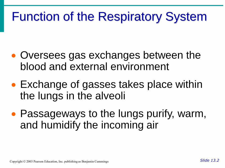
Function of the Respiratory System
Slide 13.2Copyright © 2003 Pearson Education, Inc. publishing as Benjamin Cummings
Oversees gas exchanges between the blood and external environment
Exchange of gasses takes place within the lungs in the alveoli
Passageways to the lungs purify, warm, and humidify the incoming air
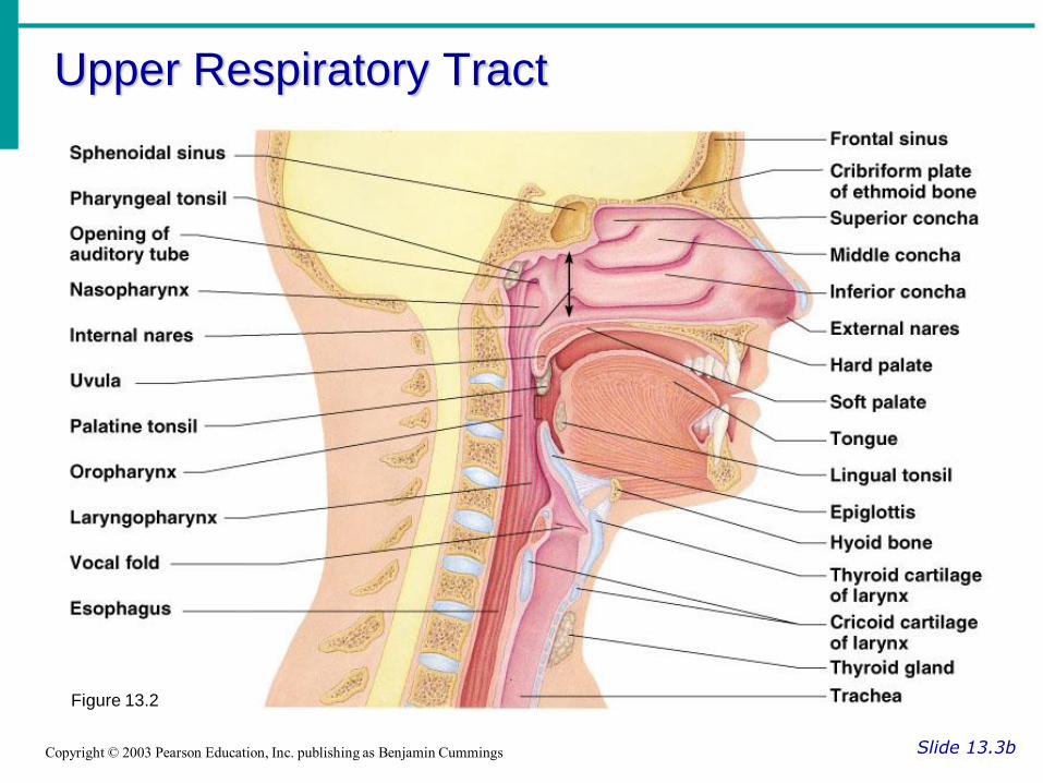
Slide 13.3bCopyright © 2003 Pearson Education, Inc. publishing as Benjamin Cummings
Figure 13.2
Upper Respiratory Tract
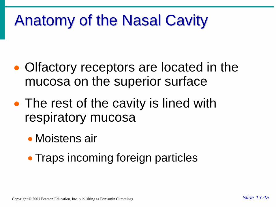
Anatomy of the Nasal Cavity
Slide 13.4aCopyright © 2003 Pearson Education, Inc. publishing as Benjamin Cummings
Olfactory receptors are located in the mucosa on the superior surface
The rest of the cavity is lined with respiratory mucosa
Moistens air
Traps incoming foreign particles
Paranasal Sinuses
Slide 13.5aCopyright © 2003 Pearson Education, Inc. publishing as Benjamin Cummings
Cavities within bones surrounding the nasal cavity
Frontal bone
Sphenoid bone
Ethmoid bone
Maxillary bone
Paranasal Sinuses
Slide 13.5bCopyright © 2003 Pearson Education, Inc. publishing as Benjamin Cummings
Function of the sinuses
Lighten the skull
Act as resonance chambers for speech
Produce mucus that drains into the nasal cavity

Structures of the Larynx
Slide 13.9bCopyright © 2003 Pearson Education, Inc. publishing as Benjamin Cummings
Vocal cords (vocal folds)
Vibrate with expelled air to create sound (speech)
Glottis – opening between vocal cords
Lungs
Slide 13.12aCopyright © 2003 Pearson Education, Inc. publishing as Benjamin Cummings
Occupy most of the thoracic cavity
Apex is near the clavicle (superior portion)
Base rests on the diaphragm (inferior portion)
Each lung is divided into lobes by fissures
Left lung – two lobes
Right lung – three lobes
Coverings of the Lungs
Slide 13.13Copyright © 2003 Pearson Education, Inc. publishing as Benjamin Cummings
Pulmonary (visceral) pleura covers the lung surface
Parietal pleura lines the walls of the thoracic cavity
Pleural fluid fills the area between layers of pleura to allow gliding

Gas Exchange
Slide 13.19Copyright © 2003 Pearson Education, Inc. publishing as Benjamin Cummings
Gas crosses the respiratory membrane by diffusion
Oxygen enters the blood
Carbon dioxide enters the alveoli
Macrophages add protection
Surfactant coats gas-exposed alveolar surfaces
Events of Respiration
Slide 13.20aCopyright © 2003 Pearson Education, Inc. publishing as Benjamin Cummings
Pulmonary ventilation – moving air in and out of the lungs
External respiration – gas exchange between pulmonary blood and alveoli

Events of Respiration
Slide 13.20bCopyright © 2003 Pearson Education, Inc. publishing as Benjamin Cummings
Respiratory gas transport – transport of oxygen and carbon dioxide via the bloodstream
Internal respiration – gas exchange between blood and tissue cells in systemic capillaries

Mechanics of Breathing
(Pulmonary Ventilation)
Slide 13.21aCopyright © 2003 Pearson Education, Inc. publishing as Benjamin Cummings
Completely mechanical process
Depends on volume changes in the thoracic cavity
Volume changes lead to pressure changes, which lead to the flow of gases to equalize pressure

Mechanics of Breathing
(Pulmonary Ventilation)
Slide 13.21bCopyright © 2003 Pearson Education, Inc. publishing as Benjamin Cummings
Two phases
Inspiration – flow of air into lung
Expiration – air leaving lung

Inspiration
Slide 13.22aCopyright © 2003 Pearson Education, Inc. publishing as Benjamin Cummings
Diaphragm and intercostal muscles contract
The size of the thoracic cavity increases
External air is pulled into the lungs due to an increase in intrapulmonary volume

Inspiration
Slide 13.22bCopyright © 2003 Pearson Education, Inc. publishing as Benjamin Cummings
Figure 13.7a
Exhalation
Slide 13.23aCopyright © 2003 Pearson Education, Inc. publishing as Benjamin Cummings
Largely a passive process which depends on natural lung elasticity
As muscles relax, air is pushed out of the lungs
Forced expiration can occur mostly by contracting internal intercostal muscles to depress the rib cage

Exhalation
Slide 13.23bCopyright © 2003 Pearson Education, Inc. publishing as Benjamin Cummings
Figure 13.7b
Pressure Differences in the
Thoracic Cavity
Slide 13.24Copyright © 2003 Pearson Education, Inc. publishing as Benjamin Cummings
Normal pressure within the pleural space is always negative (intrapleural pressure)
Differences in lung and pleural space pressures keep lungs from collapsing
Nonrespiratory Air Movements
Slide 13.25Copyright © 2003 Pearson Education, Inc. publishing as Benjamin Cummings
Can be caused by reflexes or voluntary actions
Examples
Cough and sneeze – clears lungs of debris
Laughing
Crying
Yawn
Hiccup
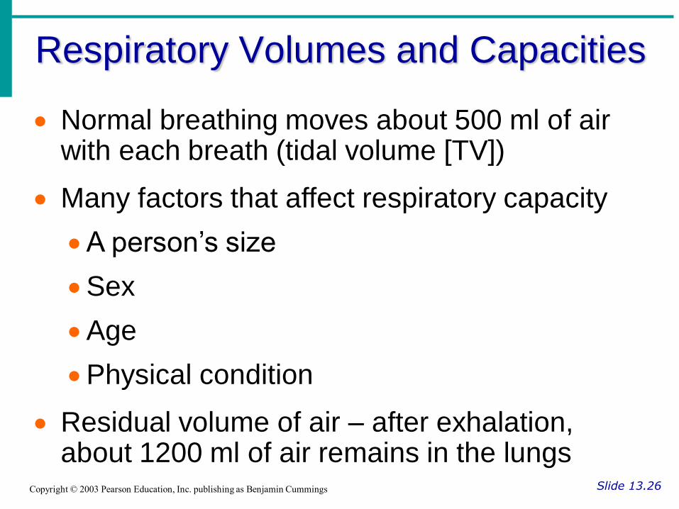
Respiratory Volumes and Capacities
Slide 13.26Copyright © 2003 Pearson Education, Inc. publishing as Benjamin Cummings
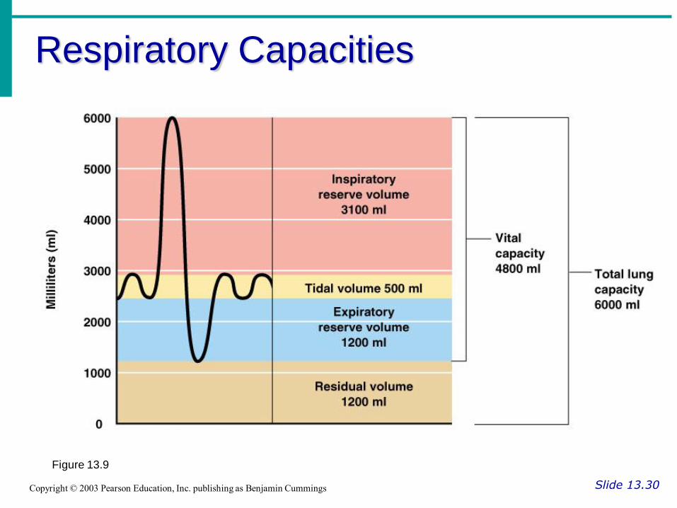
Normal breathing moves about 500 ml of air with each breath (tidal volume [TV])
Many factors that affect respiratory capacity
A person’s size
Sex
Age
Physical condition
Residual volume of air – after exhalation, about 1200 ml of air remains in the lungs
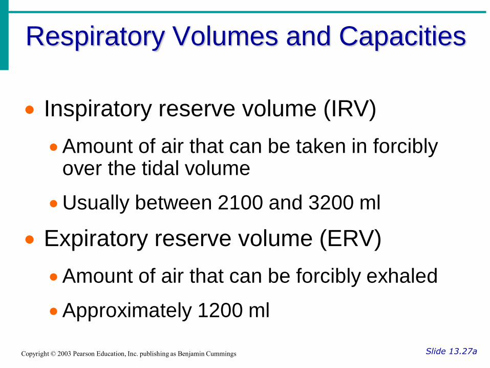
Respiratory Volumes and Capacities
Slide 13.27aCopyright © 2003 Pearson Education, Inc. publishing as Benjamin Cummings
Inspiratory reserve volume (IRV)
Amount of air that can be taken in forcibly over the tidal volume
Usually between 2100 and 3200 ml
Expiratory reserve volume (ERV)
Amount of air that can be forcibly exhaled
Approximately 1200 ml
Respiratory Volumes and Capacities
Slide 13.27bCopyright © 2003 Pearson Education, Inc. publishing as Benjamin Cummings
Residual volume
Air remaining in lung after expiration
About 1200 ml
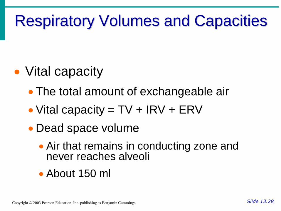
Respiratory Volumes and Capacities
Slide 13.28Copyright © 2003 Pearson Education, Inc. publishing as Benjamin Cummings
Vital capacity
The total amount of exchangeable air
Vital capacity = TV + IRV + ERV
Dead space volume
Air that remains in conducting zone and never reaches alveoli
About 150 ml
Respiratory Volumes and Capacities
Slide 13.29Copyright © 2003 Pearson Education, Inc. publishing as Benjamin Cummings
Functional volume
Air that actually reaches the respiratory zone
Usually about 350 ml
Respiratory capacities are measured with a spirometer
Respiratory Capacities
Slide 13.30Copyright © 2003 Pearson Education, Inc. publishing as Benjamin Cummings
Figure 13.9