Embed Size (px)
Citation preview

Human papillomavirus E7 requires the protease calpain to degrade the
retinoblastoma protein
Grant A Darnell1, Wayne A. Schroder1, Toni M. Antalis2, Eleanore Lambley1, Lee Major1, Joy Gardner1, Geoff Birrell3, Angel Cid-Arregui4, Andreas Suhrbier1
1 Immunovirology Laboratory, Queensland Institute of Medical Research, Brisbane, Queensland 4029,
Australia. 2 Center for Vascular Biology and Inflammatory Diseases and the Department of Physiology,
University of Maryland School of Medicine, MD 21201, USA. 3 Radiation Biology & Oncology Laboratory, Queensland Institute of Medical Research, Brisbane,
Queensland 4029, Australia. 4 Angewandte Tumorvirologie, Deutsches Krebsforschungszentrum, Heildberg, D-69012, Germany.
Running Title: HPV E7 degradation of Rb requires calpain
Address correspondence to: Dr A. Suhrbier, Queensland Institute of Medical Research, Post Office Royal Brisbane Hospital, Queensland 4029. Australia. Tel 61-7-33620415. Fax 61-7-33620107. E mail [email protected]
Cervical cancers transformed by high-risk human papilloma virus (HPV) express the E7 oncoprotein, which accelerates the degradation of the retinoblastoma protein (Rb). Here we show that the E7-mediated degradation of Rb requires the calcium-activated cysteine protease, calpain. E7 bound and activated μ-calpain and promoted cleavage at Rb810, with mutation of this residue preventing E7-mediated degradation. The calpain cleavage product, Rb1-810, was unable to mediate cell cycle arrest, but retained the ability to repress E6/E7 transcription. E7 also promoted the accelerated proteasomal degradation of Rb1-
810. Calpain inhibitors reduced the viability of HPV transformed cells and synergized with cisplatin. Calpain thus emerges as a central player in E7-mediated degradation of Rb, and represents a potential new drug target for the treatment of HPV associated lesions. Infection with high risk human papillomavirus (HPV) types is responsible for virtually all cases of cervical cancer, which is currently the second most common cause of death from cancer among women worldwide (1). Vaccination will likely have a substantial impact; however current modeling suggests that it may take 40-50 years to reach a ≈50%
reduction in mortality (2). Integration of part of the HPV genome is believed to be central to the transformation process (3) and often results in the increased expression of the two viral oncoproteins, E6 and E7. A principle activity of E6 and E7 is promotion of the accelerated degradation of p53 and the retinoblastoma protein (Rb), respectively (4,5). Hypophosphorylated Rb binds the E2F transcription factor resulting in repression of E2F-dependant gene transcription and G1 cell cycle arrest (6). The promotion of Rb degradation mediated by E7 is believed to involve a two-step process (7). Firstly, E7 binds hypophosphorylated Rb (8), which leads to the displacement of E2F and entry into the cell cycle. How E2F is displaced by E7 remains unclear as E7 and E2F binding sites on Rb have recently been shown to be quite distinct (9). The second step involves E7 targeting Rb for accelerated degradation via the proteasome (10-12). A new insight into the mechanism of E7-mediated degradation of Rb was recently provided by our observation that the serine protease inhibitor, SerpinB2 was able to inhibit E7-mediated degradation of Rb in HeLa cells (13,14). There are no reports that SerpinB2 inhibits the proteasome, and it has recently emerged that SerpinB2 inhibits calpain-mediated degradation of Rb (Tonnetti et al., submitted).
1
http://www.jbc.org/cgi/doi/10.1074/jbc.M706860200The latest version is at JBC Papers in Press. Published on October 31, 2007 as Manuscript M706860200
Copyright 2007 by The American Society for Biochemistry and Molecular Biology, Inc.
by guest on June 13, 2018http://w
ww
.jbc.org/D
ownloaded from
Calpains represent a group of calcium activated cysteine proteases, two of which, μ-calpain and m-calpain, are ubiquitously expressed and found both in the cytoplasm and the nucleus (15). Herein we provide evidence that the first step in the E7-mediated degradation of Rb involves calpain cleavage. E7 was shown to bind and activate μ-calpain and promoted cleavage of full length Rb1-928 to yield Rb1-810. Rb1-810 was unable to promote cell cycle arrest, suggesting that this cleavage event was sufficient to displace E2F from Rb. The second step involved the promotion by E7 of the proteasomal degradation of Rb1-810. Calpain inhibitors are being developed for a number of diseases including cancer (16,17) and were able to inhibit E7-mediated degradation of Rb. They also reduced the viability of HPV transformed cells through upregulation of p53, and synergized with cisplatin, a drug frequently used to treat cervical cancer. Calpain inhibitors may thus find application in the treatment of HPV associated malignancies. EXPERIMENTAL PROCEDURES Cell line- CAPN4-/- embryonic fibroblasts were provided by John Elce (Queen's University, Canada) (18). The remaining cells lines were obtained from ATCC (Manassas, VI, USA) and maintained as described (13). DNA plasmids and cell transfection- The mammalian expression plasmid encoding CAPN4 was purchased from ATCC. pIRES-Neo2-eE7 has been described previously (19). GST-eE7 was generated by subcloning the eE7 gene from pIRES-Neo2-eE7 into pGEX-2TK (Invitrogen). Plasmids encoding Rb1-928, Rb394-
928, GST-Rb768-928 and SerpinB2 have been described previously (14). Plasmids encoding RbK810A and Rb1-810 were generated from pCDNA3.1 Rb1-928 by site-directed mutagenesis (14). Dominant negative p53 (pRc/CMV-R273H) has been described previously (20). Vector controls used pcDNA3.1 (Invitrogen). Cells were transfected using GeneJammer transfection reagent (Stratagene, La Jolla, CA). Western Blots- Western blot analyses and preparation of nuclear and cellular extracts was undertaken as described (13) using antibodies specific for Rb (G3-245) (BD PharMingen, Heidelberg, Germany) and C-15 (Santa Cruz Biotechnology, Santa Cruz, CA), FLAG
(Sigma-Aldrich, Sydney, Australia), actin (C-11), p53 (D0-1), CAPN4 regulatory subunit (N-19), calpain-1 (N-9), HPV-18 E7 (N-19) (Santa Cruz), GAPDH (Chemicon, Temecula, CA) and horseradish peroxidase-conjugated secondary antibody (Chemicon). N19 was incubated with the nitrocellulose for 3 days at 4°C, prior to washing and secondary antibody. Reprobing of membranes was undertaken after treatment with Restore Western Blot Stripping Buffer (Pierce). Protein loading was quantified using BCA Protein assay (Peirce). Calpain cleavage of Rb in vitro- Recombinant calpain (Sigma-Aldrich) (0.1 IU/ml, >98% pure by SDS PAGE) was incubated with recombinant Rb (QED Biosciences) (0.5 μg/ml, >90% pure) or GST-Rb768-928 with or without recombinant GST-eE7 in the presence of calpain cleavage buffer (25 mM Tris pH 7.0, 100 mM NaCl) for 30 mins at 37ºC. GST fusion proteins were expressed in XL-10 Blue (Stratagene), extracted using lysis buffer containing protease inhibitor cocktail (Roche), and purified using glutathione-agarose beads (Sigma-Aldrich). Drug treatments in vitro- Cells (105) were treated with (i) lactacystin 10 μM (EMD Biosciences) overnight, (ii) calpain inhibitor PD 150606 or its inactive homologue PD 145305 (EMD Biosciences), (iii) Calpain inhibitor VI (EMD Biosciences), or (iv) Cisplatin (Sigma-Aldrich). Cells were then analyzed by Western Blotting or assayed for cell viability (see below). Senescence activated β-galactosidase assay-SAOS-2 cells (105) were transfected using GeneJammer (Stratagene) with indicated Rb plasmids (0.6 μg) alone, or in combination with pIRES-Neo2-eE7 (0.6 μg) and/or SerpinB2 (0.6 μg). Cells were grown and SA-β-gal detected as described previously (21). HPV-18 URR luciferase assays- SAOS-2 cells (105) were transfected using GeneJammer (Stratagene) with the HPV-URR reporter plasmid (0.6 μg) (14), a pCMV β-gal construct (0.6 μg) as a transfection control (14), the indicated Rb expression plasmid (0.6 μg), plus pIRES-neo2-eE7 or a control plasmid (0.6 μg). At 72 h post-transfection cell lysates were analyzed for LUC and β-gal activity as described previously (14). Immunoprecipitation- CAPN4-/- fibroblasts or HEK293 cells were transfected with pIRES-neo2-eE7 or HIS-eE7-FLAG, respectively. After 48-72 h cells were lysed in lysis buffer
2
by guest on June 13, 2018http://w
ww
.jbc.org/D
ownloaded from

containing protease inhibitor cocktail (Roche) (13) and FLAG-eE7 was immunoprecipited from CAPN4-/- fibroblasts using the FLAG Immunoprecipitation Kit according to the manufacture’s instructions
®
(Sigma-Aldrich) and from HeLa cells using anti-FLAG antibody M2 (Sigma-Aldrich). Bound proteins were washed, eluted, and detected by Western blotting. HEK293 cells expressing HIS-eE7-FLAG were transfected and lysed as above. Pull down of HIS-eE7-FLAG was performed using Ni-NTA-agarose beads (EMD Biosciences), and bound proteins were washed, eluted, and analyzed by Western blotting.
Immunoprecipitation of calpain and E7 were undertaken using nuclear extracts of ≈107 fully confluent HeLa cells cultured overnight with 10 μM lactacystin, and the Seize immunoprecipitation kit (Pierce, Rockford, IL) with goat anti-calpain-1 (N-19), -HPV-18 E7 (N-19) and -actin (C-11). GST-binding assays- GST-eE7 fusion protein was incubated with recombinant calpain (Sigma-Aldrich) (0.1 IU/ml), in the presence of LICHT buffer for 2 h at 4ºC. GST-eE7 was then captured with glutathione agarose beads, washed, eluted and analyzed by Western blotting as described previously (13). Calpain activity assays- In vitro calpain activity was determined using the fluorogenic calpain activity assay kit and a fluorogenic μ-calpain substrate H-K(FAM)-EVY~GMMK(DABCYL)-OH (EMD Biosciences). Recombinant calpain (Sigma-Aldrich) (0.1 IU/ml) was incubated with recombinant GST-eE7 in the presence of calpain cleavage buffer, with the indicated concentration of calcium, with or without EGTA (10 mM) or PD 150606 (150 μM), and the fluorogenic calpain substrate, which was used according to the manufacturer’s instructions. Fluorescence was measured using a FLUROstar Optima fluorometer (BMC Lab tech, Offenburg, Germany). In vivo calpain activity in nuclear lysates was measured using Calpain-GloTM Protease Assay (Promega) according to manufactures instructions. Nuclear lysate were prepared as described from cells washed in PBS (13). Quantitative real-time reverse transcription-PCR (RT-PCR)- HeLa and Caski cells (106) were incubated with the indicated concentrations of either PD 150606 or PD 14305 for 5 days. cDNA was prepared and real time RT-PCR was performed as described
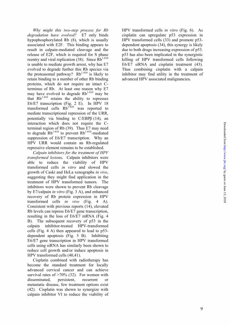
(13). PCR analysis used the following nucleotide primers (Sigma-Aldrich): for HPV-18 E7; 5′ GCTGAACCACAACGTCACAC-3′ and 5′-GGTCGTCTGCTGGAGCTTTCT-3′; for HPV-16 E7; 5′-CCGGACAGAGCCCATTACAAT-3′ and 5′-ACGTGTGTGCTTTGTACGCAC-3′; for GAPDH, 5′-GGTCGGTGTGAACG-GATTT-3′ and 5′-GTCGTTGATGGCAACAATCT-3′. Quantitation was based on a standard curve established using dilutions of untreated cell GAPDH cDNA. Cell viability assays- Cell survival was quantified by crystal violet staining assays as described (22). Cell were plated into 96-well plates at 2x10⁴ cells per well in triplicate or quadruplicate and allowed to adhere overnight. After transfection of dominant negative p53 and/or the addition of calpain inhibitors, the plates were incubated at 37°C overnight prior to staining with crystal violet. RESULTS Calpain is required for E7-mediated degradation of Rb. Calpains exist as heterodimers comprising a large 80 kDa catalytic subunit and a smaller 28 kDa regulatory subunit. The 28 kDa subunit is identical for μ- and m-calpain, is required for protease activity in vivo, and is encoded by the CAPN4 gene (23). Murine embryonic fibroblasts (MEFs) from CAPN4-/- mice were used to determine whether calpain plays a role in E7-mediated degradation of Rb. (Expression of the large subunits of μ- and m-calpain are not changed in these cells (23)). A plasmid encoding FLAG-tagged codon-optimized HPV-16 E7 (pIRES-Neo2-eE7) was transfected into CAPN4-/- MEFs, and Rb levels were determined by Western blotting. Increasing amounts of eE7 expression had little effect on Rb protein levels in these calpain-defective cells (Fig. 1 A, top panel, compare lane 1 with lanes 2-5). In contrast, when calpain activity was restored by co-transfection with a plasmid encoding CAPN4 (Fig. 1 A, anti-CAPN4, lanes 6-10), Rb protein levels dropped as E7 levels increased (Fig. 1 A, top panel, compare lane 6 with lanes 7-10). These experiments indicated that E7 requires calpain activity to mediate the accelerated degradation of Rb. (The lower levels of Rb in CAPN4-/-
MEFs, lane 6 compared to lane 1, also supports the view that calpain is involved in degrading
3
by guest on June 13, 2018http://w
ww
.jbc.org/D
ownloaded from

Rb in the absence of E7 (Tonnetti et al. submitted)). Calpain cleavage of Rb in vitro. To determine in a cell free system whether E7 enhances calpain-mediated cleavage of Rb, purified full length recombinant Rb1-928 was incubated with recombinant human µ-calpain in the presence and absence of GST-eE7. The addition of calpain to Rb resulted in the generation of a faster migrating Rb species with a lower molecular weight of ≈95 kDa (Fig. 1 B, lane 2, Cleaved Rb), which could be distinguished from the 110 kDa full length Rb (Rb1-928). The addition of GST-eE7 to this mixture resulted in almost complete loss of Rb1-928 (Fig. 1 B, lane 3), suggesting that eE7 enhanced calpain activity. The ≈95 kDa Rb species was also further degraded; an activity also seen when increasing levels of calpain are added to such reactions in the absence of eE7 (Tonnetti et al., submitted), an observation again consistent with eE7 increasing calpain activity. However, only the ≈95 kDa species was detected in vivo, suggesting that the other calpain cleavage products are only generated under in vitro conditions (Tonnetti et al., submitted).
Rb is C-terminally cleaved in HPV cell lines prior to proteosomal degradation. To determine whether a cleaved Rb species might be present in HPV transformed cells, the Rb species in control cells was compared to the species found in HPV transformed cells. Fully confluent, largely non-replicating cells were used to maximise the levels of hypophosphorylated Rb, which is the species targeted by E7 (8). As expected a 110 kDa Rb species could be clearly detected in control Jurkat cells (non HPV transformed) and S1a cells, a SerpinB2-expressing HeLa cell line in which E6/E7 transcription is silenced (14). This Rb species was detected by both the G3245 antibody (that binds towards the N terminus of Rb) and the C-15 antibody (that binds the C-terminus of Rb) (Fig. 1 C, lanes 1 and 2). When HeLa (HPV-18 transformed) and Caski (HPV-16 transformed) cell extracts were probed with the G3245 antibody, bands were also detected, but with a lower molecular weight of ≈95 kDa (Fig. 1 C, top panel lanes 3 and 5). Incubation of HeLa and Caski cell lines with the proteasomal inhibitor lactacystin resulted in a substantial increase in the levels of this ≈95 kDa Rb species, but not the 110
kDa Rb species (Fig. 1 C top panel, lanes 4 and 6). Importantly, in neither cell line could the ≈95 kDa Rb species be detected with the C-15 antibody (Fig. 1 C bottom panel, lanes 4 and 6). Thus in HPV transformed cell lines an Rb species of ≈95 kDa can be detected that is missing its C-terminus. Furthermore, lactacystin inhibited degradation of this ≈95 kDa Rb species, but did not restore expression of the 110 kDa Rb species. (Migration of the 110 and ≈95 kDa Rb species was not affected if the samples were first treated with phosphatases; Fig. S1 A). Taken together with Figs. 1 A and B, these data suggest that E7 promotes the calpain-mediated cleavage of Rb, and that the calpain-cleaved Rb species is then further degraded by the proteasome.
Treatment of cervical cancer cells with the proteasomal inhibitor MG-132 has previously also been shown to increase Rb protein levels, although a reduced molecular weight of this Rb species was not observed (11,12). This may be due to the fact that MG-132 also inhibits calpain (24), so generation of the ≈95 kDa Rb species might also be inhibited. This contention is supported by the observation that treatment of HeLa cells with both lactacystin and calpain inhibitors, which should mimick MG-132 treatment, resulted in restoration of full length Rb (Fig. S1B).
Mapping of the C-terminal cleavage site in Rb. To identify the postulated calpain-cleavage site within the C-terminus of Rb, a C-terminal fragment of Rb (Rb768-928) was expressed as a GST-fusion protein (Fig. 1 D, lane 2, GST-Rb768-928). With the addition of calpain and GST-eE7 this 55 kDa GST-Rb768-
928 was cleaved to yield protein species running at ≈30 kDa (Fig. 1 D, lane 3, Cleaved GST-Rb768-928). The top band was excised from the gel and analyzed by mass spectrometry. Molecular weight analyses suggested that the calpain cleavage site was located around Rb809 to Rb816 (Fig. S2 A, B). This region contains an almost perfect match for the preferred P3, P2, P1, P1′, P2′ and P3′ amino acid residues for calpain cleavage described by Tompa et al. (25) (Fig. S2 C). This match suggests that the cleavage site lies between P1 at Rb810 (lysine) and P1′ at Rb811 (serine) (Fig. S2 D). This site was also identified as a calpain cleavage site in Rb in the absence of E7 (Tonnetti et al. submitted).
E7-mediated degradation of Rb requires Rb amino acid 810. To confirm the importance of
4
by guest on June 13, 2018http://w
ww
.jbc.org/D
ownloaded from

the P1 residue (Rb810) for E7/calpain-mediated degradation of Rb1-928, the lysine in this position was replaced with alanine to generate RbK810A (Fig. 2 A). Plasmids encoding Rb1-928 and RbK810A were transfected into HeLa cells and Rb protein levels were analyzed by Western blot. As expected given the presence of endogenous E7, the Rb1-928 protein expression level was low in HeLa cells (Fig. 2 B, lane 2). In contrast, RbK810A was expressed at high levels (Fig. 2 B, top panel lane 3), indicating that it was resistant to E7-mediated degradation. Importantly, RbK810A was also detected by the C-15 anti-Rb antibody (Fig. 2 B, middle panel, lane 3), indicating that no C-terminal cleavage had occurred. These data indicate that mutation of the P1 residue at Rb810 renders E7 unable to promote Rb degradation.
Proteasomal degradation of Rb cleaved at amino acid 810 is enhanced by E7. Numerous studies have suggested that E7 reduces Rb stability by enhancing its proteasomal degradation (10-12). We thus sought to test the hypothesis that E7 first promotes calpain-mediated cleavage of Rb at P1 Rb810, and then E7 enhances the proteasomal degradation of the calpain cleavage product, Rb1-810. Rb1-928 (Rb), RbK810A, and Rb1-810 were expressed in Rb-defective SAOS-2 cells, and in the absence of eE7 they were expressed at comparable levels (Fig. 2 C, anti-Rb G3245, lanes 2-4). As expected Rb and RbK810A, but not Rb1-810, were recognized by the C-15 anti-Rb antibody (Fig. 2 C, anti-Rb C15, lanes 2-4). When eE7 was co-expressed with the three different Rb species, Rb and Rb1-810, but not RbK810A, were undetectable (Fig. 2 C, anti-Rb, lanes 6-8). This suggested that E7 promotes the degradation of the calpain cleavage product, Rb1-810 and again illustrated the importance of the Rb810 residue for E7-mediated degradation.
To determine the role of the proteasome in the degradation process, lactacystin was added to the eE7 and Rb expressing SAOS-2 cells. In cells expressing Rb1-928, protein levels were restored, but the restored Rb species migrated at ≈95 kDa (in the same position as Rb1-810) and was not recognized by the C-15 antibody (Fig. 2 C, lane 10). This is similar to the result seen in Fig. 1 C (lanes 4 and 6), and both data suggest that eE7/E7 promotes the proteasomal degradation of Rb1-810. As might be expected the levels of RbK810A protein were largely unaffected by lactacystin treatment (Fig. 2 C,
lane 11). To test directly the role of the proteasome in the E7-mediated degradation of Rb1-810, lactacystin was added to cells expressing Rb1-810 and eE7. This resulted in restoration of Rb1-810 protein levels (Fig. 2 C, top panel, compare lanes 8 and 12), indicating that E7-mediated degradation of Rb1-810 is dependent on the proteasome. Taken together these data suggest that E7-mediated degradation is a two-step process. First, E7 promotes calpain-mediated cleavage of Rb, and then E7 enhances the proteasomal degradation of the calpain cleavage product, Rb1-810.
Rb1-810 is inactive for mediating growth arrest. Helt et al. (7) proposed that the first stage of E7-mediated degradation of Rb involves the release of E2F from Rb. If this first stage involves calpain cleavage, then Rb1-
810 should be unable to bind E2F effectively or mediate growth arrest. After transfection with full length Rb SAOS-2 cells undergo growth arrest, adopt a flat cell phenotype and express endogenous senescence-activated-β-galactosidase (SA-β-gal) (Fig. 2 D, Rb1-928, black bar). As expected, co-expression of eE7 reduced this Rb-mediated SA-β-gal expression back to control levels (Fig. 2 D, Rb1-928, white bar). Expression of RbK810A in SAOS-2 cells also resulted in SA-β-gal induction (Fig. 2 D, RbK810A, black bar). However, this induction was not lost when RbK810A was co-expressed with eE7 (Fig. 2 D, RbK810A, white bar), again illustrating the importance of the Rb810 residue for E7 activity. Importantly, Rb1-810 expression was unable to induce SA-β-gal expression (Fig. 2 D, Rb1-810), indicating that Rb1-810 was unable to inhibit E2F activity and promote growth arrest. This suggests that the E7-promoted calpain cleavage of Rb results in loss of E2F binding to Rb. This observation is consistent with the recent reports showing that the C-terminal region of Rb is required for high affinity binding of E2F to Rb (26). Interestingly, Wang et al. (11) reported that the Rb species accumulating in lactacystin-treated Caski cells also did not bind E2F, and one might now propose that this Rb species represents Rb1-810 (see Fig. 1 C, lane 6).
The expression of SerpinB2 inhibited eE7’s ability to inhibit SA β-gal induction in these SAOS-2 cells (Fig. 2 D, Rb1-928 + SerpinB2). Similar to the K810A mutant, SerpinB2 has been shown to inhibit Rb degradation by E7
5
by guest on June 13, 2018http://w
ww
.jbc.org/D
ownloaded from
(14). As SerpinB2 also inhibits turnover of Rb and increases Rb levels in the absence of E7 (13), the percentage SA-β-gal expression for Rb1-928 + SerpinB2 exceeds that seen for Rb1-
928. The N-terminal region of Rb (Rb8-62)
contains a high scoring PEST sequence (+14.93 using PESTfind) and such sequences are often found in proteins targeted by calpain (27). However, the removal of the N-terminal region of Rb did not diminish Rb degradation by E7 (Fig. 2 D, Rb394-928) in agreement with Gonzalez et al. (21). This suggests that Rb’s potential PEST sequence is not required for E7-enhanced calpain-mediated cleavage of Rb.
Rb1-810 is able to repress transcription from the HPV URR. We have previously shown that Rb protein expression is able to repress transcription of the polycistronic E6/E7 mRNA from the integrated HPV upstream regulatory region (URR) (14). In agreement with this report, expression of Rb1-928 was shown to repress transcription from a HPV-18 URR reporter in Rb-defective SAOS-2 cells (Fig. 2 E, compare black bars in Vector and Rb1-928). As expected, eE7 co-expression relieved this Rb-mediated repression (Fig. 2 E, Rb1-928, white bar). RbK810A was as active as Rb1-928 in repressing the HPV URR and eE7 co-expression did not relieve this repression (Fig. 2 E, RbK810A, white bar), again illustrating the importance of the 810 residue for E7-mediated degradation of Rb.
As Rb1-810 was no longer active for promoting growth arrest (Fig. 2 D, Rb1-810), it is unclear why E7 has evolved to promote further degradation of Rb1-810 via the proteasome. To determine whether Rb1-810 can repress the HPV URR, Rb1-810 was expressed in the SAOS-2 cells together with the HPV URR reporter plasmid. Rb1-810 was as active as Rb1-928 in repressing the HPV URR (Fig. 2 E, black bar, Rb1-810), and eE7 co-expression did relieve this repression (Fig. 2 E, Rb1-810, white bar). Thus Rb1-810 appears to retain the ability to repress E6/E7 transcription.
E7 reduces the calcium requirement of µ-calpain in vitro. To determine whether E7 is able to activate calpain at physiological calcium concentrations (50-300 nM) in vitro, GST-eE7 was incubated with recombinant µ-calpain in the presence of 0-1 µM calcium. Calpain activity was measured using a fluorogenic calpain peptide substrate. Without eE7 µ-calpain was essentially inactive (Fig. 3
A, -eE7). However, in the presence of eE7 significant calpain activity was seen at calcium concentration as low as 50 nM (Fig. 3 A, +eE7). This activity was inhibited by EGTA and calpain inhibitor PD 150606. At 10 µM calcium the µ-calpain activity was measured as 1020 ± 53 and 1200 ± 113 RFU for –eE7 and +eE7, respectively (data not shown in Fig.), suggesting that at least in vitro E7 does not increase the maximal activity of µ-calpain.
E7 enhances nuclear calpain activity in vivo. To determine whether E7 expression would increase nuclear calpain activity in cells, a HPV-16 E7 (pIRES-Neo2-eE7) or control plasmid was transfected into C33A cells and nuclear lysates were assayed for calpain activity. C33A cells transfected with eE7 showed a 6.7 fold increase in nuclear calpain activity when compared to C33A cells transfected with a control plasmid (Fig. 3 B, compare C33A + eE7 with C33A + Vector). The addition of 10 mM EGTA to the assay inhibited all calpain activity, indicating that the assay is measuring calcium-dependent calpain activity (Fig. 3 B, + EGTA). These results illustrate that eE7 expression significantly enhances nuclear calpain activity.
E7 binds µ-calpain in vitro- To determine whether E7 can bind calpain, recombinant calpain was incubated in vitro with either GST or GST-eE7 immobilized onto glutathione agarose beads. Bead-bound proteins were analyzed by Western blotting using an anti-µ-calpain antibody. µ-Calpain bound to GST-eE7 (Fig. 3 C, lane 2), but not GST (Fig. 3 C, lane 1), indicating that in vitro E7 is able to bind µ-calpain. eE7 also bound the 37 kDa autocatalytic fragment of µ-calpain (Fig. 3 C lane 2, lower band), which represents the N-terminal region of µ-calpain.
E7 binds µ-calpain in vivo. To determine whether E7 associates with the µ-calpain catalytic subunit in vivo, CAPN4-/- fibroblasts were transfected with FLAG-eE7, and anti-FLAG antibody was then used to immunoprecipitate proteins from nuclear lysates. Western blotting analysis illustrated that anti-FLAG antibody immunoprecipitated eE7 (Fig. 3 D, lane 3, anti-FLAG) and co-immunoprecipitated the large catalytic subunit of µ-calpain (Fig. 3 D, lane 3, anti-calpain) and Rb (Fig. 3 D, lane 3, anti-Rb). Addition of FLAG peptide prevented the immunoprecipitation of eE7, Rb and calpain (Fig. 3 D, IP: Control, lane 2). In a second
6
by guest on June 13, 2018http://w
ww
.jbc.org/D
ownloaded from

system HEK293 cells were transfected with His-eE7-FLAG and Nickel-NTA-agarose beads (Ni-NTA) were used to pull down His-eE7-FLAG. µ-Calpain and Rb were pulled down with eE7 in His-eE7-FLAG transfected cells (Fig. 3 E, lane 3), whereas these proteins were inefficiently pulled down from untransfected control HEK293 cells (Fig. 3 E, lane 2). Thus in these two transfected cell systems E7 was shown to bind calpain.
To determine whether E7 and calpain interact in untransfected cells, E7 and calpain were immunoprecipitated from nuclear HeLa cell lysates. Western blotting analysis illustrated that anti-calpain antibody immunoprecipitated the large catalytic subunit of µ-calpain (Fig. 3 F, lane 2, anti-calpain) and co-immunprecipitated E7 (Fig. 3 F, lane 2, anti-E7). Furthermore, the anti-E7 antibody immunoprecipitated E7 (Fig. 3 F, lane 3, anti-E7) and co-immunoprecipitated calpain (Fig. 3 F, lane 3, anti-calpain). Immunoprecipitation with an isotype control anti-actin antibody did not precipitate E7 or calpain (Fig. 3 F, lane 1). Under these conditions neither E7 nor calpain could be detected in 15 µg of nuclear lysates by Western blotting (data not shown). These results illustrated that endogenous E7 and calpain interact in the nucleus of a HPV transformed cell line. These binding data taken together with the previous experiments suggest that E7 activates calpain and recruits calpain to Rb, thereby promoting calpain-mediated cleavage of Rb.
A calpain inhibitor restored Rb protein levels, repressed E6/E7 transcription and restored p53 protein levels in HPV transformed cells. To determine whether calpain inhibitors might be used to inhibit E7-mediated degradation of Rb, fully confluent HeLa and Caski cells were treated with the calpain inhibitor PD 150606 and the inactive analogue PD 145305, and the Rb protein levels analyzed by Western blotting. PD 145305 had little effect on Rb levels (Fig. 4 A, lanes 2 and 8, top panel), whereas treatment with increasing levels of PD 150606 resulted in the restoration of Rb protein levels (Fig. 4 A, lanes 6 and 11, top panel). Importantly, the restored Rb was also detected by the C-15 anti-Rb antibody (Fig. 4 A, middle panel, lanes 6 and 11), indicating that the calpain inhibitor restored expression of full length Rb in both HPV-18 and HPV-16 transformed cells. E7 only promotes degradation of
hypophosphorylated Rb (8), thus loss of Rb in HPV transformed cells is only clearly evident when the cells are fully confluent and primarily in G1. The calpain inhibitor is able to restore Rb expression under these conditions, whereas it has little effect on Rb expression when cells are sub-confluent (Fig. S1 C).
We have previously shown that Rb expression can repress transcription of E6/E7 mRNA resulting in recovery of p53 protein levels (14). Treatment with the calpain inhibitor PD 150606 (but not PD 145305) similarly caused an increase in p53 protein levels (Fig. 4 A, bottom panel, lanes 6 and 11). This was unlikely to be due to the inhibition of E6 activity as the accelerated degradation of p53 mediated by E6 does not involve calpain (28). Furthermore, treatment with PD 150606 (but not PD 145305) resulted in significant reductions in E6/E7 mRNA levels in both HeLa and Caski cells (Fig. 4 B). These data suggest that the calpain inhibitor inhibited E7-mediated degradation of Rb, the elevated Rb levels then repressed HPV E6/E7 oncogene transcription resulting in recovery of p53.
Calpain inhibitor induces p53 dependent cell death in HPV positive tumor lines. Expression of E6 and E7 is generally believed to be essential for survival of HPV transformed cells (29,30), thus reduction in E6/E7 mRNA might be expected to reduce cell viability. To determine whether calpain inhibitors might have utility as chemotherapeutic agents for HPV transformed cells, calpain inhibitor VI was used to treat HeLa and Caski cells, and the HPV negative cell lines C33A (cervical carcinoma) and A549 (lung carcinoma). Calpain inhibitor VI is a commercially available and has been tested in humans and has an IC50 ≈125 fold higher than PD 150606 (31). After 24 h incubation both HeLa and Caski cells displayed a dose dependant decrease in viability, while the drug had little effect on the HPV negative C33A and A549 cells at these concentrations (Fig. 5 A).
To determine whether the reduced survival of calpain inhibitor treated HeLa and Caski cells (seen in Fig. 5 A) was due to the recovery of p53 (see Fig. 4 A), HeLa and Caski cells were transiently transfected with a dominant negative p53 mutant (20). Expression of the dominant negative p53 effectively inhibited the ability of the calpain inhibitor to reduce viability of HeLa and Caski cells (Fig. 5 B),
7
by guest on June 13, 2018http://w
ww
.jbc.org/D
ownloaded from

indicating that this drug induces p53-dependent apoptosis in HPV positive cells.
Calpain inhibitor VI synergized with cisplatin. Cisplatin is extensively used in the treatment of cervical cancer (32), is able to upregulate p53 expression in HPV transformed cells (33), and promotes p53-mediated apoptosis (34). Calpain inhibitors are being developed to treat a range of diseases (16) including cancer (17), and we have shown that calpain inhibitor VI is able to inhibit the growth of HeLa or Caski cell xenographs in Foxn1nu mice (Fig. S3). Since inhibition of calpain was associated with p53-dependent cell death (Fig. 5 B), we sought to determine whether calpain inhibitor VI might synergize with cisplatin to kill HPV transformed tumor cells. For both HeLa and Caski cells synergistic cytotoxic effects were observed (Fig. 6). For instance, 50 μg/ml cisplatin killed 48% of HeLa cells, whereas with the addition of 12.5 μM of calpain inhibitor VI, 100% of HeLa cells were killed. Synergistic effects were not seen in A549 cells (Fig. 6). These data suggest that calpain inhibitors might be used in conjunction with cisplatin for the treatment of HPV transformed lesions. DISCUSSION This manuscript illustrates the requirement for calpain in the E7-mediated degradation of Rb. The proposed new mechanism is summarized in Fig. 7. Herein we show that E7 was unable to degrade Rb in calpain defective cells, and in normal cells was shown to bind and activate calpain resulting in cleavage of Rb1-928 to yield Rb1-810. Rb1-810 was unable to mediate cell cycle arrest, suggesting that this cleavage resulted in loss of E2F binding to Rb. E7 also promoted the proteasomal degradation of Rb1-
810, an activity that may have evolved to prevent Rb1-810 from repressing E6/E7 transcription. The manuscript also provided evidence that calpain inhibitors can prevent E7-mediated degradation of Rb, suggesting that such drugs could find utility in the treatment of HPV associated malignancies. .
E7 and µ-calpain. Calpains cleave a range of proteins (25) including the Rb family member, p107 (35). The ubiquitously expressed µ-calpain requires µM calcium concentrations for its activation in vitro. However, this calcium concentrations is rarely reached in vivo and exactly how this enzyme is
activated in vivo remains unclear (36,37). The current study demonstrates that HPV E7 is able to bind µ-calpain and can activate its proteolytic activity in vitro in the presence of only 50 nM calcium (Fig. 3). (The physiological calcium concentration range is 50-300 nM). This represents the first evidence for a calpain-binding protein that can reduce to physiological levels the calcium concentration required to activate calpain’s protease activity. Yet to be addressed is whether E7 has similar activity for m-calpain and whether E7 from other HPV types have similar activity. There are no mammalian proteins that show homology with E7, also leaving open the question of how calpains are activated in HPV negative cells. The ability to co-immunoprecipitate E7, Rb and calpain (Fig. 3) suggests that in HPV associated lesions, E7 both activates calpain and recruits calpain to Rb to promote calpain-mediated cleavage of Rb.
Rb degradation by E7 involves calpain cleavage followed by proteasomal degradation. Previous reports have suggested that the proteasome system represents the pathway utilized by E7 to enhance the degradation of Rb (10-12). Nevertheless, mutational analyses prompted Helt and Galloway (2003) to suggest a two-step process for Rb inactivation whereby E7 first displaces E2F from Rb, and then targets Rb for proteosomal degradation. The evidence presented herein suggests that calpain cleavage is responsible for E2F displacement since the cleavage product, Rb1-810, was unable to induce growth suppression (Fig. 2). This is consistent with the observations that (i) the C-terminal region of Rb, Rb829-874 (which is missing in Rb1-810), is necessary for the high affinity interaction between E2F and Rb, and (ii) that this high affinity interaction is required for Rb-mediated growth suppression (26).
The second step in the E7-mediated degradation of Rb appears to involve the proteasomal degradation of the calpain-cleavage product, Rb1-810. This species is likely to retain binding to E7 since Rb379-792 has been shown to be sufficient for E7 binding and proteasomal degradation (21). The proteasome thus appears to be involved in degrading Rb1-
810, rather than Rb1-928, and E7-promoted calpain cleavage of Rb1-928 appears an essential prerequisite before E7 is able to promote further proteasomal degradation of Rb1-810.
8
by guest on June 13, 2018http://w
ww
.jbc.org/D
ownloaded from
Why might this two-step process for Rb degradation have evolved? E7 only binds hypophosphorylated Rb (8), which is usually associated with E2F. This binding appears to result in calpain-mediated cleavage and the release of E2F, which is required for S phase reentry and viral replication (38). Since Rb1-810 is unable to mediate growth arrest, why has E7 evolved to degrade further this Rb species via the proteasomal pathway? Rb1-810 is likely to retain binding to a number of other Rb binding proteins, which do not require an intact C-terminus of Rb. At least one reason why E7 may have evolved to degrade Rb1-810 may be that Rb1-810 retains the ability to represses E6/E7 transcription (Fig. 2 E). In HPV 18 transformed cells Rb1-928 was reported to mediate transcriptional repression of the URR, potentially via binding to C/EBPβ (14), an interaction which does not require the C-terminal region of Rb (39). Thus E7 may need to degrade Rb1-810 to prevent Rb1-810-mediated suppression of E6/E7 transcription. Why an HPV URR would contain an Rb-regulated repressive element remains to be established.
Calpain inhibitors for the treatment of HPV transformed lesions. Calpain inhibitors were able to reduce the viability of HPV transformed cells in vitro and slowed the growth of Caski and HeLa xenographs in vivo, suggesting they might find application in the treatment of HPV transformed tumors. The inhibitors were shown to prevent Rb cleavage by E7/calpain in vitro (Fig. 3 A), and enhanced recovery of Rb protein expression in HPV transformed cells in vivo (Fig. 4 A). Consistent with previous reports (14), elevated Rb levels can repress E6/E7 gene transcription, resulting in the loss of E6/E7 mRNA (Fig. 4 B). The subsequent recovery of p53 in the calpain inhibitor-treated HPV-transformed cells (Fig. 4 A) then appeared to lead to p53-dependent apoptosis (Fig. 5 B). Inhibiting E6/E7 gene transcription in HPV transformed cells using siRNA has similarly been shown to reduce cell growth and/or induce apoptosis in HPV transformed cells (40,41).
Cisplatin combined with radiotherapy has become the standard treatment for locally advanced cervical cancer and can achieve survival rates of >70% (32). For women with disseminated, persistent, recurrent or metastatic disease, few treatment options exist (42). Cisplatin was shown to synergize with calpain inhibitor VI to reduce the viability of
HPV transformed cells in vitro (Fig. 6). As cisplatin can upregulate p53 expression in HPV transformed cells (33) and promote p53-dependent apoptosis (34), this synergy is likely due to both drugs increasing expression of p53. p53 has also been implicated in the synergistic killing of HPV transformed cells following E6/E7 siRNA and cisplatin treatment (43). Thus combining cisplatin with a calpain inhibitor may find utility in the treatment of advanced HPV associated malignancies.
9
by guest on June 13, 2018http://w
ww
.jbc.org/D
ownloaded from

REFERENCES 1. Lowy, D. R., and Schiller, J. T. (2006) J Clin Invest 116, 1167-1173 2. Garnett, G. P., Kim, J. J., French, K., and Goldie, S. J. (2006) Vaccine 24 Suppl 3,
S178-186 3. Hebner, C. M., and Laimins, L. A. (2006) Rev Med Virol 16, 83-97 4. Longworth, M. S., and Laimins, L. A. (2004) Microbiol Mol Biol Rev 68, 362-372 5. Munger, K., and Howley, P. M. (2002) Virus Res 89, 213-228 6. Frolov, M. V., and Dyson, N. J. (2004) J Cell Sci 117, 2173-2181 7. Helt, A. M., and Galloway, D. A. (2003) Carcinogenesis 24, 159-169 8. Dyson, N., Howley, P. M., Munger, K., and Harlow, E. (1989) Science 243, 934-937 9. Liu, X., Clements, A., Zhao, K., and Marmorstein, R. (2006) J Biol Chem 281, 578-
586 10. Scheffner, M., and Whitaker, N. J. (2003) Semin Cancer Biol 13, 59-67 11. Wang, J., Sampath, A., Raychaudhuri, P., and Bagchi, S. (2001) Oncogene 20, 4740-
4749 12. Boyer, S. N., Wazer, D. E., and Band, V. (1996) Cancer Res 56, 4620-4624 13. Darnell, G. A., Antalis, T. M., Johnstone, R. W., Stringer, B. W., Ogbourne, S. M.,
Harrich, D., and Suhrbier, A. (2003) Mol Cell Biol 23, 6520-6532 14. Darnell, G. A., Antalis, T. M., Rose, B. R., and Suhrbier, A. (2005) J Virol 79, 4246-
4256 15. Tremper-Wells, B., and Vallano, M. L. (2005) J Biol Chem 280, 2165-2175 16. Carragher, N. O. (2006) Curr Pharm Des 12, 615-638 17. Guan, N., Korukonda, R., Hurh, E., Schmittgen, T. D., Donkor, I. O., and Dalton, J. T.
(2006) Int J Oncol 29, 655-663 18. Postovit, L. M., Dutt, P., Dourdin, N., Park, M., Greer, P. A., Graham, C. H., and Elce,
J. S. (2002) Biochem Biophys Res Commun 297, 294-301 19. Cid-Arregui, A., Juarez, V., and zur Hausen, H. (2003) J Virol 77, 4928-4937 20. Downing, S., Bumak, C., Nixdorf, S., Ow, K., Russell, P., and Jackson, P. (2003) Mol
Carcinog 38, 130-140 21. Gonzalez, S. L., Stremlau, M., He, X., Basile, J. R., and Munger, K. (2001) J Virol 75,
7583-7591 22. Antalis, T. M., La Linn, M., Donnan, K., Mateo, L., Gardner, J., Dickinson, J. L.,
Buttigieg, K., and Suhrbier, A. (1998) J Exp Med 187, 1799-1811 23. Arthur, J. S., Elce, J. S., Hegadorn, C., Williams, K., and Greer, P. A. (2000) Mol Cell
Biol 20, 4474-4481 24. Mailhes, J. B., Hilliard, C., Lowery, M., and London, S. N. (2002) Cell Chromosome
1, 2 25. Tompa, P., Buzder-Lantos, P., Tantos, A., Farkas, A., Szilagyi, A., Banoczi, Z.,
Hudecz, F., and Friedrich, P. (2004) J Biol Chem 279, 20775-20785 26. Rubin, S. M., Gall, A. L., Zheng, N., and Pavletich, N. P. (2005) Cell 123, 1093-1106 27. Rechsteiner, M., and Rogers, S. W. (1996) Trends Biochem Sci 21, 267-271 28. Kubbutat, M. H., and Vousden, K. H. (1997) Mol Cell Biol 17, 460-468 29. Psyrri, A., DeFilippis, R. A., Edwards, A. P., Yates, K. E., Manuelidis, L., and
DiMaio, D. (2004) Cancer Res 64, 3079-3086 30. Hall, A. H., and Alexander, K. A. (2003) J Virol 77, 6066-6069 31. Volbracht, C., Chua, B. T., Ng, C. P., Bahr, B. A., Hong, W., and Li, P. (2005) J
Neurochem 93, 1280-1292 32. Rojas-Espaillat, L. A., and Rose, P. G. (2005) Curr Opin Oncol 17, 485-492
10
by guest on June 13, 2018http://w
ww
.jbc.org/D
ownloaded from

33. Wesierska-Gadek, J., Schloffer, D., Kotala, V., and Horky, M. (2002) Int J Cancer 101, 128-136
34. Siddik, Z. H. (2003) Oncogene 22, 7265-7279 35. Jang, J. S., and Choi, Y. H. (1999) Int J Mol Med 4, 487-492 36. Fernandez-Montalvan, A., Assfalg-Machleidt, I., Pfeiler, D., Fritz, H., Jochum, M.,
and Machleidt, W. (2006) Biol Chem 387, 617-627 37. Goll, D. E., Thompson, V. F., Li, H., Wei, W., and Cong, J. (2003) Physiol Rev 83,
731-801 38. Banerjee, N. S., Genovese, N. J., Noya, F., Chien, W. M., Broker, T. R., and Chow, L.
T. (2006) J Virol 80, 6517-6524 39. Chen, P. L., Riley, D. J., Chen-Kiang, S., and Lee, W. H. (1996) Proc Natl Acad Sci U
S A 93, 465-469 40. Niu, X. Y., Peng, Z. L., Duan, W. Q., Wang, H., and Wang, P. (2006) Int J Gynecol
Cancer 16, 743-751 41. Yamato, K., Fen, J., Kobuchi, H., Nasu, Y., Yamada, T., Nishihara, T., Ikeda, Y.,
Kizaki, M., and Yoshinouchi, M. (2006) Cancer Gene Ther 13, 234-241 42. Dupont, N. C., and Monk, B. J. (2006) Clin Adv Hematol Oncol 4, 279-286 43. Putral, L. N., Bywater, M. J., Gu, W., Saunders, N. A., Gabrielli, B. G., Leggatt, G.
R., and McMillan, N. A. (2005) Mol Pharmacol 68, 1311-1319
FOOTNOTES
The authors would like to thank Dr John Elce (Queen's University, Canada) for CAPN4-/- fibroblasts and Dr P. Jackson (Prince of Wales Hospital, Randwick, Sydney, Australia) for the dominant negative p53 plasmid. We would also like to thank Profs P. Lambert (University of Wisconsin, USA) and I.H. Fraser (University of Queensland, Australia) for critical review of the manuscript prior to submission. The work was funded by the NH&MRC (A.S. & G.A.D) (Australia) and NIH R01 CA098369 (T.M.A & A.S.). Abbreviations used are HPV, human papillomavirus; Rb, retinoblastoma
FIGURE LEGENDS
Fig. 1. E7 and calpain-mediated cleavage of Rb A. E7 fails to degrade Rb in calpain defective fibroblasts. Calpain defective CAPN4-/- fibroblasts were transfected with 0 (No eE7), 1, 0.5, 0.1 or 0.05 μg of a DNA plasmid encoding FLAG-tagged eE7 (lanes 1-5), or were co-transfected with FLAG-eE7 and a plasmid encoding CAPN4 (lanes 6-10). Cell lysates were analyzed after 72 h by Western blotting using antibodies specific for Rb (G3245), FLAG, CAPN4 and GAPDH. B. E7 enhances C-terminal Rb cleavage by µ-calpain in vitro. Recombinant calpain (0.1 IU/ml) was incubated with recombinant Rb (0.5 μg/ml), with or without GST-eE7. The mixture was separated on a 4-20% gradient PAGE gel and analyzed by Western blot using anti-Rb antibody (G3245). C. HPV transformed cells contain a C-terminally truncated Rb species. Nuclear lysates of Jurkat cells and SerpinB2-expressing HeLa cells (S1a) (10 μg of nuclear lysate loaded), and HeLa and Caski cells with and without lactacystin treatment (100 μg of nuclear lysate loaded) were resolved by SDS PAGE using 4-20% gradient gel. Rb was detected by Western blotting using the anti-Rb antibody G3245, and after stripping of the nitrocellulose was reprobed using the C-terminal specific anti-Rb antibody, C-15. D. µ-Calpain cleavage of a C-terminal fragment of Rb. GST-Rb768-928 (lane 2) was incubated with GST-eE7 and calpain (lane 3). The cleaved GST-Rb768-928 species (lane 3) were excised from the Coomassie stained gel and analyzed by mass spectroscopy (Fig. S1).
11
by guest on June 13, 2018http://w
ww
.jbc.org/D
ownloaded from

Fig. 2. E7, the calpain cleavage resistant RbK810A, and the calpain cleavage product Rb1-810. A. Representation of Rb constructs. Wild type full length Rb1-928 (Rb), Rb1-928 with lysine substituted with alanine at position 810 (RbK810A), and Rb C-terminally truncated at amino acid 810 (Rb1-810). B. RbK810A is not degraded in HPV transformed cells. HeLa cells were transfected with Rb1-928 or RbK810A and cell lysates were analyzed by Western blotting using anti-Rb (G3245 and C-15) and anti-actin antibodies. C. Rb1-810 is degraded by eE7 via the proteasome. SAOS-2 cells were transfected with a control plasmid (Vector), or plasmids encoding Rb1-928, RbK810A, or Rb1-810 (lanes 1-4). The same plasmids were also co-transfected with a plasmid encoding eE7 (lanes 5-6), and this was repeated in the presence of lactacystin (lanes 9-12). Lysates were analyzed by Western blotting using anti-Rb antibodies G3245 and C-15. D. Rb1-810 is inactive for cell cycle arrest. SAOS-2 cells were transfected with a control plasmid (vector) or a plasmid encoding the indicated Rb species, together with a control plasmid (-eE7) or a plasmid encoding eE7 (+eE7). Data from three independent experiments are expressed as the relative number of SA-β-gal positive colonies ± standard deviation compared with the number of SA-β-gal positive colonies for SAOS-2 cells expressing Rb1-928 (which was set to 1). E. Rb1-810 represses E6/E7 transcription. A HPV-18 URR LUC reporter representing the integrated URR from HeLa cells was used in SAOS-2 cells. The reporter plasmid was cotransfected with a control plasmid (Vector) or a plasmid encoding Rb1-928, RbK810A or Rb1-810, together with a control plasmid (-eE7) or a plasmid encoding eE7 (+eE7). LUC activity was normalized to β-gal activity and expressed as relative LUC units (RLU). Data from three independent experiments are expressed as the mean RLU ± standard deviation. Fig. 3. E7 both reduces the calcium requirement for μ-calpain activation and binds calpain. A. eE7 reduces the calcium concentration required to activate μ-calpain. GST-eE7 was mixed with μ-calpain in the presence of a range of calcium concentrations and calpain activity measured with the fluorogenic calpain peptide substrate. The data represents the mean relative fluorescence units (RFU) of 3 independent experiments for +/- eE7, and 2 independent experiments for EGTA (10 mM) and PD 150606 (150 uM). B. eE7 enhances nuclear calpain activity. C33A cells were transfected with either eE7 (C33A + eE7) or a control plasmid (C33A + vector) and nuclear lysates were assayed for calpain activity. The data represents calpain activity as the mean relative luciferase units per μg of protein of 2 independent experiments. C. eE7 binds µ-calpain in vitro. µ-Calpain (0.1 IU/ml) was incubated with glutathione-agarose beads bound to either GST (GST + calpain) or GST-eE7 (GST-eE7 + calpain). After washing, bead-bound protein was analyzed by Western blotting using anti-calpain antibody. A 1 in 10 dilution of the µ-calpain suspension is shown in lane 3. GST-eE7 ran at the expected ≈50 kDa molecular weight on Coomassie gels (data not shown). D. eE7 binds μ-calpain and Rb in CAPN4-/- fibroblasts. CAPN4-/- fibroblasts were transfected with FLAG-eE7 and nuclear lysates (lane 1) were incubated with anti-FLAG antibody beads (IP: anti-FLAG) or the same beads in the presence of excess FLAG peptide (Control). The immunoprecipitated proteins were analyzed by Western blotting using anti-calpain, anti-Rb and anti-FLAG antibodies. E. eE7 binds calpain and Rb in HEK293 cells. HEK293 cells were mock transfected (Control) or were transfected with Hexa-His and FLAG-tagged eE7 (His-eE7-FLAG). Nuclear lysates (lane 1) were incubated Nickel-NTA-agarose beads (Ni NTA) (lanes 2 and 3) and pulled down proteins were analyzed as in C. F. HPV-E7 binds calpain in HeLa cells. HeLa cells were incubated with lactacystin (10 μM) overnight. Nuclear lysates were incubated with either anti-actin (lane 1), anti-calpain (lane 2) or anti-HPV 18 E7 (lane 3). Immunoprecipitated proteins were analyzed by Western blotting using anti-calpain and E7 antibodies. Fig. 4. Calpain inhibitor restores Rb and p53 expression and reduces E6/E7 mRNA in HPV transformed cells A. HeLa and Caski cells were treated for 5 days with either no drug (lanes 1 and 7), control drug PD 145305 at 150 μM (lanes 2 and 8), or calpain inhibitor PD 150606 (50-100/150 μM). Cell lysates
12
by guest on June 13, 2018http://w
ww
.jbc.org/D
ownloaded from

were analyzed by Western blotting using anti-Rb G3245 and C-15, and anti-actin antibodies. B. Quantitative Real Time RT-PCR analysis of E7 mRNA expression in HeLa cells treated with no drug, PD 145305 (150 μM), or PD 150606 (150 μM) for 5 days. E7 mRNA expression was quantified by PCR using actin as a standard. Data is expressed as mRNA levels as a percentage of control levels and represents the average of 3 experiments. Fig. 5. Calpain inhibitors reduce viability of HPV transformed cells via p53 A. HPV negative Rb positive A549 cells, HPV and Rb negative C33A cells, and HPV positive HeLa and Caski cells were treated with the indicated concentrations of calpain inhibitor VI in duplicate for 24 h. Cell viability was determined by crystal violet staining and is expressed as a % of control untreated cells. The mean from five independent experiments is shown. B. HeLa and Caski cells were transfected with a plasmid encoding a dominant negative p53 mutant (+ dom neg p53) or a control plasmid (Control). The cells were then treated as in A. Fig. 6. Synergy between calpain inhibitor VI and cisplatin HeLa, Caski and the HPV negative A549 cells were exposed to cisplatin and calpain inhibitor VI at the indicated concentrations for 24 h. Percentage loss of cell viability was determined using crystal violet staining and calculated relative to untreated cells (0%) and wells free of cells (100%). The mean of 5 independent experiments is shown. The mean SD was ± 7%, range 2-10%. Synergistic effects resulting in >30% loss of viability is indicated by shading. Synergistic effects, where treatment with both drugs exceeded by >1.5 fold or >2 fold the sum of the effects of each drug alone, are indicated by light grey and darker grey shading, respectively. Fig. 7. The proposed model of the promotion of Rb degradation by E7. E7 binds to and recruits calpain to Rb. E7 activates calpain cleavage of Rb at P1 Rb810, which results in the release of E2F. This cleavage event is inhibited by calpain inhibitors. E7 then promotes the proteasomal degradation of Rb810, a process inhibited by proteasomal inhibitors.
13
by guest on June 13, 2018http://w
ww
.jbc.org/D
ownloaded from
Figure 1
1 2 3 4 5 6 7 8 9 10
anti-Rb (G3245)
C
anti-Rb(C-15)
S1a
HeL
aH
eLa+
Ltn
79110
79110
100 ug10 ugNuclear lysate
1 2 3 4 5 6
Jurk
at
Cas
kiC
aski
+Ltn
D
GST
eE7GST-R
b768
-928
GST-Rb7
68-92
8
Calpain- +
++
-+
GST-Rb768-928
Cleaved GST-Rb768-928
55493324
kDa
1 2 3
Rb1-928
Cleaved Rb
anti-FLAG-eE7
anti-Rb
B
kDa Calpain
eE7++
- +-
-
Rb +++AeE7
No
eE7
1 0.5 0.1 0.05
anti-GAPDH
anti-CAPN4
eE7
+ CAPN4
No
eE7
1 0.5 0.1 0.05
1 2 3
79
110
4836
by guest on June 13, 2018http://w
ww
.jbc.org/D
ownloaded from

Figure 2
A
E2F bindingE7 binding
SerpinB2 binding
928
RbK810ARb
Rb1-810
1 394
572
646
786
840
A B CA BA B
C
B
1 2 3
Con
trol
Rb1
-928
RbK
810A
anti-actin
anti-Rb (G3245)anti-Rb(C-15)
+eE7+lactacystin
1 2 3 4 5 6 7 8 9 10 11 12
Vect
orR
b
RbK
810A
Rb1
-810
+eE7
Vect
or
Rb1
-928
RbK
810A
Rb1
-810
anti-Rb (G3245)
anti-Rb (C-15)
anti-actin
Vect
or
Rb1
-928
RbK
810A
Rb1
-810
Rel
ativ
e SA
β-g
al.
posi
tive
cells
D
Vector Rb1-928 RbK810A Rb1-810 Rb1-928+
SerpinB2
+eE7-eE7
0
0.5
1
1.5
2
2.5
Rb394-928
anti-FLAG-eE7
E
0
50
100
150
200
250
300
+eE7-eE7
Vector Rb1-928 RbK810A Rb1-810
Nor
mal
ised
HPV
UR
RLU
C a
ctiv
ity x
103
by guest on June 13, 2018http://w
ww
.jbc.org/D
ownloaded from

Figure 3
C
GS
T-eE
7+c
alpa
in
Cal
pain
(1
/10)
GS
T+ c
alpa
in
72100
4055
331 2 3
A
050
100150200250
0 0.2 0.4 0.6 0.8 1.0
RFU
±SD
Calcium concentration, µM
-eE7
+eE7
+eE7 + EGTA +eE7 + PD 150606
Lysa
te
IP: a
nti-F
LAG
IP: C
ontro
l
D
1 2 3
anti-FLAG (eE7)
anti-Rb
anti-calpain
FLAG-eE7
CAPN4-/-
fibroblasts
1 2 3
HEK293
Lyst
ate
Ni-N
TA
Ni-N
TA
E
79110
1724
72100kDa
HIS-eE7-FLAG
Control
Westernantibodies
B
Cal
pain
act
ivity
x10
3±
SD
0
2
4
6
8
10
12
14
C33A + Vector
C33A + eE7
+ EGTA
C33A + Vector
C33A + eE7
F
IP: a
nti-a
ctin
IP: a
nti-c
alpa
in
IP: a
nti-E
7
anti-E7
anti-calpain
Westernantibodies
11
17
72
100
1 2 3
kDa
by guest on June 13, 2018http://w
ww
.jbc.org/D
ownloaded from

Figure 4
Anti-Rb(G3245)
No
drug
PD 1
4530
5
50 75 100 150
PD 150606 uM
A HeLa
Anti-p53N
o dr
ug
PD 1
4530
5
50 75 100
PD 150606 uM
Caski
1 2 3 4 5 6 7 8 9 10 11
Anti-Rb(C15)
B
0
20
40
60
80
100
120
140 HeLa Caski
% E
7 m
RN
A o
f con
trol
No
drug
No
drug
PD 1
4530
515
0 μM
PD 1
4530
510
0 μM
PD 1
5060
615
0 μM
PD 1
5060
610
0 μM
Anti-GAPDH
by guest on June 13, 2018http://w
ww
.jbc.org/D
ownloaded from
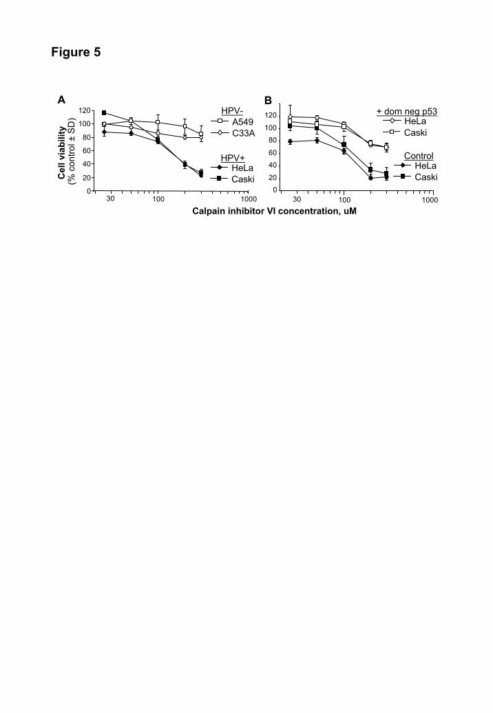
Figure 5
A
Cel
l via
bilit
y(%
con
trol ±
SD)
Calpain inhibitor VI concentration, uM
0
20
40
60
80
100
120
100 100030
HeLaCaski
HPV+
A549C33A
HPV-B
020
40
60
80
100
120
30 100 1000
HeLaCaski
Control
CaskiHeLa
+ dom neg p53
by guest on June 13, 2018http://w
ww
.jbc.org/D
ownloaded from

Figure 6C
alpa
in in
hibi
tor V
I con
cent
ratio
n, μ
M
>2>1.5Synergy
100
100
400 74
0
Caski
A549
969083777878787476808467524135313227263235200856445292823910122224100856544262014551011195076533515650005725755537191110246101212.5724734119550101100
100502512.56.33.21.60.80.40.20
10010010097999910099999910040099958677828692949594932009275655449363629474725100905952475236162115110508962484026317957325886853463827111670012.587564733174062000
100502512.56.33.21.60.80.40.20
Cisplatin concentration, μg/ml
100100100100100100100100100100400
HeLa
1001009081829210010010010010020010096654336465266564724100100100654232244243273113501001006138231433284165251001005933185271500112.510091462712224210106.788483126171215180000
502512.56.33.21.60.80.40.2
by guest on June 13, 2018http://w
ww
.jbc.org/D
ownloaded from

RbRb
E2F
CalpainE7
E2F
Rb CalpainE7
Rb
E7
ProteasomeCalpain inhibitors
Proteasomeinhibitors
Figure 7
by guest on June 13, 2018http://w
ww
.jbc.org/D
ownloaded from
Joy Gardner, Geoff Birrell, Angel Cid-Arregui and Andreas SuhrbierGrant A. Darnell, Wayne A. Schroder, Toni M. Antalis, Eleanore Lambley, Lee Major,
retinoblastoma proteinHuman papillomavirus E7 requires the protease calpain to degrade the
published online October 31, 2007J. Biol. Chem.
10.1074/jbc.M706860200Access the most updated version of this article at doi:
Alerts:
When a correction for this article is posted•
When this article is cited•
to choose from all of JBC's e-mail alertsClick here
Supplemental material:
http://www.jbc.org/content/suppl/2007/11/01/M706860200.DC1
by guest on June 13, 2018http://w
ww
.jbc.org/D
ownloaded from