Embed Size (px)
Citation preview

arbete och hälsa | vetenskaplig skriftserie
isbn 91-7045-646-1 issn 0346-7821 http://www.niwl.se/
nr 2002:10
The Nordic Expert Group for Criteria Documentationof Health Risks from Chemicals and The Dutch Expert
Committee on Occupational Standards
130. Tin and inorganic tincompounds
Bente Westrum and Yngvar Thomassen
National Institute for Working Life
Nordic Council of Ministers

ARBETE OCH HÄLSAEditor-in-chief: Staffan MarklundCo-editors: Mikael Bergenheim, Anders Kjellberg,Birgitta Meding, Bo Melin, Gunnar Rosén and EwaWigaeus Tornqvist
© National Institut for Working Life & authors 2002
National Institute for Working LifeS-112 79 StockholmSweden
ISBN 91–7045–646–1ISSN 0346–7821http://www.niwl.se/Printed at Elanders Gotab, Stockholm
Arbete och Hälsa
Arbete och Hälsa (Work and Health) is ascientific report series published by theNational Institute for Working Life. Theseries presents research by the Institute’sown researchers as well as by others, bothwithin and outside of Sweden. The seriespublishes scientific original works, disser-tations, criteria documents and literaturesurveys.
Arbete och Hälsa has a broad target-group and welcomes articles in differentareas. The language is most often English,but also Swedish manuscripts arewelcome.
Summaries in Swedish and English as wellas the complete original text are availableat www.niwl.se/ as from 1997.

Preface
An agreement has been signed by the Dutch Expert Committee on OccupationalStandards (DECOS) of the Health Council of the Netherlands and the NordicExpert Group for Criteria Documentation of Health Risks from Chemicals (NEG).The purpose of the agreement is to write joint scientific criteria documents whichcould be used by the national regulatory authorities in both the Netherlands and inthe Nordic countries.
The document on health effects of tin and inorganic tin compounds was written byMD Bente Westrum and Dr Yngvar Thomassen, both at the National Institute ofOccupational Health, Norway and has been reviewed by DECOS as well as by NEG.
Editorial work was performed by NEG’s scientific secretary, Jill Järnberg, andtechnical editing by Karin Sundström, both at the National Institute for WorkingLife in Sweden.
We acknowledge the Nordic Council of Ministers for its financial support ofthis project.
G.J. Mulder G. JohansonChairman ChairmanDECOS NEG

Abbreviations
ALA δ-aminolevulinic acidALAD δ-aminolevulinic acid dehydrataseCHO Chinese hamster ovaryLD50 lethal dose for 50% of the exposed animals at single administrationLOEL lowest observed effect levelNOEL no observed effect level

Contents
Abbreviations
1. Introduction 1
2. Substance identification 1
3. Physical and chemical properties 1
4. Occurrence, production and use 3
5. Occupational exposure data 5
6. Measurements and analysis of workplace exposure 6
7. Toxicokinetics 67.1 Uptake 6
7.1.1 Oral uptake 67.1.2 Uptake by inhalation 77.1.3 Skin uptake 7
7.2 Distribution 87.2.1 Animal data 87.2.2 Human data 97.2.3 Conclusion 10
7.3 Biotransformation 107.4 Excretion 10
7.4.1 Animal data 107.4.2 Human data 11
8. Biological monitoring 11
9. Mechanism of toxicity 11
10. Effects in animals and in vitro studies 1410.1 Irritation and sensitisation 1410.2 Effects of single exposure 1410.3 Effects of short-term exposure 1610.4 Effects of long-term exposure and carcinogenicity 1810.5 Mutagenicity and genotoxicity 2010.6 Reproductive and developmental studies 2210.7 Other studies 22
11. Observations in man 2311.1 Effects by contact and systemic distribution 23
11.1.1 General effects 2311.1.2 Skin 2311.1.3 Respiratory system 2411.1.4 Gastrointestinal tract 24
11.2 Effects of repeated exposure 2411.2.1 General effects 2411.2.2 Respiratory system 2511.2.3 Conclusion 26
11.3 Genotoxic effects 26

11.4 Carcinogenic effects 2611.5 Reproductive and developmental effects 26
12. Dose-effect and dose-response relationships 2612.1 Single/short-term exposures 26
12.1.1 In vitro 2612.1.2 Animals 2712.1.3 Humans 27
12.2 Long-term exposures 2712.2.1 Animals 2712.2.2 Humans 32
13. Previous evaluations by national and international bodies 32
14. Evaluation of human health risks 3214.1 Groups at extra risk 3214.2 Assessment of health risks 32
14.2.1 Exposure 3214.2.2 Effects 3214.2.3 Assessment 33
14.3 Scientific basis for an occupational exposure limit 33
15. Research needs 34
16. Summary 35
17. Summary in Norwegian 36
18. References 37
19. Data bases used in search of literature 47
Appendix 1 48

1
1. Introduction
Bronze, an alloy of copper and tin, has been known since 2500 BC. The firstinclusion of tin (Sn) in bronze was probably an accidental result of tin ore beingfound in copper ore; pure tin was most likely obtained at a later date (7). For thefirst time a workable metal with a low-melting point was available to fabricatedurable weapons, ornaments, coins, cooking utensils, bells and statuary. Miningand melting became established industries, adjacent cities grew wealthy, thescience of navigation advanced, trade flourished. Tin was mined in Spain, Britainand Central Europe. This thousand-year-old multifaceted civilisation declinedwith the advent of ironmongering.
The second great revolution began in 1810, when tin plate was first used forcanning foods as a result of the military needs of the Napoleonic wars. Fivedecades were to elapse before canning of food became prevalent. Concurrently, anenormous increase in human exposures to tin spread throughout the civilisedworld (159).
This document reviews the literature on tin and its inorganic compounds. SnH4
(stannane) is only referred to as a basic compound for the manufacturing of alarge number of organotin compounds and is therefore not included in thisdocument.
2. Substance identification
Pure tin is a silver-white, shiny metal with the atomic symbol Sn and belongs tothe carbon group (group IVA). Tin’s atomic number is 50 and it has an atomicweight of 118.71. Tin occurs naturally as the stable isotopes 112Sn (0.97%), 114Sn(0.65%), 115Sn (0.36%), 116Sn (14.5%), 117Sn (7.7%), 118Sn (24.2%), 119Sn (8.6%),120Sn (32.6%), 122Sn (4.6%) and 124Sn (5.8%) (43).
The most commercially significant inorganic tin compounds include tin di-andtetrachloride, tin dioxide, potassium and sodium stannates, tin difluoride, tindifluoroborate and tin pyrophosphate.
Chemical formulas, molecular weights and CAS numbers of some tin com-pounds are listed in Table 1.
3. Physical and chemical properties
The melting point (232°C) of pure tin is low compared with those of the commonstructural metals, whereas the boiling point (2602°C) exceeds that of most metalsexcept tungsten and the platinum group. Therefore, loss by volatilisation during

2
Table 1. Chemical identification of some tin compounds.Chemical name Synonyms Chemical
formulaMolecular weight
CAS-No
Potassium stannate K2Sn(OH)6 298.9 12125-03-0
Sodium stannate Na2Sn(OH)6 266.7 12209-98-2
Tin Sn 118.7 7440-31-5
Tin(IV) bromide, tin tetrabromide,stannic bromide
SnBr4 438.3 7789-67-5
Tin(II) chloride, tin dichloride,stannous chloride
SnCl2 189.6 7772-99-8
Tin(IV) chloride tin tetrachloride,stannic chloride
SnCl4 260.5 7646-78-8
Tin(IV) chloride iodide tin dichloride diiodide,stannic dichloridediiodide
SnCl2I2 443.4 13940-16-4
Tin(II) difluoroborate stannous fluoroborate Sn(BF4)2a 292.3 13814-97-6
Tin(II) fluoride tin difluoride,stannous fluoride
SnF2 156.7 7783-47-3
Tin(II) iodide tin diiodide,stannous iodide
SnI2 372.5 10294-70-9
Tin(IV) iodide tin tetraiodide,stannic iodide
SnI4 626.3 7790-47-8
Tin(IV) oxide tin dioxide,stannic oxide,
SnO2 150.69 18282-10-5
Tin(II) pyrophosphate stannous pyrophosphate Sn2P2O7 411.32 15578-26-4
Tin(II) sulphate stannous sulphate SnSO4 214.75 7488-55-3aAvailable only in solution, as the solid form has not been isolated.
melting and alloying with other metals is insignificant. Only small quantities ofsome metals can be dissolved in pure liquid tin near its melting point but inter-metallic compounds are freely formed of which some are of metallurgicalimportance. Copper, nickel, silver and gold are appreciably soluble in liquid tin.
Tin coatings can be applied to most metal surfaces by electrodeposition whilemolten tin wets and adheres readily to clean iron, steel, copper, and copper-basealloys. This tin coating provides protection against oxidation of the base metal/alloy and aids in subsequent fabrication because it is ductile and solderable (114).
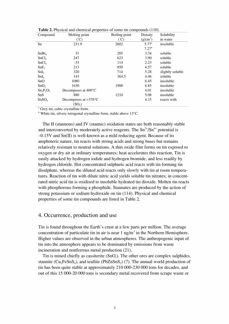
3
Table 2. Physical and chemical properties of some tin compounds (110).Compound Melting point
(°C)Boiling point
(°C)Density(g/cm3)
Solubilityin water
Sn 231.9 2602 5.77a
7.27binsoluble
SnBr4 31 205 3.34 solubleSnCl2 247 623 3.90 solubleSnCl4 -33 114 2.23 solubleSnF2 213 850 4.57 solubleSnI2 320 714 5.28 slightly solubleSnI4 143 364.5 4.46 solubleSnO 1080 6.45 insolubleSnO2 1630 1900 6.85 insolubleSn2P2O7 Decomposes at 400°C 4.01 insolubleSnS 880 1210 5.08 insolubleSnSO4 Decomposes at >378°C
(SO2)4.15 reacts with
a Grey tin, cubic crystalline form.b White tin, silvery tetragonal crystalline form, stable above 13°C.
The II (stannous) and IV (stannic) oxidation states are both reasonably stableand interconverted by moderately active reagents. The Sn2+/Sn4+ potential is–0.15V and Sn(II) is well-known as a mild reducing agent. Because of itsamphoteric nature, tin reacts with strong acids and strong bases but remainsrelatively resistant to neutral solutions. A thin oxide film forms on tin exposed tooxygen or dry air at ordinary temperatures; heat accelerates this reaction. Tin iseasily attacked by hydrogen iodide and hydrogen bromide, and less readily byhydrogen chloride. Hot concentrated sulphuric acid reacts with tin forming tindisulphate, whereas the diluted acid reacts only slowly with tin at room tempera-tures. Reaction of tin with dilute nitric acid yields soluble tin nitrates; in concent-rated nitric acid tin is oxidised to insoluble hydrated tin dioxide. Molten tin reactswith phosphorous forming a phosphide. Stannates are produced by the action ofstrong potassium or sodium hydroxide on tin (114). Physical and chemicalproperties of some tin compounds are listed in Table 2.
4. Occurrence, production and use
Tin is found throughout the Earth’s crust at a few parts per million. The averageconcentration of particulate tin in air is near 1 ng/m3 in the Northern Hemisphere.Higher values are observed in the urban atmospheres. The anthropogenic input oftin into the atmosphere appears to be dominated by emissions from wasteincineration and nonferrous metal production (21).
Tin is mined chiefly as cassiterite (SnO2). The other ores are complex sulphides,stannite (Cu2FeSnS4), and teallite (PbZnSnS2) (7). The annual world production oftin has been quite stable at approximately 210 000-230 000 tons for decades, andout of this 15 000-20 000 tons is secondary metal recovered from scrape waste or

4
Table 3. Production of tin, unwroughta. Metric tons. Major tin producing countries (187).Country 1994 1995 1996
Bolivia 15 539 17 709 16 733Brazil 20 700 19 800 19 800China 67 764 67 659 71 500Indonesia 39 000 44 218 48 960Malaysia 37 990 39 433 38 051Thailand 7 634 8 246 10 983United States 11 700 11 600 11 000World production 215 598 223 703 229 046a Production of virgin metal (primary) and tin derived from scrap (secondary). Tin alloys areincluded.
detinning. The major tin producing countries today are shown in Table 3. In theWestern World, the tin produced is mainly secondary.
Metallic tin is obtained by smelting tin ore. The ore is mixed with salt androasted at about 600°C, washed in water and then mixed with anthracite as areducing agent and smelted at about 1500°C. After refining the tin is cast into bars(146, 147).
Because of its resistance to corrosion, tin is used as a protective coating forother metals. Another important property of tin is its ability to form alloys withother metals. SnCl4 is used as a dehydrating agent in organic synthesis, a stabiliserfor plastics, and as a chemical intermediate for other tin compounds. SnCl2 servesas a reducing agent in manufacturing ceramics, glass and inks (85).
Dental amalgams contain varying proportions of tin (12-30%) (11). SnF2 hasbeen used as a prophylactic agent in preventive dentistry for decades. Sn(II) ionshave a profound and long-lasting inhibiting effect on the oral micro flora in vivo(4). Topical application of SnF2 appears to provide dentine with a layer of tin andfluoride, which may provide mechanical and chemical protection and may be ofclinical significance in restorative dentistry. Sn(II) ions possess antibacterialactivity whereas Sn(IV) ions do not (57, 61, 150, 152, 177).
The reducing agent Sn(II) is important in nuclear medicine as an essentialcomponent in diagnostic agents used to visualise blood, heart, lung, kidney andbone. Sn(II) has nearly ideal redox properties for the reduction of the visualisinglabel technetium-99m (65, 136, 143).
Most of the operations associated with the extraction of tin ore are wet pro-cesses, but tin dust and oxide fumes may escape during bagging of concentrate, inore rooms and during smelting operations (mixing-plant and furnace tapping), aswell as during the periodic cleaning of bag filters used to remove particulatematter from smelter furnace flue gas (85).
Tin reclamation from tin plated steel trimmings, rejects from tin-canmanufacturing companies, rejected plating coils from the steel industry, tindrosses and sludges, solder drosses and sludges, used bronze and bronze rejects

5
Table 4. Common uses and sources of exposure to tin and inorganic tin compounds (193).Substance Use Occupations
Tin metal Tin plating, solder and alloyproduction (common alloys arebronze, brass, gunmetal, bearingmetal, type metal and pewter),manufacture of food cans
Workers in brass and bronze foundries,makers of pewter, solder, babbit metal,and type metal; manufacturers of cansand metal containers
Inorganic tincompounds
Manufacture of toothpaste, ceramics,drill glass, porcelain, enamel, textiles(used as mordant), and ink
Production workers
and metal type scrap also involve possible exposure to tin dusts and fumes (86).Common uses and exposures to tin today are shown in Table 4.
Tin production may also involve exposure to silica, lead and arsenic in themining of the sulphide ores of tin, and to bismuth and antimony as well in theroasting and smelting process. Similarly, the preparation and use of tin alloys andsolders present an exposure to these heavy metals. Tin mining may involveexposure to radon, thorium and uranium (7, 63, 64, 83, 130, 138, 139, 180).
Studies in the United Kingdom showed mean concentrations of Sn in diet of 1-2mg/kg (60) or approximately 3 mg/day (166). The primary sources of tin weresaid to be canned goods (60).
5. Occupational exposure data
Analysis of dust samples collected from tin smelting works (see chapter 11.2.2and (149)) showed that the dust fraction with particle size < 5 µm in diametercontained more than 33% of metallic tin and no silica. The concentrations of Sn(in mg/m3) measured in the workroom air were: check sampling shed 2.22, dracco(filters for furnace gases) 1.10, smelting furnace man 1.55, refining furnace man0.82, orehouse skipman 0.34, plumber 0.12, electrician 0.05 and engineer 0.02.The methods of sampling and analysis were not described (147).
An environmental survey to determine the type of exposure in a Chilean tinfoundry showed air concentrations of metal tin between 8.6 and 14.9 mg/m3 (131).
Tin concentrations in house dust were increased in homes of electric-cablesplicers suggesting that the splicers were contaminating their homes with tin fromwork (145).
No systematic data on occupational exposure levels in tin production orprocessing are available.
The Norwegian occupational exposure database EXPO contains data from allsamples analysed at the National Institute of Occupational Health in Oslo since1984. Most of these samples have been collected due to the wish of differententerprises to control their exposures and are likely to represent "worst casemeasurements" (141). Of the 3407 air filters (8-hours personal monitoring)

6
Table 5. Branches and job functions in EXPO with air concentrations > 0.05 mg Sn/m3.Branch/job functions No. of
samplesMean
(mg Sn/m3)Range
(mg Sn/m3)Defence activities/spraying 3 0.20 0.01-0.46Metal coating/surface coating 2 0.32 0.20-0.45Electronic production/surface coating 2 1.51 0.09-2.93Railway repair/termite welding 6 1.07 0.01-5.68Metal casting/cleaning 2 0.29 0.25-0.34
analysed for tin, 420 contained amounts above the detection limit (0.002 mgSn/m3). In Table 5, the branches and job functions with tin levels > 0.05 mg Sn/m3
are listed. If some samples contained > 0.05 mg Sn/m3, all values in the analysisare included to show the mean and range of concentrations.
6. Measurements and analysis of workplace exposure
When selecting samplers for aerosol collection their sampling characteristicsshould comply with internationally accepted sampling criteria (ISO, CEN,ACGIH). Presently, most measurements are not standardised and do not complywith these sampling criteria.
The method recommended by NIOSH for measuring airborne inorganic tin andits compounds, except oxides, is filter acid digestion and atomic absorption orinductively coupled plasma atomic emission spectrometry. If the aerosol phase isbelieved to contain SnO2, the acid solution is centrifuged and the tin compoundsin the supernatant are determined as above. The precipitate is then treated withalkali, rendering SnO2 to a soluble stannate, and the determination is made asabove (7). Other acid digestion procedures (aqua regia + hydrogen fluoride) areavailable for simultaneous measurements of total tin and other elements by e.g.inductively coupled plasma atomic emission, and mass spectrometry, respectively(20, 157). Radiochemical neutron activation analysis has been used for themeasurement of tin in human biological materials at background levels (189). Afield portable X-ray fluorescence spectrometer has been developed as a rapid,nondestructive, on site alternative for analysis of membrane filters used in NIOSHmethod 7300 for metals (10).
7. Toxicokinetics
7.1 Uptake
7.1.1 Oral uptakeIn vitro experiments on the rat small intestine suggested that absorption of tin(SnCl4) occurs by passive diffusion. The absorption of SnCl4 was 7.65% after asingle oral dose in rats. The presence of some organic acids resulted in enhancedabsorption of tin from the gastrointestinal tract (98).

7
From a single oral dose of 20 mg/kg body weight of radioactive 113Sn(II) or113Sn(IV) given to 24-hour fasted rats, an absorption of 2.8% and 0.6%, respec-tively, was estimated. Changing the anion complement from fluoride to citrate hadno effect on the absorption. When the anion was pyrophosphate, the absorptionwas lowered. This was explained by the greater tendency of pyrophosphate toform insoluble complexes with tin (82).
References concerning the absorption of SnCl2 report limited absorption,usually less than 5% (66, 67, 100, 176).
In rats and cats, no tin was recovered from the urine 24 hours after the ingestionof orange juice containing high levels of tin (7-20 mg/kg body weight) derivedfrom containers. 99% of the tin ingested was recovered from the faeces in the ratsindicating a very limited oral uptake (8).
Male Wistar rats were given SnCl2 in their drinking water at three differentconcentration levels (0.44, 1.11, 2.22 mM) for 1-18 weeks. The cumulative dosewas 17.7 mmol/kg body weight (corresponding to 2100 mg Sn/kg body weight) inthe highest dose group. Blood tin increased significantly after 1 week at thehighest dose and remained at a level of 16-60 pmol/g (~16-60 nmol/l) or 2-5 timesthe concentrations in the control group. In conclusion, despite the fact thatmucosal barrier mechanisms effectively prevent tin absorption they can be over-come by very high tin doses (155).
Rabbits fed 2 mg SnCl2/kg body weight for 5 days had blood concentrations of2.3 µg Sn/l (19.4 nmol/l) after 24 hours. After 120 hours the concentration was0.7 µg/l (5.9 nmol/l). Tin was not detected in the controls (208).
Approximately 50% of the dose was absorbed by man, when 0.11 mg Sn/daywas ingested with the diet (control). From a test diet containing an additional50 mg Sn/day as SnCl2, only 3% was absorbed (90).
Four human volunteers with Sn blood levels of < 2 ng/ml (17 nmol/l) eachconsumed 60 mg Sn in the form of fruit juice from an unlacquered can. Bloodsamples were taken after 2, 5 and 24 hours. The 2 females had detectable tinblood levels of 3 ng/ml (25 nmol/l) only in the 5-hour samples. The 2 males hadpeak blood tin concentrations of 4.7 ng/ml (40 nmol/l) after 2 hours, and 3.9ng/ml (33 nmol/l) after 24 hours, respectively. Remaining blood samples had un-detectable amounts of tin (22).
Generally, data suggest that the oral uptake of tin is low, but may depend ondose, anion, and the presence of other substances.
7.1.2 Uptake by inhalationNo valid data on the uptake of inhaled inorganic tin are available.
7.1.3 Skin uptakeNo data on the absorption of tin from dermal exposure are available.

8
7.2 Distribution
7.2.1 Animal dataA single oral dose of 20 mg/kg body weight of radioactive 113Sn(II) or 113Sn(IV) asfluoride or citrate was given to female Charles River and Cox Charles River rats.The tissue distribution of tin after 48 hours as a percentage of the administeredSn(II) or Sn(IV), respectively, was as follows: skeleton 1.0 and 0.24%, liver 0.08and 0.02% and kidneys 0.09 and 0.02%. Comparing tin in tissues after the 1st andafter the 28th days of oral administration of 20 mg/kg body weight, increasedlevels were seen only in bone, and were approximately proportional to the totalamount of systemic exposure. Regarding soft tissues, the authors concluded thatonly liver and kidneys are likely to accumulate significant amounts of tin as aresult of the oral ingestion of tin salts. No 113Sn was found deposited in the brainsof rats 48 hours after a single oral dose of 4 mg, after oral daily doses of 20 mg/kgbody weight, 6 days/week for 4 weeks or after a single intravenous dose of 0.4 mgSn(II) or Sn(IV) (82). Apparently, the blood-brain barrier for the most partexcludes tin (67, 77, 82, 155).
Studies on the retention of the radionucleide 113Sn administered as SnCl2 intra-peritoneally in rats showed that most of the tin retained in the body was depositedin the bone, followed by muscle, pelt, liver and kidney. In contrast to all otherorgans, the relative amount of tin (as % of administered 113Sn) in bone increasedconsiderably (from 50% on day 1 to 65% on day 141) during this experiment (67).A study of the pharmacodynamics of several tin compounds in rabbits, usingSn(II) chelates administered with technetium-99m-labelled chelates showed thatfree Sn(II) ions localise mainly in bone. The distribution of 113Sn in bone wassimilar to that of calcium and other bone-seeking metal ions (49).
Chiba et al reported Sn concentrations in unexposed mice of 0.1-0.29 mg/kgwet weight, and 0.69 mg/kg dry weight in bone (32).
The concentration of tin in the tibias of rats fed diets supplemented with tin(> 100 mg Sn/kg diet) were more than 5 times greater than the concentrations inthe kidneys and nearly 20 times greater than the concentrations of tin in the liver.No other organs were analysed. Tin accumulated in the tibia and kidney in a dose-dependent manner at ≥ 100 mg/kg diet (92).
After lifelong administration of 0.4 mg Sn/kg body weight/day as SnCl2 in thedrinking water of rats, there was no significant increase in tin concentrations ofexamined organs; liver, kidney, heart, lung and spleen. Bone was not examined(160). In mice, a similar experiment showed tin levels of 1.2-4.5 mg/kg tissue inkidneys, liver, heart, lung, spleen and thyroid as opposed to less than 0.5 mg/kg inthe control group (158).
A 2-year carcinogenesis study of SnCl2 administered in feed (1000 or 2000 mgSnCl2/kg) showed a dose-dependent difference in the concentration of tin inexamined organs, i.e bone, liver and kidneys. Tin levels in bone were 9 and38 mg/kg (low and high dose) in male rats, and 23 and 41 mg/kg, respectively infemale mice. Tin concentrations in the kidneys were 17 and 30 mg/kg in malerats, and 0.7 and 0.9 mg/kg in female mice. In the liver Sn concentrations were 0.2

9
and 0.4 mg/kg (male rats) and 0.4 and 0.5 mg/kg (female mice). Untreated ratsand mice had tin concentrations that were below the detectable limits (124). Doseestimates in mg/kg body weight are given in chapter 10.4.
Some data indicate that the tin content in the thymus is higher than in otherrepresentative organs. In 4 young adult dogs the tin concentration in thymus wasabout twice the concentrations in the spleen or muscle (168). Analysis in un-exposed adult Lewis rats, adult COBS mice and adult A/KI mice showed thymusSn concentrations of 20, 5.5 and 4.3 mg/kg, respectively. Tin concentrated in thethymus gland as the gland atrophied with age (169).
In pregnant rats fed 20 mg Sn/kg body weight/day as radioactive SnF2 or SnF4,no tin was found in foetal or placental tissues on the 10th day of pregnancy. Onlysmall amounts of tin were found in the foetuses on day 21 (82).
In contrast, foetal tin values were elevated (0.8-1.3 mg Sn/kg) in Sprague-Dawley rats on the 20th day of gestation when the maternal diets contained tinsalts (125 mg-625 mg Sn/kg feed). Untreated rats had foetuses containing 0.64 mgSn/kg (182). Assuming a daily feed intake of 20 g/day and a body weight of 250 gthe doses given correspond to 10-50 mg Sn/kg body weight/day.
7.2.2 Human dataWith increasing age, tin levels seem to increase in the human lung, possiblybecause of inhalation of tin from polluted air. The tin content in human tissueswas high in the United States and low in Africa, and seldom present in newbornbabies in the United States (159).
Hamilton and co-workers determined the Sn content in tissue samples fromadults who had died in accidents and found the highest concentrations in lymphnodes, lung, liver and kidney (1.5, 0.8, 0.4, 0.2 mg/kg wet weight, respectively),while levels in muscle and brain were lower (0.07 and 0.06 mg/kg wet weight,respectively). In bone 4.1 mg/kg ash was reported (76).
Median tin content in adult subjects of the United States in adrenals, lung, liver,kidney, spleen, muscle, and brain was 5.1, 3.4, 1.8, 1.5, 0.8 and < 0.4, < 0.3 mg/kgwet weight, respectively (183). In healthy Japanese males, concentrations of 9.8mg/kg dry weight in hilar lymph nodes and 1.5 mg/kg dry weight in lung tissuewere reported (181).
Chiba et al reported tin concentrations determined by atomic absorptionspectrometry in several human organs. Mean concentrations (mg/kg dry weight,males, n=11-13) were: liver 1.05, kidney cortex 0.83, heart 0.75, lung 0.45, bone(rib) 0.61, testis 2.08 (32). The Sn concentration in human liver specimens fromthe United States (n=11) ranged from 0.14-0.17 mg/kg wet weight (determined byneutron activation analysis), and in Japanese human liver specimens (n=23) from0.08 to 1.12 mg/kg wet weight (determined by atomic absorption spectrophoto-metry) (27). Sherman et al found that human thymus had an average tin concent-ration of 12.8 mg Sn/kg wet weight in two children (167).
In unexposed humans, blood tin concentrations of 2-9 µg/l (17-76 nmol/l) arereported (detection limit 2 µg/l) (76, 97). Corrigan et al found background tin

10
concentrations of 11.6±4.4 nmol/l (mean±SD) in plasma and 21.7±6.7 nmol/l inred blood cells in 12 humans (8 women, 4 men, mean age 77.8 years) (38).
Background concentrations below 1 µg Sn/l (8.4 nmol/l) in serum and urine arereported (157, 189) and a 95th upper percentile of 20 µg Sn/l (168 nmol/l) in urinein a group of 496 United States residents (132).
Marked concentrations of Sn were found in the hilar lymph nodes (100 mg/kgdry weight) and lungs (100 mg/kg dry weight) of one chromate refining worker inautopsy analysis of the internal organs of 7 metallic workers and 12 unexposedmales in Japan. Elevated concentrations of Sn compared to unexposed were alsoobserved in lung, spleen, liver and kidney of chromate plating and chromaterefining workers (181).
7.2.3 ConclusionInorganic tin distributes mainly to bone but also to the lung, liver, kidney, andlymph nodes. Some data indicate that tin may have a higher affinity to thymusthan to other organs. Animal data suggest that inorganic tin does not easily passthe blood-brain barrier.
7.3 Biotransformation
Tin cations are not rapidly reduced or oxidised in the organism (87). Hiles foundthat the differences in the relative affinity of the kidneys and liver for Sn(II) andSn(IV) indicate a valence stability of the administered tin. He concluded that tinwas not rapidly oxidised or reduced during absorption and systemic transportation(82).
According to the authors, the marked differences observed between SnCl2 andSnCl4 in their effects on the immune response in C57BL/6J mice (see chapter10.7) suggested that these two oxidation states are not readily interconverted invivo (50).
7.4 Excretion
7.4.1 Animal dataAbsorbed tin is mainly excreted via the kidneys (67, 82, 87, 100, 192).
After a single oral dose of 20 mg/kg body weight of 113Sn(II) or 113Sn(IV) asfluoride or citrate given to female Charles River and Cox Charles River rats,approximately 50% of the absorbed tin was excreted within 48 hours. After asingle intravenous dose of 2 mg/kg of 113Sn(II) or 113Sn(IV), 35 and 40%, respec-tively, was excreted in the urine. 12% of the Sn(II) appeared in the faeces, butonly 3% of the Sn(IV), indicating that the biliary route is more important forSn(II) than for Sn(IV) compounds (82).
113Sn was given as SnCl2 orally, intraperitoneally or intravenously to mice, rats,monkeys and dogs. After parenteral administration, whole-body activities couldbe described by 4-component exponential expressions, similar for all species

11
studied. Intravenously injected SnCl2 in rats (0.3 µCi/rat) was eliminated withhalf-times of 0.4, 4.9, 25 and 90 days (67).
The biological half-time has been estimated to 10-20 days for Sn(II) in rat liverand kidney. In bone the half-time of Sn(II) and Sn(IV) is approximately 20-100days (17, 18, 66, 82).
7.4.2 Human dataEight adult males ate food containing 0.11 mg or 50 mg of Sn/day (as SnCl2).Their urinary excretion was 29±13 µg (mean±standard deviation) and 122 ±52µg/day, respectively (90).
In a review article by Magos, it is stated that in humans, 20% of absorbed tinwas cleared with a half-time of 4 days, 20% with 25 days, and 60% with 400days. No further details are given (111).
8. Biological monitoring
Up to the present very little information is available about tin in human biologicalmaterials even though adequate ultra-sensitive analytical techniques (inductivelycoupled plasma mass spectrometry and radiochemical neutron activation) havebeen developed for the measurement of tin at background levels (157, 189).
A method of biological monitoring demands knowledge of the relationshipbetween exposure, external dose, toxicokinetics, internal dose and effects. Suchrelationships have not yet been established for inorganic tin and no relevantmethods of biological monitoring are available.
9. Mechanism of toxicity
Even though tin is ubiquitous in animal tissues, no essential function has yet beenshown beyond doubt to be tin-dependent (2, 24, 54, 82, 121, 162-164, 169, 185,204).
Studies in animals show that inorganic tin interferes with the status of copper,iron and zinc, which may be due to impaired absorption of these metals (91, 134).
Haem is essential for cell respiration, energy generation and oxidative bio-transformation. Metal ions directly regulate cellular content of haem and haemproteins by controlling production of δ-aminolevulinic acid (ALA) syntethase andhaem oxygenase. Thus, metal ions may impair the oxidative function of cells,particularly those dependent on cytochrome P-450. As a result, the biologicalimpact of chemicals that are detoxified or metabolically transformed by the P-450system is greatly altered (55, 112). Chelation of the metal ion into the porphyrinring is not necessary in order to regulate the enzymes of haem synthesis andoxidation (113).
SnF2 and other tin dihalides form complexes with haemoproteins such ashepatic cytochrome P-450 and haemoglobin (41).

12
Substitution of tin for the central iron atom of haem leads to a synthetic haemanalogue (tin(IV)-protoporphyrin) that regulates haem oxygenase in a dualmechanism, which involves competitive inhibition of the enzyme for the naturalsubstrate haem and simultaneous enhancement of new enzyme synthesis (51,154).
SnCl2 (~ 3-30 mg Sn/kg body weight, single subcutaneous dose) and Sn(II)tartrate (~ 9 mg Sn/kg body weight, single intraperitoneal dose) induce haemoxygenase in rat liver and kidney (55, 96, 99, 112). The Sn(II) ion is more potentas an inducer of haem oxygenase-1 in rat cardiac tissue than is the Sn(IV) ion,administered subcutaneously as tin citrate (single dose, 60 mg/kg of Sn) (119).
Treatment of young spontaneously hypertensive rats with SnCl2 (63 mg/kgbody weight/day of Sn, subcutaneously, twice weekly for 8 or 15 weeks), whichselectively depletes renal cytochrome P-450 through increasing renal haemoxygenase activity, restores elevated blood pressure to normal (59, 102, 153).
Shargel and Masnyj found that the inhibition of hepatic mixed function oxidaseenzyme activity in Charles River CD albino rats by SnF2 (30 mg Sn/kg bodyweight, single intraperitoneal dose) was primarily due to the Sn(II) cation (165).
The activity of δ-aminolevulinic acid dehydratase (ALAD) in the erythrocytesof Harlan-Sprague-Dawley rats fed 2 g SnCl2/kg diet for 21 days was 55% of thatfound in the controls (92). ALAD activity was clearly decreased in Wistar ratsafter 2 doses (total 4 mg Sn/kg) of SnCl2 subcutaneously, intraperitoneally orintragastrically every other day, whereas 7 doses (total 14 mg/kg) resulted inalmost complete enzyme inhibition (206). ALAD was inhibited by SnCl2 but notby SnCl4. The inhibition was rapidly reversed (28, 30). ALA synthetase andALAD were inhibited by tin(II) tartrate (55).
Sn(II) concentrations of 1.5 µmol/l increased the activity of isolated andpurified ALAD from human red blood cells by approximately 30%. At greaterconcentrations, tin was an inhibitor of the enzyme, probably due to binding toallosteric sites (48).
A protective effect of zinc with respect to ALAD activity in blood and ALAlevels in urine was observed after combined administration of SnCl2 and ZnSO4 inrabbits (33). One subunit of the ALAD enzyme contains one zinc atom and eightsulphhydryl groups (186). Chiba and Kikuchi postulated that tin attacks onesulphhydryl group and binds weakly at the zinc-binding site of the enzyme (29).Injection of selenium (Na2SeO3) intraperitoneally simultaneously with SnCl2 inICR mice, completely protects ALAD from being inhibited by Sn. It has beensuggested that selenium protects essential thiol groups in ALAD that are other-wise blocked by tin (25, 26, 31).
Adverse effects of feeding rats diets containing SnCl2 (100 mg Sn/kg of food)for 4 weeks include copper depletion and reduction in hepatocellular antioxidantmetalloenzyme activities of superoxide dismutase and glutathione peroxidase.Impairment in hepatocellular antioxidant protection favours the peroxidation offatty acids (144).

13
Tin(II) tartrate (20 mg Sn/kg, single intraperitoneal injection) caused a decreasein glutathione in partially hepatoectomised Sprague-Dawley rats, allowing anincrease in lipid peroxidation damaging the hepatocytic membranes (53). Theinhibitory effect of tin on SH-containing enzymes, particularly hepatic glutathionereductase and glucose 6-phosphate dehydrogenase, may be caused by the SH-group forming a metal mercaptide complex with coordinate covalent bondsleading to decreased catalytic activity. The depression in enzyme levels may alsobe due to the interaction of tin with the biological ligands not directly involved inthe active centre of the enzyme but through the formation of an unacceptablesubstrate complex for enzyme catalysis (56).
SnCl2 given intravenously in mice resulted in significant inhibition of the P-450cytochrome dependent hepatic drug metabolising enzymes such as azo-reductaseand aromatic hydroxylase (19).
Pretreatment of mice with SnCl2 (50 mg/kg body weight, daily for 2 days)induced the coumarin 7-hydroxylase in liver and kidney (58).
SnCl2 (2 mg Sn/kg body weight/day) given orally had inhibitory effects oncalcium content, acid and alkaline phosphatase activity and collagen synthesis inWistar rat femoral bone (197-200). SnCl2 orally (60 mg Sn/kg body weight/dayfor 3 days) in rats also suppressed insulin secretion and inhibited hepatic phospho-rylase activity (201, 202), active transport of calcium and mucosal alkalinephosphatase activity in the duodenum and increased bile calcium contents (195,203).
SnCl2 exposure caused a dose-dependent increase in the cerebral and muscleacetylcholinesterase activity in rats given 1.11 and 2.22 mM in drinking water(highest cumulative dose corresponding to 2100 mg Sn/kg body weight) whereasno effect was seen at 0.44 mM. The authors concluded that neurochemical effectsof SnCl2 seem to occur only with extreme brain tin burdens and are thereforeprobably not relevant (155).
Studies of frog neuromuscular transmission suggest that activation of theN-type calcium channel is involved in the SnCl2 induced increase in calcium entryinto the nerve terminals (79). SnCl2 itself may facilitate the transmitter releasefrom nerve terminals in the mammalian (mouse) as well as in the amphibian(frog) species (80).
An intraperitoneal dose of SnCl2 (5-30 mg Sn/kg) suppressed gastric secretion.The mechanism of inhibition was assumed to be associated with an inhibition ofnerve transmission as well as reduction of gastrin release from G cells (194, 196).
Corrigan et al reported significant higher plasma and red blood cell Sn con-centrations in patients with Alzheimer's disease (plasma 21.6 and red blood cells32 nmol/l) than in those with multiinfarct dementia (12.4 and 19.9 nmol/l) andcontrols (11.6 and 21.7 nmol/l) (38, 39). There were negative correlationsbetween tin levels and the red blood cell polyunsaturated fatty acid levels in theAlzheimer patients, and the authors suggested that Sn is involved in lipid per-oxidation in that illness (39).

14
In conclusion, tin cations have the ability to influence the biosynthesis andinduce the biodegradation of cytochrome P-450. Some data indicate that Sn(II)may be more potent in that respect than Sn(IV). In addition, tin seems to have aninhibitory effect on the activity of several other enzymes. Thus, tin may alter drugmetabolism. An effect of SnCl2 on nerve transmission is reported.
10. Effects in animals and in vitro studies
10.1 Irritation and sensitisation
Solutions of 1% or 2% SnCl2 and 0.25% or 0.5% SnF2 in distilled water on piecesof gauze were applied to the abraded skin of rabbits for 18 hours. Intraepidermalpustules with complete destruction of the epidermis were induced. The stratumcorneum remained intact. Sites patch tested with 0.5% SnCl2 or 0.1% SnF2 (water)showed no pustules but a polymorphonuclear infiltration of leucocytes. When thesolutions were applied to intact skin there was no effect (174).
Larsson et al determined non-irritant levels of SnCl2 and SnCl4 in alcohol onskin (5%) and on oral mucosa (3% and 0.05% respectively) in Sprague-Dawleyrats. Each test solution was openly applied to the test site for 1 minute, followed 6hours later by histologic examination of the tissue response. Lesions of allergiccontact type could not be found in the oral rat mucosa (103).
An irritating effect of SnCl2 feeding on the alimentary tract of Wistar rats wasreflected by a diffusely reddened gastric and duodenal mucosa as well as bymucosal hypertrophy and hyperplasia visible in the entire small bowel at autopsy(47) (for dose regimen see chapter 10.3).
Janssen et al found ridge-like villi, increased migration of epithelial cells alongthe villus, a decreased number of villi per unit surface and increased total lengthof the rat small intestine after feeding rats 250 and 500 mg SnCl2/kg diet (88).
Metallic tin was non-toxic in a study using human epithelium-fibroblast co-culture for assessing mucosal irritancy of metals used in dentistry. Cell viabilityand prostaglandin E2 release from the cultures were used as markers for theirritative potential of the test materials and these markers were not significantlyreduced compared to untreated controls (156).
When guinea-pigs were exposed by inhalation to SnCl4 at 3000 mg/m3 for10 minutes daily for "several months", only transient irritation of the eyes andnose developed (133).
10.2 Effects of single exposure
SnCl2 (100 µmol/l) as well as SnO2 (up to 1000 µmol/l) were nontoxic towardsrabbit alveolar macrophages in single-element incubations (101), but there was asynergistic effect on the depression of superoxide anion radical release in solu-tions where Sn2+ was combined with Cd2+ or Ni2+ (71).

15
Silica containing 97% crystalline SiO2 (50 mg), SnO2 (50 mg) or a mixture ofSnO2-SiO2 (25 mg each in 1 ml saline) dust were instilled intratracheally in rats.Autopsies were performed on day 10, 20 and 30. The in vivo cytotoxicity (cellularmetabolic activity, lysozyme content and total protein content in rat broncho-alveolar lavage), interleukin-1 release from rat pulmonary cells and fibrogeniceffects (lung dry weight, collagen content of the whole lung and pathologicalgrading) after dusting correlated well with the free SiO2 content in the dusts. SnO2
tended to inhibit the effect of SiO2 in the rat lungs. The effects of SnO2 alone werepoorly described (190).
Rats were experimentally exposed to 50 mg of metallic tin dust in saline from atin smelting works by a single intratracheal administration. Four months later theauthors described X-ray changes of widespread tiny densities throughout the ratlungs; changes which they found similar to the X-ray changes earlier seen inworkers exposed to the same compound. Histologically, there was no fibrousresponse of any kind up to a year in the rats (146).
Intraperitoneal injections in guinea pigs with dusts from various stages in theprocess of a foundry reducing Bolivian tin ore concentrate to metal bars caused"an inert type of reaction", defined by the authors as dust gathered in flattenednodules with no change for months, histologically abundant macrophages withsubsequent fibroblastic reaction, and no necrosis (131).
Data on the lethal dose for 50% of the exposed animals at single administration(LD50) for NaSn2F5 and SnCl2⋅2H2O are given in Table 6. Major toxic symptomsin rats and mice were ataxia, general depression, fore and hindleg weaknessadvancing to flaccid paralysis prior to death. Rats dosed orally developeddiarrhoea. Rats, given either Sn compound, displayed swollen and discoloratedkidneys by autopsy on the 4th day, microscopically tubular necrosis and tubularregeneration. Further data from this experiment suggest that both F and Sncontribute to the toxicity of NaSn2F5 (37).
Rats receiving NaSn2F5 (~23 mg Sn/kg) or SnCl2⋅2H2O (~23 mg Sn/kg)intraperitoneally showed necrosis of the proximal tubules and regeneration oftubular cells with scar formations, while rats given NaF showed limited focallesions (205).
A study by Chmielnicka et al demonstrated a clear derangement of the variousstages in the haem synthesis in rabbits after oral administration of a single dose ofSnCl2. This effect was seen at a dose of 100 mg Sn/kg body weight, but not at10 mg Sn/kg. A protective effect of zinc with respect to ALAD activity in bloodand ALA levels in urine was observed after the combined administration of tinand zinc (33).
Male Swiss Webster mice given single intravenous doses of some radiopharmaceuticals containing SnCl2 as a reducing agent, showed a significantinhibition of P-450 cytochrome dependent hepatic drug metabolising enzymessuch as azo-reductase and aromatic hydroxylase at a dose level of ~ 0.1 mg Sn/kgbody weight of SnCl2⋅2H2O. The cytochrome P-450 content was also significantlyreduced (19).

16
Table 6. 24-hour LD50-data in mice and rats treated with NaSn2F5 or SnCl2⋅2H2O (37).Species Administration mode LD50
(mg Sn/kg)NaSn2F5
Rats, male intravenous 9Rats, female intravenous 9Mice, male intravenous 13
Rats, male intraperitoneal 50Rats, female intraperitoneal 43Mice, male intraperitoneal 54a
Rats, male oral 383Mice, male oral 396
Rats, male oral (fasted) 149Rats, female oral (fasted) 146
SnCl2 ·H2ORats, male intravenous 15Rats, male intraperitoneal 136Rats, male oral 1678Rats, male oral (fasted) 1197a 48-hour data given.
Gross and histological examination of various tissues from three species ofanimals showed no fibrosis, neoplasia, or other adverse effects followingintravenous administration of SnO2 or Sn particles at high doses. The species,doses, and times from dosing to examination were: rats 250-1000 mg SnO2/kg,200-800 mg Sn/kg, 4-26 months; rabbits 250 mg SnO2/kg, 200 mg Sn/kg, 6-26months; and dogs, similar doses as rabbits, 4-5 years (62).
10.3 Effects of short-term exposure
Weanling Wistar rats were fed diets containing 0, 0.03, 0.10, 0.30 or 1.00% ofvarious salts or oxides of tin ad libitum for periods of 4 or 13 weeks. End pointsexamined included mortality, body-weight change, diet utilisation, measurementsof blood, urine and biochemical parameters, organ weights and gross and micro-pathology. No adverse effects were noted with any levels of Sn(II) oleate, SnS,SnO or SnO2. Severe growth retardation, decreased food efficiency, slightanaemia and slight histological changes in the liver were observed with ≥ 0.3%SnCl2, Sn(II) orthophosphate, sulphate, oxalate and tartrate. The no observedeffect level (NOEL) of tin salts examined was 0.1%, or 22-33 mg Sn/kg bodyweight/day, in an unsupplemented diet, which contained a liberal amount of ironand copper. The authors stated that the level might be lower in diets marginal iniron and copper. Dietary supplements of iron had a markedly protective effectagainst tin-induced anaemia, whereas a decrease in dietary iron aggravated thecondition. The growth depression caused by tin was not alleviated by enrichingthe diet with iron and copper (45, 46).
17
In rats, ≤ 100 mg Sn/kg diet as SnCl2 (~ 7 mg Sn/kg body weight/day) for 27days had little effect on the metabolism of copper. Rats fed 500 mg Sn/kg diet(~39 mg Sn/kg body weight/day) had reduced levels of copper in plasma, liverand kidneys. Only small changes in iron metabolism were observed. Tin levels of≥ 500 mg/kg diet were associated with numerous disturbances in the metabolismof zinc. Moderate variations in dietary zinc levels did not significantly affect thelevels of minerals in tissues (91, 92).
Dietary levels of 100 mg Sn/kg diet in weanling rats for 4 weeks reducedcopper levels significantly in the duodenum, liver, kidney and femur, and zinclevels in the kidney and femur (140, 144).
Oral administration of SnCl2 (2 mg Sn/kg body weight/day) in rabbits for 1month decreased zinc and copper concentration in bone marrow and increasediron concentrations in liver and kidneys (207). Beynen et al found that iron status(tissue iron, haemoglobin, hematocrit, red blood cell count, plasma iron, total ironbinding capacity and transferrin saturation) in rabbits was not influenced bydietary tin concentrations < 100 mg Sn/kg diet as SnCl2 for 28 days. Higherdietary intake of tin caused a decrease in these parameters. Food intake and bodyweights were not reported (12).
A study in Wistar rats fed on diets containing various concentrations of tin (1,10, 50, 100 and 200 mg Sn/kg as SnCl2) for 28 days showed that iron, copper andzink tissue and plasma concentrations were seemingly unaffected at 1 mg andslightly decreased at 10 mg Sn/kg diet (~ 0.7 mg Sn/kg body weight/day). Greatereffects were seen at 50 mg/kg diet (~ 3.5 mg Sn/kg body weight/day). The bloodhaemoglobin concentration and percentage transferrin saturation decreased in alinear manner as the level of dietary Sn increased. Analysis of variance and testfor linear trend was used for the statistical evaluation (134).
Growth retardation, slight anaemia, increased relative weights of the kidneysand liver, irritation of the gastrointestinal tract, "mild" histological changes in theliver and varying degrees of pancreatic atrophy were observed in Wistar rats fedSnCl2 for 13 weeks (gradually increased from 163 mg Sn/kg body weight/day inweek 0-4 to 310 mg Sn/kg/day in week 8-13) (47).
Janssen et al investigated the effects of 0, 250 or 500 mg Sn/kg diet (as SnCl2)in a 4-week study on weanling Wistar rats. Haemoglobin was decreased and bodyweights reduced in a dose-related way in the tin-fed groups. Crypt depth, villuslength and cell turnover were increased in parts of the intestine. In week 4, theestimated doses of tin were about 25 and 50 mg Sn/kg body weight/day, respec-tively (88).
Oral doses of 2 mg Sn/kg body weight/day as SnCl2 for 5 days did not affect theprocess of haem biosynthesis in rabbits. Examined indices were ALAD in wholeblood, liver, kidneys, brain, spleen, and bone marrow, concentrations of freeerythrocyte protoporphyrins, activity of ALA synthetase in the liver and bonemarrow, urine ALA, and co-proporphyrins (208).
In rabbits, a daily oral dose of 10 mg Sn/kg body weight as SnCl2⋅2H2O for4 months caused transient anaemia in the 6-10th week. A transient high iron

18
serum concentration, a high total iron binding capacity and saturation index werealso observed (34).
A 30-day toxicity study of NaSn2F5 in albino Wistar rats resulted in depressedgrowth in a dose-related manner after 15 and 30 days. The daily oral doses ofNaSn2F5 were 20, 100 and 175 mg/kg body weight. Degenerative changes of theproximal tubular epithelium of the kidneys were observed in 15-20% of theanimals in the groups receiving 175 mg/kg. At 15 days a dose-related decrease inhaemoglobin was found, significant only in males in the two highest dose groups.Serum glucose levels were decreased at both 15 and 30 days, possibly related toreduced food intake. The dose of 20 mg/kg (~13.4 mg Sn/kg body weight)produced only minimal toxicity according to the authors (36).
The distal epiphysis compressive strength decreased significantly in the femoralbone in Wistar rats administered 300 mg Sn/kg drinking water (as SnCl2) andlaboratory chow contaminated with 52.4 mg Sn/kg of diet for 4 weeks. Feed andwater intake was not reported (127).
The calcium content in the tibia of rats fed 100 mg Sn/kg diet as SnCl2 (~7 mgSn/kg body weight/day) for 28 days was decreased (92).
Oral doses of 1.0 mg Sn/kg at 12-hours intervals for 28 days given to maleWistar rats produced an increase in the Sn content of the femoral diaphysis andepiphysis, thus resulting in decreased calcium content in bone, and also indecreased acid and alkaline phosphatase activities in the femoral epiphysis (198).
The dose-effect relationship of oral doses of SnCl2 on biochemical indices inWistar rats was studied by Yamaguchi et al. Oral doses of 0.3, 1.0 and 3.0 mgSn/kg body weight were given twice daily for 90 days. The 6.0 mg/kg/day dosecaused significant decreases in femur weight, calcium concentration, lacticdehydrogenase and alkaline phosphatase activities in serum, succinate dehydro-genase activity in the liver, and calcium content and acid phosphatase activity inthe femoral diaphysis and epiphysis. The 2.0 mg/kg/day dose produced asignificant reduction in succinate dehydrogenase activity in the liver, and thecalcium content and acid phosphatase activity in the femoral diaphysis. At the0.6 mg/kg/day dose, a slight non-significant decrease in calcium content in thefemoral epiphysis was observed. The results suggested that the LOEL of inorganictin orally administered would be 0.6 mg/kg body weight/day (197).
In conclusion, depressed growth, anaemia, a decreased calcium content in bone,interference with the status of iron, copper and zink, and decreased enzymeactivities are reported in animals after short term administration of some tincompounds including SnCl2. The anion may influence toxicity.
10.4 Effects of long-term exposure and carcinogenicity
Long Evans rats fed 5 µg Sn/ml (~0.4 mg/kg body weight/day) as SnCl2 indrinking water from weaning until natural death were compared to an equalnumber of controls. Growth was not affected. Significantly lessened longevitywas found in female rats given tin. There were increased incidences of fatty

19
degeneration of the liver and of vacuolar changes in the renal tubules in theanimals fed tin. These effects were not observed in Charles River mice. Tin wasnot tumorigenic or carcinogenic (158, 160).
From life term studies on the effect of trace elements on spontaneous tumoursin Long-Evans rats it was concluded that the oral ingestion of tin cannot be con-sidered carcinogenic at the given dose, 5 µg SnCl2/ml drinking water or 0.34-0.38mg Sn/kg body weight/day. According to the authors this corresponds to approxi-mately 25 mg of tin daily for a 70-kg man (95).
Stoner et al reported no significant difference in lung tumour production instrain A mice compared to controls after multiple intraperitoneal injections ofSnCl2 for 30 weeks. Total doses given were 240, 600 and 1200 mg/kg bodyweight and the numbers of surviving animals/initial number were 18/20, 12/20,4/20, respectively (175).
Male and female Fischer F344 rats and B6C3F1 mice received 1000 (low dose)or 2000 mg (high dose) SnCl2/kg food for 105 weeks. Feed and water wereavailable ad libitum. Mean body weight gain and feed consumption of dosed andcontrol rats were comparable. Doses in mg Sn/kg body weight/day are calculatedfrom feed consumption and body weights for male rats and female mice (Table 7).Survival appeared to depend on the dose for female mice (controls 38/50, lowdose 33/50, high dose 28/50). For male rats survival rates were 37/50, 39/50,30/50, respectively. Primary tumours occurring with statistically significantchanges in incidence are summarised in Tables 8 and 9. Such tumours occurred inmale rats and female mice only. C-cell adenomas of the thyroid gland weresignificantly increased in low-dose male rats. Thyroid C-cell adenomas andcarcinomas (combined) occurred in male rats with a significant positive trend andthe incidence in either dosed group was significantly higher than seen in thecontrols. Adenomas of the lung in male rats occurred with a significant positivetrend. The incidence of female mice with either hepatocellular adenomas orcarcinomas exhibited a significant dose-related trend, and histiocytic lymphomasin female mice also occurred with a significant positive trend. However,compared to historic control incidences for this laboratory in about 300 animals,the tumour incidence was not significantly increased except for male rats at thelow dose.
Lack of dose-response relationship, variable (7-20%) occurrence of thesetumours in untreated animals and no increase in hyperplastic changes in the tin-treated animals weaken a possible carcinogenic effect of inorganic tin.
Based on this study, SnCl2 given orally in feed was judged by the NTP not to becarcinogenic for male or female Fischer 344 rats or B6C3F1 mice, although C-celltumours of the thyroid gland in male rats may have been associated with theadministration of the chemical (124).
Tin foil imbedded subcutaneously in Wistar rats did not cause any tumourinduction (129).

20
Table 7. Calculated doses in the NTP 105-week study (124).Species Week Low dose
(mg Sn/kg bw/day)High dose
(mg Sn/kg bw/day)Male rats 5 41 89
25 30 6862 26 55
104 20 35Female mice 5 182 348
26 134 27265 92 203
104 137 290
Table 8. Primary tumours in male rats fed SnCl2 in the NTP 105-week study (124).Male rats Control diet Low dose High doseC-cell-adenoma (thyroid) 2/50 9/49a 5/50C-cell-adenoma or carcinoma (thyroid) 2/50 13/49a 8/50a
Lung adenomab 0/50 0/50 3/50a p<0.05, Fisher´s Exact test.b p<0.05, Cochran-Armitage Trend.
Table 9. Primary tumours in female mice fed SnCl2 in the NTP 105-week study (124).Female mice Control diet Low dose High doseHepatocellular adenoma or carcinomab 3/49 4/49 8/49Histiocytic malignant lymphomab 0/50 0/49 4/49b p<0.05, Cochran-Armitage Trend-
Intracranial implants of metallic tin cylinders in Marsh mice gave a localresponse of gliosis, but no fibrous capsules or related neoplasms (13). Intra-thoracic injection of tin needles in Marsh mice resulted in persisting engulfedneedles by giant cells with some adjacent nodular fibroplasia and a new networkof capillaries. The metal particles were not tumorigenic (14). Intraperitonealimplantation of tin cylinders led to development of fibrous capsules (15).
The available animal data suggest that metal tin and SnCl2 are not carcinogenicalthough one study concludes that C-cell tumours of the thyroid gland in male ratsmay have been associated with the administration of SnCl2.
10.5 Mutagenicity and genotoxicity
In the Ames test using various Salmonella strains (TA 1535, TA 100, TA 1538,TA 98 and TA 1537), SnF2 was slightly mutagenic in strain TA 100, but only inthe presence of a metabolic activation system (S9 liver fraction from Aroclor-pretreated rats) and on one type of medium (72). SnCl2 tested in the same strains,with or without the activation system, was not mutagenic in doses of 0.033-10mg/plate (137).
Kada et al reported the absence of effect (SnCl2, SnCl4 and SnSO4) in the Rec-assay in Bacillus subtilis, but indicated a high toxic effect of SnCl2 and SnCl4 on

21
bacteria (94). SnCl4 tested in the Rec-assay test with Bacillus subtilis in concen-trations up to 10 mg/test showed no genotoxicity (75).
Strains of Escherichia coli presenting mutations on specific genes for the repairof DNA were treated with SnCl2 (5-75 µg/ml, corresponding to 26-395 µmol/l).The results indicate that SnCl2 could be capable of inducing and/or producinglesions in DNA. This capability was confirmed by the lysogenic induction of E.coli K 12 and by microscopic observation of E. coli B filamentation (9).
The presence of catalase, reactive oxygen scavengers or metal-ion chelators inE. coli cultures treated with SnCl2 abolished the lethal effect and suggested theparticipation of reactive oxygen species in the toxicological effect of SnCl2 (42).
Survival rates in E. coli were most affected in the strain double mutant onspecific genes for the repair of DNA damage after incubation with SnCl2. Near-UV illumination inhibited the lethal effect of SnCl2 in E. Coli AB 1157 (wild typestrain) (170).
The SOS chromotest, a simple colorimetric assay of the induction of thebacterial gene sfiA in E. coli, indicated effects at 2-3 mmol/l of SnCl2, but theinterpretation was difficult because of a clear cyotoxic effect on the bacteria(128). SnCl4 did not produce DNA-damage in the SOS chromotest (75).
SnCl2 at concentrations of 50, 150, 350 and 500 µmol/l produced dose-relatedDNA damage, as detected by alkaline sucrose gradient analysis in Chinesehamster ovary (CHO) cells. Treatment of cells with Sn(IV) as SnCl4 produced nosuch DNA damage. There was no loss in colony formation 6 days after eithertreatment (116).
Tin(II) as SnCl2 (5, 10, 25 or 50 µmol/l) was readily taken up by human whiteblood cells and caused a dose-dependent increase in DNA strand breaks that wasmore extensive than equimolar amounts of chromium(VI), a known carcinogenand DNA damaging agent. Exposure to tin(II) also interfered with the lympho-cytes´ ability to be stimulated by the polyvalent mitogen Concanavalin A. Tin(IV)as SnCl4 did not cause DNA damage and, in contrast to other studies, was nottaken up by cells. The authors state that the relevance of these findings to humanhealth is not known. The values for tissue bound tin from environmental exposureare 2-3 orders of magnitude below the levels at which DNA damage was observedin vitro after an exposure of 30 minutes (115).
SnCl4 at 10 and 20 µg/ml (38-76 µmol/l) increased the frequency of chromo-somal aberrations, micronuclei and sister chromatide exchanges to a statisticallysignificant level in human lymphocytes in vitro when compared to the untreatedcontrol. Mitotic index and cell cycle kinetics were depressed. The effects weredirectly proportional to the concentrations used (179).
Cultures of human peripheral blood lymphocytes from 27 male donors wereincubated with 2 or 4 µg SnCl4·5 H2O/ml (5.7-11.4 µmol/l) for 70 hours. Bothdoses of SnCl4 induced chromosome aberrations in the cells. Approximately 11and 13% of the cells, respectively, were damaged compared to 4.5% in the controlcultures. Sister chromatid exchanges were about twice as frequent in both tincultures compared with controls. A reduction in cell cycle kinetics (replicative

22
index) was observed (70). Incubation of human lymphocytes from 52 donors withSnCl4, 4 µg/ml (11.4 µmol/l, assuming crystallisation water) for 48 hours, resultedin significant elevations of damaged cells, chromosome aberrations, micronucleiformation and depression of the mitotic index in both sexes (69).
Intraperitoneal doses of 9.8-39.5 mg SnF2/kg body weight at 0 and 24 hours inNMRI-mice gave no significant increase in micronucleated polychromaticerythrocytes from mouse bone marrow. In Drosophila melanogaster fed 1.25 mMSnF2 in 5% saccharose, no significant increase in sex-linked recessive mutationswas observed (72). SnCl2 was non-genotoxic in the Drosophila wing spot test(184).
To conclude, in vitro studies have shown that SnCl2 causes DNA damage inhuman white blood cells, CHO cells, and in E. coli. The effects on human whiteblood cells and CHO cells were dose-dependent. DNA damage in human lympho-cytes in vitro has been shown after treatment with SnCl4.
10.6 Reproductive and developmental studies
Theuer et al investigated the placental transfer in Sprague-Dawley rats givenvarious fluorides and tin salts (SnF2, NaSn2F5, NaSn2Cl5) (125 mg-625 mg Sn/kgfeed). Untreated rats had foetuses containing 0.64 mg Sn/kg. Foetal tin valueswere found to be elevated (0.8-1.3 mg Sn/kg) when the maternal diets containedtin salts. The greatest number of foetal resorptions was found in groups fedsodium pentafluorostannite, but the observation was not considered toxicologi-cally significant (182).
10.7 Other studies
Intraperitoneal or intravenous injections of metallic tin powder (200 mg in saline)in Lewis rats produced a striking plasmacellular hyperplasia in the draining lymphnodes and spleen (106, 108). Depending on the genetic characteristics of thesubjects, the lymph node response to metallic tin in rats varied from a slight banalresponse to insoluble foreign particles, to an exuberant granulomatous hyperplasia(August rats), and intense plasmacellular hyperplasia (Lewis rats and F1 hybridsof Lewis rats) (105). Pretreatment with tin salts in drinking water prevented theplasmacellular response to subsequently injected metallic tin as long as 2 monthsafter the pretreatment (107). The production of plasma cell hyperplasia bymetallic tin and the prevention of such response by tin salts are unique to thismetal (104).
The effect of tin compounds on the immune response to sheep red blood cells inmice was studied. SnCl2 and SnCl4 (approximately 5 and 3.5 mg Sn/kg bodyweight, respectively) intraperitoneally in mice had a number of effects on para-meters reflecting humoral and cell-mediated immune responses (50).
Subacute administration of SnCl2 (~ 20 mg Sn/kg body weight for 3 days) intra-peritoneally in mice suppressed parameters of both the primary and secondaryimmune response, suggesting that tin supresses part of the immune response in

23
which IgM antibody production is important, and that the IgG production in theprimary response is suppressed or delayed (81).
Jones et al found a significant increase in LD50 values in ICR mice followingintraperitoneal injections of salts of several metal ions including Sn(II) (as SnCl2)subsequent to pretreatment with the same ion (93).
Intratracheally injected SnCl2 in saline followed by bacterial infection (aero-solised Group C Streptococcus sp.) increased the mortality with 36 and 87% inmice at a single dose of 0.01 and 0.1 mg, respectively (~ 6 and 60 µg Sn, corre-sponding to 0.24 and 2.4 mg Sn/kg body weight). Similar effects were seen withe.g. fly ashes, carbon, bentonite, and a number of metal oxides. According to theauthors, in the case of soluble metals, inhalation exposure yields similar results(78).
In conclusion, data imply that tin chlorides have an effect on mouse immuneresponse. Injections of metallic tin in the rat seem to produce a plasmacellularhyperplasia in lymph nodes and spleen, an effect that may be prevented bypretreatment with tin salts.
11. Observations in man
11.1 Effects by contact and systemic distribution
11.1.1 General effectsIn 10-11 humans, SnCl2 (36 mg Sn) given with 65ZnCl2 solutions (0.5, 4 and 6 mgof Zn, respectively) or with turkey test meals (4 mg of 65Zn) inhibited 65Zn absorp-tion, measured by whole body counting of the retention of 65Zn after 7-10 days.According to the authors, the dose required to inhibit Zn absorption under theconditions in this study were well in excess of those ordinarily found in the diet(188). Solomons et al were unable to demonstrate clear inhibition of the plasmaappearance of zinc after 1- 4 hours in human volunteers ingesting a single dose of12.5 mg Zn and 25, 50 and 100 mg Sn given as SnCl2 (172).
11.1.2 SkinPatch tests with metallic tin in 73 nickel-sensitive patients revealed 6 positiveallergic and 4 doubtful reactions. The low frequency of doubtful reactions made itunlikely that metallic tin is irritant (117).
Patch testing with 1% SnCl2 in petrolatum and a tin disc suggested that somepatients are sensitised to tin. A low frequency of doubtful reactions suggested thatmetallic tin and SnCl2 at 1% were non-irritating. However, irritant reactions werefrequent in patients tested with 10% and 5% SnCl2 in petrolatum (44).
In 199 patients with suspected allergic reactions to metals, 13 had positivepatch tests with 2% SnCl2 in petrolatum (26 metals tested) (142).
One out of 50 craftsmen in the ceramics industry had a positive reaction bypatch testing with 2.5% elementary tin in petrolatum (68).

24
A worker producing metal patterns for body parts on trucks, exposed to air-borne dust from an alloy that used to contain tin, had dermatitis around the eyes,forehead and wrists. He had a positive patch test to 1% SnCl2 in petrolatum. Thecase is reported as an occupational allergic dermatitis due to tin (122).
In conclusion, in a few studies positive patch test reactions to metallic tin andSnCl2 but only one case of occupational allergic dermatitis are reported. Con-sidering the widespread use of tin and the unclear clinical relevance of thepositive patch test reactions it is unlikely that tin is a contact allergen.
11.1.3 Respiratory systemNo clinical or experimental reports on acute effects by inhalation of inorganic tinhave been identified. Metal fume fever caused by inorganic tin has not beendocumented (16).
11.1.4 Gastrointestinal tractSeveral authors report acute gastrointestinal illness following the intake of cannedfruit or fruit juice.
As a result of the detinning of unlacquered cans by corrosion 200-2000 mgSn/kg of food have been reported (23, 73). Estimated doses ingested are 30-200mg during a short period of time, i.e. drinking a glass of tomato juice (5, 118, 178,191). Symptoms most frequently reported are nausea, abdominal cramps, vomi-ting and diarrhoea. Median incubation period was 1 hour (range 15 minutes to 14hours), and median duration of symptoms 12 hours (range 0.5 hours to 3 weeks)(5).
Toxic signs (nausea and diarrhoea) followed the drinking of tin-containing fruitjuices by 5 human volunteers only at Sn levels of approximately 1400 mg/l,corresponding to a single dose of approximately 330 mg or 4.4-6.7 mg/kg bodyweight (LOEL) and above. No effects were observed after the ingestion ofapproximately 130 mg Sn, or 1.7-2.6 mg Sn/kg (NOEL). The authors stated thatthere was no evidence from their experiments that toxicity was due to theabsorption of tin and that the most likely cause was local irritation of the mucousmembranes of the alimentary tract (8).
11.2 Effects of repeated exposure
11.2.1 General effectsEight adult males were given mixed diets containing 0.11 mg Sn/day (controldiet) and 50 mg Sn/day (test diet, 50 mg of additional Sn as SnCl2 in fruit juice)for 20 days each in a cross-over design. There was no effect on the faecal andurinary excretion rates of copper, iron, manganese, magnesium and calcium. Zincand selenium excretion rates were moderately changed. Hematocrit and serumferritin levels were not affected (74, 89, 90). A mean body weight of 76 kg leadsto an estimated LOEL of 0.7 mg Sn/kg/day in this study.
The uremic patient might be especially prone to accumulate trace elementsfrom the usual environmental sources. Elevated tin levels have been found in

25
muscle, serum, liver and kidney of these patients. As tin affects kidney enzymeactivity in animals (chapter 9), it is suggested that tin might be involved in adegenerative feedback effect in uremic patients (125, 151).
In a Belgian case-control study (n=272), a significantly increased risk (oddsratio 3.72, 95% confidence interval 1.22-11.3) of chronic renal failure was foundfor occupational exposure to tin. Exposures were scored independently by 3industrial hygienists (126).
11.2.2 Respiratory systemThere are several case reports of workers exposed to SnO2 dust and fumes for 3years or more in tin smelting works, scrap metal recovery plants and hearthtinning. The only positive finding is the chest roentgenograms presenting apneumoconiosis called stannosis (6, 35, 40, 52, 135, 161, 171, 173). In general, noinformation on exposure levels is available.
Dundon and Hughes presented a case report of a "peculiar widespread mottlingof both lung fields by discrete shadows" in the chest roentgenogram in a man who10 years later died from cancer of the prostate. He had been employed in industrytending a detinning furnace for 18 years. The employment was terminated 18years before death. At autopsy, 1100 mg Sn/kg wet weight of lung tissue wasfound (52).
Robertson et al examined the employees, including pensioners, from a tinsmelting works and described chest X-ray changes in 121 out of 215 workers. Thechanges were widespread, tiny, dense shadows; or softer, larger, more nodularopacities. Typical changes were found in workers handling raw ore, smeltingfurnace house workers and refinery furnace men. The employment time was atleast 3 years, and up to nearly 50 years. None of the men had any clinicalsymptoms or signs referable to pneumoconiosis. None of the films suggestedfibrosis or significant emphysema (146, 149). Lung function studies of forcedexpiratory volume and airway resistance showed no disability, whatever theradiographic category. The population at the tin smelting works had lowermortality (131 deaths) than expected when compared to the male population in theUnited Kingdom (expected 166) in the period 1921-55 (147). Dust concentrationsfrom the tin smelting works were given in chapter 5, e.g. 2.22 mg tin/m3 in thecheck sampling shed, but the methods of sampling and analysis were notdescribed (147).
Autopsy findings were given by Robertson et al for 7 workers with abnormalradiographs. None had died of pulmonary disease. Aggregates of macrophagescontaining dust were seen around respiratory bronchioles and less commonlyaround segmental bronchi, in the alveoli, in the interlobular septa and in theperivascular lymphatics. The mild focal emphysema observed was assumed to beclinically insignificant and was considerably less severe than that seen in coalworkers pneumoconiosis. No fibrosis was present. Chemical and X-ray diffractionanalysis showed that the lungs contained SnO2. X-ray emission microanalysisidentified tin in a minute particle of dust in lung phagocytes (148).

26
Hlebnikova (1957, cited in (87)) made a survey over a number of years ofworkers exposed to condensation aerosols formed during the smelting of tin andconsisting mainly of SnO2. Total silica concentration in the aerosols did notexceed 3%. Total dust concentration in air varied between 3 and 70 mg/m3.Workers developed pneumoconiosis after 6-8 years of employment. No cases ofpneumoconiosis were observed 10 years after the dust concentration had beenreduced to 10 mg/m3. No further details are given (87).
Symptoms like wheezing, cough, chest pain and dyspnoea on exertion reportedin workers handling SnCl4 were probably due to elevated levels of hydrogenchloride formed by the combination of SnCl4 and water in the presence of heat(109).
11.2.3 ConclusionOccupational exposure to SnO2 dust or fumes induces stannosis with no indicationof fibrosis or apparent disability beyond chest X-ray opacities. An increased riskof chronic renal failure is reported. Excretion rates of zinc and selenium weremoderately changed in subjects given 0.7 mg Sn/kg body weight/day.
11.3 Genotoxic effects
No data are available.
11.4 Carcinogenic effects
Some reports from China are concerned with the health of tin miners. There were1724 lung cancer cases registered at the Yunnan Tin Corporation in the period1954-1986, of which 90% had a history of working underground. Assumedcontributing factors included diet, arsenic, radon and tobacco (63, 138, 139, 180).Tin was not considered a carcinogenic factor in these studies.
An increased mortality from cancer of the lung was present among Cornish tinminers. There was a clear relation between exposure to radon and death from lungcancer (64, 83).
11.5 Reproductive and developmental effects
No data are available.
12. Dose-effect and dose-response relationships
12.1 Single/short-term exposures
12.1.1 In vitroSnCl2 causes DNA damage in human white blood cells, CHO cells, and in E. coli.The effects on human white blood cells and CHO cells are dose-dependent. DNA
27
damage in human lymphocytes has been shown after treatment in vitro with SnCl4
(115, 116).
12.1.2 AnimalsOral, short-tem studies where the dose is given or possible to estimate aresummarised in table 10.
The LOEL for SnCl2 given orally with respect to calcium content in rat bone, is~ 0.6 mg Sn/kg body weight/day for 90 days (197). The decrease in calciumcontent is dose-dependent.
The LOEL for SnCl2 orally with respect to interference with the status of iron,copper and zink in rats is ~ 0.7 mg Sn/kg body weight/day for 28 days. Also,blood haemoglobin levels decrease with increasing Sn levels (0.07-13.2 mg/kgbody weight). Analysis of variance and test for linear trend suggests that there is adose-effect relationship for these effects (134).
In mice, an increased susceptiblility to bacterial infection was observed after asingle intratracheal injection of SnCl2 at a dose corresponding to 0.24 mg/kg bodyweight (78).
12.1.3 HumansThe NOEL of acute ingestion of SnCl2 with respect to gastrointestinal illness isapproximately 1.7-2.6 mg Sn/kg body weight (8).
The LOEL for SnCl2 administered orally for 20 days with respect to inter-ference with the excretion of zink and selenium is 0.7 mg Sn/kg body weight/day.Excretion rates are moderately changed at this level (74, 89, 90).
Positive patch test reactions to tin have been reported (44, 68, 117, 122, 142).However, only one case of occupational allergic contact dermatitis is published(122). 5% SnCl2 in petrolatum is irritant, whereas 1% SnCl2 in petrolatum is not(44). In view of the widespread occurrence of tin, it is probably not a relevantcontact allergen.
12.2 Long-term exposures
12.2.1 AnimalsOral long-term studies where the dose is given or possible to estimate aresummarised in Table 11.
The LOEL for SnCl2 orally in rats in life-long exposures with respect todegenerative changes in the liver and kidney is ~ 0.4 mg Sn/kg body weight/day(160).
Available data suggest that metal tin and SnCl2 are not carcinogenic althoughone study concludes that C-cell tumours of the thyroid gland in male rats mayhave been associated with the administration of SnCl2 (124).

Tab
le 1
0. D
ose-
effe
ct r
elat
ions
hips
fro
m o
ral,
shor
t-te
rm, a
nim
al s
tudi
esE
xpos
ure
regi
men
Dos
eSp
ecie
sN
o of
ani
mal
spe
r do
se g
roup
(con
trol
s)
Eff
ect
Ref
eren
ce
SnC
l 2 f
or 9
0 da
ys,
2 da
ily d
oses
0.6
mg
Sn/k
g bw
/day
2.0
mg
Sn/k
g bw
/day
6.0
mg
Sn/k
g bw
/day
Wea
nlin
gm
ale
Wis
tar
rats
6 (6
)N
on-s
igni
fica
nt d
ecre
ase
in f
emor
al e
piph
ysis
cal
cium
cont
ent.
Sign
ific
antly
dec
reas
ed f
emor
al c
alci
um c
onte
nt, s
ucci
nate
dehy
drog
enas
e ac
tivity
in li
ver,
aci
d ph
osph
atas
e ac
tivity
in f
emur
.
Sign
ific
antly
dec
reas
ed f
emor
al w
eigh
t and
cal
cium
cont
ent,
and
inhi
bitio
n of
sev
eral
enz
ymes
.T
he d
ecre
ase
in f
emor
al c
alci
um c
onte
nt a
nd e
nzym
eac
tivity
was
dos
e-de
pend
ent.
(197
)
SnC
l 2 in
die
tfo
r 28
day
s0.
07, 0
.7, 3
.5, 7
, 13.
2m
g Sn
/kg
bw/d
ay(1
, 10,
50,
100
, 200
mg
Sn/k
g di
et)
Mal
eW
ista
r ra
tsag
ed 3
wee
ks
7 (7
)Pl
asm
a, k
idne
y, s
plee
n an
d tib
ia ir
on, h
aem
oglo
bin
and
tran
sfer
rin
satu
ratio
n de
crea
sed
in a
line
ar d
ose-
effe
ctm
anne
r. S
igni
fica
nt in
vers
e re
spon
se o
f pl
asm
a, li
ver,
kidn
ey, s
plee
n an
d tib
ia C
u le
vels
and
pla
sma,
kid
ney
and
tibia
Zn
leve
ls. E
ffec
ts f
rom
~ 0
.7 m
g/kg
bw
/day
on
min
eral
sta
tus.
(134
)
SnC
l 2 f
or 2
8 da
ys,
2 da
ily d
oses
2.0
mg
Sn/k
g bw
/day
Wea
nlin
gm
ale
Wis
tar
rats
5 (5
)D
ecre
ased
cal
cium
con
tent
and
dec
reas
ed p
hosp
hata
seac
tiviti
es in
fem
oral
bon
e.(1
98)
SnC
l 2 f
or 5
day
s2
mg
Sn/k
g bw
/day
Fem
ale
rabb
its5
(5)
No
adve
rse
effe
cts
on h
aem
bio
synt
hesi
s ob
serv
ed.
(208
)
Tab
le 1
0. C
ont.
Exp
osur
e re
gim
enD
ose
Spec
ies
No
of a
nim
als
per
dose
gro
up(c
ontr
ols)
Eff
ect
Ref
eren
ce
SnC
l 2 f
or 1
mon
th2
mg
Sn/k
g bw
/day
Mal
era
bbits
5 (5
)D
ecre
ased
zin
c an
d co
pper
con
cent
ratio
ns in
bon
em
arro
w, i
ncre
ased
iron
con
cent
ratio
ns in
live
r an
dki
dney
s.
(207
)
SnC
l 2 f
or 4
mon
ths
10 m
g Sn
/kg
bw/d
ayFe
mal
era
bbits
6T
rans
ient
ana
emia
, hig
h s-
iron
, tot
al ir
on b
indi
ng c
apac
ityan
d sa
tura
tion
inde
x.(3
4)
Sn(I
I) o
leat
e, S
nS o
rSn
O2
in d
iet f
or 4
wee
ks, o
r Sn
O in
die
tfo
r 13
wee
ks
0.03
, 0.1
0, 0
.3 o
r 1.
0%of
com
poun
dW
eanl
ing
Wis
tar
rats
10 m
ales
,10
fem
ales
(10+
10)
No
adve
rse
effe
cts
note
d.(4
6)
SnC
l 2, S
n(II
) or
tho-
phos
phat
e, s
ulph
ate,
oxal
ate
and
tart
rate
indi
et f
or 4
wee
ks, o
rSn
Cl 2
in d
iet f
or 1
3w
eeks
~7-1
0 m
g Sn
/kg
bw/d
ay(0
.03%
of
com
poun
d)
~22-
33 m
g Sn
/kg
bw/d
ay(0
.1%
of
com
poun
d )
~70-
100,
~22
0-33
0m
g Sn
/kg
bw/d
ay(0
.3, 1
.0%
of
com
poun
d)
Mal
e ra
ts,
Spra
gue-
Daw
ley
10 m
ales
,10
fem
ales
(10+
10)
No
adve
rse
effe
cts
note
d.
No
adve
rse
effe
cts
note
d.
≥ 70
-100
mg
Sn/k
g bw
/day
gro
wth
ret
arda
tion,
red
uced
food
eff
icie
ncy,
ana
emia
, his
tolo
gica
l liv
er c
hang
es, m
ore
seve
re in
the
grou
ps g
iven
220
-330
mg
Sn/k
g bw
/day
.
(46)
Tab
le 1
0. C
ont.
Exp
osur
e re
gim
enD
ose
Spec
ies
No
of a
nim
als
per
dose
gro
up(c
ontr
ols)
Eff
ect
Ref
eren
ce
SnC
l 2 in
die
tfo
r 21
-27
days
~7 m
g Sn
/kg
bw/d
ay(1
00 m
g Sn
/kg
diet
,15
or
52 m
g Z
n/kg
die
t)
Mal
e ra
ts,
Spra
gue-
Daw
ley
7 (7
)≥
7 m
g Sn
/kg
bw d
ecre
ased
cal
cium
con
tent
of
the
tibia
s.(9
1, 9
2)
~13-
14 m
g Sn
/kg
bw/d
ay(2
00 m
g Sn
/kg
diet
)15
, 30
or 5
2 m
g Z
n/kg
die
t)
≥ 13
-14
mg
Sn/k
g bw
dec
reas
e in
abs
orpt
ion
of z
inc.
~39
mg
Sn/k
g bw
/day
(500
mg
Sn/k
g di
et,
15 o
r 30
mg
Zn/
kg d
iet)
≥ 39
mg
Sn/k
g bw
dec
reas
ed r
eten
tion
of z
inc
in ti
bia,
kidn
ey, l
iver
and
pla
sma,
dec
reas
ed c
oppe
r le
vels
inpl
asm
a, li
ver
and
kidn
ey.
~142
mg
Sn/k
g bw
/day
(200
0 m
g Sn
/kg
diet
,15
mg
Zn/
kg d
iet)
SnC
l 2 in
die
tfo
r 4
wee
ks25
and
50
mg
Sn/k
g bw
/day
(250
and
500
mg
Sn/k
g di
et)
Wea
nlin
gm
ale
Wis
tar
rats
10 (
10)
Dec
reas
ed h
aem
oglo
bin,
dos
e-de
pend
ent d
ecre
ase
in b
ody
wei
ght,
grow
th, f
eed
inta
ke, l
engt
h an
d w
eigh
t of
smal
lin
test
ine.
Inc
reas
ed in
test
inal
cry
pt d
epth
and
vill
us le
ngth
.
(88)
NaS
n 2F
5 by
gav
age
for
30 d
ays
13.4
mg
Sn/k
g bw
/day
(20
mg
NaS
n 2F
5/kg
bw
/day
)
67 m
g Sn
/kg
bw/d
ay(1
00 m
g N
aSn 2
F5/
kgbw
/day
)
117
mg
Sn/k
g bw
/day
(175
mg
NaS
n 2F
5/kg
bw/d
ay)
Mal
e an
dfe
mal
eW
ista
r ra
ts
20 (
20)
No
adve
rse
effe
cts
note
d.
≥ 67
mg/
kg b
w d
ose-
depe
nden
t gro
wth
ret
arda
tion.
15-2
0% o
f ra
ts h
ad d
egen
erat
ive
chan
ges
in k
idne
y tu
bula
rep
ithel
ium
.
(36)
Tab
le 1
1. D
ose-
effe
ct a
nd d
ose-
resp
onse
rel
atio
nshi
ps. L
ong-
term
exp
osur
e, o
ral s
tudi
es in
ani
mal
s on
ly.
Exp
osur
edo
seSp
ecie
sN
o of
ani
mal
spe
r do
se g
roup
(con
trol
s)
Eff
ect o
r re
spon
seR
efer
ence
SnC
l 2 in
dri
nkin
gw
ater
for
life
0.34
-0.3
8 m
g Sn
/kg
bw/d
ay(5
µg/
ml)
Lon
g-E
vans
rat
s,m
ales
and
fem
ales
94 (
82)
No
sign
ific
ant c
hang
e in
tum
our
inci
denc
e.(9
5)
SnC
l 2in
dri
nkin
gw
ater
for
life
0.4
mg
Sn/k
g bw
/day
(5 µ
g Sn
/ml)
Lon
g-E
vans
rat
s,m
ale
and
fem
ale
~100
(10
0)R
educ
ed lo
ngev
ity in
fem
ales
,in
crea
sed
inci
denc
e of
live
r an
d ki
dney
hist
opat
holo
gica
l cha
nges
, no
tum
orig
enic
or c
arci
noge
nic
effe
cts
obse
rved
.
(160
)
SnC
l 2 in
dri
nkin
gw
ater
for
life
0.4
mg
Sn/k
g bw
/day
(5 µ
g Sn
/ml)
Cha
rles
Riv
erm
ice,
mal
e an
d fe
mal
e
108
(80)
No
toxi
city
of
tin w
as d
emon
stra
ble.
(158
)
SnC
l 2 in
die
tfo
r 10
5 w
eeks
1000
or
2000
mg/
kg d
iet
Cal
cula
ted
dose
sgi
ven
in T
able
7.
Fisc
her3
44 r
ats
B6C
3F1
mic
e50
mal
es, 5
0 fe
mal
es(5
0+50
)50
mal
es, 5
0 fe
mal
es(5
0+50
)
See
Tab
les
8 an
d 9.
SnC
l 2 g
iven
ora
lly in
feed
was
judg
ed b
y th
e N
TP
not t
o be
carc
inog
enic
for
mal
e or
fem
ale
Fisc
her
344
rats
or
B6C
3F1
mic
e, a
lthou
gh C
-cel
ltu
mou
rs o
f th
e th
yroi
d gl
and
in m
ale
rats
may
hav
e be
en a
ssoc
iate
d w
ith th
ead
min
istr
atio
n of
the
chem
ical
.
(124
)

32
12.2.2 HumansSeveral case reports on inhalation exposure to SnO2 dust or fumes in humans werefound, but systematic data on exposure levels are not available. Exposure timewas 3-50 years. No symptoms or signs of impaired lung function were found, butthere were abnormal findings in radiographs and at autopsy. Aggregates ofmacrophages containing dust but no fibrosis were observed (6, 35, 40, 52, 87,135, 146, 148, 149, 161, 171, 173).
13. Previous evaluations by national and international bodies
The ACGIH and the NIOSH/OSHA recommend an occupational exposue limit of2 mg/m3 in the interest of minimising the potential of stannosis (1, 123).
In the US Environmental Protection Agency Gene-Tox Program the overallevaluation placed SnCl2 in the limited negative category for carcinogenicity (120).
SnCl2 was classified as a chemical with equivocal evidence of carcinogenicityin a review of 301 chemicals tested by the US National Toxicology Program (3).
IARC stated in 1987 that inorganic fluorides used in drinking water, includingSnF2, are not classifiable as to their carcinogenicity to humans (84).
14. Evaluation of human health risks
14.1 Groups at extra risk
Workers with renal disease might be at extra risk of renal effects by exposure totin. Johnson et al suggested that chronic exposure of man to moderate levels of tinin food probably is important mainly to those individuals who chronicallyconsume low levels of zinc and thus often are in a marginal nutritional statusregarding zinc (the elderly, children and pregnant women).
14.2 Assessment of health risks
14.2.1 ExposureOccupational exposure to tin and its inorganic compounds is described byinhalation or skin contact during the handling of tin ore, smelting and industrialprocessing of tin. Data on exposure levels are scarce.
Canned food from unlaquered cans may contain high levels of tin.
14.2.2 EffectsDeposition of tin particles in the lungs of exposed workers has been associated tostannosis, with no indication of fibrosis. No apparent disability beyond X-rayopacities is found. There are no data on the possible absorption from the lungs.

33
Irritant reactions to skin (humans and animals) and mucosa (animals) have beenshown for SnCl4 and SnCl2. Allergic dermatitis in humans has been suggested. Nodata on the possibility of skin absorption is available.
When tin is absorbed from the gut it may interfere with the status of othermetals (animals, humans) and/or cause changes in enzyme levels (animals) inblood, liver, kidney and bone and degenerative changes in liver and kidney(animals).
SnCl2 was considered not carcinogenic in rats and mice, with the possibleexception of C-cell tumours in the thyroid gland in male rats.
In vitro experiments have shown effects on enzyme activities, and DNA-damage in human white blood cells, CHO cells and E. coli.
14.2.3 AssessmentThe literature on long-term inhalation of inorganic tin consists of case reports inhumans, with poor exposure assessment and old methods of examination. Reportson effects concerning micropathology and cell toxicity in the respiratory systemare scarce. The information is insufficient to assess the health risk to the lungs.
Tin absorbed from the gastrointestinal tract may affect the mineral status inanimals and humans. On acute ingestion, the irritative symptoms of nausea anddiarrhoea are likely to prevent the ingestion of doses that might represent a furtherhealth risk. For the chronic ingestion during weeks and months, the critical effectof absorbed tin might be the decrease of calcium content in bone (rats) for short-time exposures, but interference with the status of minerals is observed at aboutthe same levels of exposure (0.6-0.7 mg/kg body weight) in both animals andhumans. In older studies, degenerative changes in liver and kidney are observed ateven lower levels (0.4 mg/kg) in life-time exposure in rats. The exact doses ofexposure in mg/kg body weight are difficult to estimate due to the study designsand lack of information in the reports. The available reports on metallic tin andSnCl2 are not sufficient to classify tin as a contact allergen.
14.3 Scientific basis for an occupational exposure limit
Data for the concentration of tin in workplace air, in body fluids and organ con-centrations in workers and toxic effects are scarce. Effects on enzymes and theconcentration of other elements in tissues and fluids are not sufficiently docu-mented and evaluated for a critical effect to be established. This is also true for invitro experiments showing cytogenetic changes in human white blood cells, CHOcells and E. coli. Although there are some indications that occupational exposuresmay cause adverse effects the available data are insufficient for forming ascientific basis for an occupational exposure limit.
34
15. Research needs
Systematic data on occupational exposure levels of inorganic tin in air are neededas are data on blood or tissue levels in workers exposed to known levels ofinorganic tin in their working atmospheres. There are also few data on back-ground blood or tissue levels of tin in unexposed humans.
If absorption from occupational exposure is confirmed in the future, furtherinvestigations into the effects on enzymes and mineral status and dose-effectrelationships of inorganic tin on the haematopoietic system, bone, liver andkidney in humans are necessary. Animal inhalation studies are also lacking.
Data on irritative effects (eye, respiratory tract) are needed. The lung effectsdescribed should be more thoroughly examined by modern laboratories of lungfunction, radiology and micropathology. The observed DNA-damage in vitroshould lead to further studies in humans. Further studies according to modernprotocols on carcinogenicity in animals are also needed.

35
16. Summary
Westrum B, Thomassen Y. The Nordic Expert Group for Criteria Documentationof Health Risks from Chemicals and the Dutch Expert Committee on Occupa-tional Standards. 130. Tin and inorganic tin compounds. Arbete och Hälsa2002;10:1-48.
Exposure to inorganic tin occurs in the mining and smelting of tin ore, in tinplating, solder, alloy, toothpaste, ceramics and textile production. Acute gastro-intestinal illness follows the intake of food containing high levels of tin. Tinabsorption from the gut is < 5%. Tin is deposited in bone, liver, kidneys, andlymph nodes. Effects of tin are growth retardation, anaemia, changes in enzymeactivities, interactions with the absorption and excretion of calcium, copper, ironand zinc, and morphological changes in liver and kidney. SnCl2 was considerednot carcinogenic in rats and mice, with the possible exception of C-cell tumours inthe thyroid gland in male rats. Tin chlorides produce DNA-damage in vitro. SnCl2
is irritating to skin. Inhalation of SnO2 may cause X-ray changes (stannosis), butno apparent pulmonary dysfunction and no fibrosis. Data are insufficient foridentification of a scientific basis for an occupational exposure limit.
Key words: bone, gastrointestinal, kidney, liver, mineral status, occupationalexposure limits, skin, SnCl2, stannosis, tin, toxicity.

36
17. Summary in Norwegian
Westrum B, Thomassen Y. The Nordic Expert Group for Criteria Documentationof Health Risks from Chemicals and the Dutch Expert Committee on Occupa-tional Standards. 130. Tin and inorganic tin compounds. Arbete och Hälsa2002;10:1-48.
Eksponering for uorganisk tinn forekommer i tinngruver og smelteverk, iproduksjon av tinnvarer, lodding, legeringer, tannkrem, keramikk og tekstiler.Inntak av mat med høyt nivå av tinn gir akutt magetarm sykdom. Absorpsjon avtinn fra tarmen er < 5%. Absorbert tinn blir avsatt i bein, lever, nyre og lymfe-knuter. Effekter av tinn er veksthemming, anemi, endringer i enzymaktivitet,interaksjon med absorpsjon og utskillelse av av kalsium, kopper, jern og sink ogmorfologiske endringer i lever og nyre. SnCl2 ble vurdert som ikke kreftfrem-kallende hos rotter og mus, med et mulig unntak for C-celle svulster i skjold-bruskkjertelen hos hannrotter. Tinnklorider gir DNA-skade in vitro. SnCl2 irritererhud. Inhalasjon av tinn kan gi røntgenologiske forandringer (stannose), mentilsynelatende ingen reduksjon i lungefunksjon og ingen fibrose. Data erutilstrekkelige for å identifisere et vitenskapelig grunnlag for en grenseverdi foreksponering i arbeidet.
Nøkkelord: bein, lever, magetarm sykdom, nyre, grenseverdi for eksponering iarbeid, SnCl2, stannose, tinn, toksisitet.

37
18. References
1. ACGIH. Documentation of the threshold limit values and biological exposure indices. 6th ed.Cincinnati: American Conference of Governmental Industrial Hygienists Inc., 1996:1550-1551.
2. Alfrey A. Aluminium and tin. In: Bronner F, Coburn J, eds. Disorders of mineral metabolismvol 1. New York: Academic Press, 1981: 353-368.
3. Ashby JT, R. W. Definitive relationships among chemical structure, carcinogenicity andmutagenicity for 301 chemicals tested by the U.S. NTP. Mutat Res 1991;257:229-306.
4. Attramadal A, Svatun B. In vivo antibacterial effect of tin on the oral microflora. Scand JDent Res 1984;92:161-164.
5. Barker W, Runte V. Tomato juice-associated gastroenteritis, Washington and Oregon, 1969.Am J Epidemiol 1972;96:219-226.
6. Barták F, Tomecka M, Tomisek O. Stanniosis (pneumokoniosis due to tin) (in Czech withFrench summary). Cas Lek Cesk 1948;87:915-920.
7. Beliles RP. The metals. Tin. In: Clayton GD, Clayton FE, eds. Patty´s industrial hygiene andtoxicology Vol. 2. 4th ed. New York: John Wiley, 1994: 2258-2276.
8. Benoy CJ, Hooper PA, Schneider R. Toxicity of tin in canned fruit juices and solid foods.Food Cosmet Toxicol 1971;9:645-656.
9. Bernardo-Filho M, Cunha M, Valsa I, Araujo A, Silva F, Fonseca A. Evaluation of potentialgenotoxicity of stannous chloride: inactivation, filamentation and lysogenic induction ofEscherichia coli. Food Chem Toxicol 1994;32:477-479.
10. Bernick M, Campagna P. Application of field-portable x-ray-fluorescence spectrometers forfield-screening air monitoring filters for metals. J Hazard Mater 1995;43:91-99.
11. Berry T, Nicholson J, Troendle K. Almost two centuries with amalgam: where are we today?J Am Dent Assoc 1994;125:392-399.
12. Beynen AC, Pekelharing HLM, Lemmens AG. High intakes of tin lower iron status in rats.Biol Trace Elem Res 1992;35:85-88.
13. Bischoff F, Bryson G. Inert foreign-body implants in the mouse CNS. Res Comm PsycholPsychiatry Behavior 1976;1:187-190.
14. Bischoff F, Bryson G. Toxicologic studies of tin needles at the intrathoracic site of mice. ResCommun Chem Pathol Pharmacol 1976;15:331-340.
15. Bischoff F, Bryson G. The pharmacodynamics and toxicology of steroids and relatedcompounds. Adv Lipid Res 1977;15:61-157.
16. Blanc P, Boushey HA. The lung in metal fume fever. Seminars in Resp Med 1993;14:212-225.
17. Brown RA, Nazario CM, De Tirado RS, Castrillón J, Agard ET. A comparison of the half-life of inorganic and organic tin in the mouse. Environ Res 1977;13:56-61.
18. Bulten EJ, Meinema HA. Tin. In: Merian E, ed. Metals and their compounds in theenvironment: Ocurrence, analysis, and biological relevance. Weinheim: VCH, 1991: 1243-1259.
19. Burba J. Inhibition of hepatic azo-reductase and aromatic hydroxylase byradiopharmaceuticals containing tin. Toxicol Lett 1983;18:269-272.
20. Butler O. Development of an international standard for the determination of metals andmetalloids in workplace air using ICP-AES: evaluation of sample dissolution proceduresthrough an interlaboratory trial. J Environ Monit 1999;1:23-32.
21. Byrd JT, Andreae MO. Tin and methyltin species in sea water. Concentration and fluxes.Science 1982;218:565-569.

38
22. Byrne AR, Kosta L. On the vanadium and tin contents of diet and human blood. Sci TotalEnviron 1979;13:87-90.
23. Capar SG, Boyer KW. Multielement analysis of food stored in their open cans. J Food Safety1980;2:105-118.
24. Cardarelli NF. Tin and the thymus gland. Silicon, germanium, tin and lead compounds1986;9:307-321.
25. Chiba M, Fujimoto N, Kikuchi M. Protective effect of selenium on the inhibition oferythrocyte 5-aminolevulinate dehydratase activity by tin. Toxicol Lett 1985;24:235-241.
26. Chiba M, Fujimoto N, Oyamada N, Kikuchi M. Interactions between selenium and tin,selenium and lead, and their effects on ALAD activity in blood. Biol Trace Elem Res1985;8:263-282.
27. Chiba M, Iyengar V, Greenberg RR, Gills T. Determination of tin in biological materials byatomic absorption spectrophotometry and neutron activation analysis. Sci Total Environ1994;148:39-44.
28. Chiba M, Kikuchi M. Effect of tin compounds on activity of 5-aminolevulinate dehydratasein blood. Biochem Biophys Res Commun 1978;82:1057-1061.
29. Chiba M, Kikuchi M. The in vitro effects of zinc and manganese on delta-aminolevulinicacid dehydratase activity inhibited by lead or tin. Toxicol Appl Pharmacol 1984;73:388-394.
30. Chiba M, Ogihara K, Kikuchi M. Effect of tin on porphyrin biosynthesis. Arch Toxicol1980;45:189-195.
31. Chiba M, Shinohara A. Inhibition of erythrocyte 5-aminolaevulinate hydrolase activity by tinand its prevention by selenite. Br J Ind Med 1992;49:355-358.
32. Chiba M, Shinohara A, Ujiie C. Tin concentrations in various organs in humans, dogs, andmice. Biomed Trace Elem Res 1991;2:257-258.
33. Chmielnicka J, Zareba G, Grabowska U. Protective effect of zinc on heme biosynthesisdisturbances in rabbits after administration per os of tin. Ecotoxicol Environ Saf 1992;24:266-274.
34. Chmielnicka J, Zareba G, Polkowska-Kulesza E, Najder M, Korycka A. Comparison of tinand lead toxic action on erythropoietic system in blood and bone marrow of rabbits. BiolTrace Elem Res 1993;36:73-87.
35. Cole CWD, Davies JVSA. Stannosis in hearth tinners. Br J Ind Med 1964;21:235-241.
36. Conine DL, Yum M, Martz RC, Stookey GK, Forney RB. Toxicity of sodiumpentafluorostannite. A new anticariogenic agent. III. 30-Day toxicity study in rats. ToxicolAppl Pharmacol 1976;35:21-28.
37. Conine DL, Yum M, Martz RC, Stookey GK, Muhler JC, Forney RB. Toxicity of sodiumpentafluorostannite, a new anticariogenic agent. I. Comparison of the acute toxicity ofsodium pentafluorostannite, sodium fluoride, and stannous chloride in mice and/or rats.Toxicol Appl Pharmacol 1975;33:21-26.
38. Corrigan FM, Crichton JS, Ward NI, Horrobin DF. Blood tin concentrations in Alzheimer'sdisease. Biol Psychiatry 1992;31:749-750.
39. Corrigan FM, Van Rhijn AG, Ijomah G, McIntyre F, Skinner ER, Horrobin DF, Ward NI.Tin and fatty acids in dementia. Prostaglandins Leukot Essent Fatty Acids 1991;43:229-238.
40. Cutter HC, Faller WW, Stocklen JB, Wilson WL. Benign pneumoconiosis in a tin oxiderecovery plant. J Ind Hyg Toxicol 1949;31:139-141.
41. Dahl AR, Hodgson E. Complexes of stannous fluoride and other group IVB dihalides withmammalian hemoproteins. Science 1977;197:1376-1378.
42. Dantas FJ, Moraes MO, Carvalho EF, Valsa JO, Bernardo-Filho M, Caldeira-de-Araujo A.Lethality induced by stannous chloride on Escherichia coli AB1157: participation of reactiveoxygen species. Food Chem Toxicol 1996;34:959-962.

39
43. de Bièvre P, Barnes IL. Table of the isotopic composition of the elements as determined bymass spectrometry. International Journal of Mass Spectrometry and Ion Processes1985;65:211-230.
44. de Fine Olivarius F, Balslev E, Menné T. Skin reactivity to tin chloride and metallic tin.Contact Dermatitis 1993;29:110-111.
45. de Groot AP. Subacute toxicity of inorganic tin as influencecd by dietary levels of iron andcopper. Food Cosmet Toxicol 1973;11:955-962.
46. de Groot AP, Feron VJ, Til HP. Short-term toxicity studies on some salts and oxides of tin inrats. Food Cosmet Toxicol 1973;11:19-30.
47. der Meulen HC, Feron VJ, Til HP. Pancreatic atrophy and other pathological changes in ratsfollowing the feeding of stannous chloride. Pathol Eur 1974;9:185-192.
48. Despaux N, Bohuon C, Comoy E, Boudene C. Postulated mode of action of metals onpurified human ALA-dehydratase (EC 4-2-1-24). Biomedicine 1977;27:358-361.
49. Dewanjee MK, Wahner HW. Pharmacodynamics of stannous chelates administered with99mTc-labeled chelates. Radiology 1979;132:711-716.
50. Dimitrov NV, Meyer C, Nahhas F, Miller C, Averill BA. Effect of tin on immune responsesof mice. Clin Immunol Immunopathol 1981;20:39-48.
51. Drummond GS, Kappas A. Chemoprevention of neonatal jaundice: potency of tin-proto-porphyrin in an animal model. Science 1982;217:1250-1252.
52. Dundon CC, Huges JP. Stannic oxide pneumoconiosis. Amer J Roentgenol Radium Ther1950;63:797-812.
53. Dwivedi RS, Kaur G, Srivastava RC, Krishna Murti CR. Lipid peroxidation in tin intoxicatedpartially hepatectomized rats. Bull Environ Contam Toxicol 1984;33:200-209.
54. Dwivedi RS, Kaur G, Srivastava RC, Krishna Murti CR. Metabolic activity of lysosomes intin-intoxicated regenerating rat liver. Toxicol Lett 1985;28:133-138.
55. Dwivedi RS, Kaur G, Srivastava RC, Murti CR. Role of tin on heme and drug biotrans-formation mechanism of partially hepatectomized rats. Ind Health 1985;23:1-7.
56. Dwivedi RS, Kaur G, Srivestava RC, Krishna MCR. Effects of tin on sulfhydryl containingenzymes in liver of rats. Chemospere 1983;12:333-340.
57. Ellingsen JE, Rolla G. Treatment of dentin with stannous fluoride - SEM and electronmicroprobe study. Scand J Dent Res 1987;95:281-286.
58. Emde B, Tegtmeier M, Hahnemann B, Legrum W. Inorganic tin - a new selective inducer ofthe murine coumarin 7-hydroxylase (CYP2A5). Toxicology 1996;108:73-78.
59. Escalante B, Sacerdoti D, Davidian MM, Laniado-Schwartzman M, McGiff JC. Chronictreatment with tin normalizes blood pressure in spontaneously hypertensive rats.Hypertension 1991;17:776-779.
60. Evans WH, Sherlock JC. Relationships between elemental intakes within the UnitedKingdom total diet study and other adult dietary studies. Food Addit Contam 1987;4:1-8.
61. Feretti GA, Tanzer JM, Tinanoff N. The effect of fluoride and stannous ions on Strepto-coccus mutans. Viability, growth, acid, glucan production, and adherence. Caries Res1982;16:298-307.
62. Fischer HW, Zimmerman GR. Long retention of stannic oxide. Lack of tissue reaction inlaboratory animals. Arch Pathol 1969;88:259-264.
63. Forman MR, Yao SX, Graubard BI, Qiao YL, McAdams M, Mao BL, Taylor PR. The effectof dietary intake of fruits and vegetables on the odds ratio of lung cancer among Yunnan tinminers. Int J Epidemiol 1992;21:437-441.
64. Fox AJ, Goldblatt P, Kinlen LJ. A study of the mortality of Cornish tin miners. Br J Ind Med1981;38:378-380.

40
65. Francis MD, Tofe AJ, Hiles RA, Birch CG, Bevan JA, Grabenstetter RJ. Inorganic tin:chemistry, disposition and role in nuclear medicine diagnostic skeletal imaging agents. Int JNucl Med Biol 1981;8:145-152.
66. Fritsch P, De Saint Blanquat G, Derade R. Effects of various dietary components onabsorption and tissue distribution of orally administered inorganic tin in rats. Food CosmetToxicol 1977;15:147-149.
67. Furchner JE, Drake GA. Comparative metabolism of radionuclides in mammals - XI.Retention of 113Sn in the mouse, rat, monkey and dog. Health Phys 1976;31:219-224.
68. Gaddoni G, Baldassari L, Francesconi E, Motolese A. Contact dermatitis among decoratorsand enamellers in hand-made ceramic decorations. Contact Dermatitis 1993;28:127-128.
69. Ganguly BB. Cell division, chromosomal aberration, and micronuclei formation in humanperipheral blood lymphocytes. Effect of stannic chloride on donor's age. Biol Trace Elem Res1993;38:55-62.
70. Ganguly BB, Talukdar G, Sharma A. Cytotoxicity of tin on human peripheral lymphocytes invitro. Mutat Res 1992;282:61-67.
71. Geertz R, Gulyas H, Gercken G. Cytotoxicity of dust constituents towards alveolar macro-phages: interactions of heavy metal compounds. Toxicology 1994;86:13-27.
72. Gocke E, King MT, Eckhardt K, Wild D. Mutagenicity of cosmetics ingredients licensed bythe European Communities. Mutat Res 1981;90:91-109.
73. Greger JL, Baier MJ. Tin and iron content of canned and bottled foods. J Food Sci1981;46:1751-1754, 1765.
74. Greger JL, Smith SA, Johnson MA, Baier MJ. Effects of dietary tin and aluminium onselenium utilization by adult males. Biol Trace Elem Res 1982;4:269-278.
75. Hamasaki T, Sato T, Nagase H, Kito H. The genotoxicity of organotin compounds in SOSchromotest and rec-assay. Mutat Res 1992;280:195-203.
76. Hamilton EI, Minski MJ, Cleary JJ. The concentration and distribution of some stableelements in healthy human tissues from the United Kingdom. An environmental study. SciTotal Environ 1972/1973;1:341-374.
77. Hasset JM, Johnson D, Myers JA, Al-Mudamgha A, Melcer ME, Kutscher CL, Sembrat MM.The exposure of rats to inorganic tin: Behavioral and systemic effects of different levels andmodes of exposure. Trace Subst Environ Health 1984; 8:487-496.
78. Hatch GE, Boykin E, Graham JA, Lewtas J, Pott F, Loud K, Mumford JL. Inhalable particlesand pulmonary host defense: In vivo and in vitro effects of ambient air and combustionparticles. Environ Res 1985;36:67-80.
79. Hattori T, Maehashi H. Interaction between stannous chloride and calcium channel blockersin frog neuromuscular transmission. Res Commun Chem Pathol Pharmacol 1992;75:243-246.
80. Hattori T, Maehashi H. Facilitation of transmitter release from mouse motor nerve terminalsby stannous chloride. Res Commun Chem Pathol Pharmacol 1993;82:121-124.
81. Hayashi O, Chiba M, Kikuchi M. The effects of stannous chloride on the humoral immuneresponse of mice. Toxicol Lett 1984;21:279-285.
82. Hiles RA. Absorption, distribution and excretion of inorganic tin in rats. Toxicol ApplPharmacol 1974;27:366-379.
83. Hodgson JT, Jones RD. Mortality of a cohort of tin miners 1941-86. Br J Ind Med1990;47:665-676.
84. IARC. Fluorides (inorganic, used in drinking-water) (group 3). In: IARC monographs on theevaluation of carcinogenic risks to humans Suppl. 7. Lyon: International Agency for researchon cancer, 1987: 208-210.
85. ILO. Tin. In: Stellman J, ed. Encyclopedia of Occupational Health and Safety. 4th ed.Geneva: International Labour Organization (ILO), 1998;63:41 pp.
41
86. ILO. Tin reclamation. In: Stellman J, ed. Encyclopedia of Occupational Health and SafetyVol 82. Geneva: International Labour Organization (ILO), 1998: 51-52.
87. IPCS. Environmental Health Criteria 15. Tin and organotin compounds: a preliminaryreview. Geneva: World Health Organization, 1980.
88. Janssen PJ, Bosland MC, van Hees JP, Spit BJ, Willems MI, Kuper CF. Effects of feedingstannous chloride on different parts of the gastrointestinal tract of the rat. Toxicol ApplPharmacol 1985;78:19-28.
89. Johnson MA, Baier MJ, Greger JL. Effects of dietary tin on zinc, copper, iron, manganese,and magnesium metabolism of adult males. Am J Clin Nutr 1982;35:1332-1338.
90. Johnson MA, Greger JL. Effects of dietary tin on tin and calcium metabolism of adult males.Am J Clin Nutr 1982;35:655-660.
91. Johnson MA, Greger JL. Absorption, distribution and endogenous excretion of zinc by ratsfed various dietary levels of inorganic tin and zinc. J Nutr 1984;114:1843-1852.
92. Johnson MA, Greger JL. Tin, copper, iron and calcium metabolism of rats fed various dietarylevels of inorganic tin and zinc. J Nutr 1985;115:615-624.
93. Jones MM, Schoenheit JE, Weaver AD. Pretreatment and heavy metal LD50 values. ToxicolAppl Pharmacol 1979;49:41-44.
94. Kada T, Hirano K, Shirasu Y. Screening of environmental chemical mutagens by the rec-assay system with Bacillus subtilis. Chem Mutagens 1980;6:149-173.
95. Kanisawa M, Schroeder HA. Life term studies on the effect of trace elements on spontaneoustumors in mice and rats. Cancer Res 1969;29:892-895.
96. Kappas A, Maines MD. Tin: a potent inducer of heme oxygenase in kidney. Science1976;192:60-62.
97. Kazantis G. Thallium and tin. In: Alessio L, Berlin A, Roi R, van der Venne MT, eds.Biological indicators for the assessement of human exposure to industrial chemicals.Luxembourg: European commission. Office for Official Publications of the Europeancommunities, 1994.
98. Kojima S, Saito K, Kiyozumi M. Studies on poisonous metals. IV. Absorption of stannicchloride from rat alimentary tract and effect of various food components on its absorption(author's transl). Yakugaku Zasshi 1978;98:495-502.
99. Kutty RK, Maines MD. Effects of induction of heme oxygenase by cobalt and tin on the invivo degradation of myoglobin. Biochem Pharmacol 1984;33:2924-2926.
100. Kutzner J, Brod KH. Resorption and excretion of tin after oral administration of tin-133 (inGerman). Nuclear Med 1971;10:286-297.
101. Labedzka M, Gulyas H, Schmidt N, Gercken G. Toxicity of metallic ions and oxides to rabbitalveolar macrophages. Environ Res 1989;48:255-274.
102. Laniado-Schwartzman M, Abraham NG, Sacerdoti D, Escalante B, McGiff JC. Effect ofacute and chronic treatment of tin on blood pressure in spontaneously hypertensive rats.Tohoku J Exp Med 1992;166:85-91.
103. Larsson Å, Kinnby B, Könsberg R, Peszkowski MJ, Warfvinge G. Irritant and sensitizingpotential of copper, mercury and tin salts in experimental contact stomatitis of rat oralmucosa. Contact Dermatitis 1990;23:146-153.
104. Levine S, Saltzman A. Tin compounds inhibit the plasma cell response to metallic tin.Transfer of inhibition by parabiosis. Biol Trace Elem Res 1991;28:165-172.
105. Levine S, Saltzman A. Metallic tin-induced lymphadenopathy in rat strains and hybrids. BiolTrace Elem Res 1996;52:303-308.
106. Levine S, Sowinski R. Plasmacellular lymphadenopathy produced in rats by tin. Exp MolPathol 1982;36:86-98.
107. Levine S, Sowinski R. Tin salts prevent the plasma cell response to metallic tin in Lewis rats.Toxicol Appl Pharmacol 1983;68:110-115.

42
108. Levine S, Sowinski R, Koulish S. Plasmacellular and granulomatous splenomegaly producedin rats by tin. Exp Mol Pathol 1983;39:364-376.
109. Levy BS, Davis F, Johnson B. Respiratory symptoms among glass bottle makers exposed tostannic chloride solution and other potentially hazardous substances. J Occup Med1985;27:277-282.
110. Lide DR, ed. CRC Handbook of chemistry and physics. 79th ed. New York: CRC Press,1998-1999.
111. Magos L. Tin. In: Friberg L, Nordberg G, Vouk V, eds. Handbook on the toxicology ofmetals Vol. 2. Amsterdam: Elsevier, 1986: 568-593.
112. Maines MD, Kappas A. Metals as regulators of heme metabolism. Science 1977;198:1215-1221.
113. Maines MD, Kappas A. Regulation of heme pathway enzymes and cellular glutathionecontent by metals that do not chelate with tetrapyrroles: blockade of metal effects by thiols.Proc Natl Acad Sci U S A 1977;74:1875-1878.
114. Mark H, ed. Encyclopedia of Chemical Technology Vol. 23. 3rd ed. New York: John Wiley &Sons, 1983:18-78.
115. McLean JR, Birnboim HC, Pontefact R, Kaplan JG. The effect of tin chloride on the structureand function of DNA in human white blood cells. Chem Biol Interact 1983;46:189-200.
116. McLean JR, Blakey DH, Douglas GR, Kaplan JG. The effect of stannous and stannic (tin)chloride on DNA in Chinese hamster ovary cells. Mutat Res 1983;119:195-201.
117. Menné T, Andersen KE, Kaaber K, Osmundsen PE, Andersen JR, Yding F, Valeur G. Tin:An overlooked contact sensitizer? Contact Dermatitis 1987;16:9-10.
118. Nehring P. Tin in peach preserves. Ind Obst Gemüssererwert 1972;57:489-492.
119. Neil TK, Abraham NG, Levere RD, Kappas A. Differential heme oxygenase induction bystannous and stannic ions in the heart. J Cell Biochem 1995;57:409-414.
120. Nesnow S, Argus M, Bergman H, Chu K, Frith C, Helmes T, McGaughy R, Ray V, Slaga TJ,Tennant R, Weisburger E. Chemical carcinogens. A review and analysis of the literature ofselected chemicals and the establishment of the Gene-Tox Carcinogen Data Base. A report ofthe U.S. Environmental Protection Agency Gene-Tox Program. Mutat Res 1987;185:1-195.
121. Nielsen FH. Ultratrace elements in nutrition. Annu Rev Nutr 1984;4:21-41.
122. Nielsen NH, Skov L. Occupational allergic contact dermatitis in a patient with a positivepatch test to tin. Contact Dermatitis 1998;39:99-100.
123. NIOSH. NIOSH Pocket Guide to Chemical Hazards. US Department of Health and HumanServices. National Institute for Occupational Safety and Health, 1997 (Publication no 97-140).
124. NTP. National Toxicology Program. Technical report series no. 231 on the caricinogenesisbioassay on stannous chloride (CAS No 7772-99-8) in F344 rats and B6C3F1/N mice (feedstudy). National Institutes of Health, Bethesda 1982; NIH Publication no. 82-1787.
125. Nunnelley LL, Smythe WR, Alfrey AC, Ibels LS. Uremic hyperstannum: elevated tissue tinlevels associated with uremia. J Lab Clin Med 1978;91:72-75.
126. Nuyts GD, Van Vlem E, Thys J, De Leersnjider D, D'Haese PC, Elseviers MM, De Broe ME.New occupational risk factors for chronic renal failure. Lancet 1995;346:7-11.
127. Ogoshi K, Kurumatani N, Aoki Y, Moriyama T, Nanzai T. Decrease in compressive strengthof the femoral bone in rats administered stannous chloride for a short period. Toxicol ApplPharmacol 1981;58:331-332.
128. Olivier P, Marzin D. Study of the genotoxic potential of 48 inorganic derivatives with theSOS chromotest. Mutat Res 1987;189:263-269.
129. Oppenheimer BS, Oppenheimer ET, Danishefsky I, Stout AP. Carcinogenic effect of metalsin rodents. Cancer Res 1956;16:439-441.
43
130. Oresegun MO, Babalola AI. Occupational radiation exposure associated with milling of Th-U-rich Sn ore in Nigeria. Health Phys 1990;58:213-215.
131. Oyanguren H, Haddad R, Maass H. Stanniosis. Ind Med Surg 1958;27:427-431.
132. Paschal DC, Ting BG, Morrow JC, Pirkle JL, Jackson RJ, Sampson EJ, Miller DT, CaldwellKL. Trace metals in urine of United States residents: reference range concentrations. EnvironRes 1998;76:53-59.
133. Pedley FG. Chronic poisoning by tin and its salts. J Ind Hyg 1927;9:43-47.
134. Pekelharing HLM, Lemmens AG, Beynen AC. Iron, copper and zinc status in rats fed ondiets containing various concentrations of tin. Br J Nutr 1994;71:103-109.
135. Pendergrass EP, Pryde AW. Benign pneumoconiosis due to tin oxide. A case report withexperimental investigation of the radiographic density of the tin oxide dust. J Ind Hyg Toxicol1948;30:119-123.
136. Popescu HI, Lessem J, Erjavec M, Fuger GF. In vivo labelling of RBC with 99mTc for bloodpool imaging using different stannous radiopharmaceuticals. Eur J Nucl Med 1984;9:295-299.
137. Prival MJ, Simmon VF, Mortelmans KE. Bacterial mutagenicity testing of 49 foodingredients gives very few positive results. Mutat Res 1991;260:321-329.
138. Qiao YL, Taylor PR, Yao SX, Erozan YS, Luo XC, Barrett MJ, Yan QY, Giffen CA, HuangSQ, Maher MM, Forman MR, Tockman MS. Risk factors and early detection of lung cancerin a cohort of Chinese tin miners. Ann Epidemiol 1997;7:533-541.
139. Qiao YL, Taylor PR, Yao SX, Schatzkin A, Mao BL, Lubin J, Rao JY, McAdams M, XuanXZ, Li JY. Relation of radon exposure and tobacco use to lung cancer among tin miners inYuannan Province, China. Am J Ind Med 1989;16:511-521.
140. Rader JI, Hight SC, Capar SG. Copper depletion in Long-Evans rats fed inorganic tin. JTrace Elem Exp Med 1990;3:193-202.
141. Rajan B, Alesbury R, Carton B, Gerin M, Litske H, Marquardt H, Olsen E, Scheffers T,Stamm R, Woldbaek T. European proposal for core information for the storage and exchangeof workplace exposure on chemical agents. Appl Occup Environ Hyg 1997;12:31-39.
142. Rammelsberg P, Pevny I. Metall-Allergien. Epicutantestergebnisse von 1981 bis 1984 (inGerman with English summary). Dermatosen 1986;34:160-162.
143. Rao SA, Knobel J, Collier BD, Isitman AT. Effect of Sn(II) ion concentration and heparin ontechnetium-99m red blood cell labeling. J Nucl Med 1986;27:1202-1206.
144. Reicks M, Rader JI. Effects of dietary tin and copper on rat hepatocellular antioxidantprotection. Proc Soc Exp Biol Med 1990;195:123-128.
145. Rinehart RD, Yanagisawa Y. Paraoccupational exposures to lead and tin carried by electric-cable splicers. Am Ind Hyg Assoc J 1993;54:593-599.
146. Robertson AJ. Pneumoconiosis due to tin oxide. In: King EJ, Fletcher CM, eds. Symposiumon industrial pulmonary diseases. Boston: Little Brown, 1960: 168-184.
147. Robertson AJ. The romance of tin. Lancet 1964;1:1229-1237, 1289-1293.
148. Robertson AJ, Rivers D, Nagelschmidt G, Duncumb P. Stannosis: Benign pneumoconiosisdue to tin oxide. Lancet 1961;1:1089-1093.
149. Robertson AJ, Whitaker PH. Radiological changes in pneumoconiosis due to tin oxide.Journal of the Faculty of Radiologists 1955;6:224-233.
150. Rolla G, Amsbaugh SM, Monell-Torrens E, Ellingsen JE, Afseth J, Ciardi JE, Bowen WH.Effect of topical application of stannous fluoride, stannous chloride and stannous tartrate onrat caries. Scand J Dent Res 1983;91:351-355.
151. Rudolph H, Alfrey AC, Smythe WR. Muscle and serum trace element profile in uremia.Trans Am Soc Artif Intern Organs 1973;19:456-465.

44
152. Rykke M, Ellingsen JE, Sonju T. Chemical analysis and scanning electron microscopy ofacquired pellicle formed in vivo on stannous fluoride treated enamel. Scand J Dent Res1991;99:205-211.
153. Sacerdoti D, Escalante B, Abraham NG, McGiff JC, Levere RD, Schwartzman ML. Treat-ment with tin prevents the development of hypertension in spontaneously hypertensive rats.Science 1989;243:388-390.
154. Sardana MK, Kappas A. Dual control mechanism for heme oxygenase: tin(IV)-protoporphyrin potently inhibits enzyme activity while markedly increasing content ofenzyme protein in liver. Proc Natl Acad Sci U S A 1987;84:2464-2468.
155. Savolainen H, Valkonen S. Dose-dependent brain tin concentration in rats given stannouschloride in drinking water. Toxicol Lett 1986;30:35-39.
156. Schmalz G, Arenholt-Bindslev D, Hiller KA, Schweikl H. Epithelium-fibroblast co-culturefor assessing mucosal irritancy of metals used in dentistry. Eur J Oral Sci 1997;105:86-91.
157. Schramel P, Wendler I, Angerer J. The determination of metals (antimony, bismuth, lead,cadmium, mercury, palladium, platinum, tellurium, thallium, tin and tungsten) in urinesamples by inductively coupled plasma-mass spectrometry. Int Arch Occup Environ Health1997;69:219-223.
158. Schroeder HA, Balassa JJ. Arsenic, germanium, tin and vanadium in mice: effects on growth,survival and tissue levels. J Nutr 1967;92:245-252.
159. Schroeder HA, Balassa JJ, Tipton I. Abnormal trace elements in man: Tin. J Chron Dis1964;17:483-502.
160. Schroeder HA, Kanisawa M, Frost DV, Mitchener M. Germanium, tin and arsenic in rats:effects on growth, survival, pathological lesions and life span. J Nutr 1968;96:37-45.
161. Schuler P, Cruz E, Guijon C, Maturana V, Valenzuela A. Stannosis. Benign pneumoconiosisowing to inhalation of tin dust and fume. Ind Med Surg 1958;27:432-435.
162. Schwarz K. New Essential Trace elements (Sn, V, F, Si): Progress report and outlook. In:Hoekstra WG, ed. Trace element metabolism in animals, No 2. Madison, Wisconsin:University Park Press, Baltimore, 1974: 355-380.
163. Schwarz K. Proceedings: Recent dietary trace element research, exemplified by tin, fluorine,and silicon. Fed Proc 1974;33:1748-1757.
164. Schwarz K, Milne DB, Vinyard E. Growth effects of tin compounds in rats maintained in atrace element- controlled environment. Biochem Biophys Res Commun 1970;40:22-29.
165. Shargel L, Masnyj J. Effect of stannous fluoride and sodium fluoride on hepatic mixed-function oxidase activities in the rat. Toxicol Appl Pharmacol 1981;59:452-456.
166. Sherlock JC, Smart GA. Tin in foods and the diet. Food Addit Contam 1984;1:277-282.
167. Sherman LR, Bilgicer KI, Cardarelli NF. Analyses of tin in mouse and human organs. J NutrGrowth Cancer 1985;2:107-115.
168. Sherman LR, Cardarelli NF. Tin in the thymus gland of dogs. Thymus 1988;12:131-134.
169. Sherman LR, Masters J, Peterson R, Levine S. Tin concentration in the thymus glands of ratsand mice and its relation to the involution of the gland. J Anal Toxicol 1986;10:6-9.
170. Silva FC, Fonseca AS, Correa AS, Lee CC, De Araujo AC, Valsa JO, Bernardo-Filho M,Favre A. Near-UV light protection effect against lethality induced by stannous chloride inEscherichia coli. Microbios 1994;79:241-244.
171. Sluis-Cremer GK, Thomas RG, Goldstein B, Solomon A. Stannosis. A report of 2 cases. SAfr Med J 1989;75:124-126.
172. Solomons NW, Marchini JS, Duarte-Favaro RM, Vannuchi H, de Oliveira JED. Studies onthe bioavailability of zinc in humans: intestinal interaction of tin and zinc. Am J Clin Nutr1983;37:566-571.
173. Spencer GE, Wycoff WC. Benign tin oxide pneumoconiosis. Arch Ind Hyg Occup Med1954;10:295-297.
45
174. Stone OJ, Willis CJ. The effect of stannous fluoride and stannous chloride on inflammation.Toxicol Appl Pharmacol 1968;13:332-338.
175. Stoner GD, Shimkin MB, Troxell MC, Thompson TL, Terry LS. Test for carcinogenicity ofmetallic compounds by the pulmonary tumor response in strain A mice. Cancer Res1976;36:1744-1747.
176. Sullivan MF, Miller BM, Goebel JC. Gastrointestinal absorption of metals (51Cr, 65Zn,95mTc, 109Cd, 113Sn, 147Pm, and 238Pu) by rats and swine. Environ Res 1984;35:439-453.
177. Svantun B, Gjermo P, Eriksen HM, Rolla G. A comparison of the plaque-inhibiting effect ofstannous fluoride and chlorhexidine. Acta Odontol Scand 1977;35:247-250.
178. Svensson V. Tin poisoning caused by canned peaches. Hygien och miljö 1975; 6:25-27.
179. Talukder G, Ghosh BB, Sharma A. Comparative clastogenic effects of organic and inorganictin salts in vitro. Environ Mol Mutagen 1989;14:197.
180. Taylor PR, Qiao YL, Schatzkin A, Yao SX, Lubin J, Mao BL, Rao JY, McAdams M, XuanXZ, Li JY. Relation of arsenic exposure to lung cancer among tin miners in YunnanProvince, China. Br J Ind Med 1989;46:881-886.
181. Teraoka H. Distribution of 24 elements in the internal organs of normal males and themetallic workers in Japan. Arch Environ Health 1981;36:155-165.
182. Theuer RC, Mahoney AW, Sarett HP. Placental transfer of fluoride and tin in rats givenvarious fluoride and tin salts. J Nutr 1971;101:525-532.
183. Tipton IH, Cook MJ. Trace elements in human tissue. Part II. Adult subjects from the UnitedStates. Health Physics 1963;9:103-145.
184. Tripathy NK, Wurgler FE, Frei H. Genetic toxicity of six carcinogens and six non-carcino-gens in the Drosophila wing spot test. Mutat Res 1990;242:169-180.
185. Tsangaris JM, Williams DR. Tin in pharmacy and nutrition. Appl Organometallic Chem1992;6:3-18.
186. Tsukamoto I, Yoshinaga T, Sano S. The role of zinc with special reference to the essentialthiol groups in delta-aminolevulinic acid dehydratase of bovine liver. Biochim Biophys Acta1979;570:167-178.
187. United Nations. Industrial commodity statistics yearbook. 1996. Production statistics (1987-1996). New York: United Nations, 1998: 683-685.
188. Valberg LS, Flanagan PR, Chamberlain MJ. Effects of iron, tin and copper on zincabsorption in humans. Am J Clin Nutr 1984;40:536-541.
189. Versieck J, Vanballenberghe L. Determination of tin in human blood serum by radiochemicalneutron activation analysis. Anal Chem 1991;63:1143-1146.
190. Wang FS, Liu LF, Chen NM, Li YR. A study on cellular reactions and fibrinogenic effects ofmineral dusts. Biomed Environ Sci 1994;7:116-121.
191. Warburton S, Udler W, Ewert RM, Haynes WS. Outbreak of foodborne illness attributed totin. Public Health Reports 1962;77:798-800.
192. Widdowson EM. Absorption, excretion and storage of trace elements: studies over 50 years.Food Chemistry 1992;43:203-207.
193. Wilkenfield M. Metal compounds and rare earths. Tin. In: Rom WN, ed. Environmental andoccupational medicine Boston: Little, Brown and Company, 1992: 824-826.
194. Yamaguchi M, Endo T, Yamamoto T. Action of tin on the secretory effect of agents whichstimulate gastric acid secretion in rats. Toxicol Appl Pharmacol 1978;43:417-423.
195. Yamaguchi M, Kubo Y, Yamamoto T. Inhibitory effect of tin on intestinal calciumabsorption in rats. Toxicol Appl Pharmacol 1979;47:441-444.
196. Yamaguchi M, Naganawa M, Yamamoto T. Decreased gastric secretion in rats treated withstannous chloride. Toxicol Appl Pharmacol 1976;36:199-200.
197. Yamaguchi M, Saito R, Okada S. Dose-effect of inorganic tin on biochemical indices in rats.Toxicology 1980;16:267-273.

46
198. Yamaguchi M, Sugii K, Okada S. Changes of mineral composition and its related enzymeactivity in the femur of rats orally administered stannous chloride. J Pharm Dyn 1981;4:874-878.
199. Yamaguchi M, Sugii K, Okada S. Inhibition of collagen synthesis in the femur of rats orallyadministered stannous chloride. J Pharmacobiodyn 1982;5:388-393.
200. Yamaguchi M, Sugii K, Okada S. Tin decreases femoral calcium independently of calciumhomeostasis in rats. Toxicol Lett 1982;10:7-10.
201. Yamaguchi M, Suzuki H, Yamamoto T. Suppression of insulin secretion by oral admini-stration of stannous chloride. Toxicol Lett 1978;2:111-113.
202. Yamaguchi M, Suzuki H, Yamamoto T. Decrease of phorphorylase activity in the liver ofrats orally administered stannous chloride. Toxicol Lett 1978;2:171-174.
203. Yamaguchi M, Yamamoto T. Effect of tin on calcium content in the bile of rats. Toxicol ApplPharmacol 1978;45:611-616.
204. Yokoi K, Kimura M, Itokawa Y. Effect of dietary tin deficiency on growth and mineral statusin rats. Biol Trace Elem Res 1990;24:223-231.
205. Yum MN, Conine DL, Martz RC, Forney RB, Stookey GK. Renal tubular injury in ratsinduced by sodium pentafluorostannite, a new anticariogenic agent. Toxicol Appl Pharmacol1976;37:363-370.
206. Zareba G, Chmielnicka J. Aminolevulinic acid dehydratase activity in the blood of ratsexposed to tin and zinc. Ecotoxicol Environ Saf 1985;9:40-46.
207. Zareba G, Chmielnicka J. Effects of tin and lead on organ levels of essential minerals inrabbits. Biol Trace Elem Res 1989;20:233-242.
208. Zareba G, Chmielnicka J. Disturbances in heme biosynthesis in rabbits after administrationper os of low doses of tin or lead. Biol Trace Elem Res 1992;34:115-122.

47
19. Data bases used in search of literature
In the search for literature the following data bases were used:
- American Chemical Society- Arbline- CISDOC- HSELINE- Medline- NIOSTIC- RILOSH- Toxline
Last search was performed in March 2002.

48
Appendix 1
Occupational exposure limits for tin and inorganic tin compounds in air.
Country mg/m3 Comments Year Ref
Denmark 2 as Sn 2000 1Finland 2 as Sn 1998 2
5 as Sn, tin oxide, fume 1998 2Germany - 2001 3Iceland 2 as Sn 1999 4Netherlands 2 as Sn, except SnO2, SnH4 2001 5
2 SnO2
Norway 2 as Sn 2001 6Sweden - - 2000 7United Kingdom 2 as Sn, except SnH4 1998 8
4 short-term 1998 8USA (ACGIH) 2 as Sn, metal 2002 9
as Sn, oxide and inorganiccompounds, except SnH4 2002 9
(NIOSH) 2 as Sn, except oxides 2000 102 as Sn, SnO, SnO2 2000 10
(OSHA) 2 as Sn, except oxides 2000 10
References
1. Grænsverdier for stoffer og materialer. København: Arbejdstilsynet, 2000 (At-vejledning.C.0.1).
2. HTP-arvot 1998. Tampere: Työministeriö, 1998 (Turvallisuustiedote 25).
3. MAK- und BAT-Werte-Liste 2001. Weinheim: Wiley-VCH, 2001. ISBN 3-527-27599-1.
4. Mengunarmörk og a∂ger∂ir til a∂ draga úr mengun á vinnustö∂um. Vinnueftirlit ríksins,1999.
5. Nationale MAC-lijst 2001. Den Haag: Sdu Uitgevers 1999. ISBN 90-12-08899-2.
6. Administrative normer for forurensning i arbeidsatmosfære. Veiledning til arbeidsmiljøloven.Oslo: Direktoratet for Arbeidstilsynet, 2001 (Bestillingsnr. 361).
7. Hygieniska gränsvärden och åtgärder mot luftföroreningar. Stockholm: Arbetarskydds-styrelsen, 2000 (AFS 2000:3) ISBN 91-7930-357-9.
8. Occupational exposure limits 2000. EH40/00. Health and Safety Executive, United Kingdom,2000, ISBN 0-7176-1730-0.
9. 2002 TLVs and BEIs. Cincinnati, Ohio: American Conference of Governmental IndustrialHygienists, 2002. ISBN 1-882417-46-1.
10. NIOSH Pocket Guide to Chemical Hazards. Washington: U.S. Department of Health andHuman Services, 2000. CD-ROM. DHHS (NIOSH) Publication No. 2000-130.