Embed Size (px)
Citation preview
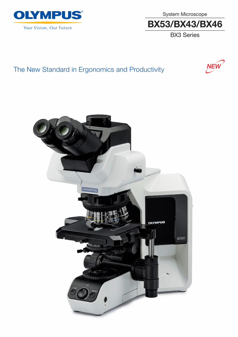
The New Standard in Ergonomics and Productivity
BX53/BX43/BX46BX3 Series
System Microscope

Olympus' BX3 series combines ergonomics with leading-edge optical technology in three
models— the BX53, BX43, and BX46 microscopes. BX3 series microscopes have an ergonomic
design that helps keep users comfortable during extended periods of use and an intuitive control
layout for fast, effi cient observation and imaging. Designed for laboratory and clinical applications,
white LED illumination has a high luminosity and color-rendering index so users can see their
samples in true-to-life colors.
Your Choice for Clinical Applications
BX46Clinical Microscope
BX43System Microscope
1
BX53System Microscope
2
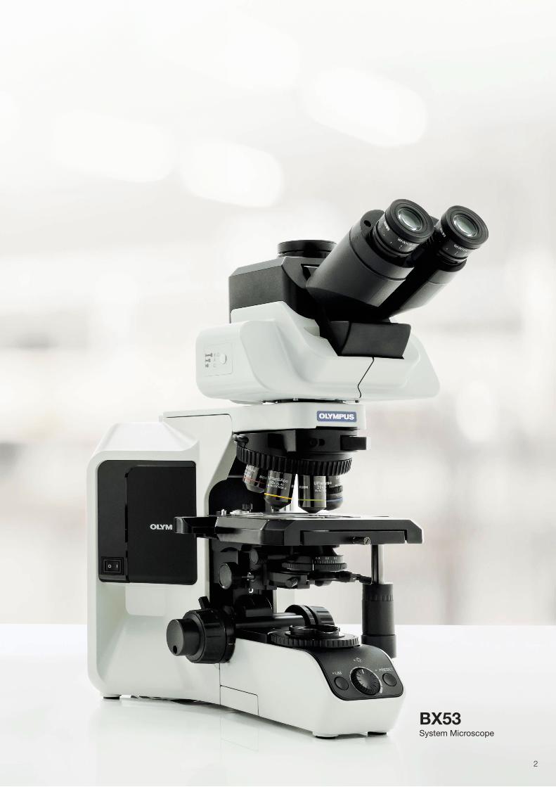
Excellent Ergonomic Tube
Our most ergonomic option moves up and down, tilts, and
extends forward and back so you can move it closer to you.
With this one component, users of nearly any height can adjust
the scope so that they're comfortable. The super ergonomic
tube is suitable for labs where multiple users share a
microscope since each can adjust it to accommodate their
height and posture.
Tilting Trinocular Tube
The tilting trinocular tube is designed for users who want the
flexibility of an ergonomic component but need to attach a
camera to their microscope. The optical path switch can be
attached to either side of the tube, so both left and right handed
users can comfor tab ly
switch from the camera to
the eyepieces.
Tilting Binocular Tubes that Meet Your Needs
Our diverse lineup of tilting observation tubes provides flexibility
in a variety of applications. From cost-effective models to tubes
for erect image observation and eyepoint adjusters that
accommodate user height differences, choose the tilting
binocular tube that suits your needs.
Abrasion Resistant and Durable Stage
Mechanical stages are coated with a durable ceramic, maximizing
abrasion resistance and helping to keep the surface smooth.
Keep Your Hands on the Desk
The stage handle extender enables users to do their work while
keeping their arms resting on the desk, resulting in less fatigue
during extended use. Users can also mount a rubber cap to the
handle so the stage can be controlled using light torque.
Rackless Stage with Enhanced Operability
The stage has a rackless, wire-driven design with no teeth in
the gear, helping to minimize injuries to users.
Comfortable and Efficient
Maintain a Natural Posture
Comfortable, Easy-to-Use Stage
U-ETBI /U-TTBI
U-TBI-3
U-EPA2
U-TBI-3-CLI
U-TTLBIU-EPAL-2
U-TTR-2
Before After
Ceramic Coated
Tilts: 0 to 27 degrees Extends: 55 mm Lifts: 45 mm
3
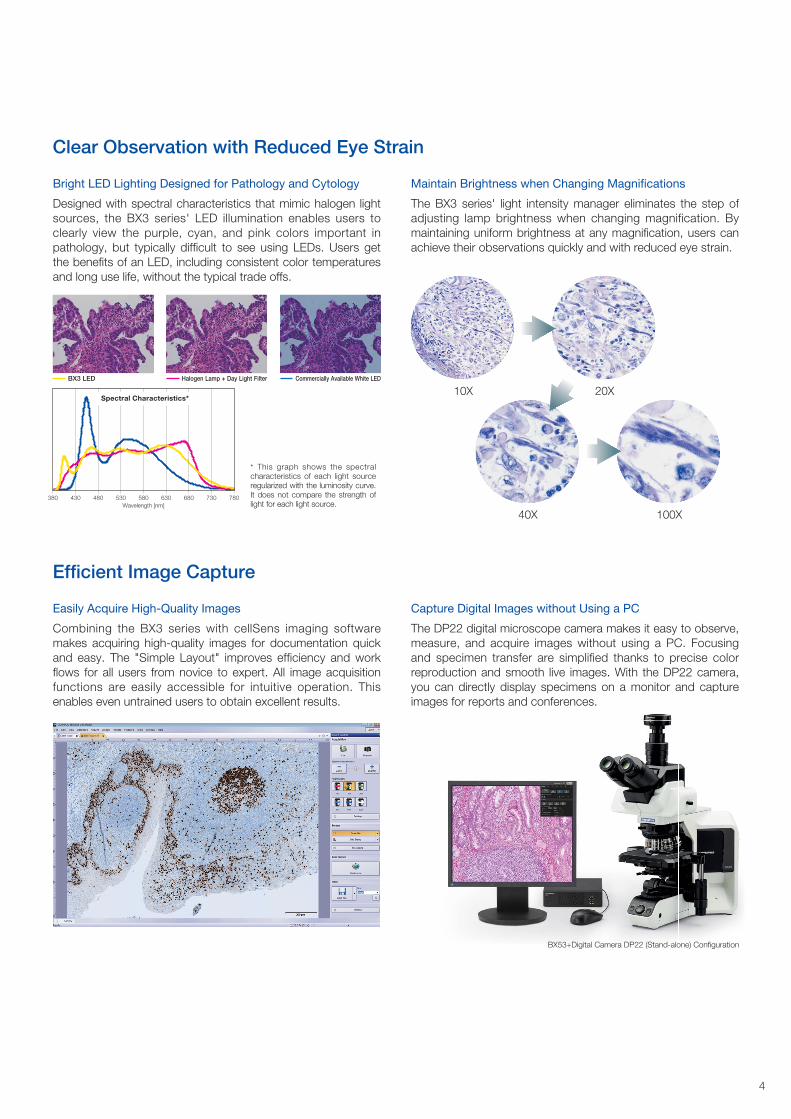
Maintain Brightness when Changing Magnifications
The BX3 series' light intensity manager eliminates the step of
adjusting lamp brightness when changing magnification. By
maintaining uniform brightness at any magnification, users can
achieve their observations quickly and with reduced eye strain.
Bright LED Lighting Designed for Pathology and Cytology
Designed with spectral characteristics that mimic halogen light
sources, the BX3 series' LED illumination enables users to
clearly view the purple, cyan, and pink colors important in
pathology, but typically difficult to see using LEDs. Users get
the benefits of an LED, including consistent color temperatures
and long use life, without the typical trade offs.
Capture Digital Images without Using a PC
The DP22 digital microscope camera makes it easy to observe,
measure, and acquire images without using a PC. Focusing
and specimen transfer are simplified thanks to precise color
reproduction and smooth live images. With the DP22 camera,
you can directly display specimens on a monitor and capture
images for reports and conferences.
Easily Acquire High-Quality Images
Combining the BX3 series with cellSens imaging software
makes acquiring high-quality images for documentation quick
and easy. The "Simple Layout" improves efficiency and work
flows for all users from novice to expert. All image acquisition
functions are easily accessible for intuitive operation. This
enables even untrained users to obtain excellent results.
10X 20X
40X 100X
Clear Observation with Reduced Eye Strain
Efficient Image Capture
* This graph shows the spectral characteristics of each light source regularized with the luminosity curve. It does not compare the strength of light for each light source.
380 430 480 530 580 630 680 730 780
Spectral Characteristics*
Wavelength [nm]
images for reports and conferences.
BX53+Digital Camera DP22 (Stand-alone) Configuration
Halogen Lamp + Day Light FilterBX3 LED Commercially Available White LED
4

With an LED illuminator equivalent to or better than a 100 W halogen lamp, the BX53 microscope delivers
outstanding brightness that's ideal for teaching and polarized light applications.
Designed for Teaching and Challenging Applications
BX53
Stomach (HE Stain) Breast (HER2, FISH)Large Intestine (EGFR)
5
White LED with High Color Rendering—Equivalent to or better
than a 100 W Halogen Lamp
Enjoy the benefits of LED illumination, such as a 50,000 hour
use life, without compromising your ability to clearly see purple,
cyan, and pink dyes. The BX53 microscope utilizes a white LED
with a luminosity equivalent to or better than a 100 W halogen
lamp. Since LEDs have a consistent color temperature, users
won't have to waste time adjusting a color filter.
Bright Images in Multi-Head Configurations
Multi-head discussion systems are essential for training and
education. With the BX53 microscope's LED illumination, up to
26 participants can view clear, bright images.
Quick Magnification Change with Motorized Functionality
Easily change objectives with a motorized nosepiece using a
hand switch. The hand switch is located near the focus handle,
enabling users to control the nosepiece without taking their
eyes off the specimen.
Advanced Optical Performance Accommodates Various
Observation Styles
Customize your BX53 microscope with modular units that
enable different observations. Choose from options including
condensers, nosepieces, a rotating stage, objectives, and
intermediate optics optimized for various observation methods,
including polarization, phase contrast, and fluorescence.
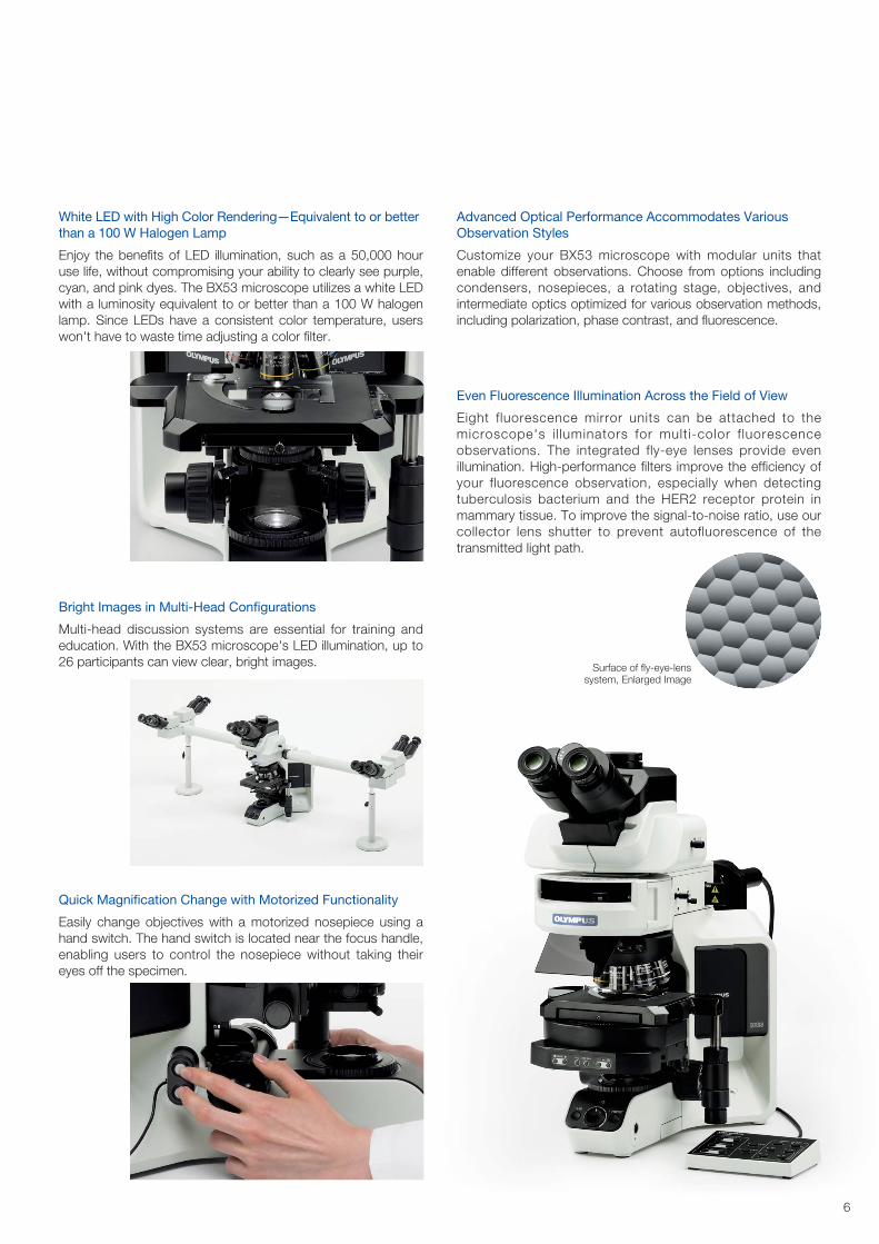
Even Fluorescence Illumination Across the Field of View
Eight fluorescence mirror units can be attached to the
microscope's i l luminators for multi-color f luorescence
observations. The integrated fly-eye lenses provide even
illumination. High-performance filters improve the efficiency of
your fluorescence observation, especially when detecting
tuberculosis bacterium and the HER2 receptor protein in
mammary tissue. To improve the signal-to-noise ratio, use our
collector lens shutter to prevent autofluorescence of the
transmitted light path.
Surface of fly-eye-lens system, Enlarged Image
6
Take advantage of the BX3 series' advanced features in a cost-effective model. Durable and easy to use, the BX43
microscope maximizes effi ciency in busy testing labs. It's easy to expand the microscope's capabilities so users
can add functionality as their needs change.
Excellent Performance in a Cost-Effective System
BX43
Hematology (Giemsa Stain)Cervical Cell (Papanicolaou Stain) Kidney (Fibrin, PTAH Stain)
7
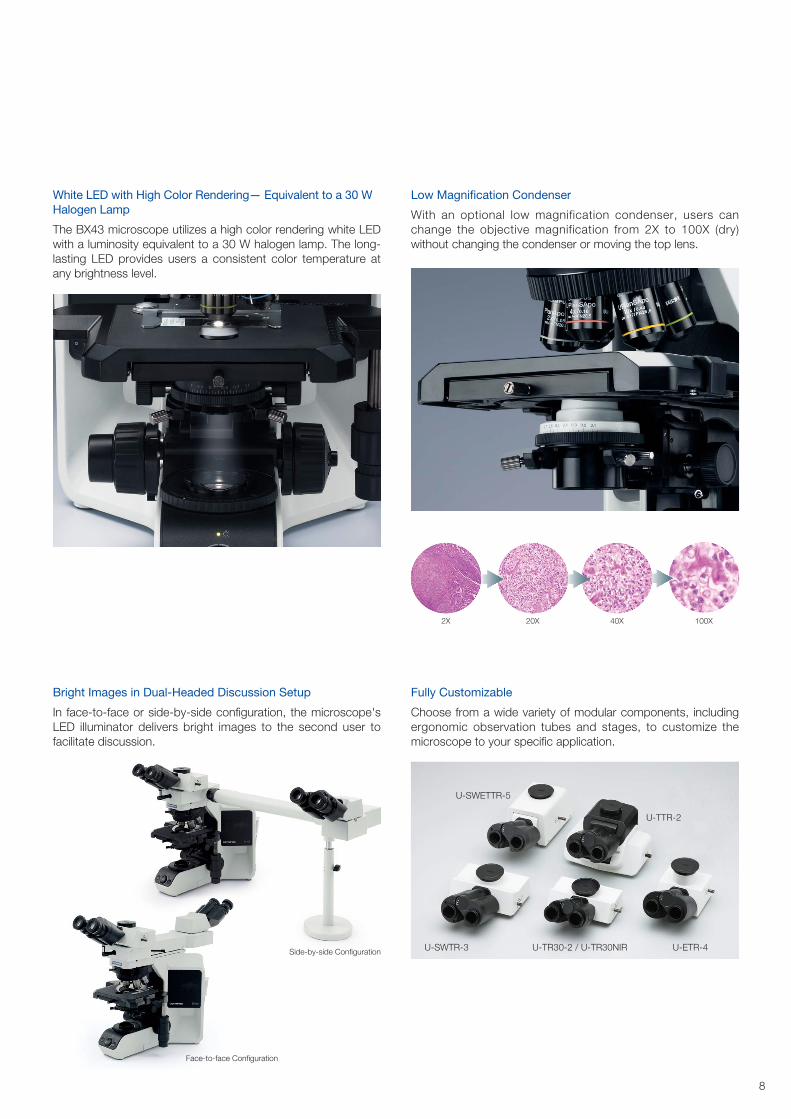
Low Magnification Condenser
With an optional low magnification condenser, users can
change the objective magnification from 2X to 100X (dry)
without changing the condenser or moving the top lens.
Fully Customizable
Choose from a wide variety of modular components, including
ergonomic observation tubes and stages, to customize the
microscope to your specific application.
White LED with High Color Rendering— Equivalent to a 30 W
Halogen Lamp
The BX43 microscope utilizes a high color rendering white LED
with a luminosity equivalent to a 30 W halogen lamp. The long-
lasting LED provides users a consistent color temperature at
any brightness level.
Bright Images in Dual-Headed Discussion Setup
In face-to-face or side-by-side configuration, the microscope's
LED illuminator delivers bright images to the second user to
facilitate discussion.
100X40X20X2X
scussion.
Face-to-face Configuration
Side-by-side ConfigurationU-SWTR-3
U-SWETTR-5
U-TTR-2
U-ETR-4U-TR30-2 / U-TR30NIR
8

With an ergonomic design and advanced features, the BX46 microscope helps keep users comfortable during
routine pathology and cytology.
Designed for Routine Pathology and Cytology
BX46
Breast (Anti HER2)Stomach (HE Stain) Cervical Cell (Papanicolaou Stain)
9
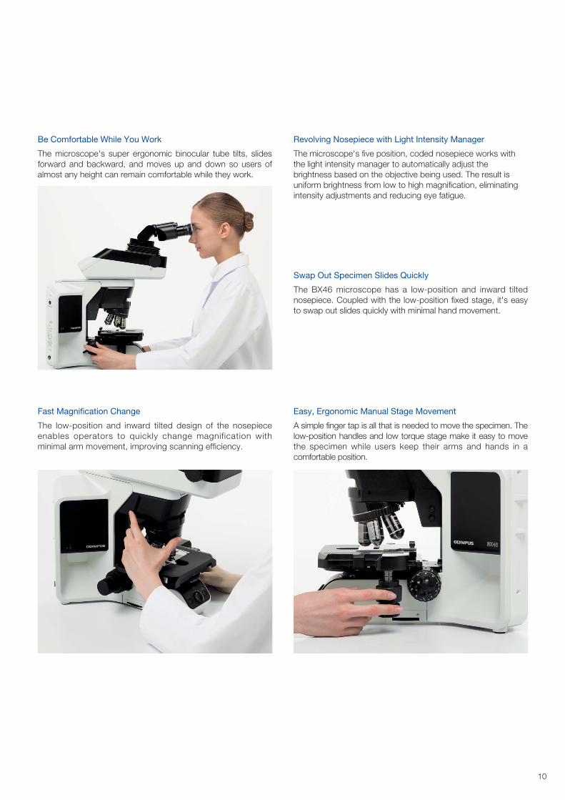
Be Comfortable While You Work
The microscope's super ergonomic binocular tube tilts, slides
forward and backward, and moves up and down so users of
almost any height can remain comfortable while they work.
Fast Magnification Change
The low-position and inward tilted design of the nosepiece
enables operators to quickly change magnification with
minimal arm movement, improving scanning efficiency.
Revolving Nosepiece with Light Intensity Manager
The microscope's five position, coded nosepiece works with
the light intensity manager to automatically adjust the
brightness based on the objective being used. The result is
uniform brightness from low to high magnification, eliminating
intensity adjustments and reducing eye fatigue.
Swap Out Specimen Slides Quickly
The BX46 microscope has a low-position and inward tilted
nosepiece. Coupled with the low-position fixed stage, it's easy
to swap out slides quickly with minimal hand movement.
Easy, Ergonomic Manual Stage Movement
A simple finger tap is all that is needed to move the specimen. The
low-position handles and low torque stage make it easy to move
the specimen while users keep their arms and hands in a
comfortable position.
10
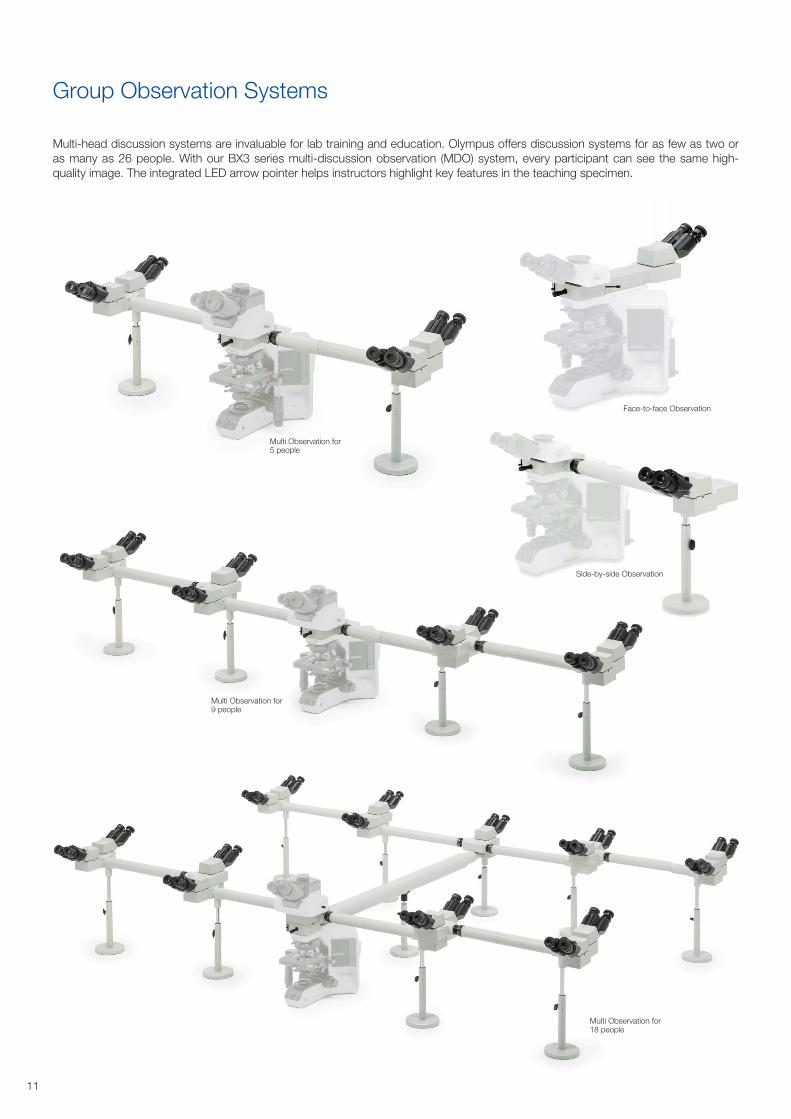
Group Observation Systems
Multi-head discussion systems are invaluable for lab training and education. Olympus offers discussion systems for as few as two or
as many as 26 people. With our BX3 series multi-discussion observation (MDO) system, every participant can see the same high-
quality image. The integrated LED arrow pointer helps instructors highlight key features in the teaching specimen.
Face-to-face Observation
Side-by-side Observation
Multi Observation for 5 people
Multi Observation for 9 people
Multi Observation for 18 people
11
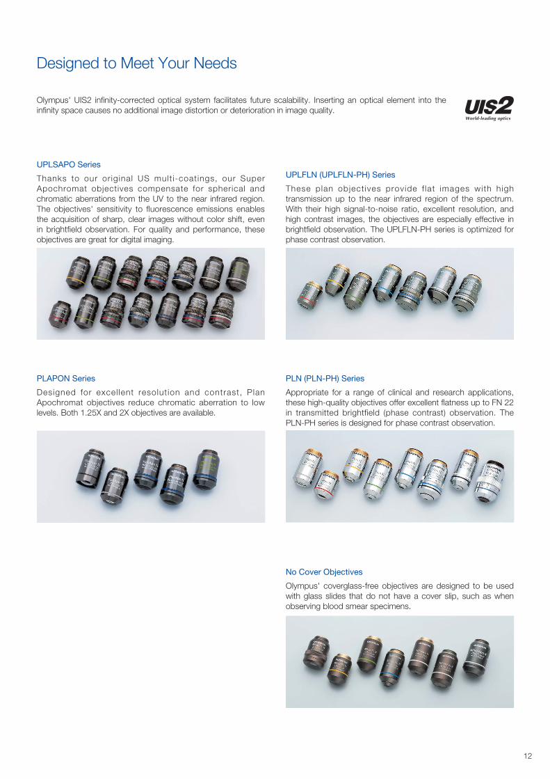
Designed to Meet Your Needs
Olympus' UIS2 infinity-corrected optical system facilitates future scalability. Inserting an optical element into the
infinity space causes no additional image distortion or deterioration in image quality.
UPLSAPO Series
Thanks to our or iginal US mult i-coat ings, our Super
Apochromat objectives compensate for spherical and
chromatic aberrations from the UV to the near infrared region.
The objectives' sensitivity to fluorescence emissions enables
the acquisition of sharp, clear images without color shift, even
in brightfield observation. For quality and performance, these
objectives are great for digital imaging.
PLAPON Series
Designed for excel lent resolut ion and contrast, Plan
Apochromat objectives reduce chromatic aberration to low
levels. Both 1.25X and 2X objectives are available.
UPLFLN (UPLFLN-PH) Series
These plan object ives provide f lat images with high
transmission up to the near infrared region of the spectrum.
With their high signal-to-noise ratio, excellent resolution, and
high contrast images, the objectives are especially effective in
brightfield observation. The UPLFLN-PH series is optimized for
phase contrast observation.
PLN (PLN-PH) Series
Appropriate for a range of clinical and research applications,
these high-quality objectives offer excellent flatness up to FN 22
in transmitted brightfield (phase contrast) observation. The
PLN-PH series is designed for phase contrast observation.
No Cover Objectives
Olympus' coverglass-free objectives are designed to be used
with glass slides that do not have a cover slip, such as when
observing blood smear specimens.
12
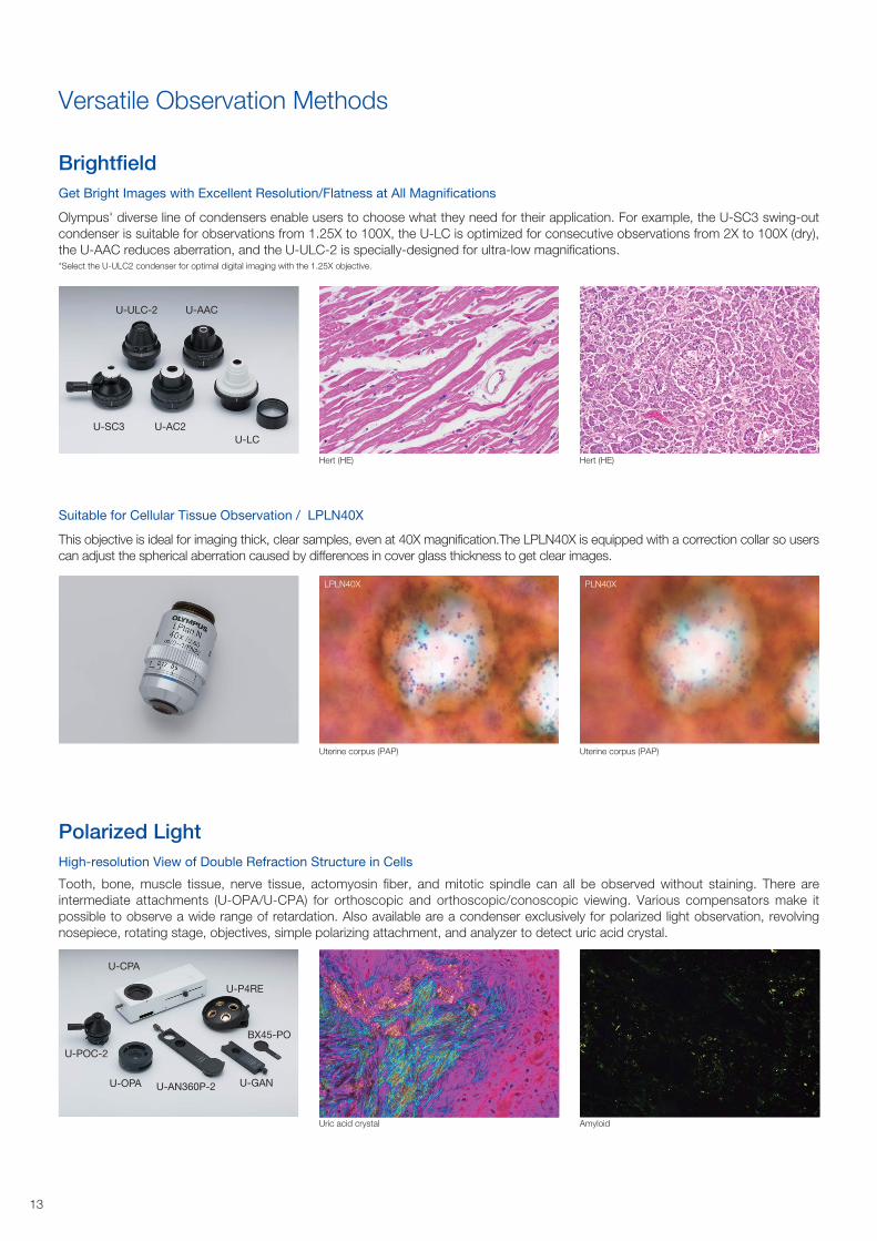
Get Bright Images with Excellent Resolution/Flatness at All Magnifications
Olympus' diverse line of condensers enable users to choose what they need for their application. For example, the U-SC3 swing-out
condenser is suitable for observations from 1.25X to 100X, the U-LC is optimized for consecutive observations from 2X to 100X (dry),
the U-AAC reduces aberration, and the U-ULC-2 is specially-designed for ultra-low magnifications.*Select the U-ULC2 condenser for optimal digital imaging with the 1.25X objective.
Suitable for Cellular Tissue Observation / LPLN40X
This objective is ideal for imaging thick, clear samples, even at 40X magnification.The LPLN40X is equipped with a correction collar so users
can adjust the spherical aberration caused by differences in cover glass thickness to get clear images.
High-resolution View of Double Refraction Structure in Cells
Tooth, bone, muscle tissue, nerve tissue, actomyosin fiber, and mitotic spindle can all be observed without staining. There are
intermediate attachments (U-OPA/U-CPA) for orthoscopic and orthoscopic/conoscopic viewing. Various compensators make it
possible to observe a wide range of retardation. Also available are a condenser exclusively for polarized light observation, revolving
nosepiece, rotating stage, objectives, simple polarizing attachment, and analyzer to detect uric acid crystal.
Versatile Observation Methods
Brightfield
Polarized Light
U-SC3
U-ULC-2
U-AC2
U-AAC
U-LC
U-P4RE
BX45-PO
U-GANU-AN360P-2
U-CPA
U-OPA
U-POC-2
Hert (HE)
Uterine corpus (PAP)
Uric acid crystal
Hert (HE)
Uterine corpus (PAP)
Amyloid
LPLN40X PLN40X
13
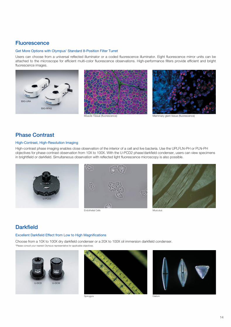
Get More Options with Olympus' Standard 8-Position Filter Turret
Users can choose from a universal reflected illuminator or a coded fluorescence illuminator. Eight fluorescence mirror units can be
attached to the microscope for efficient multi-color fluorescence observations. High-performance filters provide efficient and bright
fluorescence images.
High-Contrast, High-Resolution Imaging
High-contrast phase imaging enables close observation of the interior of a cell and live bacteria. Use the UPLFLN-PH or PLN-PH
objectives for phase contrast observation from 10X to 100X. With the U-PCD2 phase/darkfield condenser, users can view specimens
in brightfield or darkfield. Simultaneous observation with reflected light fluorescence microscopy is also possible.
Excellent Darkfield Effect from Low to High Magnifications
Choose from a 10X to 100X dry darkfield condenser or a 20X to 100X oil immersion darkfield condenser. *Please consult your nearest Olympus representative for applicable objectives.
Fluorescence
Phase Contrast
Darkfield
U-DCD U-DCW
U-PCD2
BX3-URA
BX3-RFAS
Endothelial Cells Musculus
Spirogyra Diatom
Muscle Tissue (fluorescence) Mammary glant tissue (fluorescence)
14

** * * *
U-SRPPrecision rotatable stage
B BA
ACC
C
C
G
B
D
F
U-HLS-4,U-HLST-4Specimen holder
U-HLD-4, U-HLDT-4Specimen holder
U-HRD-4, U-HRDT-4Specimen holder
U-CST Centeringtarget
U-SPPlain stage
U-FMPMechanical stage
U-SVROOil rectangularstage with right-hand control
U-SVLOOil rectangular stage with left-hand control
U-SVRB-4Mechanical stages withright-handcontrol
U-SVLB-4Mechanical stages withleft-handcontrol
U-SHGRubber grip U-SHGTRubber grip
U-SRG2Rotatable graduated stage
*1 Slight vignetting may occur in combination with an additional intermediate attachment or observation method. *2 Require an additional intermediate attachment or fluorescence illuminator.*3 Cannot be used with U-TTLBI. *4 Compatible with FN 22. *5 Cannot be used with BX3-URA. *6 Stand is a standard equipment of the U-MDOSV, BX3-MDO18R, and U-MDO10R3.
U-SWETTR-5Super widefield erect image tilting trinocular observation tube
U-CMAD3C-MountAdapter
U-BMADBayonet-mountAdapter
U-FMTF-mount Adapter
U-TMADT-mount Adapter
U-SMADSony-mountAdapter
U-TV1X-2TV Adapter U-DPTS
Multi double port tube
U-DPCADDual port tube with C mounts
U-CMDPTSC mount adapter for U-DPTS
U-CMDPTSC mount adapter for U-DPTS
U-TTR-2Tilting trinocular tube
U-SWTR-3Super widefieldtrinocular tube
U-TR30NIRTrinocular tube
U-TR30-2Trinocular tube
U-BI30-2Binocular tube
U-ETR-4*1
Erect imagetrinocular tube
U-TBI-3*1
Tilting binocular tube
TR-Adapter
U-D7RESCoded 7-positionnosepiece
U-D6RESCoded 6-positionnosepiece
U-D7REAMotorized 7-positionnosepiece
U-D6RESextuple revolvingnosepiece forDIC/simple POL
U-D7RESeptuple revolvingnosepiece forDIC/simple POL
U-ANTAnalyzer fortransmitted light
U-DICTDIC slider fortransmitted light
U-DICTSShift DIC slider fortransmitted lightU-DICTHRHigh resolution DICslider for transmitted lightU-DICTHCHigh contrast DIC sliderfor transmitted light
U-GANAnalyzer for urate crystals observation
U-TADPlate adapter
COMPENSATORS
U-ANTAnalyzer fortransmitted light
OBJECTIVES
U-POTPolarizer
Filter (ø45)
BX53F2BX53 frame
U-LHLEDC100High powerLED lamp housing
46S-LBA4LBA filter
BX3-SHTShutter for transmitted light
CX3-SHPSpecimen hold plate
BX3-SHEAStage handle extention Adapter
BX3-ARMStandard arm
U-AN-2Analyzer slider
U-AN-2Analyzer slider
U-AN-2Analyzer slider
BX3-URAUniversal reflected illuminator
BX3-RFAS*4
Coded fluorescence illuminator
BX3-RFAA*4
Motorized fluorescence illuminator
BX3-25ND6ND filterBX3-25ND25ND filter
Mirror units
WHN10X-H,CROSS WHN10XEyepiecesU-CT30-2Centering telescope
WHN10X, WHN10X-H,CROSS WHN10XEyepiecesU-CT30-2Centering telescope
SWH10X-H,CROSS SWH10X,MICRO SWH10XEyepiecesU-CT30-2Centering telescope
U-TV0.63XBB4-Mount Adapter
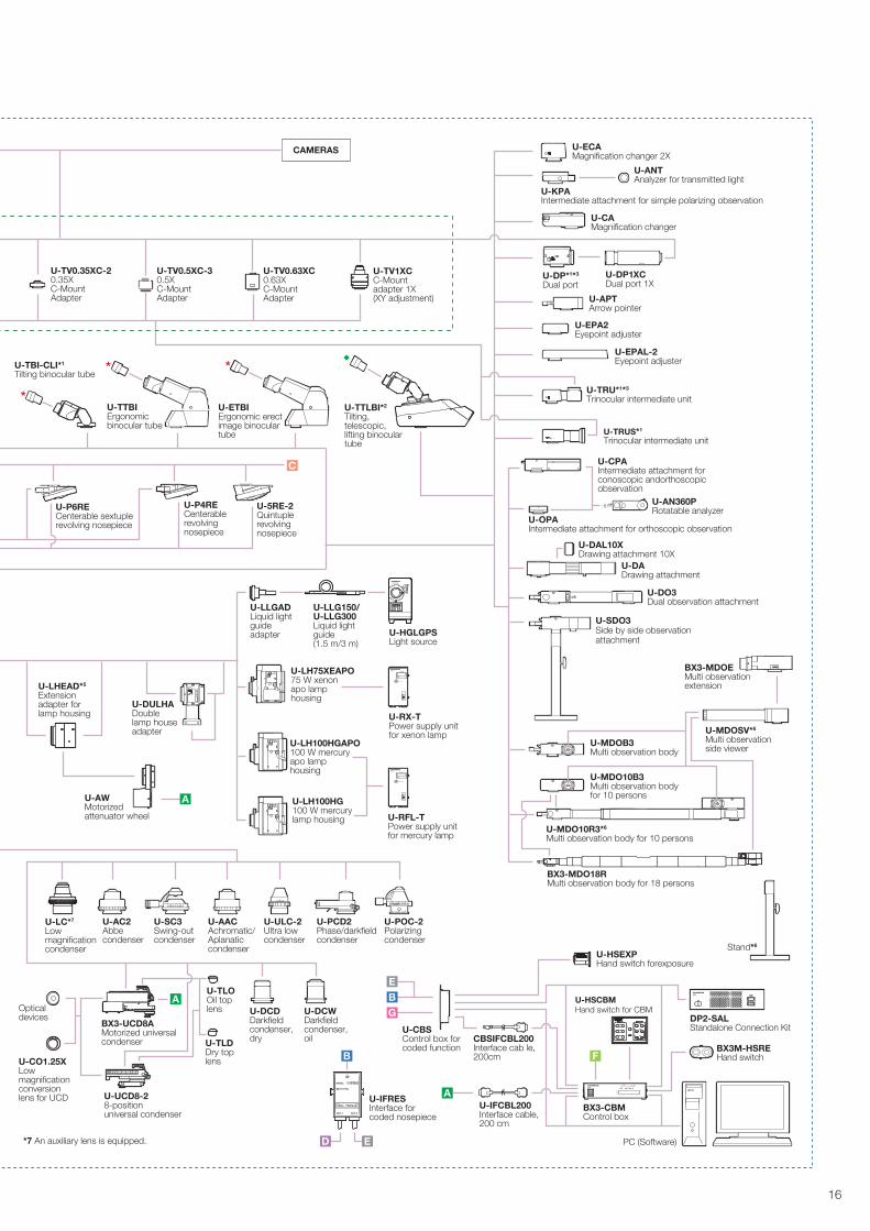
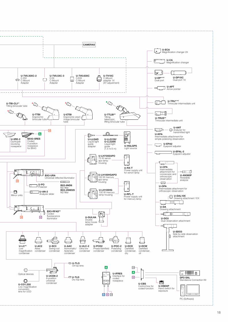
BX53 SYSTEM DIAGRAM
15
*
* *
LIFE TIME
BURNER ON
U-RFL-T
LIFE TIME
BURNER ON
U-RFL-T
A
A
A
C
B F
D E
EBG
U-CO1.25XLow magnification conversionlens for UCD
U-PCD2Phase/darkfield condenser
U-POC-2Polarizingcondenser
U-AACAchromatic/Aplanaticcondenser
U-AC2Abbe condenser
U-SC3Swing-out condenser
U-ULC-2Ultra low condenser
U-DCDDarkfieldcondenser,dry
U-DCWDarkfieldcondenser,oil
U-TLOOil top lens
U-TLDDry top lens
U-UCD8-28-positionuniversal condenser
Optical devices BX3-UCD8A
Motorized universal condenser
U-LC*7
Low magnificationcondenser
U-CBSControl box forcoded function
U-HSEXPHand switch forexposure
BX3-CBMControl box
CBSIFCBL200Interface cab le, 200cm
U-HSCBMHand switch for CBM
*7 An auxiliary lens is equipped.
U-TRUS*1 Trinocular intermediate unit
U-IFCBL200Interface cable, 200 cm
U-TBI-CLI*1
Tilting binocular tube
CAMERAS
U-TV1XCC-Mountadapter 1X(XY adjustment)
U-TV0.63XC0.63XC-MountAdapter
U-TV0.5XC-30.5XC-MountAdapter
U-TV0.35XC-20.35XC-MountAdapter
U-TTLBI*2
Tilting, telescopic, lifting binocular tube
U-ETBIErgonomic erect image binocular tube
U-TTBIErgonomic binocular tube
U-P4RECenterable revolvingnosepiece
U-P6RECenterable sextuple revolving nosepiece
U-5RE-2Quintuplerevolvingnosepiece
U-AWMotorized attenuator wheel
U-ECAMagnification changer 2X
U-TRU*1*3
Trinocular intermediate unit
U-CAMagnification changer
U-KPAIntermediate attachment for simple polarizing observation
U-ANTAnalyzer for transmitted light
U-EPA2Eyepoint adjuster
U-EPAL-2Eyepoint adjuster
U-APTArrow pointer
U-DP*1*3
Dual port U-DP1XCDual port 1X
U-CPAIntermediate attachment for conoscopic andorthoscopic observation
U-AN360PRotatable analyzer
U-OPAIntermediate attachment for orthoscopic observation
U-DO3Dual observation attachment
U-DADrawing attachment
U-DAL10XDrawing attachment 10X
U-SDO3Side by side observationattachment
U-MDO10B3Multi observation body for 10 persons
U-MDOB3Multi observation body
U-MDOSV*6
Multi observation side viewer
Stand*6
U-MDO10R3*6
Multi observation body for 10 persons
BX3-MDO18RMulti observation body for 18 persons
BX3-MDOEMulti observation extension
U-DULHADouble lamp house adapter
U-LHEAD*5
Extension adapter for lamp housing
PC (Software)
DP2-SALStandalone Connection Kit
BX3M-HSREHand switch
U-LLGADLiquid light guide adapter
U-LLG150/U-LLG300Liquid light guide (1.5 m/3 m)
U-HGLGPSLight source
U-LH100HG100 W mercury lamp housing
U-LH75XEAPO75 W xenon apo lamp housing
U-LH100HGAPO100 W mercury apo lamp housing
U-RX-TPower supply unit for xenon lamp
U-RFL-TPower supply unit for mercury lamp
U-IFRESInterface for coded nosepiece
16
*1 Slight vignetting may occur in combination with an additional intermediate attachment or observation method. *2 Require an additional intermediate attachment or fluorescence illuminator. *3 Cannot be used with U-TTLBI. *4 Compatible with FN 22. *5 An auxiliary lens is equipped.
WHN10X-H,CROSS WHN10XEyepiecesU-CT30-2Centering telescope
WHN10X, WHN10X-H,CROSS WHN10XEyepiecesU-CT30-2Centering telescope
SWH10X-H,CROSS SWH10X,MICRO SWH10XEyepiecesU-CT30-2Centering telescope
** * * *
A A
AB
U-LHLEDCLED lamp housing
U-D7RESCoded 7-positionnosepiece
TR-Adapter
U-CMAD3C-MountAdapter
U-BMADBayonet-mountAdapter
U-FMTF-mount AdapterU-TMADT-mount Adapter
U-SMADSony-mountAdapter
U-TV1X-2TV Adapter
U-ANTAnalyzer fortransmitted light
U-DICTDIC slider fortransmitted light
U-DICTSShift DIC slider fortransmitted lightU-DICTHRHigh resolution DICslider for transmitted light U-DICTHCHigh contrast DIC sliderfor transmitted light
U-GANAnalyzer for urate crystals observation
U-TADPlate adapter
COMPENSATORS
U-ANTAnalyzer fortransmitted light
U-P4RECenterable revolvingnosepiece
U-P6RECenterable revolvingnosepiece
U-D6RESextuple revolvingnosepiece forDIC/simple POL
U-D7RESeptuple revolvingnosepiece forDIC/simple POL
OBJECTIVES
U-POTPolarizer
BX43FBX43 frame
U-HLS-4,U-HLST-4Specimen holder
U-HLD-4, U-HLDT-4Specimen holder
U-HRD-4, U-HRDT-4Specimen holder
U-CST Centering target
U-SRPPrecision rotatable stage
U-SPPlain stage
U-FMPMechanical stage
U-SVROOil rectangularstage with right-hand control
U-SVLOOil rectangular stage with left-hand control
U-SVRB-4Mechanical stages withright-handcontrol
U-SVLB-4Mechanical stages withleft-handcontrol
U-SRG2Rotatable graduated stage
U-DPTSMulti double port tube
U-DPCADDual port tube with C-mounts
U-CMDPTSC mount adapter for U-DPTS
U-CMDPTSC mount adapter for U-DPTS
U-SWETTR-5Super widefield erect image tilting trinocular observation tube
U-SWTR-3Super widefieldtrinocular tube
U-TR30NIRTrinocular tube
U-TR30-2Trinocular tube
U-BI30-2Binocular tube
U-ETR-4*1
Erect imagetrinocular tube
U-TBI-3*1
Tilting binocular tube
U-D6RESCoded 6-positionnosepiece
U-TV0.63XBB4-Mount Adapter
BX3-SHTShutter for transmitted light
CX3-SHPSpecimen hold plate
U-SHGRubber grip U-SHGTRubber grip
BX3-SHEAStage handle extention Adapter
U-TTR-2Tilting trinocular tube
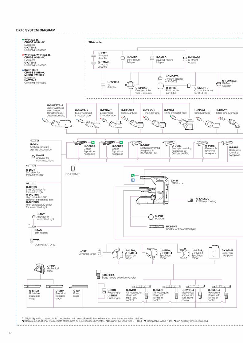
BX43 SYSTEM DIAGRAM
17
LIFE TIME
BURNER ON
U-RFL-T
LIFE TIME
BURNER ON
U-RFL-T
*
* *
A
EB
A
D
C
C
C
D
EAD
U-TRUS*3 Trinocular intermediate unit
U-AN-2Analyzer slider
U-CO1.25XLow magnification conversionlens for UCD
BX3-URAUniversal reflected illuminator
BX3-RFAS*4
Coded fluorescence illuminator
BX43-5RESCoded 5-positionnosepiece for BX43
U-LC*5
Low magnificationcondenser
BX3-6ND6ND filterBX3-25ND25ND filter
U-CBSControl box forcoded function
U-IFRESInterface for coded nosepiece
U-HSEXPHand switch forexposure
DP2-SALStandalone Connection Kit
PC (Software)
CAMERAS
U-5RE-2Quintuplerevolvingnosepiece
U-POPolarizer
U-ECAMagnification changer 2X
U-TRU*1*3
Trinocular intermediate unit
U-CAMagnification changer
U-KPAIntermediate attachment forsimple polarizing observation
U-ANTAnalyzer fortransmitted light
U-EPA2Eyepoint adjuster
U-EPAL-2Eyepoint adjuster
U-LLGADLiquid light guide adapter
U-LLG150/U-LLG300Liquid light guide (1.5 m/3 m)
U-HGLGPSLight source
U-APTArrow pointer
U-DP*1*3
Dual port U-DP1XCDual port 1X
U-CPAIntermediate attachment for conoscopic and orthoscopic observation
U-AN360PRotatable analyzer
U-OPAIntermediate attachment fororthoscopic observation
U-DO3Dual observation attachment
U-DADrawing attachment
U-DAL10XDrawing attachment 10X
U-SDO3Side by side observationattachment
U-LH100HG100 W mercury lamp housing
U-LH75XEAPO75 W xenon apo lamp housing
U-LH100HGAPO100 W mercury apo lamp housing
U-PCD2Phase/darkfield condenser
U-POC-2Polarizingcondenser
U-AACAchromatic/Aplanaticcondenser
U-AC2Abbe condenser
U-SC3Swing-out condenser
U-ULC-2Ultra low condenser
U-DCDDarkfieldcondenser,dry
U-DCWDarkfieldcondenser,oil
U-TLOOil top lens
U-TLDDry top lens
U-UCD8-28-positionuniversal condenser
Optical devices
U-DULHADouble lamp house adapter
Mirror units
U-TV1XCC-Mountadapter 1X(XY adjustment)
U-TV0.63XC0.63XC-MountAdapter
U-TV0.5XC-30.5XC-MountAdapter
U-TV0.35XC-20.35XC-MountAdapter
U-TBI-CLI*1
Tilting binocular tube
U-TTLBI*2
Tilting, telescopic, lifting binocular tube
U-ETBIErgonomic erect image binocular tube
U-TTBIErgonomic binocular tube
U-RX-TPower supply unit for xenon lamp
U-RFL-TPower supply unit for mercury lamp
18
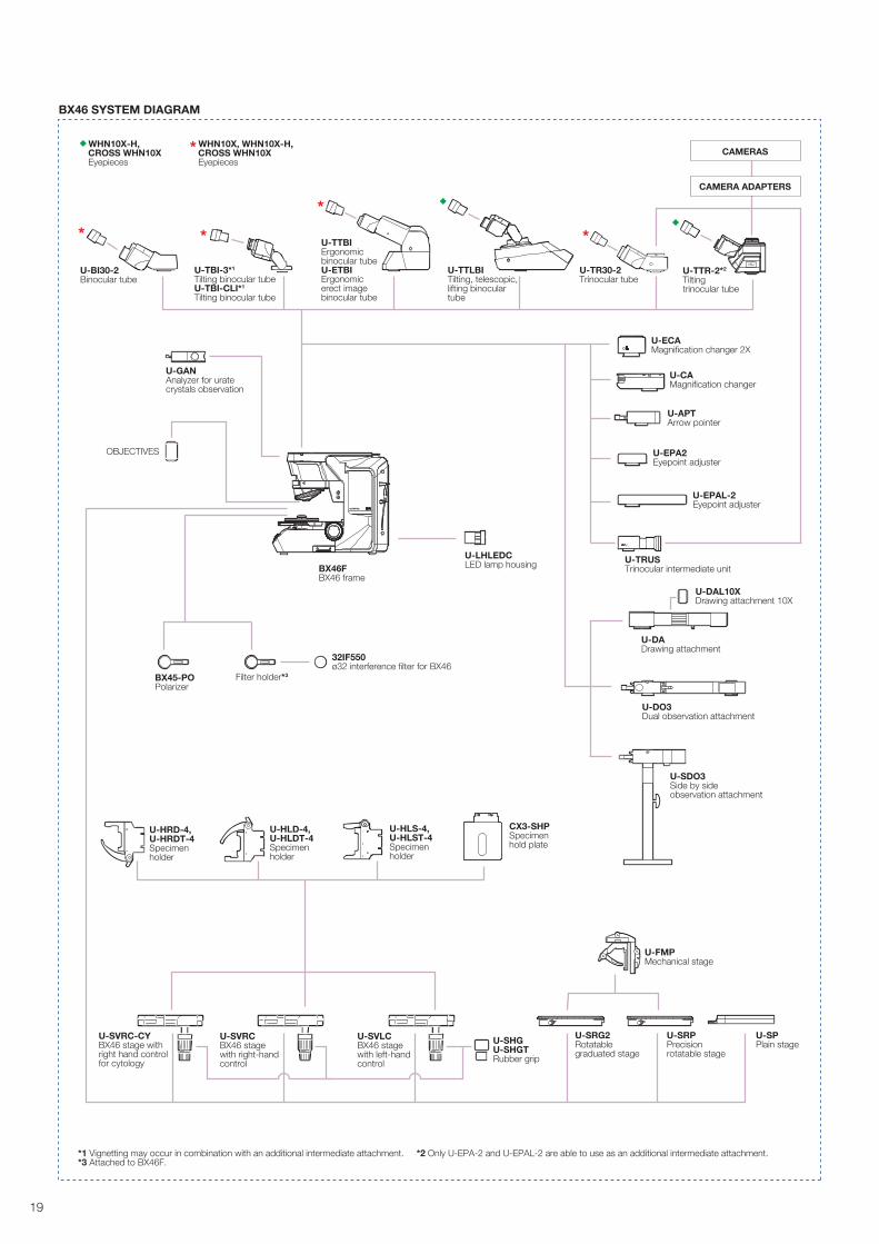
*1 Vignetting may occur in combination with an additional intermediate attachment. *2 Only U-EPA-2 and U-EPAL-2 are able to use as an additional intermediate attachment. *3 Attached to BX46F.
WHN10X-H,CROSS WHN10XEyepieces
WHN10X, WHN10X-H,CROSS WHN10XEyepieces
* * **
U-TBI-3*1
Tilting binocular tubeU-TBI-CLI*1
Tilting binocular tube
U-TTBIErgonomic binocular tubeU-ETBIErgonomic erect image binocular tube
U-TTLBITilting, telescopic, lifting binocular tube
U-TTR-2*2
Tilting trinocular tube
U-BI30-2Binocular tube
U-GANAnalyzer for urate crystals observation
U-LHLEDCLED lamp housing
U-SVRCBX46 stage with right-hand control
U-SVRC-CYBX46 stage with right hand control for cytology
U-SVLCBX46 stage with left-hand control
U-SRG2Rotatable graduated stage
U-SRPPrecision rotatable stage
U-HLS-4,U-HLST-4Specimen holder
U-HRD-4, U-HRDT-4Specimen holder
U-HLD-4, U-HLDT-4Specimen holder
U-FMPMechanical stage
BX46FBX46 frame
BX45-POPolarizer
Filter holder*3
32IF550ø32 interference filter for BX46
U-SHGU-SHGTRubber grip
CAMERAS
CAMERA ADAPTERS
U-DAL10XDrawing attachment 10X
U-DADrawing attachment
U-EPA2Eyepoint adjuster
U-EPAL-2Eyepoint adjuster
U-APTArrow pointer
U-DO3Dual observation attachment
U-SDO3Side by side observation attachment
U-CAMagnification changer
U-ECAMagnification changer 2X
U-SPPlain stage
U-TRUS Trinocular intermediate unit
U-TR30-2Trinocular tube
CX3-SHPSpecimen hold plate
OBJECTIVES
BX46 SYSTEM DIAGRAM
19
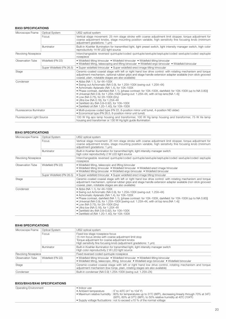
BX43 SPECIFICATIONS
Microscope Frame Optical System UIS2 optical system
Focus Vertical stage movement: 25 mm stage stroke with coarse adjustment limit stopper, torque adjustment for coarse adjustment knobs, stage mounting position variable, high sensitivity fine focusing knob (minimum adjustment gradations: 1 μm)
Illuminator Built-in Koehler illumination for transmitted light, light intensity manager switchhigh color reproductivity 2 W LED light source
Revolving Nosepiece Interchangeable reversed quintuple/coded quintuple/sextuple/septuple/coded sextuple/coded septuple nosepiece
Observation Tube Widefi eld (FN 22) • Widefi eld tilting, telescopic and lifting binocular • Widefi eld tilting trinocular • Widefi eld trinocular • Widefi eld erect image trinocular• Widefi eld tilting binocular • Widefi eld ergo binocular • Widefi eld binocular
Super Widefi eld (FN 26.5) • Super widefi eld trinocular • Super widefi eld erect image tilting trinocular
Stage Ceramic-coated coaxial stage with left or right hand low drive control: with rotating mechanism and torque adjustment mechanism, optional rubber grips and stage handle extension adapter available (non stick grooved coaxial, plain, rotatable stages are also available)
Condenser • Abbe (NA 1.1), for 4X–100X• Swing out Achromatic (NA 0.9), for 1.25X–100X (swing-out: 1.25X–4X)• Achromatic Aplanatic (NA 1.4), for 10X–100X• Phase contrast, darkfi eld (NA 1.1), [phase contrast: for 10X–100X, darkfi eld: for 10X–100X (up to NA 0.80)]• Universal (NA 0.9), for 1.25X–100X [swing-out: 1.25X–4X, with oil top lens:(NA 1.4)]• Low (NA 0.75), for 2X–100X (Dry)• Ultra low (NA 0.16), for 1.25X–4X• Darkfi eld dry (NA 0.8–0.92), for 10X–100X• Darkfi eld oil (NA 1.20–1.40), for 10X–100X
BX53 SPECIFICATIONS
Microscope Frame Optical System UIS2 optical system
Focus Vertical stage movement: 25 mm stage stroke with coarse adjustment limit stopper, torque adjustment for coarse adjustment knobs, stage mounting position variable, high sensitivity fine focusing knob (minimum adjustment gradations: 1 μm)
Illuminator Built-in Koehler illumination for transmitted light, light preset switch, light intensity manager switch, high color reproductivity 14 W LED light source
Revolving Nosepiece Interchangeable reversed quintuple/coded quintuple/sextuple/septuple/coded sextuple/coded septuple nosepiece
Observation Tube Widefi eld (FN 22) • Widefield tilting trinocular • Widefield trinocular • Widefield tilting binocular • Widefield tilting, telescoping and lifting binocular • Widefield ergo binocular • Widefield binocular
Super Widefi eld (FN 26.5) • Super widefield trinocular • Super widefield erect image tilting trinocular
Stage Ceramic-coated coaxial stage with left or right hand low drive control: with rotating mechanism and torque adjustment mechanism, optional rubber grips and stage handle extension adapter available (non stick grooved coaxial, plain, rotatable stages are also available)
Condenser • Abbe (NA 1.1), for 4X–100X• Swing out Achromatic (NA 0.9), for 1.25X–100X (swing-out: 1.25X–4X)• Achromatic Aplanatic (NA 1.4), for 10X–100X• Phase contrast, darkfi eld (NA 1.1), [phase contrast: for 10X–100X, darkfi eld: for 10X–100X (up to NA 0.80)]• Universal (NA 0.9), for 1.25X–100X [swing-out: 1.25X–4X, with oil top lens:(NA 1.4)]• Low (NA 0.75), for 2X–100X (Dry)• Ultra low (NA 0.16), for 1.25X–4X• Darkfi eld dry (NA 0.8–0.92), for 10X–100X• Darkfi eld oil (NA 1.20–1.40), for 10X–100X
Fluorescence Illuminator • Multi-purpose coded type (FN 22, 8-position mirror unit turret, 4-position ND slider)• Economical type (FN 26.5, 8-position mirror unit turret)
Fluorescence Light Source 100 W Hg apo lamp housing and transformer, 100 W Hg lamp housing and transformer, 75 W Xe lamp housing and transformer or 130 W Hg light guide illumination
BX53/BX43/BX46 SPECIFICATIONS
Operating Environment • Indoor use• Ambient temperature : 5˚ to 40˚C (41˚ to 104˚ F)• Maximum relative humidity : 80% for temperatures up to 31˚C (88˚F), decreasing linearly through 70% at 34˚C (93˚F), 60% at 37˚C (99˚F), to 50% relative humidity at 40˚C (104˚F)• Supply voltage fl uctuations : not to exceed ±10 % of the normal voltage
BX46 SPECIFICATIONS
Microscope Frame Optical System UIS2 optical system
Focus Fixed low stage nosepiece focus15 mm focus stroke with coarse adjustment limit stopTorque adjustment for coarse adjustment knobs High sensitivity fi ne focusing knob (adjustment gradations: 1 μm)
Illuminator Built-in Koehler illumination for transmitted light, light intensity manager switchHigh color reproductivity 2 W LED light source
Revolving Nosepiece Fixed reversed coded quintuple nosepiece
Observation Tube Widefi eld (FN 22) • Widefield tilting trinocular • Widefield trinocular • Widefield tilting binocular • Widefield tilting, telescopic, lifting binocular • Widefield ergo binocular • Widefield binocular
Stage Ceramic-coated coaxial stage with left or right hand low drive control, rotating mechanism and torque adjustment mechanism (low torqe, plain, rotating stages are also available)
Condenser Built-in condenser (NA 0.9) 1.25X–100X (swing out: 1.25X–2X)
20
362
82
209
4565
433*
91
274
90
179*
30°
111
65
210*
82
209
45479
482*
92362
383
220
90
275
210*
82
209
4512
4
538
541*
92362383482
90
275
220
82128
145
92362
193*
548*
90275
125
451
27º
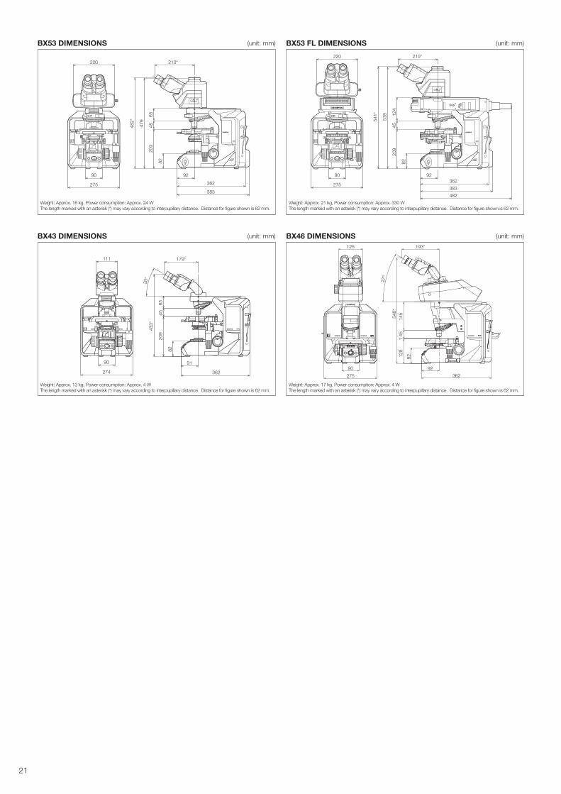
Weight: Approx. 13 kg, Power consumption: Approx. 4 W
The length marked with an asterisk (*) may vary according to interpupillary distance. Distance for figure shown is 62 mm.
Weight: Approx. 16 kg, Power consumption: Approx. 24 W
The length marked with an asterisk (*) may vary according to interpupillary distance. Distance for figure shown is 62 mm.
Weight: Approx. 21 kg, Power consumption: Approx. 330 W
The length marked with an asterisk (*) may vary according to interpupillary distance. Distance for figure shown is 62 mm.
Weight: Approx. 17 kg, Power consumption: Approx. 4 W
The length marked with an asterisk (*) may vary according to interpupillary distance. Distance for figure shown is 62 mm.
BX43 DIMENSIONS (unit: mm)
BX53 DIMENSIONS (unit: mm)
BX46 DIMENSIONS (unit: mm)
BX53 FL DIMENSIONS (unit: mm)
21
312211*
527*
4565
4520
9
82
455383362
92
644601
644 601
742 603
967
4545
31941
2
11001372
1609
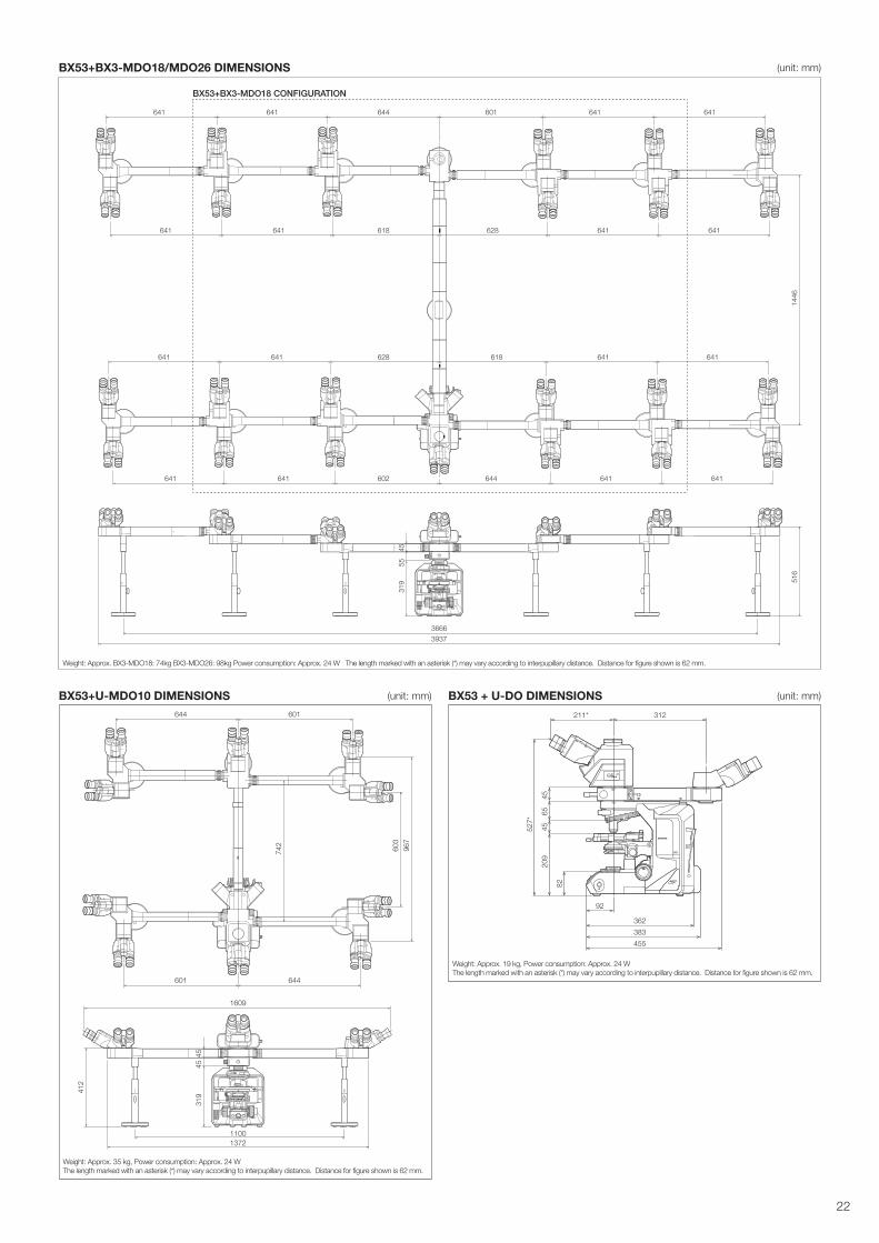
Weight: Approx. 19 kg, Power consumption: Approx. 24 W
The length marked with an asterisk (*) may vary according to interpupillary distance. Distance for figure shown is 62 mm.
Weight: Approx. 35 kg, Power consumption: Approx. 24 W
The length marked with an asterisk (*) may vary according to interpupillary distance. Distance for figure shown is 62 mm.
BX53 + U-DO DIMENSIONS (unit: mm)
BX53+BX3-MDO18/MDO26 DIMENSIONS (unit: mm)
BX53+U-MDO10 DIMENSIONS (unit: mm)
516
39373666
319
4555
1446
628 641641
601 641641
618
644641 641
641 641
641 641
641 641
618 641641628
602 644 641641
Weight: Approx. BX3-MDO18: 74kg BX3-MDO26: 98kg Power consumption: Approx. 24 W The length marked with an asterisk (*) may vary according to interpupillary distance. Distance for figure shown is 62 mm.
BX53+BX3-MDO18 CONFIGURATION
22

www.olympus-lifescience.com
Printed in Japan M1696E-062017
• is ISO14001 certifi ed.
• is ISO9001 certifi ed.
• is ISO13485 certifi ed.
• Illumination devices for microscope have suggested lifetimes. Periodic inspections are required. Please visit our website for details.
• All company and product names are registered trademarks and/or trademarks of their respective owners.
• Images on the PC monitors are simulated.
• Specifi cations and appearances are subject to change without any notice or obligation on the part of the manufacturer.
Shinjuku Monolith, 2-3-1 Nishi-Shinjuku, Shinjuku-ku, Tokyo 163-0914, Japan


![· International Journal of Industrial Ergonomics, productivity and its application to work- Ergonomics v0136, 367-377 [8] Reuben Escorpizo. 2007. Understanding work application](https://img.dokumen.tips/doc/110x75/5e896083af649d295a3339f1/international-journal-of-industrial-ergonomics-productivity-and-its-application.jpg)