Embed Size (px)
Citation preview

The Nervous System
Charles C. Cook, MD
Divisions of the Nervous System

The Central Nervous System
• Brain and Spinal cord
• Processes sensory information
• Produces a response

The Central Nervous System
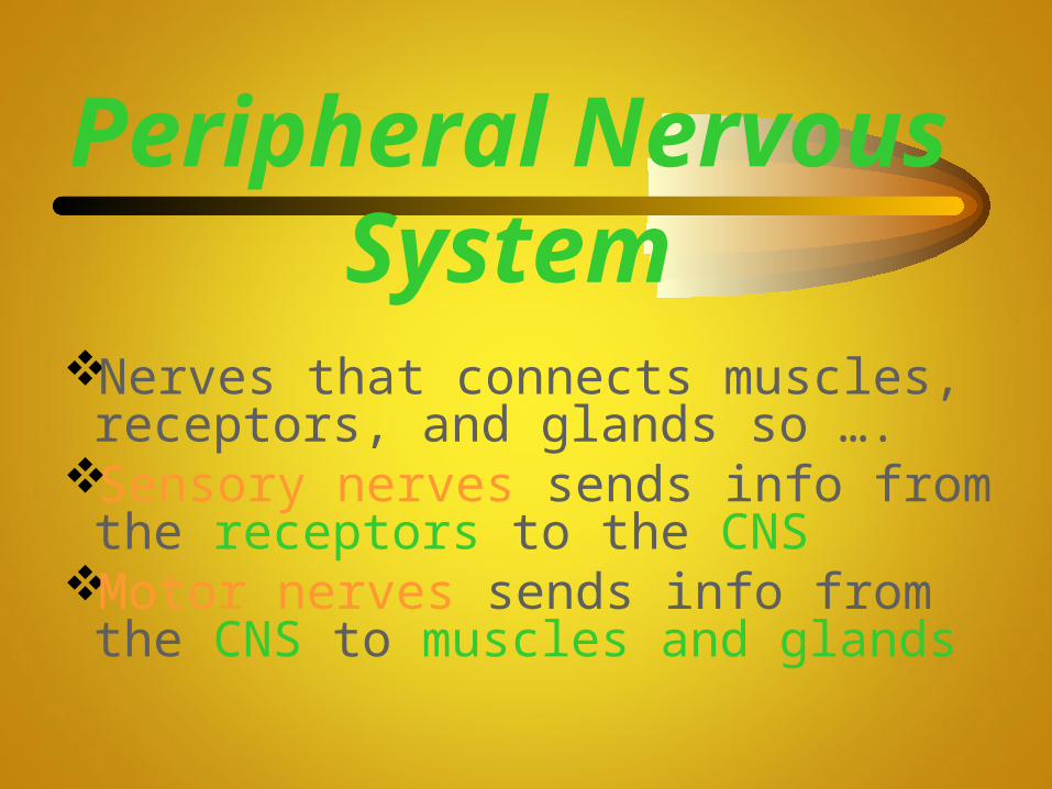
Peripheral Nervous System
Nerves that connects muscles, receptors, and glands so ….
Sensory nerves sends info from the receptors to the CNS
Motor nerves sends info from the CNS to muscles and glands

Peripheral Nervous System
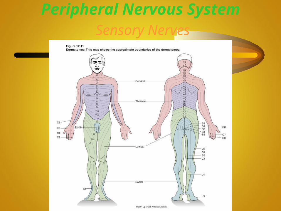
Peripheral Nervous System Sensory Nerves
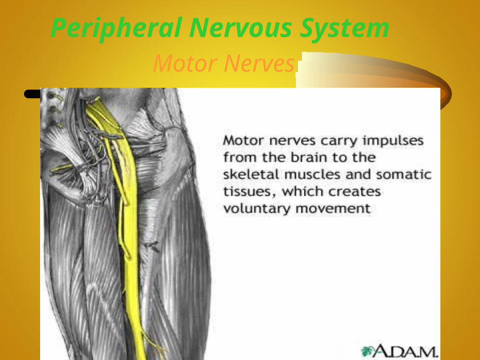
Peripheral Nervous System Motor Nerves

Peripheral Nervous System Autonomic Nervous System
PNS that controls internal organs

Autonomic Nervous System
Grouping of Neural Tissue

Nerve
Fibers located outside the CNS, held together by
connective tissue


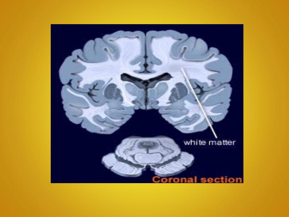
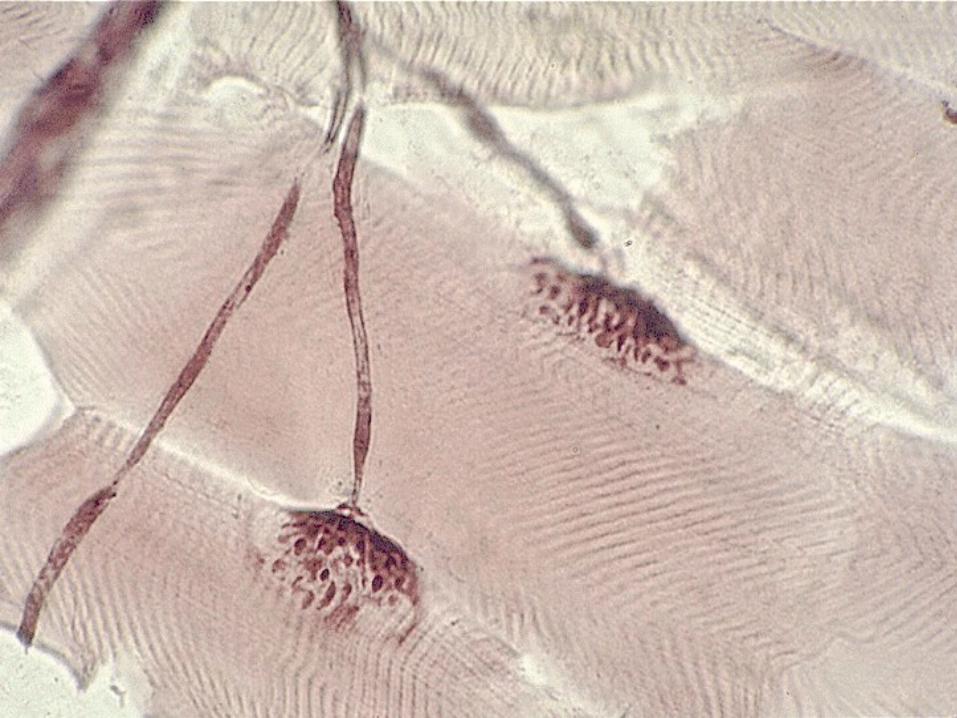
White matter
Aggregations of myelinated processes of
many neurons


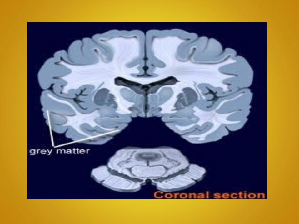
Gray Matter
Contains either nerve cell bodies or bundles of
unmyelinated nerve fibers
Reflexes
Reflex Arcs
Behavioral unit of the nervous system
Structural and functional basis for the simplest involuntary actions


Reflex Behavior
Automatic and unconsciousChanges inside or outside
Maintain HomeostasisHeart rate, breathing rate,
swallowing……

Examples of Reflexes

Protection & Covering of CNS

Cranial Bones

Vertebral Bones

Meninges
Membranes that cover brain and spinal cord
Serve as bacterial barriers
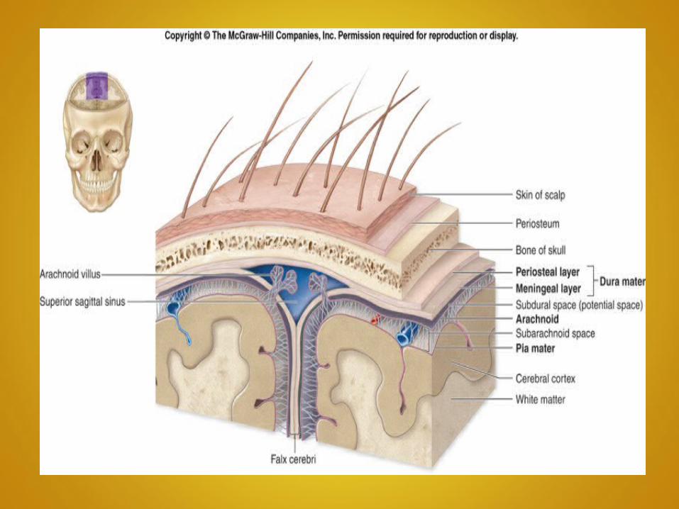

Meninges (cont)
Dura mater (tough mother)
• Outermost layer
• Composition is white fibrous connective tissue, blood vessels and nerves

Meninges (cont)
Dura mater
• Cranium —attached to bone (skull)
• Spinal cord —surrounded by adipose (fatty) tissue


Meninges (cont)
Arachnoid
• Web-like
• Lacks blood vessels
• Located between dura mater and pia mater


Meninges (cont)
Pia mater (gentle mother)• Innermost layer• Attached to organ surface
–Nerves and many blood vessels

Meninges (cont)
Subarachnoid Space
• Fluid filled
• Between arachnoid mater and pia mater


Ventricles and Cerebrospinal
fluid (CSF)

Ventricles
Cavities within the cerebral hemispheres
and brain stem


Cerebrospinal Fluid
Clear watery fluid secreted within ventricles
Circulates within ventricles and subarachnoid space
Protects and supports CNS

Spinal Cord

Structure
Base of skull to 1st lumbar vertebrae
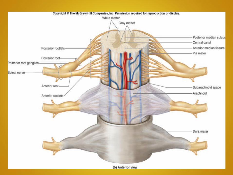
Core gray matter surrounded by white matter
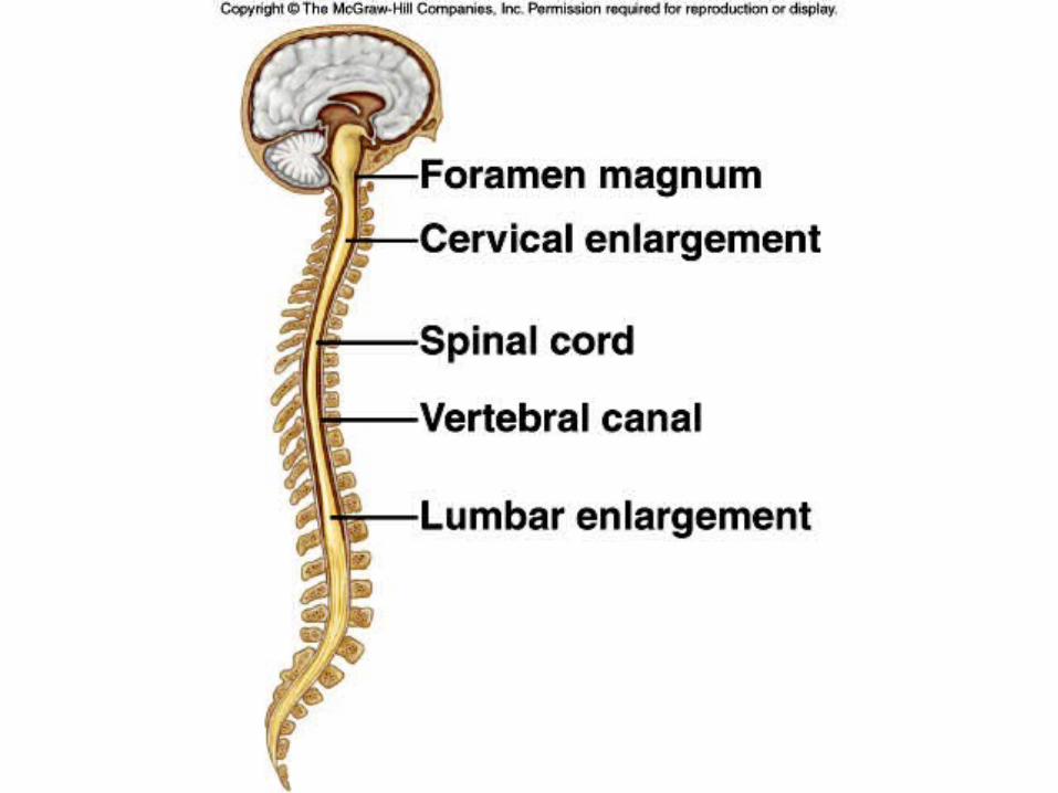

Structure
Give rise to 31Pairs of spinal nerves
Inferiorly, splits into cauda equina


Function
Conduct nerve impulsesCenter for spinal reflexes
Cerebrum
Largest part of the mature brain


Cerebrum structure
Cerebral hemispheres
(Thinking Caps)
Mirrored large masses of the brain


Cerebrum structure
Fissure
Deep furrows


Cerebrum structure(Cont)
Corpus callosum
Nerve fibers that connect hemispheres

Cerebrum structure(Cont)
Exterior - Gray matterInterior - White matter


Cerebral lobes (regions)

Frontal lobeParietal lobeTemporal lobeOccipital lobe
Cerebral Lobes

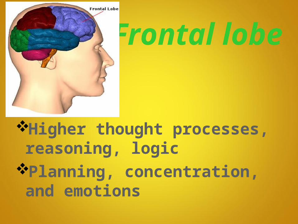
Frontal lobe
Higher thought processes, reasoning, logic
Planning, concentration, and emotions

Parietal lobe
General sensations (Hot or Cold, Pressure or Pain)
Understanding speech

Temporal lobe
Special sensations (Hearing, Taste, Smell)
Memory of visual and auditory patterns

Occipital lobe
Sight and recognition of objects

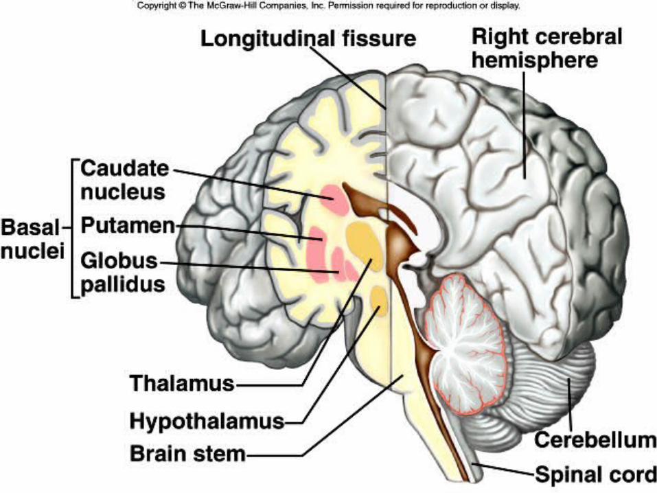
Diencephalon

Diencephalon
• Forms central core of forebrain and consists of:
--Thalamus
--Hypothalamus


Thalamus
Relays all sensory impulses to cerebral cortex (except smell)
Understanding speech, light, touch and pressure


HypothalamusControls heart rate, blood pressure, and
body temp. (Autonomic Nervous System)
Receives sensory impulses from internal organs
Connects nervous w/ endocrine system Controls release of regulating hormones
from anterior pituitary gland

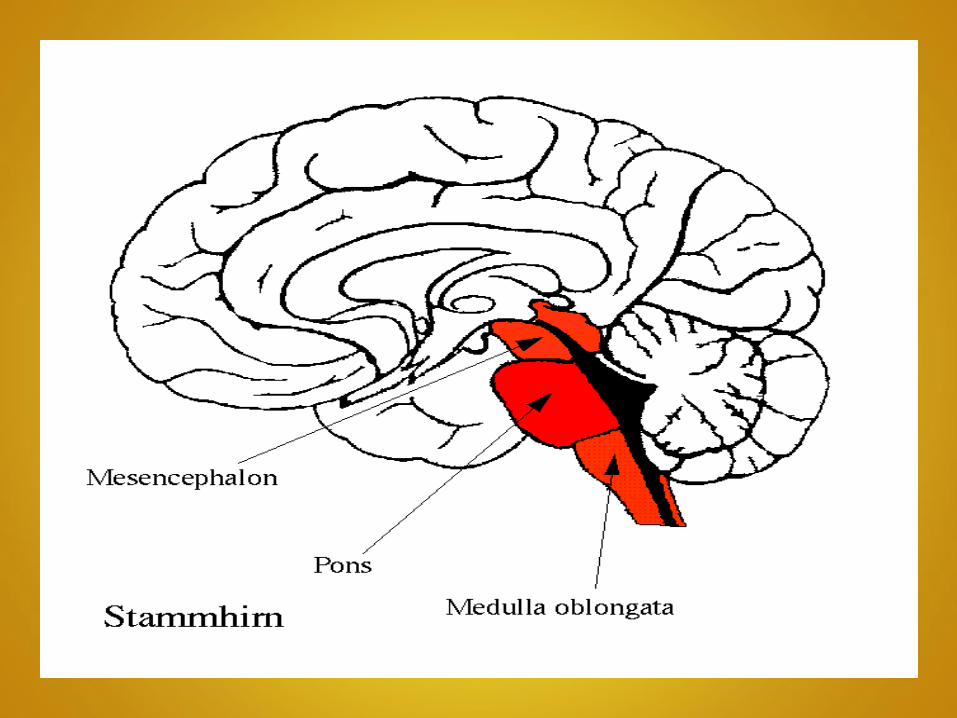
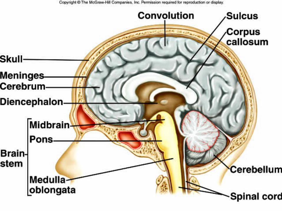
Brainstem

Brainstem
• Regions include the following:
--Midbrain
--Pons
--Medulla Oblongata

Midbrain
• Between hypothalamus and the pons
• Visual and auditory reflex centers
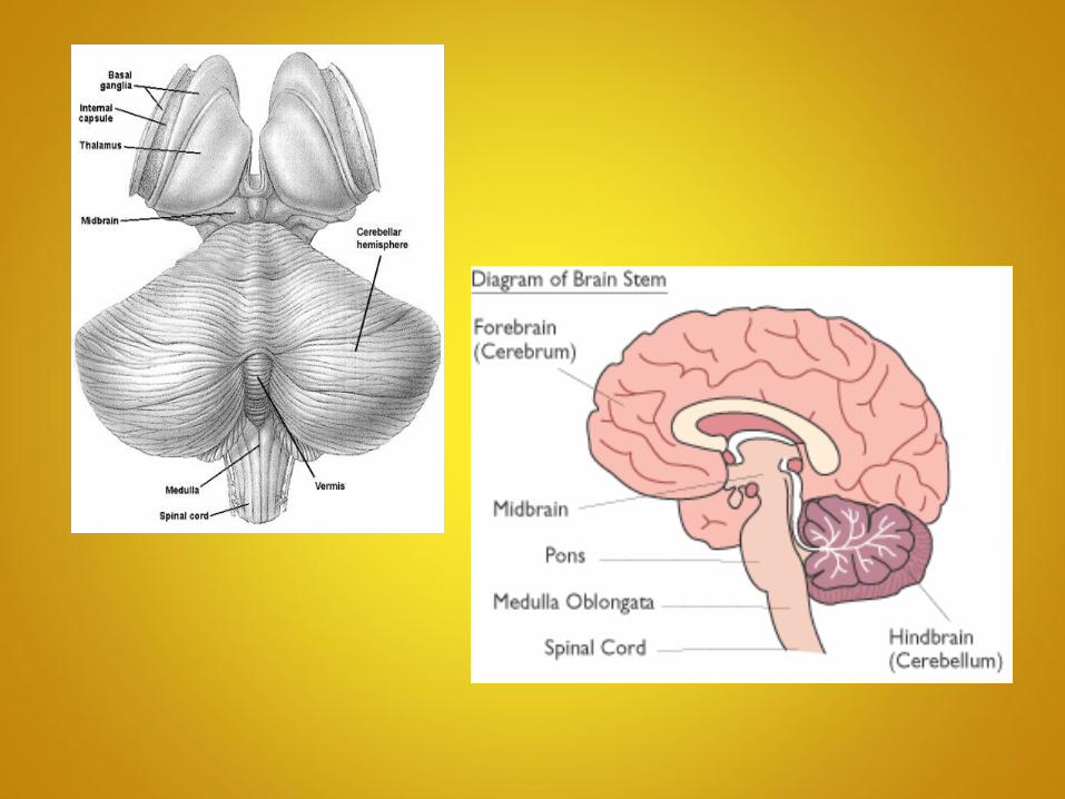
Pons
• Separates midbrain/medulla oblongata
• Helps regulate breathing

Medulla oblongata• Continuation of spinal cord
from pons to the base of skull
• Transmits impulses, Contains vital visceral centers
Medulla oblongata
• Cardiac center Vasomotor
center Respiratory
center
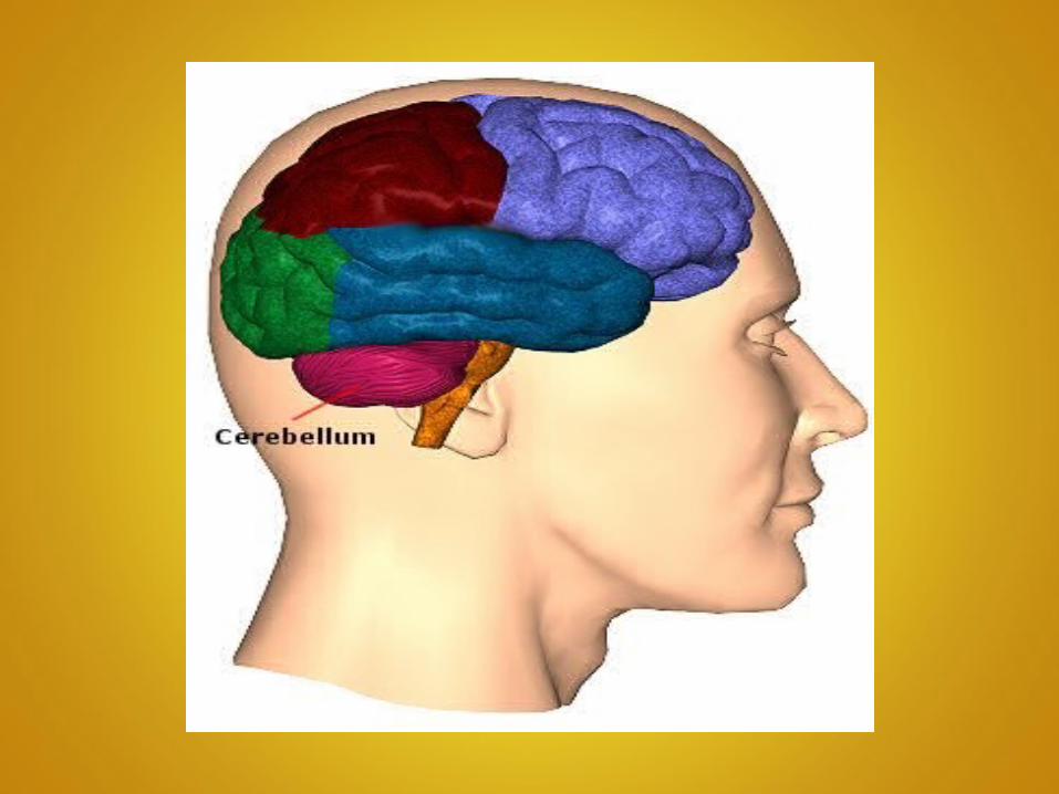
Cerebellum• Second largest structure of the brain
• Below the occipital lobes of the cerebrum, posterior to the pons and the medulla oblongata.
• Consists of two hemispheres-connects by a vermis
• Functions primarily in coordination of skeletal muscle movement and maintaining posture

Peripheral Nervous System
(PNS)
Peripheral Nervous System
• Cranial Nerves
• Spinal Nerves
• Plexus
Cranial Nerves
• 12 pair
• All arise from brainstem (Except CN 1 ---Optic Nerve)
• Most are mixed nerves


Spinal Nerves
• 31 pair
• All arise from spinal cord
• All are mixed nerves
• Emerges as Roots

Spinal Nerves
• Cervical Nerves - C1-C8
• Thoracic Nerves - T1-T12
• Lumbar Nerves - L1-L5
• Sacral Nerves - S1-S5
• 1pair of Coccygeal nerves

Plexus
Formed by combining branches of several spinal nerves
(Except in the Thoracic Region)

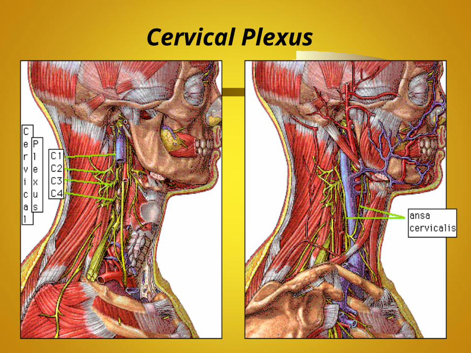
Cervical Plexus
• C-1 through C-4• Innervates muscles and skin of
neck• Forms Phrenic nerve which
innervates the Diaphragm
Cervical Plexus
Brachial Plexus
C-5 through T-1
Innervates muscles and skin of arms, forearms & hands

Brachial Plexus

Lumbosacral Plexus
T-12 through S-5Innervates lower extremity


Summary
• Divisions of the Nervous System
• Neural Tissue
• Behavior of Reflexes
• Functions of the Central Nervous System
• Functions of Spinal Cord
Summary
• Functions of Cerebrum
• Components of Diencephlon
• Functions of Brain Stem
• Functions of Cerebellum
• Components of Peripheral Nervous System


![The Nervous System. Divisions of the Nervous System Central Nervous System [CNS] = Spinal Cord Brain Peripheral Nervous System [PNS]= Spinal Nerves](https://img.dokumen.tips/doc/110x75/56649d6c5503460f94a4c71d/the-nervous-system-divisions-of-the-nervous-system-central-nervous-system.jpg)