Embed Size (px)
Citation preview
The Nervous System
Biology 12
Ms. Bowie

Divisions on the Nervous System
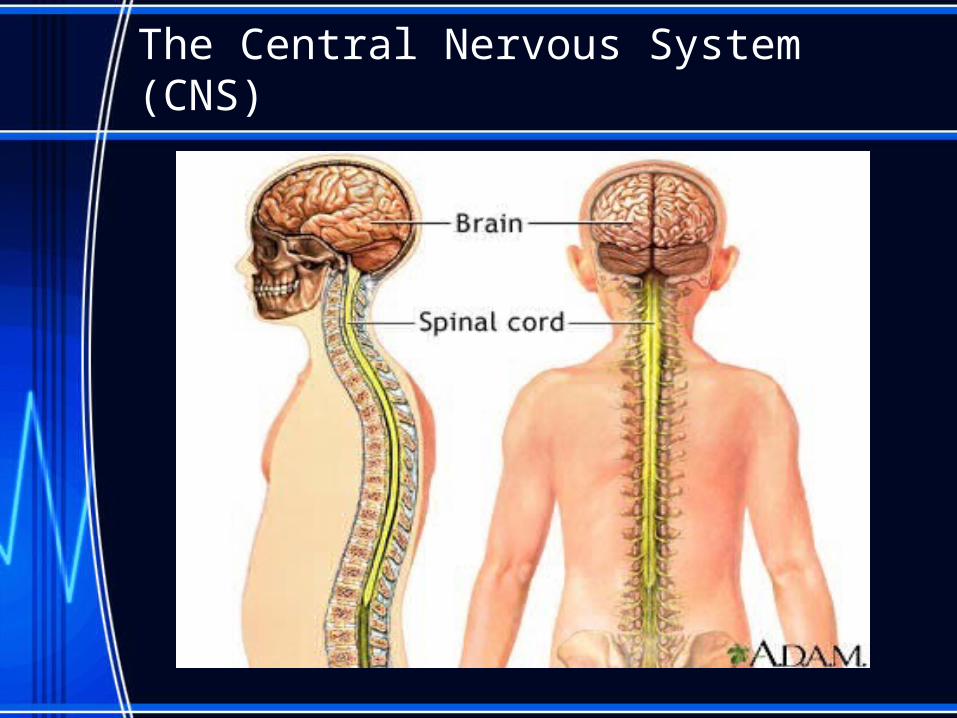
The Central Nervous System (CNS)
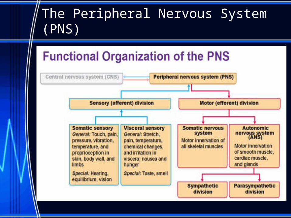
The Peripheral Nervous System (PNS)
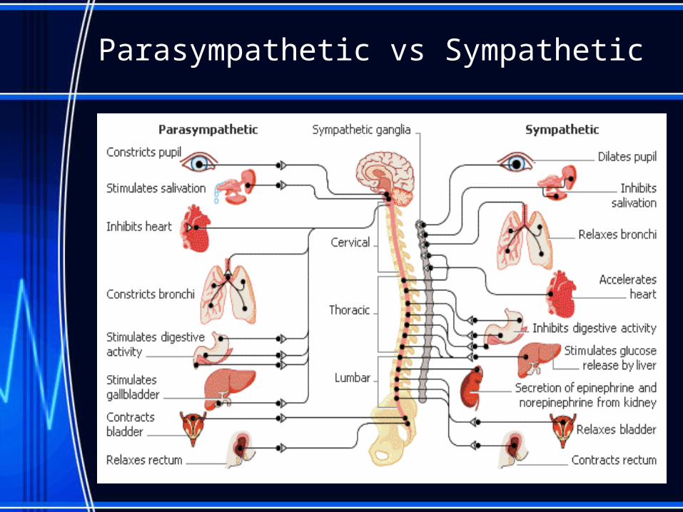
Parasympathetic vs Sympathetic
Anatomy of a Nerve Cell

Anatomy of a Nerve Cell
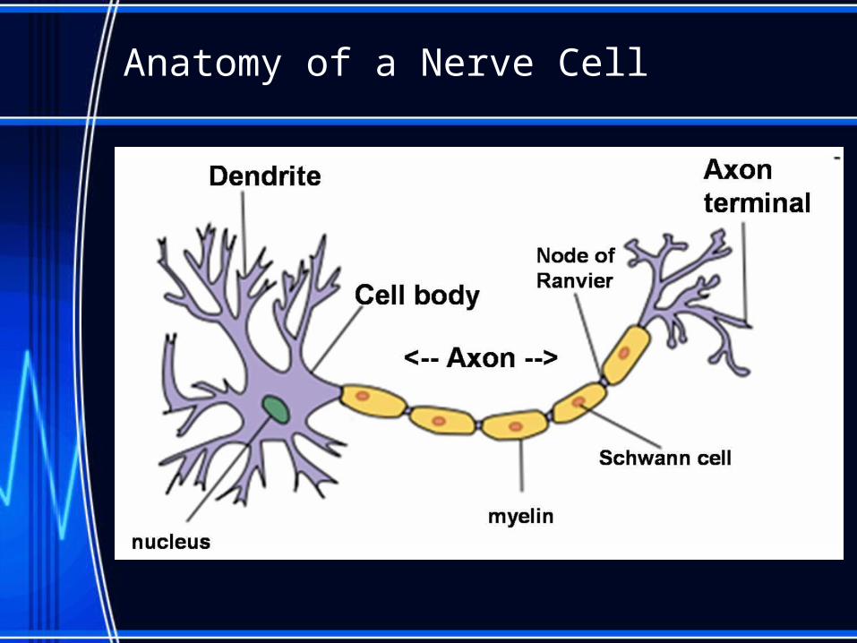
• Dendrites carry impulses toward the cell body.
• Axon are extensions of cytoplasm that carries nerve impulses away from the cell body.
• Myelin sheath is the insulated covering over the axon of a nerve cell. Myelin is formed by special cells known as Schwann cells.
• Nodes of Ranvier are the gaps between the sections of myelin sheath. Impulses can jump from node to node, speeding transmission.

Neuron Terminology
• Glial cells are non-conducting that provide support and metabolism for the nerve cells.
• Sensory neurons carry impulses from sensory receptors to the CNS; also known as afferent neurons.
• Ganglia are clusters of sensory neurons outside the CNS

Neuron Terminology
• Motor neurons carry impulses from CNS to the effector cells (muscles, organs and glands) to produce a response; also known as efferent neurons.
• Interneurons are neurons that act as links between the sensory and motor neurons. They generally make up the CNS. They interpret the sensory info and stimulate the motor neurons.

Neuron Terminology
• Neurilemma is a delicate membrane that surrounds the axon of some nerve cells;– its promotes the regeneration of the axon when
damaged.– found mostly in the PNS. – explains why feeling eventually returns to you skin
after a paper cut.
Reflex Arcs
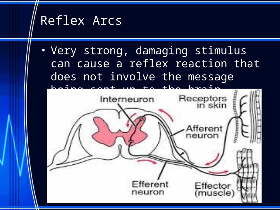
• Very strong, damaging stimulus can cause a reflex reaction that does not involve the message being sent up to the brain.

Check your understanding
1. Describe the difference between the CNS and the PNS.
2. Differentiate between sensory nerves and motor nerves.
3. Describe the function of: dendrites, myelin sheath, Schwann cells, cell body and axon.
4. Name the 5 essential parts of the reflex arc.
5. Describe the process of a reflex reaction.

The Central Nervous System in Detail
• The brain is surrounded by tough three-layer protective membrane known as the meninges. There are 3 layers:1. Dura mater – outer layer
2. Arachnoid mater – middle layer
3. Pia mater – inner layer
• The meninges form the blood-brain barrier. This barrier determines what chemicals will reach the brain.
• The brain and spinal cord float in a liquid known as the cerebrospinal fluid.
The Spinal Cord
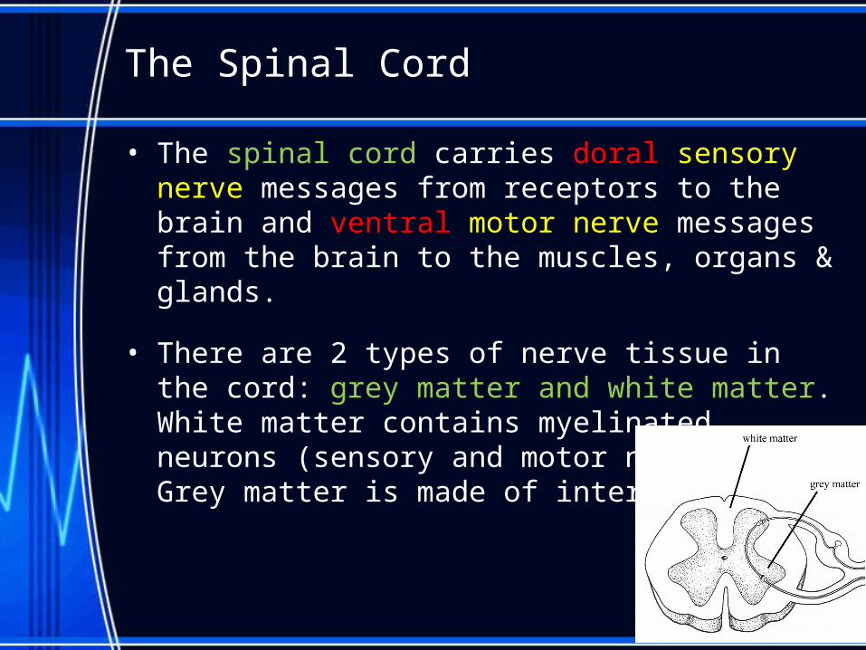
• The spinal cord carries doral sensory nerve messages from receptors to the brain and ventral motor nerve messages from the brain to the muscles, organs & glands.
• There are 2 types of nerve tissue in the cord: grey matter and white matter. White matter contains myelinated neurons (sensory and motor neurons). Grey matter is made of interneurons.

The Brain – Three Distinct Regions
– Forebrain• Olfactory lobes (smell)• Cerebrum
– Coordinates sensory & motor function
– Speech, reasoning, memory and personality
– Divides into 4 lobes: frontal, parietal, occipital, temporal
• Thalamus, hypothalamus & the pituitary gland• Cerebral cortex
– Grey matter
– Contains folds that increase the surface area
– Deep folds are known as fissures
– The corpus callosum is a bundle of nerves that connects the two hemispheres of the cortex.

The Brain – Three Distinct Regions
– Midbrain
• involved in the motor functions– vision, basic movements, and hearing.
• An area called the substantia nigra plays a role in releasing dopamine-producing neurons
• Because the midbrain is involved in muscle movement, the death of neural cells in the substantia nigra can lead to Parkinson's disease

The Brain – Three Distinct Regions
– Hindbrain• Pons
– The pons a “bridge” region– The pons also helps regulate breathing.
• Medulla oblongata – The last three centimeters of the brain stem. – Responsible for regulating our bodies heart rate,
respiration, and blood pressure.• Cerebellum
– The cerebellum is the second largest structure in the brain– Receives sensory input from a tendon, muscle, and joint
receptors. – Needed for motor learning as well as movement & muscle
tone.

The Brain – The Cerebrum
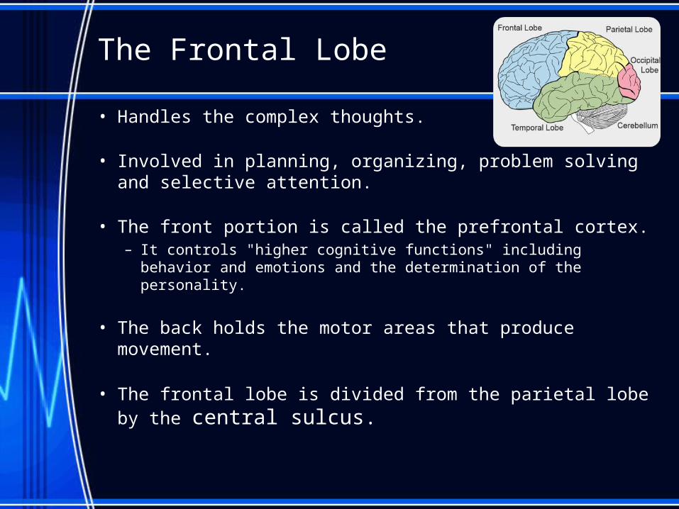
• 4 lobes– Frontal lobe– Parietal lobe– Temporal lobe– Occipital lobe
The Frontal Lobe
• Handles the complex thoughts.
• Involved in planning, organizing, problem solving and selective attention.
• The front portion is called the prefrontal cortex. – It controls "higher cognitive functions" including behavior and
emotions and the determination of the personality.
• The back holds the motor areas that produce movement.
• The frontal lobe is divided from the parietal lobe by the central sulcus.
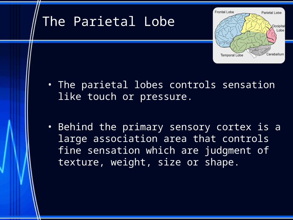
The Parietal Lobe
• The parietal lobes controls sensation like touch or pressure.
• Behind the primary sensory cortex is a large association area that controls fine sensation which are judgment of texture, weight, size or shape.
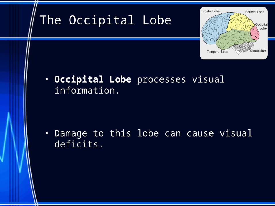
The Occipital Lobe
• Occipital Lobe processes visual information.
• Damage to this lobe can cause visual deficits.

The Temporal Lobe
• These lobes allow a person to tell one smell from another and one sound from another.
• They also help in sorting new information and are responsible for short-term memory.
• Right Lobe involved mainly in visual memory like pictures and faces while left lobe involved mainly in verbal memory such as words and names.
Check your understanding
1. List the four regions of the cerebral cortex and state the function of each.
2. If a physician cuts the corpus callosum how might this affect the patient?
3. Name the meninges and explain what they do.
4. If you wanted to learn more about how memories form and are stored, what area of the brain would you study?
Homeostasis & The Autonomic Nervous System
• The autonomic nervous system is part of the PNS.
• It works with the endocrine system to help the body adjust to changes in the external and internal environment.
• They are all motor nerves that regulate the organs of the body without conscious control.
• Made of 2 parts:– The sympathetic and parasympathetic nervous
systems.
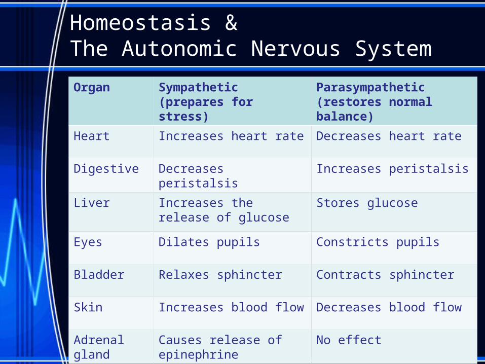
Homeostasis & The Autonomic Nervous System
Organ Sympathetic(prepares for stress)
Parasympathetic(restores normal balance)
Heart Increases heart rate Decreases heart rate
Digestive Decreases peristalsis Increases peristalsis
Liver Increases the release of glucose
Stores glucose
Eyes Dilates pupils Constricts pupils
Bladder Relaxes sphincter Contracts sphincter
Skin Increases blood flow Decreases blood flow
Adrenal gland Causes release of epinephrine
No effect