Embed Size (px)
Citation preview
Anatomy of the Central Nervous SystemAnatomy of the Central Nervous System
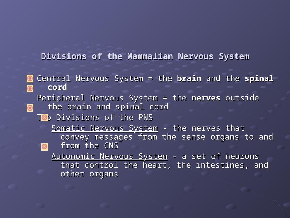
Divisions of the Mammalian Nervous SystemDivisions of the Mammalian Nervous System
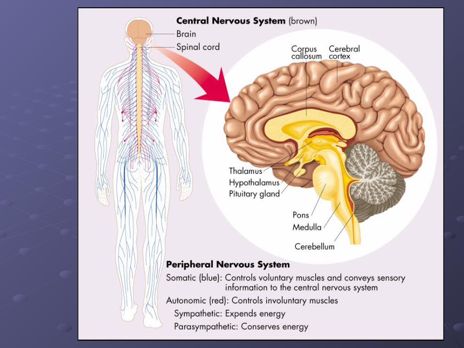
Central Nervous System = the Central Nervous System = the brainbrain and the and the spinal cordspinal cordPeripheral Nervous System = the Peripheral Nervous System = the nervesnerves outside the brain and outside the brain and
spinal cordspinal cordTwo Divisions of the PNSTwo Divisions of the PNS
Somatic Nervous SystemSomatic Nervous System - the nerves that convey messages - the nerves that convey messages from the sense organs to and from the CNS from the sense organs to and from the CNS
Autonomic Nervous SystemAutonomic Nervous System - a set of neurons that control the - a set of neurons that control the heart, the intestines, and other organsheart, the intestines, and other organs
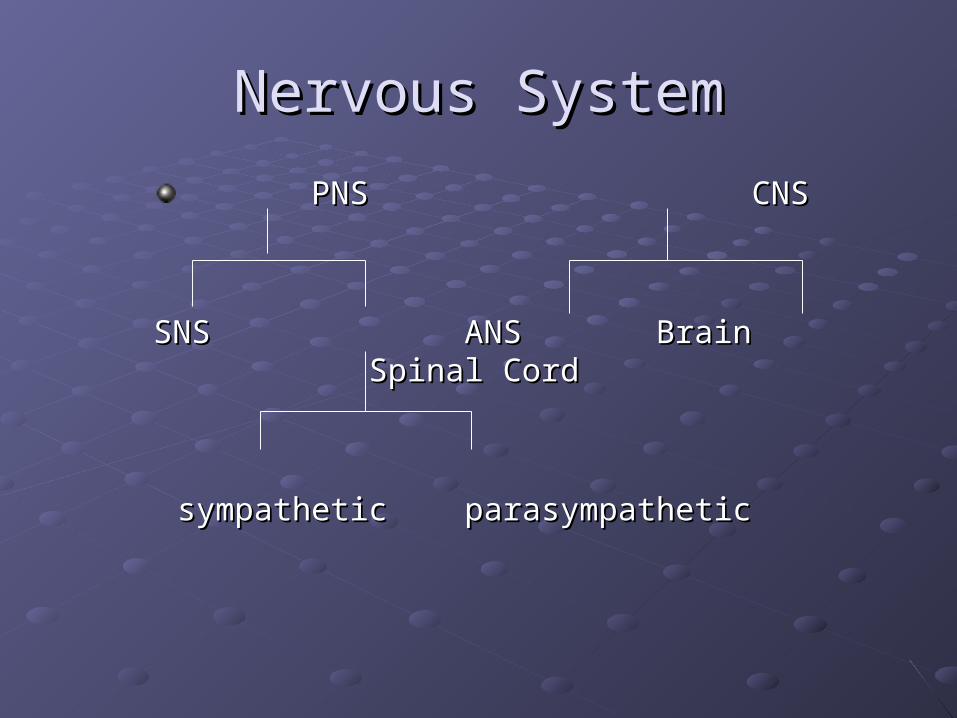
Nervous SystemNervous System
PNSPNS CNSCNS
SNSSNS ANSANS BrainBrainSpinal CordSpinal Cord
sympathetic parasympatheticsympathetic parasympathetic

The Nervous SystemThe Nervous System
The Spinal Cord-part of the CNS found within the spinal column The Spinal Cord-part of the CNS found within the spinal column
The spinal cord communicates with the sense organs and The spinal cord communicates with the sense organs and muscles below the level of the headmuscles below the level of the head
Bell-Magendie Law-the entering Bell-Magendie Law-the entering dorsal roots carry dorsal roots carry sensory informationsensory information …and the exiting …and the exiting ventral roots ventral roots carry motor informationcarry motor information to the muscles and glands to the muscles and glands
Dorsal Root Ganglia - are clusters of neurons outside the Dorsal Root Ganglia - are clusters of neurons outside the spinal cordspinal cord
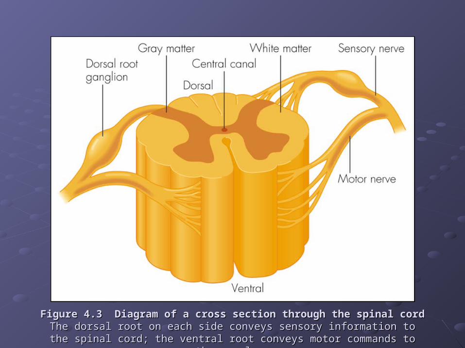
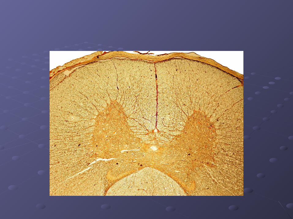
Figure 4.3 Diagram of a cross section through the spinal cordFigure 4.3 Diagram of a cross section through the spinal cordThe dorsal root on each side conveys sensory information to the spinal cord; the The dorsal root on each side conveys sensory information to the spinal cord; the
ventral root conveys motor commands to the muscles.ventral root conveys motor commands to the muscles.
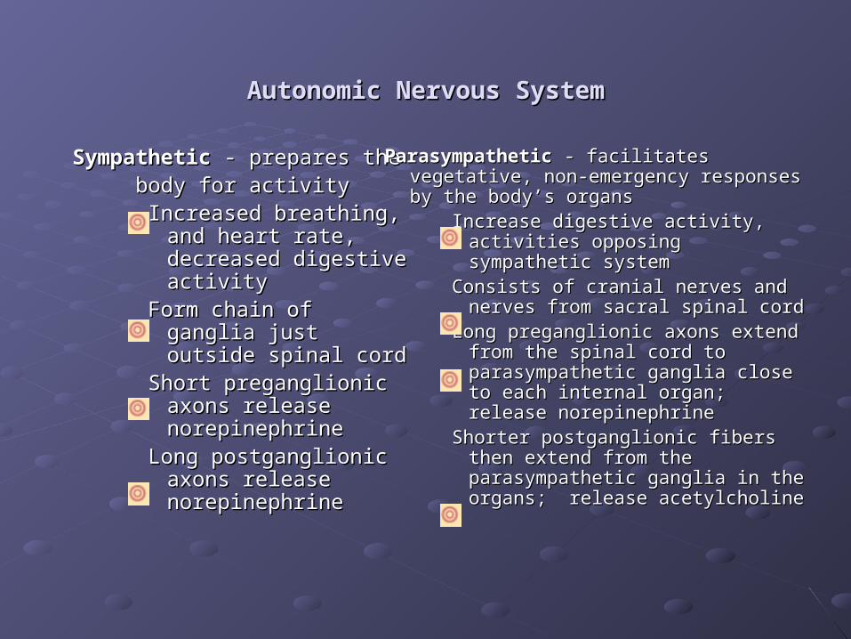
Autonomic Nervous SystemAutonomic Nervous System
Sympathetic Sympathetic - prepares the - prepares the body for activitybody for activity
Increased breathing, and Increased breathing, and heart rate, decreased heart rate, decreased digestive activitydigestive activity
Form chain of ganglia Form chain of ganglia just outside spinal cordjust outside spinal cord
Short preganglionic Short preganglionic axons release axons release norepinephrinenorepinephrine
Long postganglionic Long postganglionic axons release axons release norepinephrinenorepinephrine
Parasympathetic Parasympathetic - facilitates vegetative, - facilitates vegetative, non-emergency responses by the body’s non-emergency responses by the body’s organsorgans
Increase digestive activity, activities Increase digestive activity, activities opposing sympathetic systemopposing sympathetic system
Consists of cranial nerves and Consists of cranial nerves and nerves from sacral spinal cordnerves from sacral spinal cord
Long preganglionic axons extend Long preganglionic axons extend from the spinal cord to from the spinal cord to parasympathetic ganglia close to parasympathetic ganglia close to each internal organ; release each internal organ; release norepinephrinenorepinephrine
Shorter postganglionic fibers then Shorter postganglionic fibers then extend from the parasympathetic extend from the parasympathetic ganglia in the organs; release ganglia in the organs; release acetylcholineacetylcholine
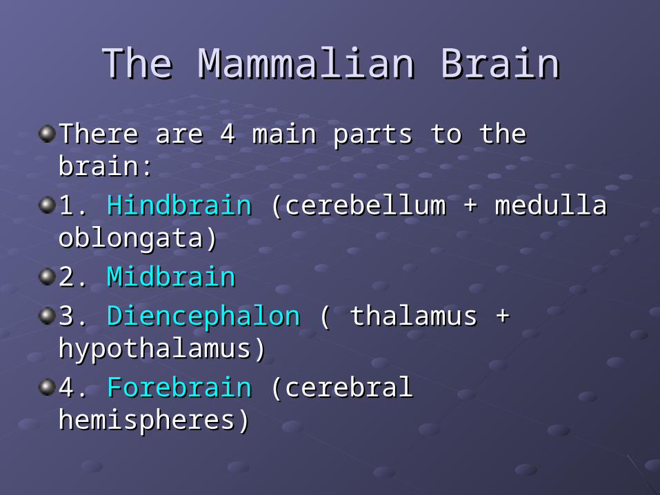
The Mammalian BrainThe Mammalian Brain
There are 4 main parts to the brain:There are 4 main parts to the brain:
1. 1. HindbrainHindbrain (cerebellum + medulla (cerebellum + medulla oblongata)oblongata)
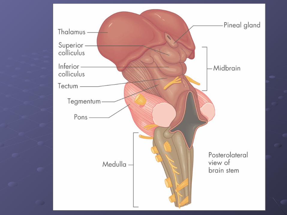
2. 2. MidbrainMidbrain
3. 3. DiencephalonDiencephalon ( thalamus + ( thalamus + hypothalamus)hypothalamus)
4. 4. ForebrainForebrain (cerebral hemispheres) (cerebral hemispheres)
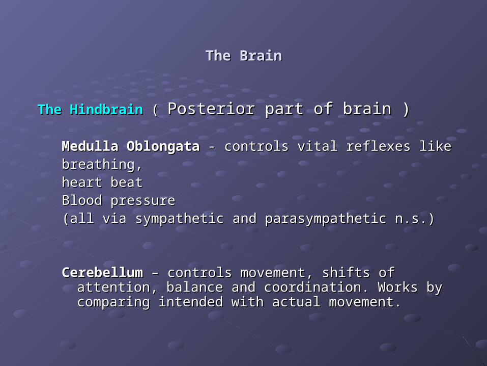
The BrainThe Brain
The HindbrainThe Hindbrain ( ( Posterior part of brain )Posterior part of brain )
Medulla OblongataMedulla Oblongata - controls vital reflexes like - controls vital reflexes like breathing,breathing,heart beat heart beat Blood pressureBlood pressure(all via sympathetic and parasympathetic n.s.)(all via sympathetic and parasympathetic n.s.)
CerebellumCerebellum – controls movement, shifts of attention, balance and – controls movement, shifts of attention, balance and coordination. Works by comparing intended with actual coordination. Works by comparing intended with actual movement.movement.
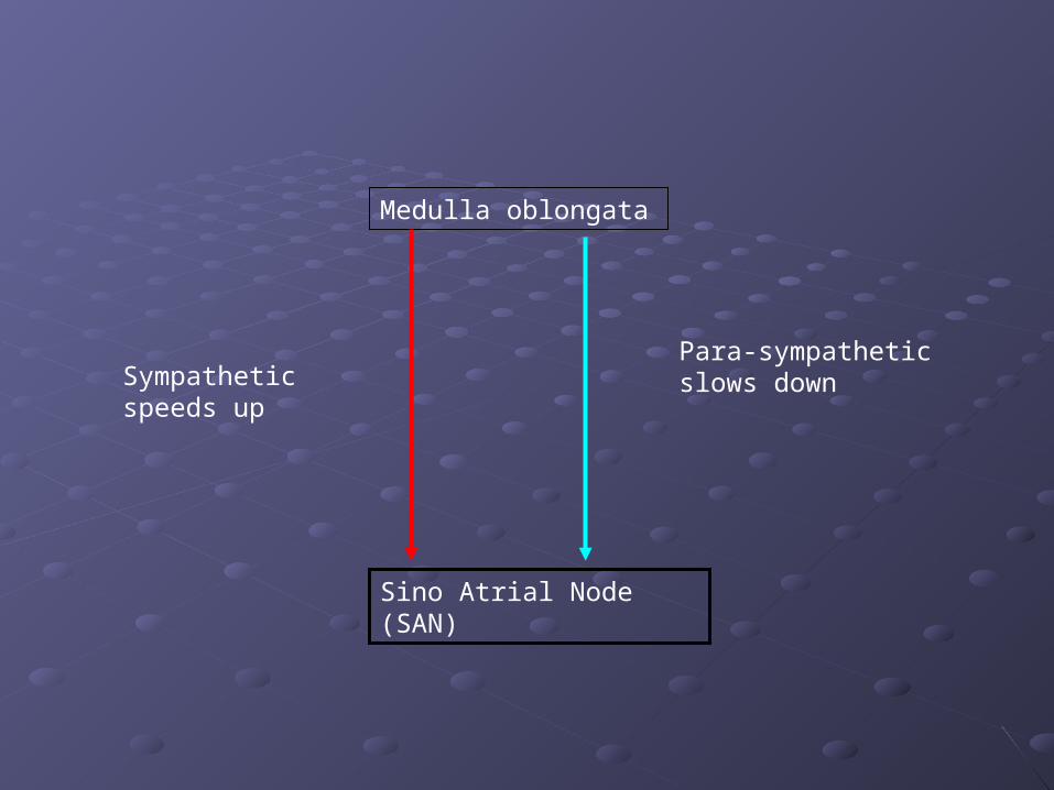
Medulla oblongata
Sino Atrial Node (SAN)
Sympathetic speeds up
Para-sympathetic slows down
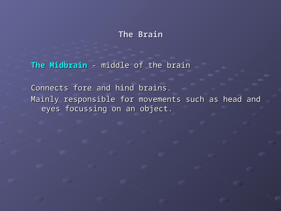
The BrainThe Brain
The MidbrainThe Midbrain - middle of the brain - middle of the brain
Connects fore and hind brains.Connects fore and hind brains.
Mainly responsible for movements such as head and eyes Mainly responsible for movements such as head and eyes focussing on an object.focussing on an object.
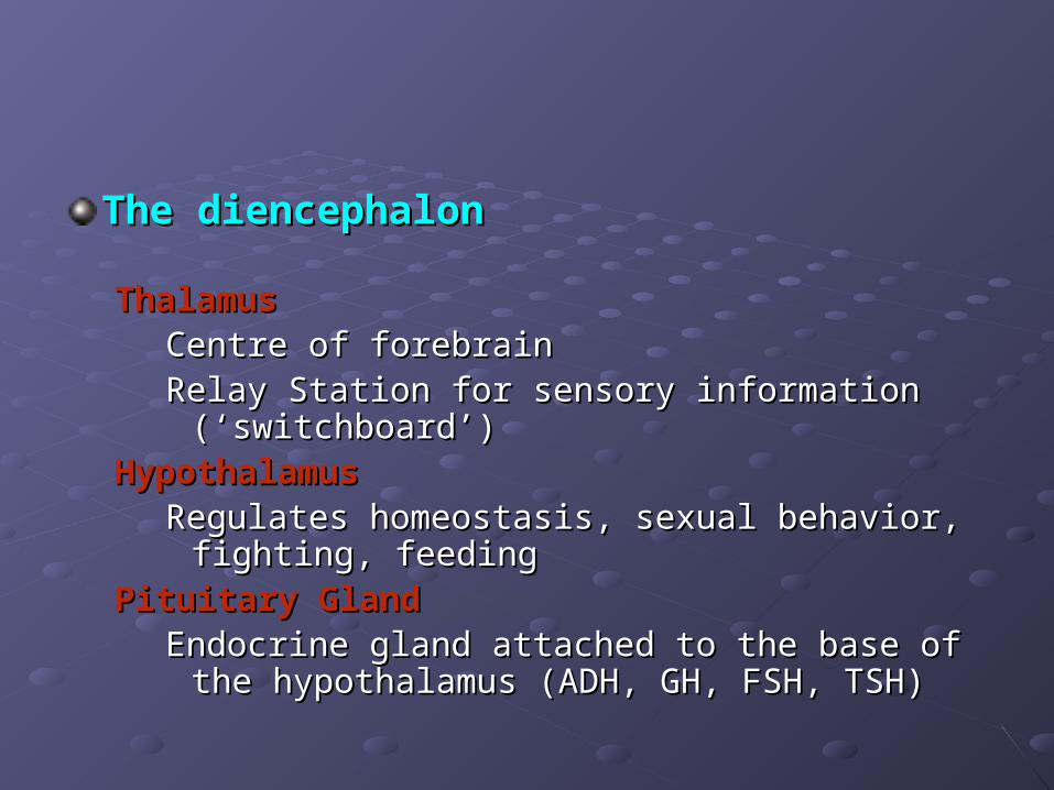
The diencephalonThe diencephalon
ThalamusThalamusCentre of forebrainCentre of forebrainRelay Station for sensory information (‘switchboard’)Relay Station for sensory information (‘switchboard’)
HypothalamusHypothalamusRegulates homeostasis, sexual behavior, fighting, Regulates homeostasis, sexual behavior, fighting,
feedingfeedingPituitary GlandPituitary Gland
Endocrine gland attached to the base of the Endocrine gland attached to the base of the hypothalamus (ADH, GH, FSH, TSH)hypothalamus (ADH, GH, FSH, TSH)
The BrainThe Brain
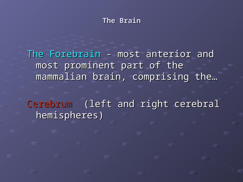
The ForebrainThe Forebrain - most anterior and most - most anterior and most prominent part of the mammalian brain, prominent part of the mammalian brain, comprising the…comprising the…
CerebrumCerebrum (left and right cerebral hemispheres) (left and right cerebral hemispheres)
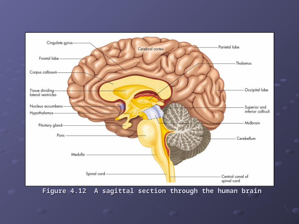
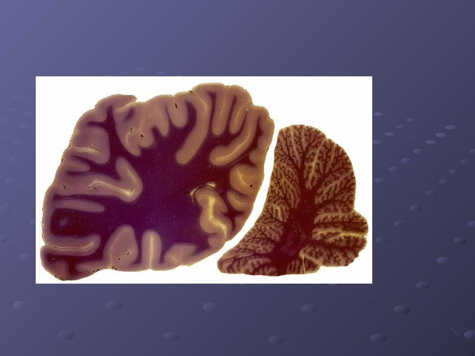
Figure 4.12 A sagittal section through the human brainFigure 4.12 A sagittal section through the human brain
The CerebrumThe Cerebrum
The cerebral hemispheres contain 10The cerebral hemispheres contain 109 9 nerve nerve cells in a layer only 3mm thick.cells in a layer only 3mm thick.
Left and Right hemispheres are linked by the Left and Right hemispheres are linked by the CORPUS CALLOSUM.CORPUS CALLOSUM.
Each hemisphere has 4 LOBES:Each hemisphere has 4 LOBES:
i) Frontali) Frontal
ii) Parietalii) Parietal
iii) Temporaliii) Temporal
iv) Occipitaliv) Occipital
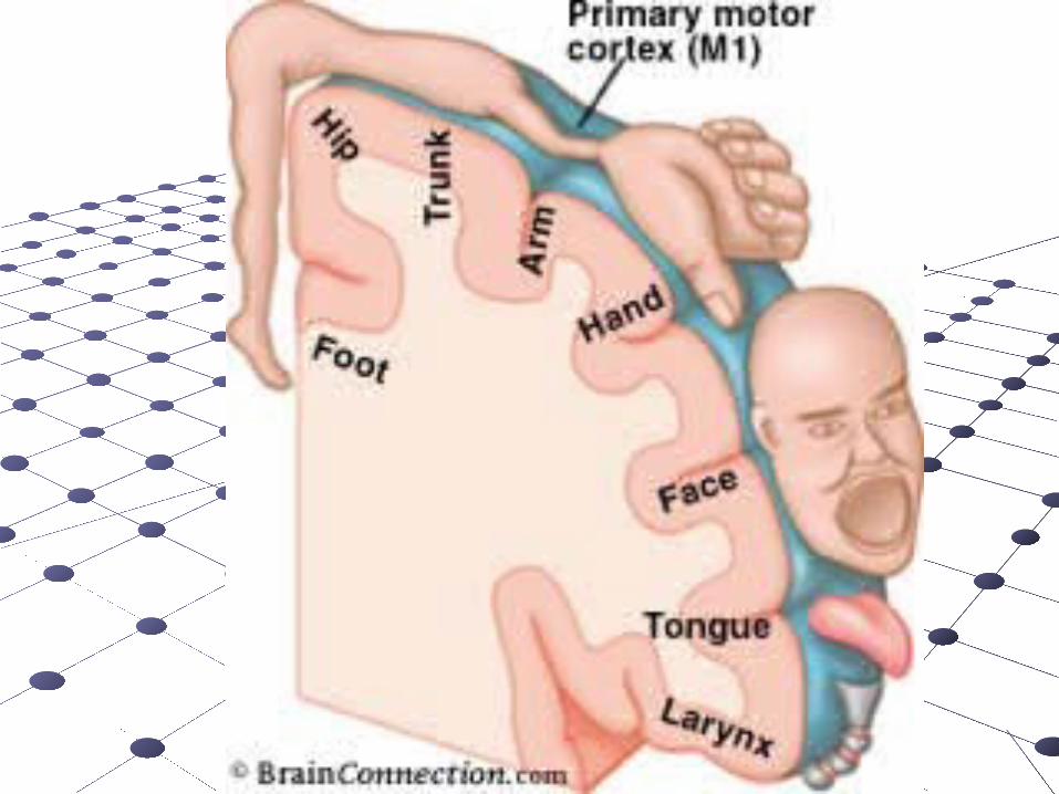
Frontal LobeFrontal Lobe
The Frontal Lobe-extends from the central sulcus The Frontal Lobe-extends from the central sulcus (groove) to the anterior limit of the brain(groove) to the anterior limit of the brain
Contains Primary Motor Cortex – responsible Contains Primary Motor Cortex – responsible for fine movementsfor fine movements
Contributes to shifting attention, planning of Contributes to shifting attention, planning of action, delayed response tasks as examplesaction, delayed response tasks as examples
Parietal LobeParietal Lobe
The Parietal Lobe - between occipital lobe and the The Parietal Lobe - between occipital lobe and the central sulcuscentral sulcus
Contains the primary somato-sensory cortex –Contains the primary somato-sensory cortex –i.e. receiving touch sensation, muscle-stretch i.e. receiving touch sensation, muscle-stretch
information and joint position informationinformation and joint position informationAlso, 3-D processing (visualisations, face Also, 3-D processing (visualisations, face
recognition etc)recognition etc)
Temporal LobeTemporal Lobe
The Temporal Lobe - lateral portion of each The Temporal Lobe - lateral portion of each hemisphere, near the templeshemisphere, near the temples
Contains targets for hearing, essential for Contains targets for hearing, essential for understanding spoken language (Wernicke’s understanding spoken language (Wernicke’s Area), complex visual processes, emotional Area), complex visual processes, emotional and motivational behaviorsand motivational behaviors
Occipital LobeOccipital Lobe
The Occipital Lobe - posterior end of cortexThe Occipital Lobe - posterior end of cortex
Contains primary visual cortexContains primary visual cortex
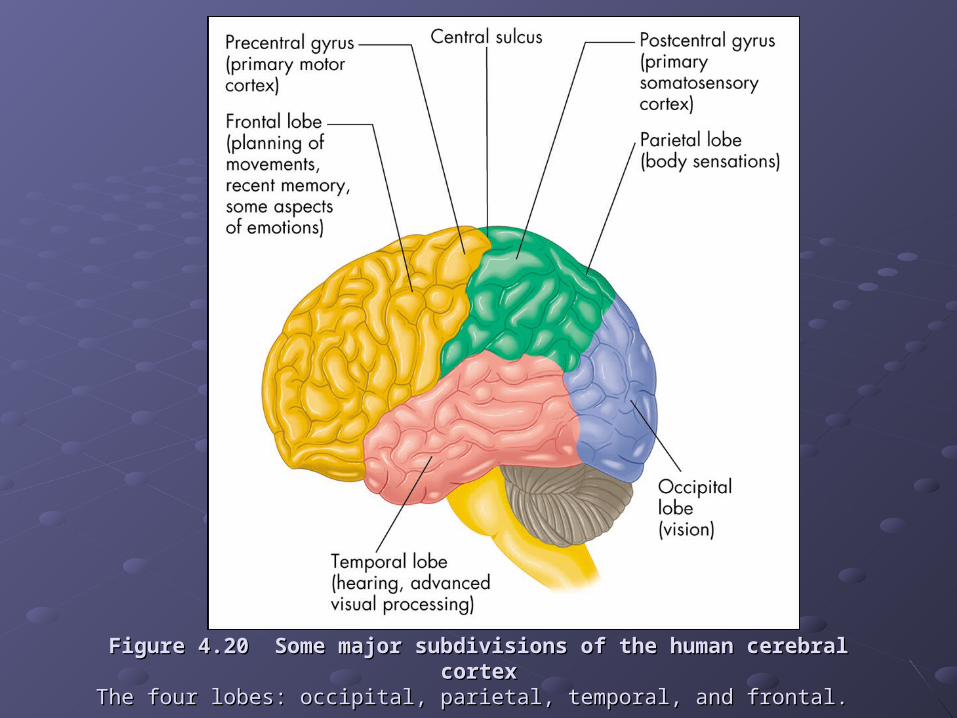
Figure 4.20 Some major subdivisions of the human cerebral cortexFigure 4.20 Some major subdivisions of the human cerebral cortexThe four lobes: occipital, parietal, temporal, and frontal. The four lobes: occipital, parietal, temporal, and frontal.
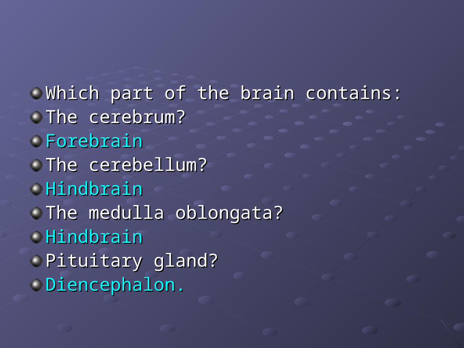
Which part of the brain contains:Which part of the brain contains:The cerebrum? The cerebrum? ForebrainForebrainThe cerebellum?The cerebellum?HindbrainHindbrainThe medulla oblongata?The medulla oblongata?HindbrainHindbrainPituitary gland?Pituitary gland?Diencephalon.Diencephalon.
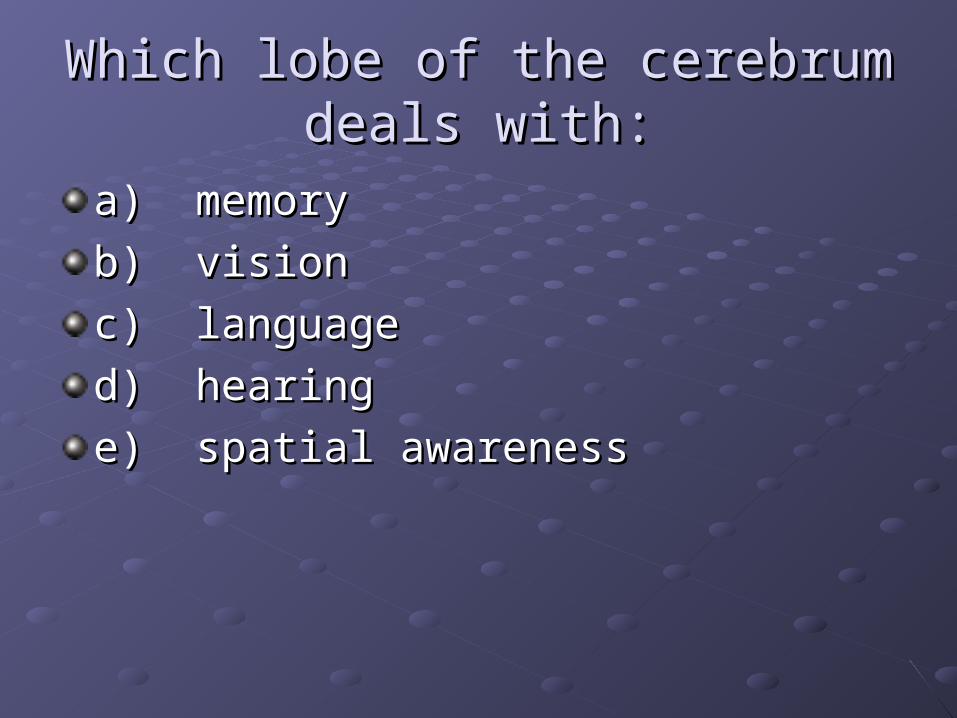
Which lobe of the cerebrum deals Which lobe of the cerebrum deals with:with:
a) memorya) memory
b) visionb) vision
c) languagec) language
d) hearingd) hearing
e) spatial awarenesse) spatial awareness

Essay for next week:Essay for next week:
a) Outline the functions of the cerebrum in a) Outline the functions of the cerebrum in the human brain.the human brain.b) Describe the changes that occur in the b) Describe the changes that occur in the cerebrum of a person with Alzheimer’s cerebrum of a person with Alzheimer’s disease and discuss the possible causes disease and discuss the possible causes of the disease.of the disease.
Try a web search eg Try a web search eg www.alz.orgwww.alz.org or or www.alzheimers.orgwww.alzheimers.org