Embed Size (px)
Citation preview

DARCY DOELLMAN MSN, RN, CRNI, VA -BC
MARCH 15, 2017
LOUISVILLE, KENTUCKY
The Impact of Catheter Occlusion in Central Line Associated
Bloodstream Infections

Cincinnati Children’s Hospital
642 beds
Rated #3 nationally
Vascular Access Team (24/7) 37 FTEs
Services provided: IV access
Midline and PICC insertions
CVC monitoring and complication management
Education and research
Learning Objectives
Describe types of catheter occlusion
Review how to identify, prevent, and manage catheter dysfunction
Discuss current guidelines and practices for reducing CLABSI rates
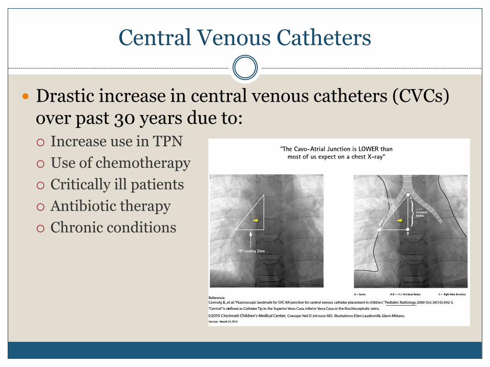
Central Venous Catheters
Drastic increase in central venous catheters (CVCs) over past 30 years due to:
Increase use in TPN
Use of chemotherapy
Critically ill patients
Antibiotic therapy
Chronic conditions
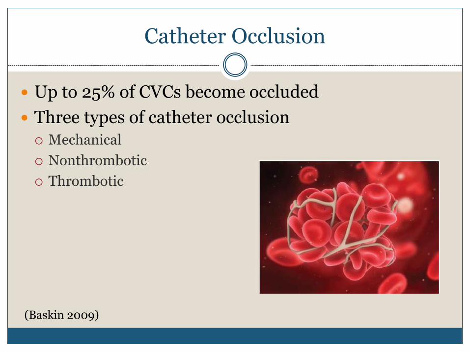
Catheter Occlusion
Up to 25% of CVCs become occluded
Three types of catheter occlusion Mechanical
Nonthrombotic
Thrombotic
(Baskin 2009)
Mechanical Occlusion
External Clamped/kinked tubing
Position of patient
Tight suture around catheter
Port needle dislodgement
Internal Catheter tip malposition
Pinch-Off Syndrome
Compression of catheter between 1st rib and clavicle
Symptom: Flushing stops/starts in a predictable manner
Occurs in 1% of patients
(Dougherty 2006; Gabriel, 2006)

Pinch-Off Syndrome

Nonthrombotic Occlusion
Precipitate Extreme pH, calcium phosphate crystals,
or lipid deposits
Crystallization of TPN
Drug precipitation
Diazepam, aminoglycosides, phenytoin

Thrombotic Occlusion
Most common type of catheter occlusion
Four types: Intraluminal
Fibrin sheath
Fibrin tail
Mural thrombus
(Stephens 1999)

Fibrin Formation
CVCs are considered a foreign body in situ After placement, CVCs become covered with plasma proteins and
fibrin
Within 24 hours, platelets and WBCs adhere to protein and allows for colonization of bacteria
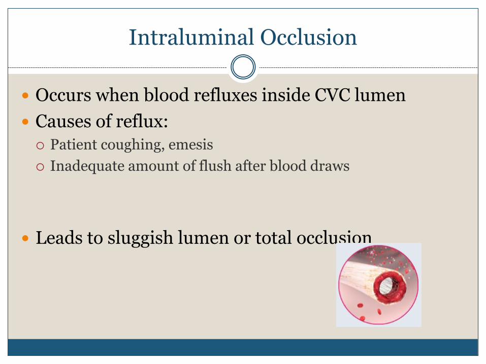
Intraluminal Occlusion
Occurs when blood refluxes inside CVC lumen
Causes of reflux: Patient coughing, emesis
Inadequate amount of flush after blood draws
Leads to sluggish lumen or total occlusion

Fibrin Sheath
Fibrin adheres to catheter surface
May completely cover the opening of the catheter tip, like a sock
Causes: injury to tunica intima
A complete fibrin sheath may lead to infiltration or extravasation
(Gabriel 2006)

Fibrin Tail
Tail extends from CVC tip
Upon aspiration, fibrin tail blocks catheter lumen
Results in an ability to infuse fluids but an inability to withdraw blood
Leads to partial occlusion

Mural Thrombus
Forms where the catheter touches the vein
Can occur anywhere along the catheter path Common with catheter tip malposition
May lead to catheter thrombosis

Identification of Catheter Occlusion
Routinely assess catheter for patency with flushing of CVC
Patency: ability to flush and aspirate blood without resistance
Sluggish lumen
May be first symptom of a catheter occlusion
May lead to a partial (withdraw) or complete occlusion
Assess CVC further if catheter is sluggish

Rule Out Mechanical Factors
Begin at infusion pump or catheter hub
Assess for closed clamps, kinked tubing or catheter
Is patient laying on the catheter?
Has PICC migrated out?
Change in port needle location?
Is needleless connector clotted?
Review MAR profile, is there a concern for precipitation?
Has mechanical factors been resolved or ruled out?

Management of Occlusion
Preferred approach is catheter salvage
Manage as thrombotic occlusion if unable to determine type of occlusion
Do not leave an occluded lumen untreated because
another lumen is functional

Symptoms
Resistance when flushing
Sluggish flow
Inability to infuse fluids
Frequent occlusion alarm on infusion pump

Treatment for Thrombotic Occlusion
Alteplase (thrombolytic) 2mg/2mL sterile water
Can instill alteplase up to 2 doses
Adults: 2 mg/2mL
Pediatrics: 110% of the catheter priming volume
Dosing: Allow 1st dose to dwell for up to 2 hours
If occlusion persists, instill 2nd dose
Efficacy: Adults: 88% patency after 2 doses
75% patency after 1 dose
Pediatrics: 83% patency after 2 doses
Treat all types of thrombotic occlusions
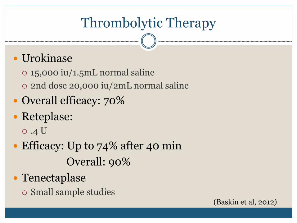
Thrombolytic Therapy
Urokinase 15,000 iu/1.5mL normal saline
2nd dose 20,000 iu/2mL normal saline
Overall efficacy: 70%
Reteplase: .4 U
Efficacy: Up to 74% after 40 min
Overall: 90%
Tenectaplase Small sample studies
(Baskin et al, 2012)
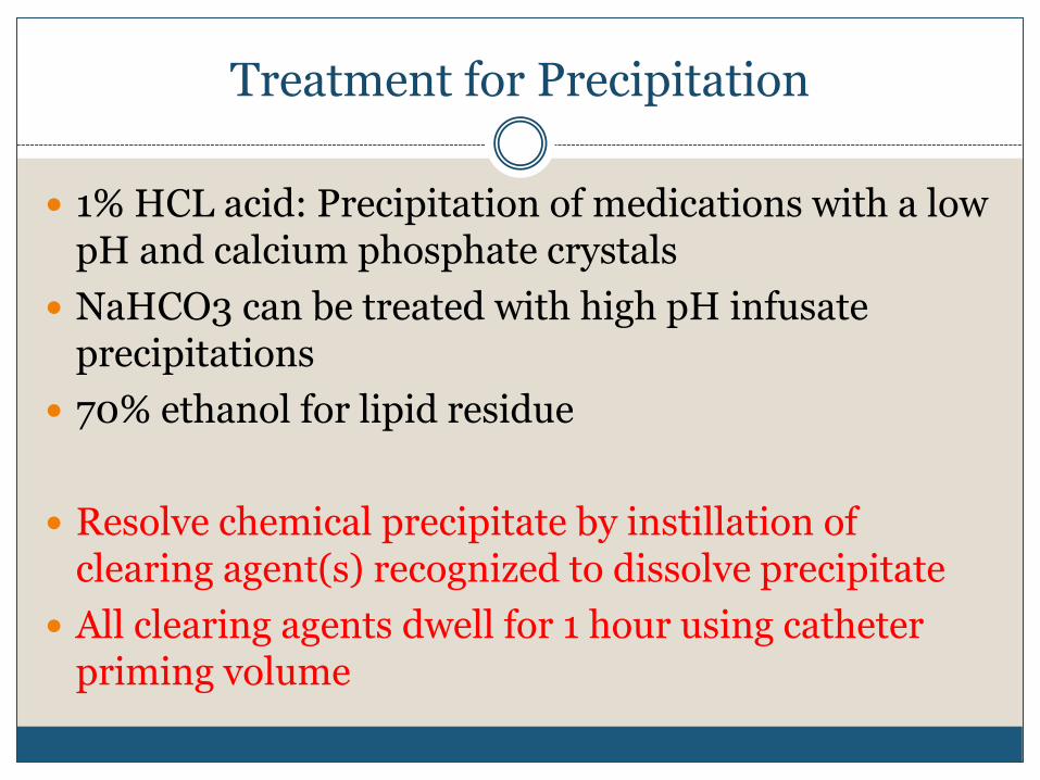
Treatment for Precipitation
1% HCL acid: Precipitation of medications with a low pH and calcium phosphate crystals
NaHCO3 can be treated with high pH infusateprecipitations
70% ethanol for lipid residue
Resolve chemical precipitate by instillation of clearing agent(s) recognized to dissolve precipitate
All clearing agents dwell for 1 hour using catheter priming volume
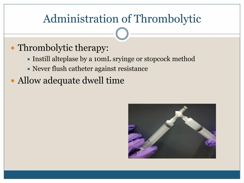
Administration of Thrombolytic
Thrombolytic therapy: Instill alteplase by a 10mL sryinge or stopcock method
Never flush catheter against resistance
Allow adequate dwell time
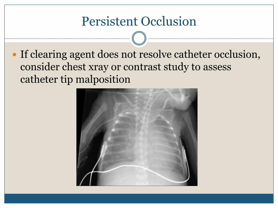
Persistent Occlusion
If clearing agent does not resolve catheter occlusion, consider chest xray or contrast study to assess catheter tip malposition
Persistent Partial (Withdraw) Occlusion
If CVC remains dysfunctional (unable to obtain blood return)
Do not continue to use CVC In rare cases, consider transfemoral approach to pull off fibrin

Risks for Catheter Occlusion
Catheter tip malposition
Number of CVC lumens
Large catheter to vessel ratio
PICCs are prone to occlusion by a factor of >2 in comparison with other CVADs
Ports have less catheter occlusion as compared to Broviac and Hickman catheters
(Moureau & Chopra, 2016)
Prevention
Proper flushing technique Volume of flush
Type of flush
Normal saline vs heparinized saline
Frequency of flush
Routine flushing and after blood draw
Push-pause flushing
Use 10mL syringe for flushing
Flush unused lumens per protocol
(Goosens 2014)

Prevention
Ensure optimal CVC tip placement
Minimizing catheter occlusions requires the tip placement for CVADs to be in the lower one-third of the superior vena cava near the junction of the
right atrium
Prevention
Review institutional data on CVC complications
Use of valved PICCs
Needleless connector
Education of the nursing staff Occlusion rate decreased in a 6-month period from 29% down to
8.5%
Dedicated PICC team Occlusion rate decreased from 32 to 15 occlusions
(Bartock 2010;Ngo, 2005)
Patient Family Experience
Delays in therapy At risk for suboptimal therapy
Delays in procedures
Delays in discharge
Increased cost
Complications
14 -36% complication rate within 2 years of CVC placement
Increase cost
Increase LOS

Link Between CLABSI and Occlusion
In animal studies, fibrin sheath formation around CVCs promoted
Colonization
Infection
Persistent bacteremia
(Mehall et al, 2004)

PICCs
Retrospective study of hospital ortho patients
180 PICCs in 136 patients Mean dwell: 21 days
36/180 (20%) removed due to complications CLABSI: 8/180 (14%)
Occlusion: 11/180 (8%)
(Valbouquet Schneider et al 2015)
Two risk factors for CLABSI:> 70 years of ageTwo lumens or greater
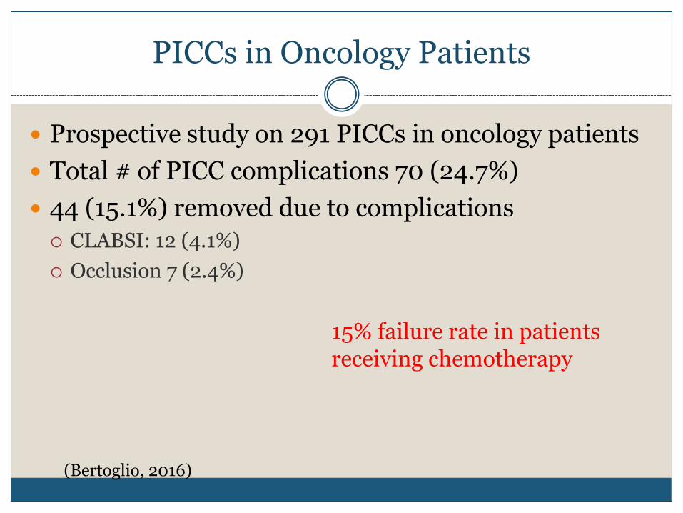
PICCs in Oncology Patients
Prospective study on 291 PICCs in oncology patients
Total # of PICC complications 70 (24.7%)
44 (15.1%) removed due to complications CLABSI: 12 (4.1%)
Occlusion 7 (2.4%)
15% failure rate in patients receiving chemotherapy
(Bertoglio, 2016)
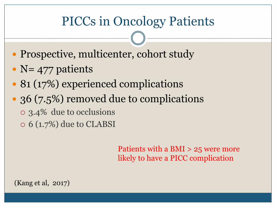
PICCs in Oncology Patients
Prospective, multicenter, cohort study
N= 477 patients
81 (17%) experienced complications
36 (7.5%) removed due to complications 3.4% due to occlusions
6 (1.7%) due to CLABSI
(Kang et al, 2017)
Patients with a BMI > 25 were morelikely to have a PICC complication

CLABSI Prevention
Two phases: Insertion
Insertion checklist
Use of ultrasound
Credentialing of clinician
Standardized CVC insertion kits
Maximal sterile barrier/chlorhexidine skin antisepsis
Maintenance
Dressing changes
Needleless connector antisepsis
Tubing changes
Chlorhexidine bathing

Avoid the Femoral Vein
Increases infection risk and deep venous thrombosis in adults
Emergent situations only
(Lorente, Henry, Martin, et al, 2005)

Hand Hygiene
Using alcohol-based waterless product or antiseptic soap and water
Compliance measurements can be obtained by actual observation
Curlej & Katrancha 2016
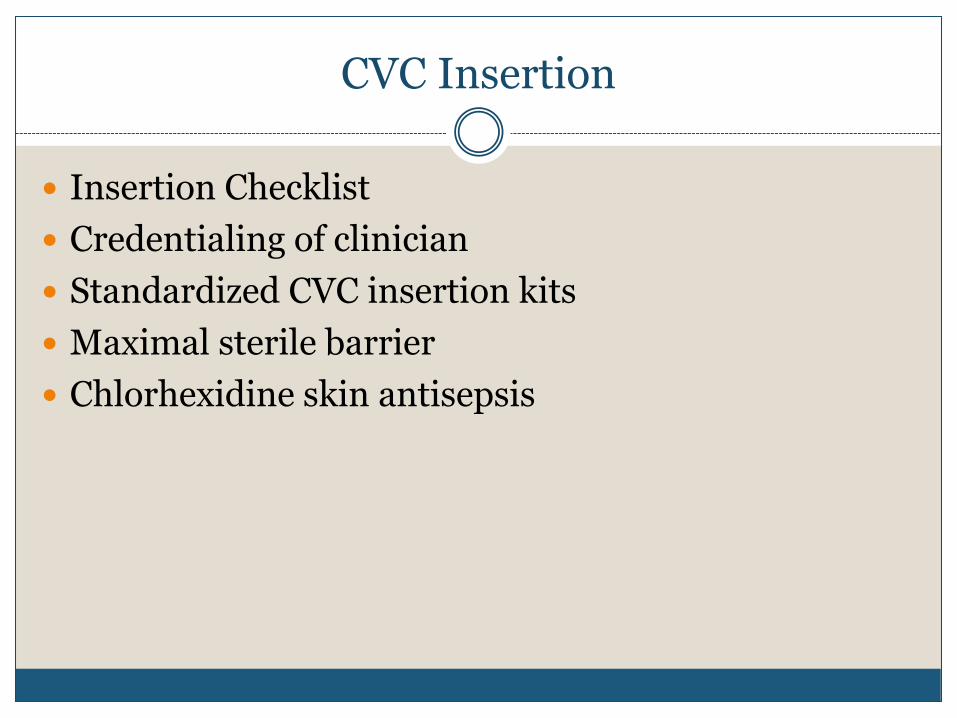
CVC Insertion
Insertion Checklist
Credentialing of clinician
Standardized CVC insertion kits
Maximal sterile barrier
Chlorhexidine skin antisepsis

Device Selection
CVADs carry significant risk to patients
Apply evidence-based guidelines for selection
Avoid overuse of PICCs
Vascular Access Teams can assist with difficult IV access, failed attempts, or specific patient conditions:
Obesity
Diabetes
(Moureau & Chopra, 2016)
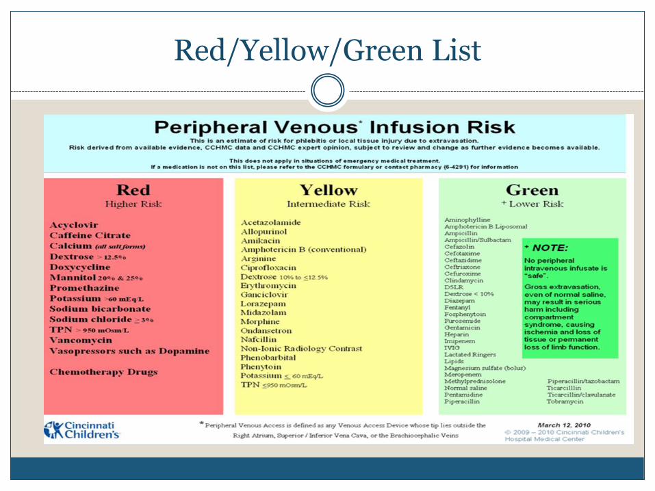
Red/Yellow/Green List
In the absence of indications for a multilumen PICC, use a single-lumen
PICC of the smallest gauge
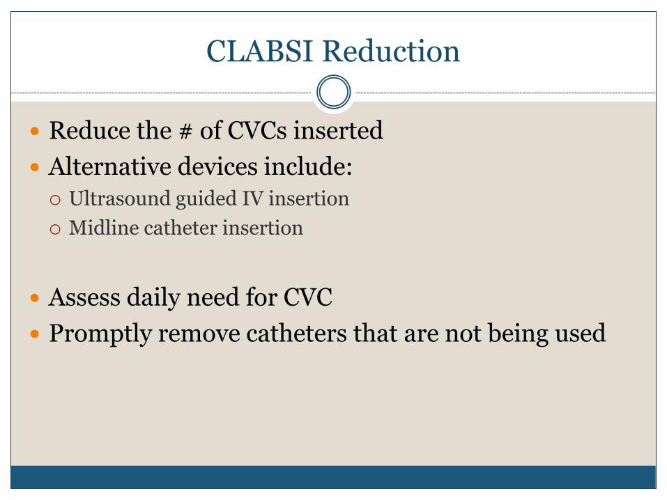
CLABSI Reduction
Reduce the # of CVCs inserted
Alternative devices include: Ultrasound guided IV insertion
Midline catheter insertion
Assess daily need for CVC
Promptly remove catheters that are not being used
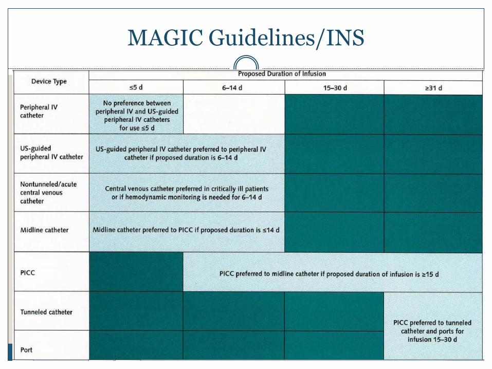
MAGIC Guidelines/INS
Antibiotic Coated Catheters
240 community hospital in California Control group: 257 patients, 8 CLABSIs with an infection rate
of 4.18/1,000 line
Study group: 260 patients, 1 CLABSI with an infection rate of 0.47/1,000 line days.
(Rutkoff, 2014)

CLABSI
The estimated mortality rate associated with CLABSI is 23.8%, with an average increase LOS up to 21 days and an estimated cost of $29,156 per patient

Single Lumen vs Multi-lumen CVCs
In the absence of indications for a multilumen PICC, use a single-lumen PICC of the smallest gauge
Insert the smallest catheter french size with the lowest number of lumens
Criteria for a Multi-lumen CVC
Continuous vesicant or irritant chemotherapy with additional lumen needs
Need for vasopressors
Milrinone
Need for simultaneous administration of multiple incompatible medications
TPN plus additional lumen needs

Senior Leadership
Senior leadership can help reduce CLABSI rates by: Driving the implementation of best practices and technologies
Promoting compliance with nursing staff
Support of staff for continuing education (conferences, certification)
Invest in EMRs that align with data collection and reporting
CLABSI task force member
Summary
Catheter occlusion is a common complication of CVCs
Assess CVC patency with routine flushing, medication administration, and blood sampling
Prevention and prompt treatment of occlusions is vital for CVCs
Choose the most appropriate VAD based on the patient’s type and length of therapy
Avoid unnecessary CVCs
Follow current CVC bundles for reducing CLABSI

Thank You