Embed Size (px)
Citation preview

Signal Transduction
The Hypoxic Tumor Microenvironment PromotesInvadopodia Formation and Metastasis throughLPA1 Receptor and EGFR CooperationKelly Harper, Roxane R. Lavoie, Martine Charbonneau, Karine Brochu-Gaudreau, andClaire M. Dubois
Abstract
Hypoxia, a common feature of solid tumors, has beencritically involved in cell invasion and metastasis, but theunderlying mechanisms remain poorly understood. Previous-ly, it has been observed that the lysophosphatidic acid receptor4 (LPA4) signaling axismediates production of the degradativesubcellular structures invadopodia, which are known to berequired for metastasis. Here, it is demonstrated that LPA1
(LPAR1) is a common and major receptor used for hypoxia-induced invadopodia production in various cancer cell lines.The widespread use of LPA1 was not due to increased LPA1
expression but rather relied on Src-mediated cross-talk with
EGFR. LPA1-mediated phosphorylation of Y845-EGFR underhypoxia led to PI3K/Akt activation, an event that increases theability of cells to produce invadopodia. Moreover, phospho-Y845-EGFRwas upregulated in hypoxic zones of tumors and acombination of EGFR and LPA1 inhibition synergisticallysuppressed metastasis in vivo.
Implications: This study uncovers an LPA1–EGFR signalingaxis that is used for cell invasion in hypoxia and suggests apotential target to impede cancer metastasis. Mol Cancer Res; 1–13. �2018 AACR.
IntroductionMetastasis is the leading cause of mortality in cancer patients.
However, effective therapies targeting the disseminated diseaseremain a major challenge in clinical management of the disease.The tumor microenvironment is increasingly recognized to play asignificant role in many of the hallmarks of cancer, notably as animportant modulator of tumor cell phenotypes driving cell inva-sion and metastasis (1). A common feature of the tumor micro-environment is hypoxia, or low concentrations of oxygen, withinsolid tumors. Tumor hypoxia arises because of inadequate deliv-ery of oxygen as a result of insufficient and defective vasculatureand high oxygen consumption rate of cancer cells (2). Hypoxictumors are aggressive, resistant to chemotherapy, and prone torecurrence (3). It has been recently reported that tumor micro-environment stimuli such as hypoxia and the associated acidic pHinduce cell invasion and metastasis through activation of sodi-um–hydrogen exchangers, growth factors, metalloproteases, orRhoGEFs (4). In these reports, the increase in cellular invasionwasassociatedwith production of invadopodia, which are specializedcell structures required for cancer cell dissemination.
Invadopodia are actin-rich and proteolytically active subcellu-lar structures generated by cancer cells that promote their migra-
tion and invasion through tumor stroma and the basementmembrane of blood vessels during the process of metastasis(5). These structures are relevant to the cell invasion process asthey have been observed in migrating cancer cells undergoingintravasion as well as in tumor cells invading through tissues(6, 7). More recently, invadopodia have been shown to beessential for cancer cell extravasion andmetastasis in vivo throughgenetic analysis and pharmacologic inhibition of molecules spe-cifically involved in their initiation, maturation, or function (8).These studies have provided direct in vivo evidence of the func-tional role of invadopodia in metastasis, making them relevanttherapeutic targets to counter pathologic cell invasion. Genera-tion of invadopodia requires tight coordination between polar-ized cellular trafficking, signaling events and cytoskeletal remo-deling. Whereas these events have been extensively studied,manyof the upstream inducers that drive invadopodia formation andactivity remain unknown (9).
An emerging participant in cancer progression is the ATX-LPAreceptor signaling axis. Lysophosphatidic acid (LPA) is a bioactivelipid produced mainly by autotaxin (ATX) that signals through 6known LPA G-protein–coupled receptors (GPCR; LPA1-6). Thesereceptors activate various signaling components that includecAMP, PLCb, PLCe, small rhoGTPases, Ras, ERK, and PI3K thatregulatemany pathophysiologic processes, including cancer (10).We have previously reported that LPA4, through the cAMP–Rap1–Rac1 axis, was involved in invadopodia production by cancer cellsand that this event correlated with their metastatic efficiency (11).The ability of LPA to stimulate invadopodia production has sincebeen confirmed in prostate and melanoma cells (12, 13).Furthermore, overexpression of ATX or LPA1-3 has been shownto induce tumorigenesis and metastasis in a mouse model (14),whereas their pharmacologic inhibition decreased cell migrationin vitro and caused tumor regression in mice (15). Although thesefindings suggested the critical implication of LPA receptors in
Immunology Division, Department of Pediatrics, Faculty of Medicine and HealthSciences, Universit�e de Sherbrooke, Sherbrooke, Qu�ebec, Canada.
Note: Supplementary data for this article are available at Molecular CancerResearch Online (http://mcr.aacrjournals.org/).
Corresponding Author: Claire M. Dubois, Universit�e de Sherbrooke, 3001 12thNorthAvenue, Sherbrooke, Qu�ebec J1H5N4, Canada. Phone: 819-564-5289; Fax:819-564-5215; E-mail: [email protected]
doi: 10.1158/1541-7786.MCR-17-0649
�2018 American Association for Cancer Research.
MolecularCancerResearch
www.aacrjournals.org OF1
on April 15, 2020. © 2018 American Association for Cancer Research. mcr.aacrjournals.org Downloaded from
Published OnlineFirst June 4, 2018; DOI: 10.1158/1541-7786.MCR-17-0649

tumor progression, the interplay between LPARs and hypoxiawithin the context of cell invasion has not yet been addressed.Here, we report that hypoxia promotes cell invasion through amechanism that involves Src-mediated cross-communicationbetween LPA receptor 1 and EGFR. Our findings suggest amechanistic rationale for targeting both LPA1 and EGFR to coun-teract metastasis.
Materials and MethodsReagents
1-Oleoyl-sn-glycerol-3-phosphate 18:1 (LPA) sodium salt andLPA receptor antagonist (Ki16425) were purchased from Sigma-Aldrich. Inhibitors of EGFR (Tyrphostin AG 1478), PLC(U-73122), and PI3K (LY294002) were purchased from BiomolInternational. Inhibitors of MEK (PD 98059), ROCK (Y-27632),Src (PP2), and RAP1 (GGTI-298) were from Calbiochem (EMDChemical Inc). LPA1 antagonist AM095 was from APExBIO.CorningMatrigel Basement MembraneMatrix was fromCorning.The antibody directed against LPA1was fromAbnova and, againstLPA4, from Novus Biologicals. Antibodies directed against EGFR(D38B1), pTyr, phospho-EGFR (Y845), phospho-AKT1 (Ser473),pan-AKT, Src 32G6 and phospho-Src (416) were purchased fromCell Signaling Technology. The anti-GRK2 antibody was fromSanta Cruz Biotechnology. The anti-tubulin antibody was fromSigma-Aldrich and, Texas Red-conjugated phalloidin, DAPI (40,6-diamidino-2-phenylindole), and all secondary antibodies werefrom Invitrogen (Molecular Probes). Hypoxyprobe kit containingpimonidazole HCl and anti-pimonidazole mouse antibody wasfrom Hypoxyprobe.
Cell culture and transfectionsHT1080 human fibrosarcoma, MDA-MB-231 human breast
cancer, and U87 human glioblastoma cells were obtained fromthe American Type Culture Collection. All cell lines wereroutinely tested for mycoplasma using the MycoSEQ Mycoplas-ma Detection Kit (all negative; Thermo Fisher Scientific). Cellswere grown for no more than 25 passages in total for anyexperiment. Cells were cultured in minimal essential medium(MEM; Wisent) supplemented with 10% FBS (Gibco BRL) and40 mg/mL of gentamicin (Wisent) in a humidified 95% air/5%CO2 incubator at 37�C. For hypoxic stimulations, cells werecultured in an INVIVO2 400 hypoxic chamber (Ruskinn)under an atmosphere of 1% O2 and 5% CO2. In the caseof stable transfections with shRNA against LPA1 or LPA4
(SABiosciences), cells were seeded at a density of 1 � 105 cellsper well in a 6-well culture plate the day before transfection.Transfections were performed using the FuGENE reagent(Roche Diagnostics), according to the manufacturer's protocol.Stable transfectants were obtained by selection with puromycin2 mg/mL (Invivogen). In the case of lentiviral transductions,cells were seeded at a density of 3 � 105 cells per 10 cm2
Petri dish and infected with 1 mL of viral stock in 2 mL ofoptiMEM supplemented with 2 mL Polybrene (10 mg/mL;EMD Millipore). Mission lentiviral shRNA targeting EGFR(TRCN0000010329), Src (TRCN0000195339), ADAM12(TRCN0000047033), ADAM17 (TRCN0000052172), or ascramble sequence were used (Sigma-Aldrich). Viral particleswere generated by transient transfection of 293T cells using aViraPower lentiviral expression system (Invitrogen ThermoFisher Scientific).
Real-time RT-PCRTotal RNA was isolated using the TRIzol (Invitrogen) protocol,
as described (11), and 1 mg of RNA was reverse transcribed tocomplementary DNA (cDNA) using a QuantiTect reverse tran-scription kit (Qiagen). cDNAwas then analyzed by real-time PCRusing a hot start SYBR Green qPCR master mix (BiMake). Primerpairs are described in the Supplementary Material and Methodssection. Quantitative real-time PCR was performed on a Rotor-Gene 3000 (Corbett Research). The cycling program was asfollows: the initial denaturation was performed at 95�C for15 minutes, followed by 40 amplification cycles with annealingtemperature of 55�C for 30 seconds, and final extension at 72�Cfor 30 seconds. For calculation of copy numbers, cloned plasmidDNA was used to generate a standard curve for each target, andsamples were normalized with a reference gene (RPLP0) aspreviously described (16).
Western blottingCells were lysed in a RIPA buffer. Supernatant samples were
recovered by centrifugation (13,000 rpm for 30 minutes at 4�C),and protein concentrations were determined using the BCAreagent (Biolynx Inc.). Immunoblotting was performed as previ-ously described (11). In the case of immunoprecipitation experi-ments, 1 mg of total protein was immunoprecipitated using ananti-EGFR antibody (dilution, 1:100). The membranes wereprobed overnight with primary antibodies. The secondary anti-body was a peroxidase-conjugated anti-rabbit or anti-mouseantibody, depending on the source of primary antibody used(Amersham, Baie-d'Urf�e, QC). Immunoblots were revealed usingthe Luminata Western HRP Chemiluminescence substrate(Millipore). Band intensities were analyzed using the QuantityOne software (Bio-Rad Laboratories).
Invadopodia assayCoverslips were prepared as described (11), using Oregon-
Green488-conjuagted gelatin (Invitrogen). Thirty thousand cellswere seeded on each coverslip and allowed to adhere. Following a10-hour or 16-hour incubation period under the conditionsdescribed in figure legends, cells were fixed with 2% paraformal-dehyde for 10 minutes at room temperature. Nuclei were stainedwith DAPI and F-actin was stained using Texas-Red-conjugatedphalloidin. Cells were visualized and images were taken using aZeiss Axioskop fluorescence microscope. Cells formingECM-degrading invadopodia were identified based on cells withat least one F-actin-enriched area of matrix degradation (charac-terized by loss of green fluorescence). Three fields of 100 cells(magnification, �40) were counted per coverslip to quantify thepercentage of cells forming ECM-degrading invadopodia.
RTK phosphorylation arrayHT1080 cellswere incubated under normoxia or hypoxia for 30
or 60minutes in serum-free medium. Proteins were extracted andanalyzed using the human PathScan RTK Signaling AntibodyArray kit (Cell Signaling Technology), according to the manufac-turer's instructions. Fluorescent images were captured using anOdyssey Infrared Imaging System and fluorescence intensitiessemi-quantitated using the ImageJ software.
ELISA assaysHT1080 cells were plated at a density of 1�106 cells per 10 cm2
Petri dish. The next day, the medium was removed and replaced
Harper et al.
Mol Cancer Res; 2018 Molecular Cancer ResearchOF2
on April 15, 2020. © 2018 American Association for Cancer Research. mcr.aacrjournals.org Downloaded from
Published OnlineFirst June 4, 2018; DOI: 10.1158/1541-7786.MCR-17-0649
with serum-freemedium for overnight starvation. Cells were thenincubated for 8 hours in an atmosphere of 21%O2 or 1%O2, andsupernatants were collected and centrifuged to remove cellulardebris and concentrated. EGFR ligandswere detected usingQuan-tikine ELISA kits for human EGF, TGF-alpha (R&D Systems) orHB-EGF (Abcam), according to the manufacturer's instructions.
Chorioallantoic membrane assaysFertilized eggs fromwhite leghorn chicken were obtained from
the Public Health Agency of Canada (Nepean, ON). The projectwas approved by the Ethics Committee on Animal Research of theUniversity of Sherbrooke (Protocol #054-13), and all experimen-tal procedures involving embryos were conducted in accordancewith regulations of the Canadian Council on Animal Care.Chorioallantoic membrane (CAM) assays were performed as wedescribed (17), with the following modifications. Cell suspen-sions were mixed with the appropriate inhibitors prior to graftingon the CAM, and the eggs were then returned to the incubator foranother 6 days. The cells were treated with DMSO (vehicle),AM095, AG1478, or a combination of the two inhibitors. At theend of the experiments, livers from the chick embryos wereremoved and immediately snap frozen in liquid nitrogen andstored at �80�C until DNA extraction. Genomic DNA extractionwas performed using DNAzol reagent (Invitrogen), according tothe manufacturer's instructions. Amplification of Alu repeats byqPCR was performed as described (18), using the hot start TaqPCR kit (Qiagen), and relative changes in metastasis were thencalculated as 2DDCT.
ImmunohistochemistryImmunohistochemistry of tumor hypoxia in tumors grown on
CAM was performed as we described (17). Tumor sections weredouble stained for pimonidazole, in combination with phospho-Y845-EGFR antibody (1:50). For quantification of phospho-Y845-EGFR staining intensity, at least 6 representative areas fromat least 3 separate tumors for each condition were captured usingan Axioskop 2 phase-contrast/epifluorescence microscope (CarlZeiss Inc.). Fluorescence intensitieswere analyzed using the ImagePro software (Media Cybernetics), and results are expressed as thesum of labeling intensity (density) relative to the total area.
Statistical analysisThe GraphPad software was used for statistical analysis. Paired
or unpaired Student t test were used to assess statistical signifi-cance, which was set at a P value of <0.05.
ResultsLPA1 is themajor LPA receptor used in hypoxia for invadopodiaproduction
We have previously reported that LPA4 signaling was involvedin invadopodia production inHT1080 cancer cells cultured undernormoxic conditions (11). Because LPARs can have cell type– andcontext-dependent effects on cell motility and invasion (19), andbecause several GPCRs are known to play a role in hypoxia-mediated signaling (20), we sought to investigate which LPARand downstream signaling were involved in invadopodia forma-tion under hypoxic conditions. To address this issue, we firstexamined the impact of LPA4 knockdown on the percentageof cells forming ECM-degrading invadopodia under normoxicor hypoxic conditions using various cancer cell lines, namely
MDA-MB-231 breast cancer, U87 glioblastoma, and HT1080fibrosarcoma cells. Unexpectedly, shRNA-mediated knockdownof LPA4 in MDA-MB-231 or U87 cells had no effect on thepercentage of cells with invadopodia in hypoxia and had onlya partial effect in HT1080 cells. As previously reported, LPA4
knockdown resulted in complete inhibition of LPA-inducedinvadopodia-forming cells in normoxic HT1080 cells (11),whereas it had no effect under these conditions in MDA-MB-231 andU87 cells (Fig. 1A; Supplementary Fig. S1A). Because oneinterpretation of these results is the utilization of LPA receptorsother than LPA4, for invadopodia production in hypoxia, cellswere incubated with the LPAR inhibitor Ki16425, which inhibitspreferentially LPA1 > LPA3 >> LPA2 (21). Interestingly, low con-centrations of Ki16425, which are selective for LPA1, were effec-tive at reducing the increase in the percentage of cells producinginvadopodia under hypoxia in all three cells lines. In MDA-MB-231 and U87 cells, Ki16425 treatment also diminished LPA-induced invadopodia under normoxic conditions (Fig. 1B). Theseresults suggest a commonusage of LPA1under hypoxic conditionsand a selective usage under normoxia by MDA-MB-231 and U87cells. To confirm the role of LPA1, we silenced its expression usingLPA1-targeted shRNA (Supplementary Fig. S1B). LPA1 knock-down decreased the percentage of invadopodia-forming cellsinduced by hypoxia in all three cell lines, further underscoringthe prevailing LPA1 usage under this condition. In addition, LPA1
knockdown also decreased the percentage of LPA-induced inva-dopodia-forming cells in MDA-MB-231 and U87 cells (Fig. 1C).Immunofluorescence pictures revealed a marked difference inmatrix degradation between scrambled and LPA1 shRNA trans-fected HT-1080 cells cultured under hypoxic conditions com-pared with the similar levels of degradation observed in LPA-stimulated cells under normoxic conditions (Fig. 1D). Overall,these results suggested that LPA1 is a common and major LPAreceptor used for the production of hypoxia-induced ECM-degrading invadopodia in various cancer cell lines.
LPA1 usage in hypoxic HT1080 cells is not related to changes inLPA1 or GRK2 levels of expression
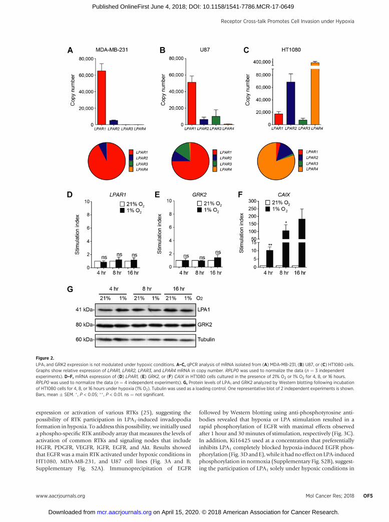
The differential use of LPA receptors under normoxic andhypoxic conditions led us to investigate the gene expressionprofile of LPA receptors in HT1080, MDA-MB-231, and U87 celllines. Results from qPCR experiments showed that bothMDA-MB-231 and U87 cells expressed predominantly mRNA forthe LPA receptor 1, which is consistent with the use of thisreceptor for invadopodia production under both normoxic andhypoxic conditions (Fig. 2A and B). In contrast, and as previouslydescribed (22), HT1080 cells expressed mainly LPA4, whichcorrelates with its role in invadopodia formation in normoxia.Of interest, these cells displayed low levels of LPA1, despite themajor use of this receptor in hypoxia (Fig. 2C). To furtherinvestigate this issue, we examined whether modulation of LPA1
expressionor activity could explain themainusage of this receptorin hypoxic HT1080 cells. Analysis of both mRNA and proteinlevels showed no significant increases in LPA1 expression underhypoxic conditions, despite a significant increase inmRNAexpres-sion of CAIX, a well-recognized hypoxia-regulated gene (Fig. 2D,F, and G). Because the G-protein–regulatory kinase GRK2 hasbeen shown to be decreased in hypoxic brain cells resulting inLPA1 overactivation (23), we next examined whether GRK2 levelswere modulated in hypoxic HT1080 cells. No significant changesin either mRNA or protein expression of GRK2 were observed in
Receptor Cross-talk Promotes Cell Invasion under Hypoxia
www.aacrjournals.org Mol Cancer Res; 2018 OF3
on April 15, 2020. © 2018 American Association for Cancer Research. mcr.aacrjournals.org Downloaded from
Published OnlineFirst June 4, 2018; DOI: 10.1158/1541-7786.MCR-17-0649

HT-1080 cells subjected to hypoxic conditions for up to 16 hours(Fig. 2E and G). Taken together, these results indicated thatmodulation of LPA1 or GRK2 expression did not account forLPA1 usage in hypoxic HT-1080 cells.
LPA1 usage in hypoxic cells involves cross-talk with EGFRGPCRs and LPARs are known to cross-communicate with RTKs
to mediate additional signaling events and growth factor–likeeffects (24). Furthermore, hypoxia has been shown to increase the
Figure 1.
LPA1 is essential for hypoxia-induced cancer cell invasion. A–C, MDA-MB-231, U87, or HT1080 cells were cultured on fluorescently labeled gelatin for 10 hoursor 16 hours, under normoxia (21% O2) or hypoxia (1% O2) in the presence/absence of LPA (10 mmol/L). The percentages of cells forming ECM-degrading invadopodiain the case of (A) cells stably transfected with shRNA against LPA4, (B) cells treated with the LPA receptor inhibitor Ki16425, or (C) cells stably transfected withshRNA against LPA1 are shown (n � 3 independent experiments). D, Representative immunofluorescence images of matrix degradation by scramble or LPA1
shRNA-transfected HT1080 cells cultured for 10 hours on fluorescently labeled gelatin in an atmosphere of 21%O2, 21% O2 in the presence of LPA, or 1% O2 are shown(magnification, �40). F-actin staining (red) and Oregon Green488-conjugated gelatin (green). Bars, mean � SEM. � , P < 0.05; �� , P < 0.01; ��� , P < 0.001.
Harper et al.
Mol Cancer Res; 2018 Molecular Cancer ResearchOF4
on April 15, 2020. © 2018 American Association for Cancer Research. mcr.aacrjournals.org Downloaded from
Published OnlineFirst June 4, 2018; DOI: 10.1158/1541-7786.MCR-17-0649
expression or activation of various RTKs (25), suggesting thepossibility of RTK participation in LPA1-induced invadopodiaformation in hypoxia. To address this possibility, we initially useda phospho-specific RTK antibody array that measures the levels ofactivation of common RTKs and signaling nodes that includeHGFR, PDGFR, VEGFR, IGFR, EGFR, and Akt. Results showedthat EGFR was a main RTK activated under hypoxic conditions inHT1080, MDA-MB-231, and U87 cell lines (Fig. 3A and B;Supplementary Fig. S2A). Immunoprecipitation of EGFR
followed by Western blotting using anti-phosphotyrosine anti-bodies revealed that hypoxia or LPA stimulation resulted in arapid phosphorylation of EGFR with maximal effects observedafter 1 hour and 30minutes of stimulation, respectively (Fig. 3C).In addition, Ki16425 used at a concentration that preferentiallyinhibits LPA1 completely blocked hypoxia-induced EGFR phos-phorylation (Fig. 3DandE),while it hadno effect on LPA-inducedphosphorylation in normoxia (Supplementary Fig. S2B), suggest-ing the participation of LPA1 solely under hypoxic conditions in
Figure 2.
LPA1 and GRK2 expression is not modulated under hypoxic conditions. A–C, qPCR analysis of mRNA isolated from (A) MDA-MB-231, (B) U87, or (C) HT1080 cells.Graphs show relative expression of LPAR1, LPAR2, LPAR3, and LPAR4 mRNA in copy number. RPLP0 was used to normalize the data (n ¼ 3 independentexperiments). D–F, mRNA expression of (D) LPAR1, (E) GRK2, or (F) CAIX in HT1080 cells cultured in the presence of 21% O2 or 1% O2 for 4, 8, or 16 hours.RPLP0 was used to normalize the data (n ¼ 4 independent experiments). G, Protein levels of LPA1 and GRK2 analyzed by Western blotting following incubationof HT1080 cells for 4, 8, or 16 hours under hypoxia (1% O2). Tubulin was used as a loading control. One representative blot of 2 independent experiments is shown.Bars, mean � SEM. � , P < 0.05; �� , P < 0.01. ns ¼ not significant.
Receptor Cross-talk Promotes Cell Invasion under Hypoxia
www.aacrjournals.org Mol Cancer Res; 2018 OF5
on April 15, 2020. © 2018 American Association for Cancer Research. mcr.aacrjournals.org Downloaded from
Published OnlineFirst June 4, 2018; DOI: 10.1158/1541-7786.MCR-17-0649

HT1080 cells. Similar inhibition of hypoxia-induced EGFR phos-phorylation by Ki16425 was observed in MDA-MB-231 and U87cell lines (Supplementary Fig. S2C and S2D), indicating thatLPA1–EGFR cross-talk in hypoxic cells is not associated with aparticular cell type.
The involvement of EGFR in hypoxia-induced invadopodiaproduction downstream of LPA1 was next investigated. EGFRknockdown in HT1080 cells was found to significantly block theincrease in the percentage of cells forming invadopodia underhypoxic conditions, whereas this procedure did not affect inva-dopodia induced by LPA under normoxic conditions (Fig. 4A;Supplementary Fig. S3). Similar findings were observed inMDA-MB-231 and U87 cells (Fig. 4A). In contrast, in cells whereLPA1 had been knocked down by shRNA treatment, inhibition ofEGFR had no effect on invadopodia production whether cellswere grown under normoxic or hypoxic conditions or werestimulated with LPA (Fig. 4B). Overall, these results suggestedthat EGFR was preferentially involved in the ability of cells toproduce invadopodia under hypoxic conditions, and that thisevent occurred downstream of LPA1.
Src is a mediator of LPA1–EGFR cross-talkGPCR-mediated transactivation of RTKs can involve different
mechanisms that include increased ligand availability, throughprotease-mediated shedding of the ligands, or direct phosphor-ylation of the RTK by a downstream kinase (26). To address thefirst possibility, we investigated whether hypoxic HT1080 cellsshowed increased release of the major EGFR ligands involved inGPCR-RTK cross-talk, EGF, HB-EGF, and TGF-alpha. Results fromELISA assays showed the absence of increases in the release ofEGF, HB-EGF, or TGF-alpha under hypoxic conditions (Fig. 5A).
Furthermore, shRNA-mediated knockdown of ADAM17 orADAM12, two major proteases involved in the shedding of EGFRligands, did not affect hypoxia-induced EGFR phosphorylation(Fig. 5B–D). These results indicate that increased ligand avail-ability is unlikely to be the mechanism responsible for the rapidincrease in EGFR phosphorylation under hypoxia.
Because one of the main kinases known to be involved inphosphorylation-induced transactivation of EGFR is Src (26), wenext examined the contribution of this kinase to the hypoxia-induced LPA1–phosphoEGFR axis. Western blot results showed aclear increase in Src phosphorylation after 45minutes of exposureto hypoxia and that upregulated Src phosphorylationwas reducedto basal levels following inhibition of LPA1 (Fig. 5F and G).Similarly, shRNA-mediated knockdown of Src or inhibition ofits activity by PP2 strongly repressed EGFR phosphorylationinduced by hypoxia (Fig. 5B and E; Supplementary Fig. S4). Thesefindings clearly indicate that Src-mediated phosphorylation ofEGFR is the likely mechanism of EGFR transactivation by LPA1 inhypoxic cells.
To gain insight into the significance of the Src-mediated EGFRtransactivation mechanism in the ability of cells to formECM-degrading invadopodia under hypoxic conditions, SrcshRNA-transduced cells were tested in invadopodia assays. Srcknockdown cells failed to affect the increase in the percentage ofinvadopodia forming cells induced by LPA under normoxicconditions, which is consistent with our previous results that hadidentified an alternativemechanism downstream of LPA4 (11). Incontrast, Src knockdown resulted in inhibition in the percentageof hypoxia-induced invadopodia producing cells, in agreementwith its involvement in EGFR transactivation. As a control, knock-down of ADAM17 or ADAM12 did not affect invadopodia
Figure 3.
EGFR is transactivated by LPA1 under hypoxic conditions. A and B, HT1080 cells were incubated under 21% O2 or 1% O2 for 30 or 60 minutes, and cell lysateswere subjected to phospho-RTK array analysis. A, One representative array, showing phospho-EGFR and phospho-Akt, of two independent experiments is shown.B, The associated graph shows the mean phosphoprotein fluorescence intensities for each cell lysate from two independent experiments. C–E, HT1080 celllysates were immunoprecipitated using an anti-EGFR antibody and immunoblotted with anti-phospho-tyrosine (pY) and total EGFR antibodies. C, Onerepresentativeblot that showsa time course of LPA (10mmol/L) or hypoxic (1%O2) stimulations.D,One representativeblot showinghypoxic stimulation (30minutes)in the presence/absence of Ki16425 (1 mmol/L). E, Corresponding graph showing densitometric analysis of p-Y/EGFR ratios of 3 independent experiments.Bars, mean � SEM (� , P < 0.05). � , P < 0.05.
Harper et al.
Mol Cancer Res; 2018 Molecular Cancer ResearchOF6
on April 15, 2020. © 2018 American Association for Cancer Research. mcr.aacrjournals.org Downloaded from
Published OnlineFirst June 4, 2018; DOI: 10.1158/1541-7786.MCR-17-0649

production under hypoxia (Fig. 5H), consistent with theirobserved lack of involvement in EGFR phosphorylation (Fig. 5B).
LPA1–EGFR axis signals through PI3K/Akt to promote hypoxia-induced invadopodia production
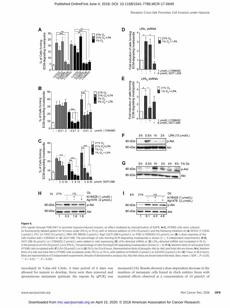
We next investigated the involvement of the main LPA1 andEGFR downstream signaling pathways in hypoxia-induced inva-dopodia production using pharmacologic inhibitors of ROCK(Y-27632), PLC (U-73122), MEK (PD 98059), or PI3K(LY294002). Inhibition of Rap1 (GGTI-298), which has previ-ously been reported to be downstream of LPA4 for invadopodiaproduction (11), was used as a control. Among the pathwaystested, only inhibition of MEK or Rap1 signaling significantlydecreased the percentage of cells forming invadopodia induced byLPA in normoxia or induced by hypoxia. In contrast, PI3Kinhibition exclusively decreased invadopodia productioninduced by hypoxia (Fig. 6A). Increasing the concentration ofthe PI3K inhibitor did not affect LPA-stimulated invadopodia-forming cells in normoxia, but efficiently blocked the stimulatoryeffect of hypoxia alone or in the presence of LPA (Fig. 6B). Asexpected, Rap1 inhibition reduced the percentage of cells forminginvadopodia under both normoxic and hypoxic conditions(Fig. 6C). Given that cells mostly used LPA1 in hypoxia(Fig. 1C), these results suggest that HT-1080 cells use PI3Ksignaling downstream of the LPA1–EGFR axis for invadopodiaproduction only under hypoxic conditions. To support this
interpretation, we compared the effect of PI3K inhibition in LPA1-or LPA4-knockdownHT1080 cells. Results showed that inhibitionof the PI3K pathway in hypoxic LPA1-knockdown cells did notaffect invadopodia production, whereas Rap1 inhibition resultedin a strong inhibitory effect (Fig. 6D). Conversely, in LPA4-knockdown cells, PI3K inhibition decreased invadopodia pro-duction,whereas Rap1hadnoeffect (Fig. 6E),which is in linewithour previously published results (11). These findings clearlyindicate that invadopodia production in hypoxic cells requiresPI3K signaling downstream of LPA1.
Akt, a major effector downstream of PI3K, has been reportedto be implicated in invadopodia generation as well as inproduction of several matrix metalloproteases important fortheir ability to degrade the extracellular matrix (27, 28). Toconfirm activation of the PI3K/Akt pathway under hypoxicconditions, Akt phosphorylation was assessed by Western blot-ting. Results showed that Akt was phosphorylated in responseto LPA after 30 minutes, whereas a sustained phosphorylationfrom 1 hour up to 6 hours was observed following hypoxicstimulation (Fig. 6F and G). Furthermore, addition of the LPA1
inhibitor Ki16425 or the EGFR inhibitor AG1478 resulted in areduction of Akt phosphorylation induced by 1 hour or 6 hoursof hypoxic stimulation (Fig. 6H and I). These results suggestthat PI3K/Akt activation under hypoxia is mediated by LPA1-induced transactivation of EGFR, an event that promotes inva-dopodia production.
Figure 4.
EGFR is necessary for hypoxia-induced invadopodia formation downstream of LPA1. A and B, Cells were cultured on fluorescently labeled gelatin for 10 hours,and the percentages of cells forming ECM-degrading invadopodia are shown. A, HT-1080, MDA-MB-231, or U87 cells transduced with scramble or EGFR-targetingshRNA were incubated in 21% O2 in the presence/absence of LPA (10 mmol/L) or in 1% O2. B, Cells transfected with scramble or LPA1-targeting shRNA wereincubated in 21% O2, 1% O2, or 1% O2 in the presence of LPA (10 mmol/L) with or without the addition of the EGFR inhibitor AG1478. Bars, mean � SEM. � , P < 0.05;�� , P < 0.01; ��� , P < 0.001.
Receptor Cross-talk Promotes Cell Invasion under Hypoxia
www.aacrjournals.org Mol Cancer Res; 2018 OF7
on April 15, 2020. © 2018 American Association for Cancer Research. mcr.aacrjournals.org Downloaded from
Published OnlineFirst June 4, 2018; DOI: 10.1158/1541-7786.MCR-17-0649

A combined inhibition of LPA1 and EGFR reduces metastasis inan in vivo model
To assess whether our observations that hypoxia promoted cellinvasion through cross-talk between LPA1 andEGFRwere relevantto cancer metastasis therapy in vivo, we used an ex ovo CAMxenograft model in chicken embryos (29). Because HT1080tumors grown in the CAM assay had been previously shown todevelop hypoxia (17), the CAM model allows us to evaluate theeffects of LPAR and EGFR receptor inhibition on spontaneousmetastasis arising from tumors containing hypoxic areas. First,CAM bearing HT1080 tumors (grown for 6 days) were injected
with pimonidazole. After 30 minutes, the tumors were excisedand we determined the presence of phospho-Y845-EGFR withinhypoxic zones of the tumors. Results showed a significant increasein phospho-Y845-EGFR staining within pimonidazole-positiveregions of the tumors (Fig. 7A). This finding was consistent withincreased EGFR phosphorylation that was observed in hypoxiccells (Fig. 3A–C). Next, we analyzed the effect of targeting LPA1
alone or in combination with EGFR inhibition using the EGFRinhibitor (AG1478) and/or AM095, a potent and selective LPA1
inhibitor used for in vivo studies (30). HT1080 cells, in thepresence of increasing concentrations of AM095 or AG1478, were
Figure 5.
Mechanism of transactivation of EGFR by LPA1 under hypoxia. A, Detection of HB-EGF, TGFa, and EGF by ELISA was performed on concentrated cellsupernatants from HT1080 cells incubated for 8 hours in the presence of 21% O2 or 1% O2. Results are presented in pg/mL (n¼ 3). ND¼ nondetectable. B,Westernblot of phospho-Y845 EGFRand total EGFR inHT1080cells transducedwith scramble, ADAM17, ADAM12, or Src-targeted shRNAand incubated under 21%O2or 1%O2
for 30 minutes. Densitometric analysis of phospho-Y845/EGFR ratio is shown below the blots. One representative blot of 3 independent experiments is shown.C–E, Quantification of (C) ADAM17. D, ADAM12 and (E) SRC mRNA levels by qPCR using RPLP0 as a reference gene (n ¼ 3). F, HT1080 cell lysates were analyzedby Western blotting using anti-phospho-Src and Src antibodies. Densitometric analysis of phospho-Src/Src ratio is shown below the blots. One representativeblot of a time course of hypoxic (1% O2) stimulations is shown. G, HT1080 cells were incubated for 45 minutes in 1% O2 with or without Ki16425 (1 mmol/L).One representative blot is shown. H, HT1080 cells transfected with scramble, ADAM17, ADAM12, or SRC shRNA were cultured on fluorescently labeled gelatinfor 10 hours in 21% O2 or 1% O2 in the presence or absence of LPA (10 mmol/L). The percentage of cells forming ECM-degrading invadopodia is shown (n ¼ 3).Bars, mean � SEM. � , P < 0.05; �� , P < 0.01; ��� , P < 0.001.
Harper et al.
Mol Cancer Res; 2018 Molecular Cancer ResearchOF8
on April 15, 2020. © 2018 American Association for Cancer Research. mcr.aacrjournals.org Downloaded from
Published OnlineFirst June 4, 2018; DOI: 10.1158/1541-7786.MCR-17-0649
inoculated in 9-day-old CAMs. A time period of 6 days wasallowed for tumors to develop, livers were then removed andspontaneous metastasis (primate Alu repeats by qPCR) was
measured (18). Results showed a dose-dependent decrease in thenumbers of metastatic cells found in chick embryo livers withmaximal effects observed at a concentration of 10 mmol/L of
Figure 6.
LPA1 signals through PI3K/AKT to promote hypoxia-induced invasion, an effect mediated by transactivation of EGFR. A–C, HT1080 cells were culturedon fluorescently labeled gelatin for 10 hours under 21% O2 or 1% O2 with or without addition of LPA (10 mmol/L) and the following inhibitors of (A) ROCK (Y-276322 mmol/L), PLC (U-73122 0.5 mmol/L), MEK (PD 98059 2 mmol/L), Rap1 (GGTI-298 6 mmol/L), or PI3K (LY294002 0.5 mmol/L) or (B) a dose response of thecells treated with LY294002 or (C) GGTI-298. The percentage of cells forming ECM-degrading invadopodia is shown (n � 3 independent experiments). D–E,GGTI-298 (6 mmol/L) or LY294002 (1 mmol/L) were added to cells expressing (D) LPA1-directed shRNA or (E) LPA4-directed shRNA and incubated in 1% O2
in the presence of LPA (10mmol/L) or in 21%O2. The percentage of cells forming ECM-degrading invadopodia is shown (n¼4). F–G,Western blots of cell lysates fromHT1080 cells incubated with (F) LPA (10 mmol/L) or in (G) 1%O2 for 0 to 6 hours. Representative blots of phospho-Akt (p-Akt) and total Akt are shown.H–I,Westernblots of p-Akt and total Akt in HT1080 cells incubated under 21% O2 or 1% O2 with addition of Ki16425 (1 mmol/L) or AG1478 (2 mmol/L) for (H) 1 hour or (I) 6 hours.Blots are representative of 3 independent experiments. Results of densitometric analysis of p-Akt/Akt ratios are shownbelow the blots. Bars,mean�SEM. � ,P<0.05;�� , P < 0.01; ��� , P < 0.001.
Receptor Cross-talk Promotes Cell Invasion under Hypoxia
www.aacrjournals.org Mol Cancer Res; 2018 OF9
on April 15, 2020. © 2018 American Association for Cancer Research. mcr.aacrjournals.org Downloaded from
Published OnlineFirst June 4, 2018; DOI: 10.1158/1541-7786.MCR-17-0649

AM095 and 10 mmol/L of AG1478 (Fig. 7B and C). To definewhether these inhibitors can provide therapeutic gain whencombined, tumors were treated with suboptimal concentrationsof AM095 (0.2 mmol/L) and AG1478 (0.2 mmol/L). A synergisticeffect on metastasis was observed with a combined inhibitionof EGFR and LPA1 (Fig. 7D). We next analyzed the levels ofphospho-EGFR in hypoxic zones of tumors treatedwith the aboveinhibitors. Maximum EGFR inhibition drastically decreased
EGFR phosphorylation, as expected. Similarly, the maximumdose of AM095 decreased p-EGFR, confirming its role insuppressing hypoxia-induced EGFR phosphorylation as observedin in vitro assays (Fig. 7E). Finally, suboptimal concentrations ofboth inhibitors induced a strong reduction of EGFR phosphor-ylate in hypoxic zones of tumors, similar to the diminutionobserved with the highest concentration of EGFR inhibitor(Fig. 7F and G).
Figure 7.
LPA1 and EGFR are implicated inspontaneous metastasis in a CAMxenograft assay. A, Representativeimmunohistochemistry images ofHT1080 xenograft tumors showinghypoxic regions [pimonidazole(pimo); green] and phospho-EGFR(red) staining. Nuclei werestained with DAPI (blue). Scale bars,100 mm. The associated graphshows phospho-EGFR relativelabeling intensities in pimoþ andpimo� zones of tumors. B–G, HT1080cells (3 � 105) were inoculated inthe presence/absence of the LPA1
inhibitor AM095 or the EGFR inhibitorAG1478, to the CAM of chick embryos.Embryonic livers were harvested after6 days of tumor growth for genomicDNA extraction, and tumors wereprocessed for immunohistochemistry.The ability of HT1080 cells todisseminate in embryonic livers wasquantitated as the relative amount ofmetastasis normalized to the amountof host DNA. Dose response to(B) AG1478, (C) AM095, and (D) acombined treatment with AM095(0.2mmol/L) andAg1478 (0.2mmol/L).E and F, Relative labeling intensitiesof phospho-EGFR in hypoxic(pimoþ) zones of tumors treatedwith (E) AM095 (10 mmol/L) orAG1478 (10 mmol/L) or (F) acombined treatment with AM095(0.2 mmol/L) and Ag1478(0.2 mmol/L). G, Representativeimmunohistochemistry images ofHT1080 xenograft tumors showingphospho-EGFR staining in hypoxic(pimoþ) zone of tumors treated withAM095 (0.2 mmol/L) and Ag1478(0.2 mmol/L). Hypoxic regions(pimo, green) and phospho-EGFR(red) staining.Nucleiwere stainedwithDAPI (blue). Scale bars, 50 mm.Bars, mean � SEM. � , P < 0.05;�� , P < 0.01; ��� , P < 0.001.
Harper et al.
Mol Cancer Res; 2018 Molecular Cancer ResearchOF10
on April 15, 2020. © 2018 American Association for Cancer Research. mcr.aacrjournals.org Downloaded from
Published OnlineFirst June 4, 2018; DOI: 10.1158/1541-7786.MCR-17-0649

DiscussionIn this study, we uncovered an essential role for LPA1 in
hypoxia-induced invadopodia production and that this eventwas dependent on Src-mediated cross-communication withEGFR. Furthermore, we found that hypoxic cells use PI3K/AKTsignaling downstream of the LPA1–EGFR axis as a dominantinvadopodia-inducing signaling node. Tumor xenograft experi-ments further support the requirement of LPA1 and EGFR forin vivo metastasis and suggest that targeting both receptors couldbe an effective strategy to improve metastasis.
Hypoxia, a prevalent feature of the tumormicroenvironment, isa potent inducer of cancer cell aggressiveness, invasion, andmetastasis. However, the nature of the mechanisms involved inthese processes remains to be fully understood (3). Althoughthere have been a few clues that hypoxia and LPA signaling areintertwined (23, 31, 32), the observations reported here are, to thebest of our knowledge, the first study that identifies LPA1 as themain receptor involved in invadopodia production triggered bythe hypoxicmicroenvironment. The ability to establish the role ofa particular LPAR is of importance because these receptors canhave redundant or opposing effects on cell motility and cellinvasion. These cellular properties are cell type– and context-dependent, suggesting that the therapeutic use of pan-LPARantagonists ought to be considered with caution. For example,both LPA1 and LPA3 can be positive or negative regulators of cellmotility and invasion, depending on the cell type involved (19).LPA4 has also been shown to inhibit cell motility and invasion inmouse embryonic fibroblasts while promoting invasion andmetastasis in human fibrosarcoma cells (11, 33). Our findingsof the common use of LPA1 in a predominant tumor microen-vironment highlight the potential benefit of LPA1-directed ther-apy to fight cancer metastasis and progression.
A discrepancy between LPA receptor gene expression levelsand usage led us to investigate the influence of hypoxia on LPA1
expression and activity. Long-term exposure to hypoxia hasbeen shown to increase LPA1 expression in retinal ganglioncells (23) and to induce overactivation of LPA1 in fetal brainthrough downregulation of the G-protein regulatory kinaseGRK2 (34). To our surprise, exposure of the cells to hypoxiafor an extended period of time (16 hours) did not modulatemRNA or protein expression of LPA1 or GRK2. These findingsprompted us to explore alternatives for the common use ofLPA1 under hypoxia. We found that the RTK, EGFR, wastransactivated by LPA1, leading to invadopodia productionsolely under hypoxia (as compared with normoxia), suggestingthat receptor cross-talk is a mechanism by which LPA1 pro-motes invadopodia in this condition.
LPA1-mediated transactivation of EGFR is in concordance withprevious studies that identified both ligand-dependent andligand-independent mechanisms of LPA-induced transactivation,depending on cell type and physiologic context (35). Underhypoxic conditions, we found a ligand-independent mechanismof EGFR transactivation that involved Src kinase-mediatedphosphorylation of tyrosine-845 (Y845) on EGFR. Phosphoryla-tion of this tyrosine residue has been associated with survival,proliferation, and malignancy of various cancer cells, includingglioma and breast cancer cells, as well as being a highly predictivevalue for prognosis correlating with worse prognosis andpoor response to chemotherapy (36). In addition, colocalizationof p-EGFR and hypoxic markers in human tumor biopsies wasassociated with a critical subpopulation prone to local recurrence,
metastasis formation or treatment resistance (37). The findingthat Y845-EGFR is phosphorylated in response to LPA1 activationunder hypoxia in vitro and localized in hypoxic zones of tumorssuggests the possibility that this could be used, in conjuncturewith hypoxic markers, as predictive tools of the clinical outcomeof LPA1- and EGFR-directed therapy.
We also uncovered PI3K/Akt signaling as an effector of LPA1
and EGFR cross-talk that leads to invadopodia production inhypoxia. PI3K acts through Akt and has previously been impli-cated in the regulation of invadopodia formation, as well as theirability to degrade the extracellular matrix (27). Although ourfindings showed that PI3K/Akt signaling was only involved inhypoxia-induced invadopodia production, we also observedphosphorylation of Akt in response to LPA under normoxicconditions. This apparent discrepancy may be partly explainedby the fact that Akt phosphorylation in normoxic cells wastransient with a peak effect reached after 30 minutes of LPAstimulation. In contrast, we observed a sustained activation (upto 6 hours) in response to hypoxia. Although few publicationshave directly compared the effects of acute versus sustainedAkt phosphorylation, Goel and colleagues found differentialeffects on G1 progression, suggesting distinct consequences onbiological responses (38). Furthermore, sustained Akt phosphor-ylation has been associated with enhanced tumor survival as wellas increased cytoskeletal rearrangement and migration (39, 40),suggesting that under hypoxic conditions, this event also plays arole in invadopodia formation, as these structures require actinremodeling (5).
The effect of hypoxia on Src-mediated EGFR–LPA1 cross-talkand sustained Akt phosphorylation may be due to relocaliza-tion of both receptors and Src in specific subcellular domainssuch as lipid rafts, thus facilitating their interactions. Hypoxiahas been reported to rapidly increase cellular cholesterol levels,thereby influencing the composition, number, and size of lipidrafts (41). Lipid rafts function as organizing centers for theassembly of signaling molecules; their increased rigidity opti-mizes spatiotemporal interactions and compartmentalizes pro-teins involved in specific signaling tasks (42, 43). Interestingly,both RTKs and GPCRs can localize to lipid rafts, which providea platform that facilitates interactions between EGFR andGPCRs, therefore enhancing the efficiency of transactivation(44). Lipid rafts have also been identified as key plasmamembrane microdomains for Src-EGFR functional interactionsas well as PI3K/Akt activation (42, 45). It is therefore conceiv-able that, under hypoxic conditions, the recruitment of LPA1,Src, and EGFR in lipid rafts facilitates their interactions and thatresults in prolonged Akt activation and invadopodia formation.This possibility is in keeping with the finding that lipid raftsand caveolin-1 are required for invadopodia production inbreast cancer cells (46).
A combination of suboptimal doses of inhibitors revealed asynergistic effect between LPA1 and EGFR inhibitors in blockingmetastasis. Combination therapy using lower doses of inhibitorsare of clear clinical interest in order to overcome toxicity andresistance, two major issues associated with the use of RTK-targeted therapies (47). It has been suggested that resistance toRTK inhibitors is linked to activation or cross-talk with receptorsthat could include IGF-1R, c-MET, and LPARs (EDGR; ref. 48).More specifically, blocking LPA1/3 receptors with Ki16425 hasbeen shown to prevent and delay resistance to the RTK inhibitorsunitinib in vivo (49), whereas inhibiting Src or Y845EGFR
Receptor Cross-talk Promotes Cell Invasion under Hypoxia
www.aacrjournals.org Mol Cancer Res; 2018 OF11
on April 15, 2020. © 2018 American Association for Cancer Research. mcr.aacrjournals.org Downloaded from
Published OnlineFirst June 4, 2018; DOI: 10.1158/1541-7786.MCR-17-0649

phosphorylation reverses resistance to anti-EGFR monoclonalantibodies (50). These results suggest that blocking LPA1-inducedtransactivation of EGFR could also overcome resistance to EGFRinhibitors. In addition, EGF has been shown to increase LPAproduction, suggesting there can be bidirectional cross-talkbetween LPA and EGF receptors (51). Preclinical studies thatcombine the therapeutic potential of LPA1 and EGFR invarious types of solid tumors should therefore be a valuableresearch direction.
In conclusion, our study provides insight into a mechanismof hypoxia-induced cell invasion, which implies a major rolefor LPA1 in activating two major players in tumorigenesis: Srcand EGFR. Targeting LPA1, alone or in combination with EGFRinhibitors, could benefit cancer patients by blocking upstreaminducers of invadopodia, a key component of the metastaticprocess. Because metastasis remains the most deadly aspectof cancer, efficient interference with this process is of theutmost importance to increase cancer patients' well-beingand survival.
Disclosure of Potential Conflicts of InterestNo potential conflicts of interest were disclosed.
Authors' ContributionsConception and design: K. Harper, C.M. DuboisDevelopment of methodology: K. Harper, C.M. Dubois
Acquisition of data (provided animals, acquired and managed patients,provided facilities, etc.): K. Harper, R. R. Lavoie, M. Charbonneau,K. Brochu-Gaudreau, C.M. DuboisAnalysis and interpretation of data (e.g., statistical analysis, biostatistics,computational analysis): K. Harper, R. R. Lavoie, M. Charbonneau,C.M. DuboisWriting, review, and/or revision of the manuscript: K. Harper, K. Brochu-Gaudreau, C.M. DuboisAdministrative, technical, or material support (i.e., reporting or organizingdata, constructing databases): C.M. DuboisStudy supervision: C.M. Dubois
AcknowledgmentsThe authors thankDr.GillesDupuis for critical readingof themanuscript and
helpful comments. We also thank Dr. L�eonid Volkov for expert assistance withconfocal microscopy and Martine Charbonneau and Anna Yatsyna for assis-tance with the CAM assay.
This work was supported by the Canadian Institutes of Health Research(CIHR) grant MOP-126173 (to C.M. Dubois). C.M. Dubois is a member of theFonds de la Recherche en Sant�e du Qu�ebec-funded Centre de RechercheClinique du Centre Hospitalier Universitaire de Sherbrooke. K. Harper isrecipient of a student scholarship from CIHR.
The costs of publication of this articlewere defrayed inpart by the payment ofpage charges. This article must therefore be hereby marked advertisement inaccordance with 18 U.S.C. Section 1734 solely to indicate this fact.
Received November 8, 2017; revised April 3, 2018; accepted May 17, 2018;published first June 4, 2018.
References1. Hanahan D, Weinberg RA. Hallmarks of cancer: the next generation.
Cell 2011;144:646–74.2. Vaupel P, Harrison L. Tumor hypoxia: causative factors, compensatory
mechanisms, and cellular response. Oncologist 2004;9:4–9.3. Hockel M, Schlenger K, Aral B, Mitze M, Schaffer U, Vaupel P. Association
between tumor hypoxia and malignant progression in advanced cancer ofthe uterine cervix. Cancer Res 1996;56:4509–15.
4. Gould CM, Courtneidge SA. Regulation of invadopodia by the tumormicroenvironment. Cell Adh Migr 2014;8:226–35.
5. Paz H, Pathak N, Yang J. Invading one step at a time: the role of invado-podia in tumor metastasis. Oncogene 2014;33:4193–202.
6. Condeelis J, Segall JE. Intravital imaging of cell movement in tumours.Nat Rev Cancer 2003;3:921–30.
7. Yamaguchi H, Lorenz M, Kempiak S, Sarmiento C, Coniglio S, Symons M,et al. Molecular mechanisms of invadopodium formation: the role ofthe N-WASP-Arp2/3 complex pathway and cofilin. J Cell Biol 2005;168:441–52.
8. Leong HS, Robertson AE, Stoletov K, Leith SJ, Chin CA, Chien AE, et al.Invadopodia are required for cancer cell extravasation and are a therapeutictarget for metastasis. Cell Rep 2014;8:1558–70.
9. Eddy RJ, Weidmann MD, Sharma VP, Condeelis JS. Tumor cell invadopo-dia: invasive protrusions that orchestrate metastasis. Trends Cell Biol2017;27:595–607.
10. KiharaY,MizunoH,Chun J. Lysophospholipid receptors indrugdiscovery.Exp Cell Res 2015;333:171–7.
11. Harper K, Arsenault D, Boulay-Jean S, Lauzier A, Lucien F, Dubois CM.Autotaxin promotes cancer invasion via the lysophosphatidic acid receptor4: participation of the cyclic AMP/EPAC/Rac1 signaling pathway in inva-dopodia formation. Cancer Res 2010;70:4634–43.
12. Hwang YS, Lee J, Zhang X, Lindholm PF. Lysophosphatidic acid activatesthe RhoA and NF-kB through Akt/IkBa signaling and promotes prostatecancer invasion and progression by enhancing functional invadopodiaformation. Tumour Biol 2016;37:6775–85.
13. Kedziora KM, Leyton-Puig D, Argenzio E, Boumeester AJ, van Butselaar B,Yin T, et al. Rapid remodeling of invadosomes by Gi-coupled receptors:DISSECTING THE ROLE OF Rho GTPases. J Biol Chem 2016;291:4323–33.
14. Liu S, Umezu-Goto M, MurphM, Lu Y, Liu W, Zhang F, et al. Expression ofautotaxin and lysophosphatidic acid receptors increases mammary tumor-igenesis, invasion, and metastases. Cancer Cell 2009;15:539–50.
15. ZhangH, XuX,Gajewiak J, TsukaharaR, Fujiwara Y, Liu J, et al.Dual activitylysophosphatidic acid receptor pan-antagonist/autotaxin inhibitor reducesbreast cancer cell migration in vitro and causes tumor regression in vivo.Cancer Res 2009;69:5441–9.
16. Li X, Wang X. Application of real-time polymerase chain reaction for thequantification of interleukin-1beta mRNA upregulaiton in brain ischemictolerance. Brain Res Brain Res Protoc 2000;5:211–7.
17. Lucien F, Pelletier PP, Lavoie RR, Lacroix JM, Roy S, Parent JL, et al.Hypoxia-induced mobilization of NHE6 to the plasmamembrane triggersendosome hyperacidification and chemoresistance. Nat Commun 2017;8:15884.
18. Zijlstra A, Mellor R, Panzarella G, Aimes RT, Hooper JD, Marchenko ND,et al. A quantitative analysis of rate-limiting steps in the metastatic cascadeusing human-specific real-time polymerase chain reaction. Cancer Res2002;62:7083–92.
19. Tsujiuchi T, Hirane M, Dong Y, Fukushima N. Diverse effects of LPAreceptors on cell motile activities of cancer cells. J Recept Signal TransductRes 2014;34:149–53.
20. Lappano R, Rigiracciolo D, De Marco P, Avino S, Cappello AR, Rosano C,et al. Recent advances on the role of G protein-coupled receptors inhypoxia-mediated signaling. AAPS J 2016;18:305–10.
21. Ohta H, Sato K, Murata N, Damirin A, Malchinkhuu E, Kon J, et al.Ki16425, a subtype-selective antagonist for EDG-family lysophosphatidicacid receptors. Mol Pharmacol 2003;64:994–1005.
22. Kishi Y, Okudaira S, Tanaka M, Hama K, Shida D, Kitayama J, et al.Autotaxin is overexpressed in glioblastoma multiforme and contributesto cell motility of glioblastoma by converting lysophosphatidylcholine tolysophosphatidic acid. J Biol Chem 2006;281:17492–500.
23. Herr KJ, Herr DR, Lee CW, Noguchi K, Chun J. Stereotyped fetal braindisorganization is induced by hypoxia and requires lysophosphatidic acidreceptor 1 (LPA1) signaling. Proc Natl Acad Sci USA 2011;108:15444–9.
24. Wang Z.Transactivation of epidermal growth factor receptor by G protein-coupled receptors: recent progress, challenges and future research. Int JMolSci 2016;17.
Harper et al.
Mol Cancer Res; 2018 Molecular Cancer ResearchOF12
on April 15, 2020. © 2018 American Association for Cancer Research. mcr.aacrjournals.org Downloaded from
Published OnlineFirst June 4, 2018; DOI: 10.1158/1541-7786.MCR-17-0649
25. Gl€uck AA, Aebersold DM, Zimmer Y, Medov�a M. Interplay betweenreceptor tyrosine kinases and hypoxia signaling in cancer. Int J BiochemCell Biol 2015;62:101–14.
26. Cattaneo F, Guerra G, Parisi M, De Marinis M, Tafuri D, Cinelli M, et al.Cell-surface receptors transactivationmediated by gprotein-coupled recep-tors. Int J Mol Sci 2014;15:19700–28.
27. Yamaguchi H, Yoshida S, Muroi E, Yoshida N, Kawamura M, Kouchi Z,et al. Phosphoinositide 3-kinase signaling pathway mediated by p110aregulates invadopodia formation. J Cell Biol 2011;193:1275–88.
28. Cho SJ, Chae MJ, Shin BK, Kim HK, Kim A. Akt- and MAPK-mediatedactivation and secretion of MMP-9 into stroma in breast cancer cells uponheregulin treatment. Mol Med Rep 2008;1:83–8.
29. Ribatti D. The chick embryo chorioallantoic membrane as a model fortumor biology. Exp Cell Res 2014;328:314–24.
30. Swaney JS, Chapman C, Correa LD, Stebbins KJ, Broadhead AR, Bain G,et al. Pharmacokinetic and pharmacodynamic characterization of an orallysophatidic acid type1 receptor-selective antagonist. J Pharmacol ExpTher2011;336:693–700.
31. Kostic I, Fidalgo-Carvalho I, Aday S, Vaz~aoH, Carvalheiro T, Gr~aosM, et al.Lysophosphatidic acid enhances survival of human CD34(þ) cells inischemic conditions. Sci Rep 2015;5:16406.
32. Pan H, Cheng L, Yang H, Zou W, Cheng R, Hu T. Lysophosphatidic acidrescueshumandental pulp cells from ischemia-induced apoptosis. J Endod2014;40:217–22.
33. Lee Z, Cheng CT, Zhang H, Subler MA, Wu J, Mukherjee A, et al. Role ofLPA4/p2y9/GPR23 in negative regulation of cell motility. Mol Biol Cell2008;19:5435–45.
34. Yang C, Lafleur J, Mwaikambo BR, Zhu T, Gagnon C, Chemtob S, et al. Therole of lysophosphatidic acid receptor (LPA1) in the oxygen-inducedretinal ganglion cell degeneration. Invest Ophthalmol Vis Sci 2009;50:1290–8.
35. Bhola NE, Grandis JR. Crosstalk between G-protein-coupled receptorsand epidermal growth factor receptor in cancer. Front Biosci 2008;13:1857–65.
36. Sato KI. Cellular functions regulated by phosphorylation of EGFR onTyr845. Int J Mol Sci 2013;14:10761–90.
37. Hoogsteen IJ, Marres HA, van den Hoogen FJ, Rijken PF, Lok J, Bussink J,et al. Expression of EGFR under tumor hypoxia: identification of a sub-population of tumor cells responsible for aggressiveness and treatmentresistance. Int J Radiat Oncol Biol Phys 2012;84:807–14.
38. Goel R, Phillips-Mason PJ, Raben DM, Baldassare JJ. alpha-Thrombininduces rapid and sustained Akt phosphorylation by beta-arrestin1-dependent and -independent mechanisms, and only the sustained Akt
phosphorylation is essential for G1 phase progression. J Biol Chem2002;277:18640–8.
39. Takahashi Y, Morales FC, Kreimann EL, Georgescu MM. PTEN tumorsuppressor associates with NHERF proteins to attenuate PDGF receptorsignaling. EMBO J 2006;25:910–20.
40. Schmid C, Ghirlanda C, Niessen M. Prevention of tumour cell apoptosisassociated with sustained protein kinase B phosphorylation is moresensitive to regulation by insulin signalling than stimulation of prolifer-ation and extracellular signal-regulated kinase. Mol Cell Biochem 2017;432:41–54.
41. Danza G, Di Serio C, Ambrosio MR, Sturli N, Lonetto G, Rosati F, et al.Notch3 is activated by chronic hypoxia and contributes to the progressionof human prostate cancer. Int J Cancer 2013;133:2577–86.
42. Liu Y, Lv JY, Shi JF, Yang M, Liu SH, Li ZW, et al. Targeting the raft-associated Akt signaling in hepatocellular carcinoma. Biomed Res Int2014;2014:836025.
43. Simons K, Toomre D. Lipid rafts and signal transduction. Nat RevMol CellBiol 2000;1:31–9.
44. Jensen DD, Godfrey CB, Niklas C, Canals M, Kocan M, Poole DP, et al.The bile acid receptor TGR5 does not interact with b-arrestins or traffic toendosomes but transmits sustained signals from plasma membrane rafts.J Biol Chem 2013;288:22942–60.
45. Irwin ME, Bohin N, Boerner JL. Src family kinases mediate epidermalgrowth factor receptor signaling from lipid rafts in breast cancer cells.Cancer Biol Ther 2011;12:718–26.
46. Yamaguchi H, Takeo Y, Yoshida S, Kouchi Z, Nakamura Y, Fukami K.Lipid rafts and caveolin-1 are required for invadopodia formation andextracellular matrix degradation by human breast cancer cells. Cancer Res2009;69:8594–602.
47. Rosenzweig SA. Acquired resistance to drugs targeting receptor tyrosinekinases. Biochem Pharmacol 2012;83:1041–8.
48. Steeg PS, Theodorescu D. Metastasis: a therapeutic target for cancer.Nat Clin Pract Oncol 2008;5:206–19.
49. Leblanc R, Peyruchaud O. New insights into the autotaxin/LPA axis incancer development and metastasis. Exp Cell Res 2015;333:183–9.
50. Lu Y, Li X, Liang K, Luwor R, Siddik ZH, Mills GB, et al. Epidermal growthfactor receptor (EGFR) ubiquitination as a mechanism of acquired resis-tance escaping treatment by the anti-EGFR monoclonal antibody cetux-imab. Cancer Res 2007;67:8240–7.
51. Snider AJ, Zhang Z, Xie Y, Meier KE. Epidermal growth factor increaseslysophosphatidic acid production in human ovarian cancer cells: roles forphospholipase D2 and receptor transactivation. Am J Physiol Cell Physiol2010;298:C163–70.
www.aacrjournals.org Mol Cancer Res; 2018 OF13
Receptor Cross-talk Promotes Cell Invasion under Hypoxia
on April 15, 2020. © 2018 American Association for Cancer Research. mcr.aacrjournals.org Downloaded from
Published OnlineFirst June 4, 2018; DOI: 10.1158/1541-7786.MCR-17-0649

Published OnlineFirst June 4, 2018.Mol Cancer Res Kelly Harper, Roxane R. Lavoie, Martine Charbonneau, et al. Receptor and EGFR CooperationInvadopodia Formation and Metastasis through LPA1 The Hypoxic Tumor Microenvironment Promotes
Updated version
10.1158/1541-7786.MCR-17-0649doi:
Access the most recent version of this article at:
Material
Supplementary
http://mcr.aacrjournals.org/content/suppl/2018/06/02/1541-7786.MCR-17-0649.DC1
Access the most recent supplemental material at:
E-mail alerts related to this article or journal.Sign up to receive free email-alerts
Subscriptions
Reprints and
To order reprints of this article or to subscribe to the journal, contact the AACR Publications
Permissions
Rightslink site. (CCC)Click on "Request Permissions" which will take you to the Copyright Clearance Center's
.http://mcr.aacrjournals.org/content/early/2018/08/01/1541-7786.MCR-17-0649To request permission to re-use all or part of this article, use this link
on April 15, 2020. © 2018 American Association for Cancer Research. mcr.aacrjournals.org Downloaded from
Published OnlineFirst June 4, 2018; DOI: 10.1158/1541-7786.MCR-17-0649