Embed Size (px)
Citation preview

Feb 2012
Hypoxic-Ischemic Encephalopathy
TW de WittUniversity of Pretoria
Department of PaediatricsNeonatology

Feb 2012
Background
� HIE remains a serious condition that causes significant mortality and long-term morbidity.

Feb 2012
Fetal response to asphyxia illustrating the initial redistribution of blood flow to
vital organs. With prolonged asphyxia insult and failure of compensatory
mechanisms, cerebral blood flow falls, leading to ischemic brain injury.
Physiology / Pathology

Feb 2012
Patho-physiology (cont.)

Feb 2012
Frequency
� HIE is the most important reason why term babies die in South Africa

Feb 2012
Mortality/Morbidity
� Grade 3 HIE � 80% DIED
� 20% SEVERE DISABILITIES
Data: Pretoria Academic Hospital 2004

Feb 2012
Guidelines for the diagnosis of HIE.
� Profound metabolic or mixed acidemia (pH <7) in an umbilical artery blood sample.
� Persistence of an Apgar score of 0-3 for longer than 5 minutes
� Neonatal neurologic sequelae (eg, seizures, coma, hypotonia)
� Multiple organ involvement (eg, kidney, lungs, liver, heart, intestines)
� However, infants may have experienced asphyxia or brain hypoxia remote from the time of delivery and may have exhibited the signs and symptoms of hypoxic encephalopathy prior to the time of birth and, therefore, may not meet all of the criteria set forth by the AAP and ACOG at birth.

Feb 2012
Clinical manifestations and course vary depending on hypoxic-ischemic
encephalopathy severity.
Feb 2012
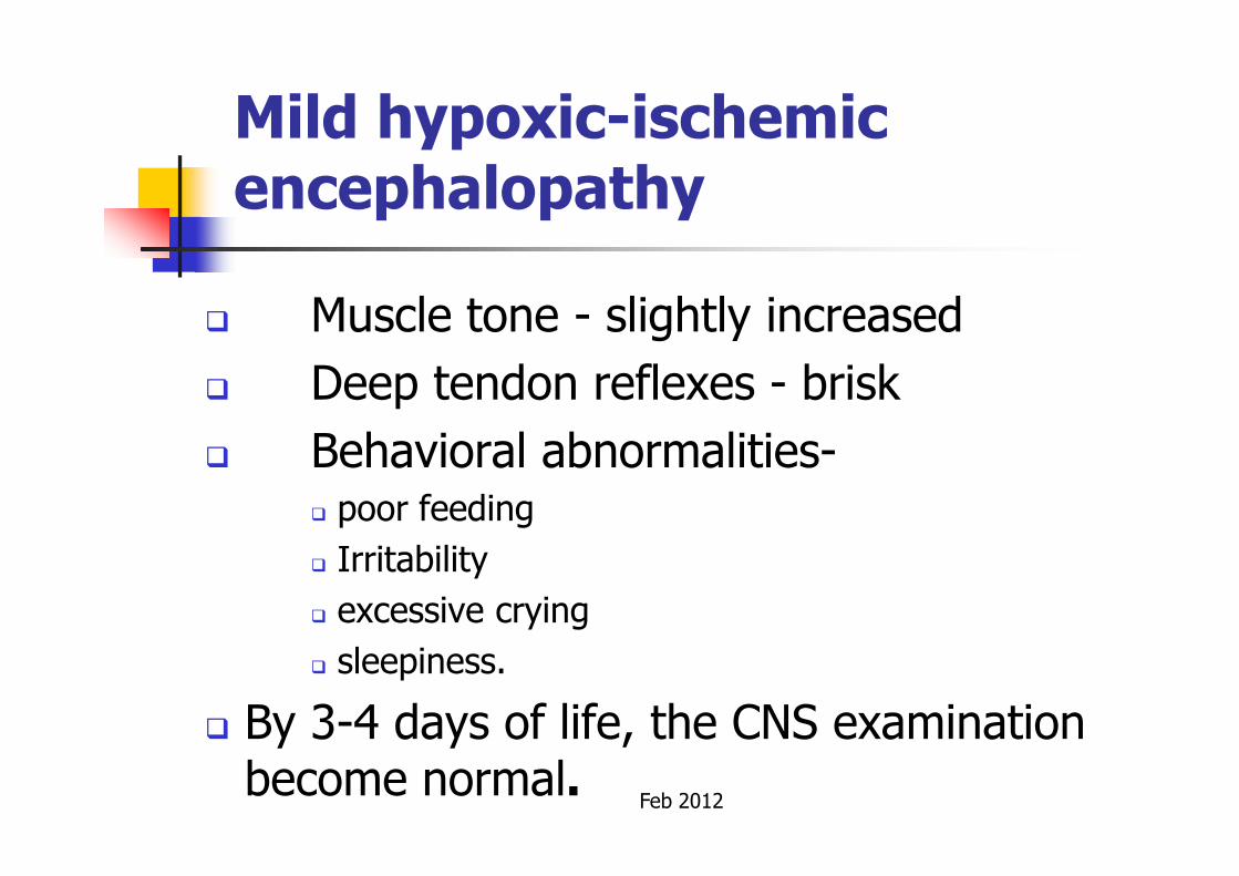
Mild hypoxic-ischemic encephalopathy
� Muscle tone - slightly increased
� Deep tendon reflexes - brisk
� Behavioral abnormalities-� poor feeding
� Irritability
� excessive crying
� sleepiness.
� By 3-4 days of life, the CNS examination become normal.

Feb 2012
Moderately severe hypoxic-ischemic encephalopathy
� Lethargic, hypotonia and diminished deep tendon reflexes.
� Primitive reflexes sluggish or absent� Moro, grasping sucking reflexes
� Periods of apnea. � Seizures within the first 24 hours of life. � Full recovery within 1-2 weeks is possible and is
associated with a better long-term outcome.
� An initial period of well-being or mild hypoxic-ischemic encephalopathy may be followed by sudden deterioration, suggesting ongoing brain cell dysfunction, injury, and death; during this period, seizure intensity might increase.

Feb 2012
Severe hypoxic-ischemic encephalopathy
� Stupor or coma. No response to any physical stimulus.
� Breathing irregular, (gasping) and the infant often requires ventilatory support
� Generalized hypotonia and depressed deep tendon reflexes
� Neonatal reflexes (eg, sucking, swallowing, grasping, Moro) are absent.
� Disturbances of ocular motion, such as a skewed deviation of the eyes, nystagmus, bobbing, and loss of "doll's eye"
� Pupils dilated, fixed, or poorly reactive to light.

Feb 2012
Severe hypoxic-ischemic encephalopathy (cont.)
� Seizures - early frequency may increase during the 24-48 hours after onset.
� As the injury progresses, seizures subside and the EEG becomes isoelectric or shows a burst suppression pattern.
� Fontanelle bulge- increasing cerebral edema.
� Irregularities of heart rate and blood pressure (BP)

Feb 2012
Infants who survive
� The level of alertness improves by days 4-5 of life
� Hypotonia and feeding difficulties persist, requiring tube feeding for weeks to months.
� Severe CP

Feb 2012
Multi-organ dysfunction
� Heart (43-78%): � Lungs (71-86%):� Renal (46-72%):. � Liver (80-85%):� GI dysfunction:� Hematologic (32-54%):� Severely depressed respiratory and cardiac functions and
signs of brainstem compression suggest a life-threatening rupture of the vein of Galen (ie, great cerebral vein) with a hematoma in the posterior cranial fossa.

Feb 2012
SARNAT CLASSIFICATION or
SHANKARAN

Feb 2012
CAUSES HIE
� South Africa –
� high incidence of intra-partum asphyxia

Feb 2012
Diagnosis
� No specific test can always confirm or exclude a diagnosis of hypoxic-ischemic encephalopathy (HIE) because the diagnosis is made based on the history and physical and neurological examinations.
� Many of the tests are performed to assess the severity of brain injury and to monitor the functional status of systemic organs.
� As always, the results of the tests should be interpreted in conjunction with the clinical history and the findings from physical examination.

Feb 2012
WORK-UP
� Serum electrolytes � Acute tubular necrosis.� Polyuria
� Renal function studies: � Serum creatinine levels, and urea.
� Coagulation system evaluation: � prothrombin time, partial thromboplastin time, and fibrinogen
levels.
� ABG: � acid-base status hyperoxia and hypoxia as well as hypercapnia
and hypocapnia.
� Cardiac and liver enzymes: � degree of hypoxic-ischemic injury

Feb 2012
Imaging studies
� MRI is the imaging modality of choice for the diagnosis and follow-up of infants diagnosed with moderate-to-severe hypoxic-ischemic encephalopathy.
Not regularly available NICU / SBAH
Feb 2012
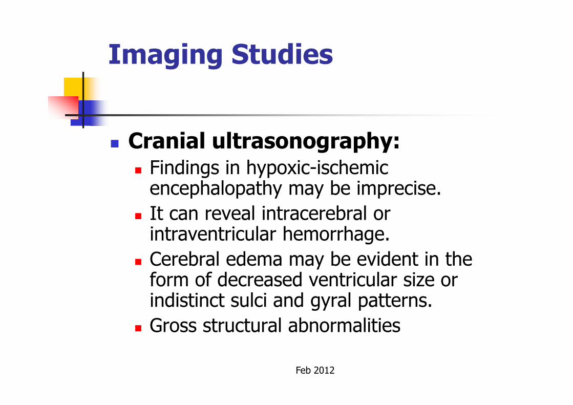
Imaging Studies
� Cranial ultrasonography: � Findings in hypoxic-ischemic encephalopathy may be imprecise.
� It can reveal intracerebral or intraventricular hemorrhage.
� Cerebral edema may be evident in the form of decreased ventricular size or indistinct sulci and gyral patterns.
� Gross structural abnormalities

Feb 2012
Amplitude-integrated electroencephalography (aEEG)
•Single-channel a-EEG
•Discontinuous tracing
•Burst suppression pattern
•Continuous low voltage
•Inactive pattern with no detectable cortical activity
•Seizures, abrupt rise in both the lower and upper margins

Feb 2012
Standard EEG
� Generalized depression varying degrees of superimposed seizures
� Burst suppression and iso-electric EEG patterns are particularly ominous.
� Note that large doses of anticonvulsant therapy may alter the EEG findings.
� Serial EEGs to assess seizure control and evolution of background abnormalities.
� Repeat EEGs obtained just prior to discharge can be helpful in establishing prognosis.
� Improvement in the EEG findings over the first week, in conjunction with improvement in the clinical condition, may help predict a better long-term outcome.

Feb 2012
Special sensory evaluation:
� Hearing test
� increased incidence of deafness
� Retinal and ophthalmic examination
� for developmental abnormalities of the brain.

Feb 2012
Bilateral acute infarctions of the frontal lobe

Feb 2012
Severe acute hypoxic-ischemic neuronal change

Feb 2012
Gliosis secondary to the hypoxic-ischemic event

Feb 2012
Treatment: Medical Care
� PREVENTION� Obstetric and Neonatal resussitation
� Supportive care � Maintain adequate ventilation � Maintain the blood gases and acid-base status in the physiological ranges
� Maintain adequate perfusion. � Maintain the mean blood pressure (BP) above 35-40 mm Hg (for term infants). Dopamine or dobutamine can be used to maintain adequate cardiac output.

Feb 2012
Treatment: Medical Care
� Maintain adequate metabolic status. � Fluid and glucose homeostasis
� Because of the concern for acute tubular necrosis (ATN) insensible water loss (40-60 mL/kg/d in term infant).
� Avoid hyperthermia.

Feb 2012
Treatment of seizures
� Seizures should be treated early and be well controlled because even asymptomatic seizures (ie, seen only on EEG) may continue to injure the brain.
� Seizures should be treated with phenobarbital or lorazepam; phenytoin may be added if either of these medications fails to control the seizures.

Feb 2012
Hypothermia treatment
� Extensive experimental data suggest that mild hypothermia (3-4°C below the baseline temperature) applied within a few hours (no later than 6 h) of hypoxia-ischemia injury is neuro-protective.
� Therapeutic hypothermia is now the standard of care in neonates with moderate-to-severe hypoxic-ischemic encephalopathy.

Feb 2012
Hypothermia treatment
� Near-term infants born at 36 weeks' gestation or less with birth weight of 1800-2000 g or less, younger than 6 hours at admission
� Evidence of acute event around the time of birth � Apgar score of 5 or less at 10 minutes after birth (In the study by
Shankaran et al, this needed to be in conjunction with either evidence of acute perinatal event or need for assisted ventilation for at least 10 min.13 )
� Severe acidosis, defined as pH level of less than 7 or base deficit of 16 mmol/L or less (cord blood or any blood gas obtained within 1 h of birth)
� Continued need for resuscitation at 10 minutes after birth� Evidence of moderate to severe encephalopathy at birth
� Clinically determined (at least 2) � Lethargy, stupor, or coma � Abnormal tone or posture � Abnormal reflexes (suck, grasp, Moro, gag, stretch reflexes) � Decrease or absent spontaneous activity � Autonomic dysfunction (including bradycardia, abnormal pupils, apneas)
� Clinical evidence of seizures� Moderately or severely abnormal aEEG background or seizures

Feb 2012
Hypothermia studies
Feb 2012
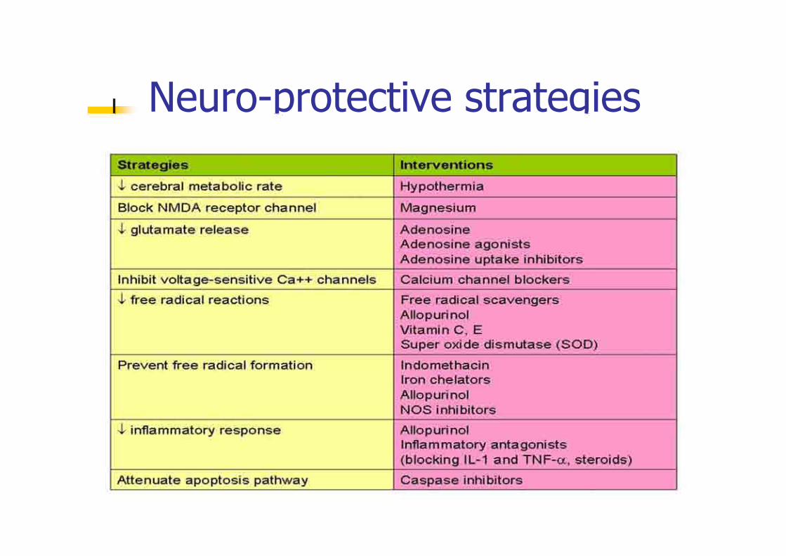
Neuro-protective strategies

Feb 2012
Surgical CarePosterior cranial fossa hematoma- surgical drainage
DietTrophic feeding with expressed breast milk about 5 mL every 3-4 hours if bowel sounds audible. Individualize increments in feeding volume and composition

Feb 2012
AnticonvulsantsThese agents are used to control seizures.
Phenobarbital Clinical or EEG seizures - continued on the basis of both EEG findings and clinical status. Weaned and stopped during the first month of life if no further convulsions.
EEG and clinical status should guide decision.

Feb 2012
Follow-up
Physical therapy and developmental evaluation are needed before discharge.
Further Outpatient CareSeverely disabled children may need to be monitored in multi-discipility clinics and by a developmental neurologist.

Feb 2012
Experimental treatment.
� None has shown consistent efficacy in infants.
� Alloperinol
� High dosis Phenobarb

Feb 2012
PrognosisAccurate prediction of the severity of long-term complications is difficult, although the following pointers may be used:
Lack of spontaneous respiratory effort within 20-30 minutes of birth almost always associated with death.
The presence of seizures is an ominous sign. The risk of poor neurological outcome is distinctly greater in such infants,
Abnormal clinical neurological findings persisting beyond the first 7-10 days of life usually indicate poor prognosis. Among these, abnormalities of muscle tone and posture (hypotonia, rigidity, weakness) should be carefully noted.

Feb 2012
Prognosis ( cont.)
An EEG done at about 7 days that has normal background activity is a good prognostic sign.
Persistent feeding difficulties, which generally are due to abnormal tone of the muscles of sucking and swallowing, also suggest significant CNS damage.
Poor head growth during the postnatal period and the first year of life is a sensitive finding predicting higher frequency of neurologic deficits.

Feb 2012
Medico-legal pitfallsBirth asphyxia, birth injury, and perinatal asphyxia are terms often used incorrectly to describe hypoxic-ischemic encephalopathy (HIE).
It is recommend using hypoxic-ischemic encephalopathy because this term accurately describes the clinical condition, encephalopathy from asphyxia, without implying the time of brain injury.
Counseling the parents with realistic explanations about their infant's clinical status and prognosis is always recommended.
Always document and sign the counseling session.

Feb 2012
References:
� American Academy of Pediatrics. Relation between perinatal factors and neurological outcome. In: Guidelines for Perinatal Care. 3rd ed. Elk Grove Village, Ill: American Academy of Pediatrics; 1992:221-234.
� Committee on fetus and newborn, American Academy of Pediatrics and Committee on obstetric practice, American College of Obstetrics and Gynecology. Use and abuse of the APGAR score. Pediatr. 1996;98:141-142. [Medline].
� Sarnat HB, Sarnat MS. Neonatal encephalopathy following fetal distress: A clinical and electroencphalographic study. Archives of Neur. 1976;33:696-705.
� Badawi N, Kurinczuk JJ, Keogh JM, et al. Antepartum risk factors for newborn encephalopathy: the Western Australian case-control study. British Medical Journal. 1998;317:1549-1553. [Medline].
� Gluckman PD, Wyatt JS, Azzopardi D, et al. Selective head cooling with mild systemic hypothermia after neonatal encephalopathy: multicenter randomised trial. Lancet. 2005;365:663-70. [Medline].
� [Best Evidence] Jacobs S, Hunt R, Tarnow-Mordi W, Inder T, Davis P. Cooling for newborns with hypoxic ischaemic encephalopathy. Cochrane Database Syst Rev. 2007;4:CD003311. [Medline].
� de Vries LS, Toet MC. Amplitude integrated electroencephalography in the full-term newborn. Clin Perinatol. 2006;33:619-632. [Medline].