Embed Size (px)
Citation preview
CompendiumVet.com | July 2009 | Compendium: Continuing Education for Veterinarians® E1©Copyright 2009 Veterinary Learning Systems. This document is for internal purposes only. Reprinting or posting on an external website without written permission from VLS is a violation of copyright laws.
CE Article
The Epiphyseal Plate: Physiology, Anatomy, and Trauma*
Pathologic conditions affecting epiphyseal (growth) plates in immature animals may result in severe
orthopedic problems such as limb shortening, angular limb deformity, or joint incongruity. Understanding growth plate anatomy and physiology enables practicing veterinarians to provide a prognosis and assess indications for surgery. Injured animals should be closely observed during the period of rapid growth.
Bone FormationBone is formed by transformation of connective tissue (intramembranous ossification) and replacement of a cartilaginous model (endochondral ossification).1 Intramem branous ossification occurs in flat bone (e.g., cranium, periosteal surface of the diaphysis of long bones), where it causes growth in bone width. Endochondral ossification is responsible for growth in bone length and forms the articular surfaces.1
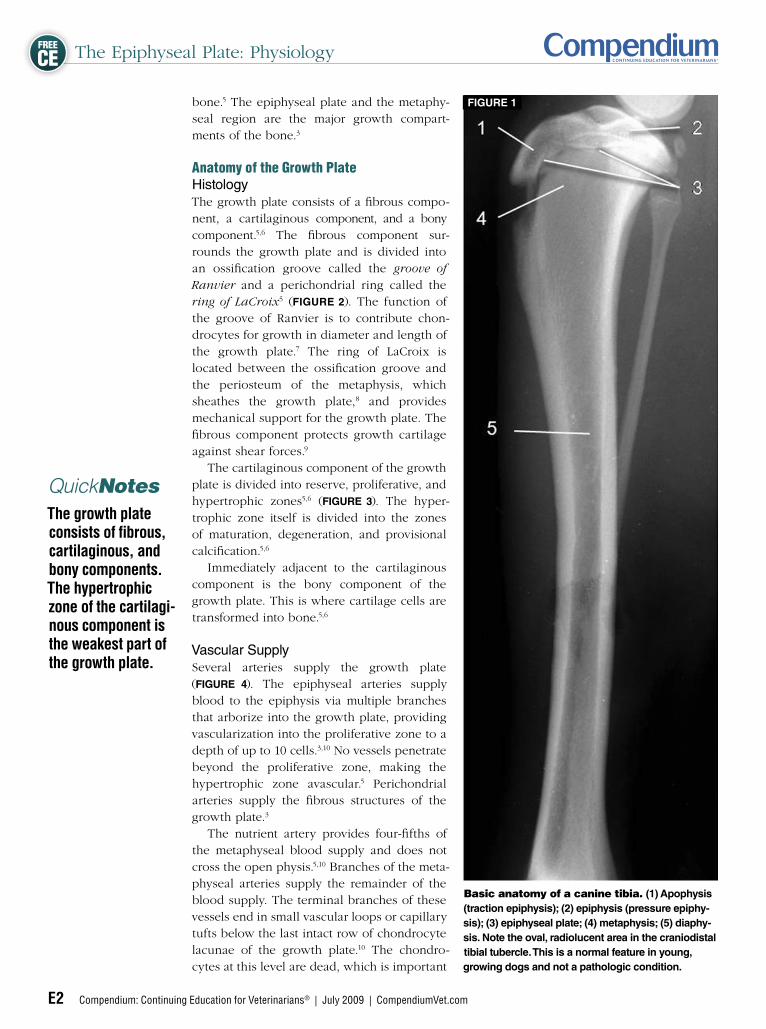
The process of endochondral ossification generates all three major areas of long bones: diaphysis, epiphysis, and metaphysis2 (Figure 1). The diaphysis develops first in the fetus. Mesenchymal cells form a cartilaginous structure containing a center of calcifying chondrocytes surrounded by a thin collar of cancellous bone.2 This area is called the primary ossification center. Vascular invasion develops at the nutrient
foramen. Growth factors and multipotent stem cells support the formation of neonatal bone consisting of a central marrow cavity surrounded by a thin periosteum.2
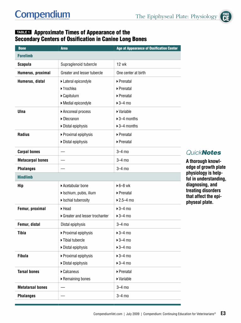
The epiphysis is a secondary ossification center in the hyaline cartilage forming the joint surfaces at the proximal and distal ends of the bones. Secondary ossification centers can appear in the fetus as early as 28 days after conception1 (Table 1). Growth of the epiphysis arises from two areas: (1) the vascular reserve zone cartilage, which is responsible for growth of the epiphysis toward the joint, and (2) the epiphyseal plate, which is responsible for growth in bone length.3 The epiphyseal plate is mostly composed of hyaline cartilage and is visible in radiographs of young animals as a radiolucent line between the epiphysis and the metaphysis2 (Figure 1). In mature animals, the epiphysis consists of cancellous bone surrounded by a thin layer of compact bone. There are two types of epiphyses: (1) pressure epiphyses, which are found at the ends of long bones, and (2) traction epiphyses (apophyses), which are sites of origin or insertion of major muscles (e.g., the greater trochanter of the femur).4
The metaphysis is an area between the diaphysis and epiphysis. Bone is developed from the growth plate, matures, and remodels in the metaphysis of growing
❯❯ Dirsko J. F. von Pfeil, Dr.med.vet, DVM, DACVS, DECVS Veterinary Specialists of Alaska Anchorage, Alaska
❯❯ Charles E. DeCamp, DVM, MS, DACVS Michigan State University
3 CECREDITS
Bone Formation Page E1
Anatomy of the Growth Plate
Page E2
Physiology of the Growth Plate
Page E4
Growth Plate Closure and Contribution to Overall Growth
Page E5
Fractures of the Growth Plate
Page E7
Osgood-Schlatter Disease Page E10
At a Glance
Abstract: This article reviews the development of long bones, the microanatomy and physiology of the growth plate, the closure times and contribution of different growth plates to overall growth, and the effect of, and prognosis for, traumatic injuries to the growth plate. Details on surgical treatment of growth plate fractures are beyond the scope of this article.
*A companion article, “The Epiphyseal Plate: Nutritional and Hormonal Influences; Hereditary and Other Disorders,” is also available on CompendiumVet.com.
The Epiphyseal Plate: Physiology
E2 Compendium: Continuing Education for Veterinarians® | July 2009 | CompendiumVet.com
FREE
CE
The growth plate consists of fibrous, cartilaginous, and bony components. The hypertrophic zone of the cartilagi-nous component is the weakest part of the growth plate.
QuickNotes
bone.5 The epiphyseal plate and the metaphyseal region are the major growth compartments of the bone.3
Anatomy of the Growth PlateHistologyThe growth plate consists of a fibrous component, a cartilaginous component, and a bony component.5,6 The fibrous component surrounds the growth plate and is divided into an ossification groove called the groove of Ranvier and a perichondrial ring called the ring of LaCroix5 (Figure 2). The function of the groove of Ranvier is to contribute chondrocytes for growth in diameter and length of the growth plate.7 The ring of LaCroix is located between the ossification groove and the periosteum of the metaphysis, which sheathes the growth plate,8 and provides mechanical support for the growth plate. The fibrous component protects growth cartilage against shear forces.9
The cartilaginous component of the growth plate is divided into reserve, proliferative, and hypertrophic zones5,6 (Figure 3). The hypertrophic zone itself is divided into the zones of maturation, degeneration, and provisional calcification.5,6
Immediately adjacent to the cartilaginous component is the bony component of the growth plate. This is where cartilage cells are transformed into bone.5,6
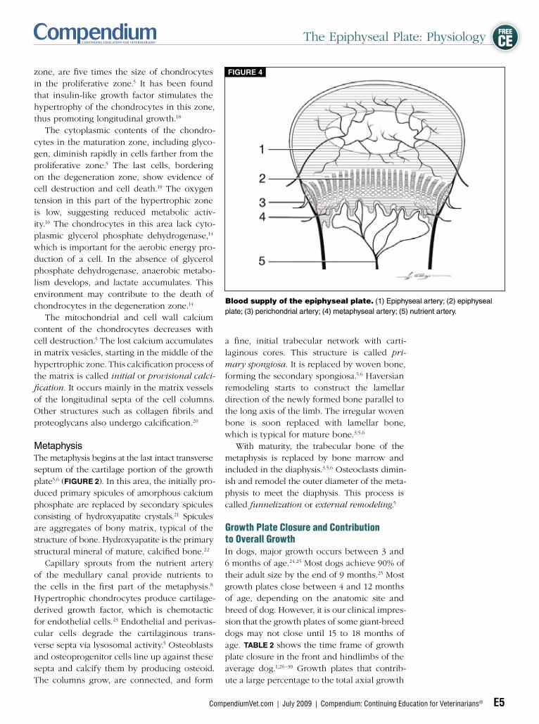
Vascular SupplySeveral arteries supply the growth plate (Figure 4). The epiphyseal arteries supply blood to the epiphysis via multiple branches that arborize into the growth plate, providing vascularization into the proliferative zone to a depth of up to 10 cells.3,10 No vessels penetrate beyond the proliferative zone, making the hypertrophic zone avascular.5 Perichondrial arteries supply the fibrous structures of the growth plate.3
The nutrient artery provides fourfifths of the metaphyseal blood supply and does not cross the open physis.5,10 Branches of the metaphyseal arteries supply the remainder of the blood supply. The terminal branches of these vessels end in small vascular loops or capillary tufts below the last intact row of chondrocyte lacunae of the growth plate.10 The chondrocytes at this level are dead, which is important
Basic anatomy of a canine tibia. (1) apophysis (traction epiphysis); (2) epiphysis (pressure epiphy-sis); (3) epiphyseal plate; (4) metaphysis; (5) diaphy-sis. Note the oval, radiolucent area in the craniodistal tibial tubercle. This is a normal feature in young, growing dogs and not a pathologic condition.
Figure 1
The Epiphyseal Plate: Physiology
CompendiumVet.com | July 2009 | Compendium: Continuing Education for Veterinarians® E3
FREE
CE
A thorough knowl-edge of growth plate physiology is help-ful in understanding, diagnosing, and treating disorders that affect the epi-physeal plate.
QuickNotes
Table 1 Approximate Times of Appearance of the Secondary Centers of Ossification in Canine Long Bones
Bone Area Age at Appearance of Ossification Center
Forelimb
Scapula Supraglenoid tubercle 12 wk
Humerus, proximal Greater and lesser tubercle One center at birth
Humerus, distal Lateral epicondyle
Trochlea
Capitulum
Medial epicondyle
Prenatal
Prenatal
Prenatal
3–4 mo
Ulna Anconeal process
Olecranon
Distal epiphysis
Variable
3–4 months
3–4 months
Radius Proximal epiphysis
Distal epiphysis
Prenatal
Prenatal
Carpal bones — 3–4 mo
Metacarpal bones — 3–4 mo
Phalanges — 3–4 mo
Hindlimb
Hip Acetabular bone
Ischium, pubis, ilium
Ischial tuberosity
6–8 wk
Prenatal
2.5–4 mo
Femur, proximal Head
Greater and lesser trochanter
3–4 mo
3–4 mo
Femur, distal Distal epiphysis 3–4 mo
Tibia Proximal epiphysis
Tibial tubercle
Distal epiphysis
3–4 mo
3–4 mo
3–4 mo
Fibula Proximal epiphysis
Distal epiphysis
3–4 mo
3–4 mo
Tarsal bones Calcaneus
Remaining bones
Prenatal
Variable
Metatarsal bones — 3–4 mo
Phalanges — 3–4 mo

The Epiphyseal Plate: Physiology
E4 Compendium: Continuing Education for Veterinarians® | July 2009 | CompendiumVet.com
FREE
CE
Components of the epiphysis and metaphysis. (1) articular cartilage; (2) epiphyseal cartilage; (3) secondary center of ossification; (4) epiphyseal plate; (5) epiphysis; (6) metaphysis; (7) fibrous layer of the periosteum; (8) ring of laCroix; (9) groove of ranvier; (10) fibrous components of the epiphyseal plate; (11) cortical bone.
Figure 2
Zones of the cartilaginous component of the epiphyseal plate. (1) reserve zone; (2) proliferative zone; (3) zone of maturation; (4) zone of degenera-tion; (5) zone of provisional calcification; (6) hypertrophic zone.
Figure 3
in understanding the development of osteochondrosis dissecans.a Venous drainage of the metaphysis occurs via the large central vein of the diaphysis.11
In humans and cats, the femoral capital growth plate can be partially supplied with blood via branches of the artery of the ligament of the femoral head (epiphysis); however, no such blood supply exists in dogs.12,13
Physiology of the Growth PlateCartilaginous ComponentAs blood supply varies in the different zones of the growth plate, so does cell metabolism. In the proliferative zone and the top half of the hypertrophic zone, it is aerobic, while in the lower half of the hypertrophic zone, it is anaerobic.14 Chondrocytes in the reserve zone are spherical, not as numerous, and separated by more matrix compared with cells in other zones.3,5,6 The cells in the reserve zone contain many lipid vacuoles and abundant endoplasmic reticulum, which is indicative of protein production.5 The oxygen tension in this area is relatively low, consistent with low cellular activity. This may indicate that oxygen and nutrients reach this area only by diffusion, which in turn may be important for the etiology of osteochondrosis dissecans and hypertrophic osteodystrophy. The function of this zone is likely the endowment of chondrocytes to the proliferative zone.13,15
In the proliferative zone, chondrocytes are flattened and aligned in columns parallel to the long axis of the bone3,5,6 (Figure 3). The oxygen tension is higher than in other zones,5 as is the cell metabolism, resulting in a high concentration of cell metabolites.16 The primary function of this zone is cellular proliferation; other functions include the formation of intracellular matrix, proteoglycan, and collagen.17 Collagen has great tensile strength and supports the mechanically weak proteoglycan gel within the cartilage of this zone.3
The hypertrophic zone is divided into the zones of maturation, degeneration, and provisional calcification5,6 (Figure 3). The beginning of the maturation zone can be accurately determined based on cell shape. The chondrocytes become spherical and, at the base of the
aOsteochondrosis dissecans is discussed in the companion article, “The Epiphyseal Plate: Nutritional and Hormonal Influences, Hereditary and Other Disorders,” also available on CompendiumVet.com.
The Epiphyseal Plate: Physiology
CompendiumVet.com | July 2009 | Compendium: Continuing Education for Veterinarians® E5
FREE
CE
zone, are five times the size of chondrocytes in the proliferative zone.5 It has been found that insulinlike growth factor stimulates the hypertrophy of the chondrocytes in this zone, thus promoting longitudinal growth.18 The cytoplasmic contents of the chondrocytes in the maturation zone, including glycogen, diminish rapidly in cells farther from the proliferative zone.5 The last cells, bordering on the degeneration zone, show evidence of cell destruction and cell death.19 The oxygen tension in this part of the hypertrophic zone is low, suggesting reduced metabolic activity.16 The chondrocytes in this area lack cytoplasmic glycerol phosphate dehydrogenase,14 which is important for the aerobic energy production of a cell. In the absence of glycerol phosphate dehydrogenase, anaerobic metabolism develops, and lactate accumulates. This environment may contribute to the death of chondrocytes in the degeneration zone.14
The mitochondrial and cell wall calcium content of the chondrocytes decreases with cell destruction.5 The lost calcium accumulates in matrix vesicles, starting in the middle of the hypertrophic zone. This calcification process of the matrix is called initial or provisional calci-fication. It occurs mainly in the matrix vessels of the longitudinal septa of the cell columns. Other structures such as collagen fibrils and proteoglycans also undergo calcification.20
MetaphysisThe metaphysis begins at the last intact transverse septum of the cartilage portion of the growth plate5,6 (Figure 2). In this area, the initially produced primary spicules of amorphous calcium phosphate are replaced by secondary spicules consisting of hydroxyapatite crystals.21 Spicules are aggregates of bony matrix, typical of the structure of bone. Hydroxyapatite is the primary structural mineral of mature, calcified bone.22
Capillary sprouts from the nutrient artery of the medullary canal provide nutrients to the cells in the first part of the metaphysis.8 Hypertrophic chondrocytes produce cartilagederived growth factor, which is chemotactic for endothelial cells.23 Endothelial and perivascular cells degrade the cartilaginous transverse septa via lysosomal activity.5 Osteoblasts and osteoprogenitor cells line up against these septa and calcify them by producing osteoid. The columns grow, are connected, and form
Blood supply of the epiphyseal plate. (1) Epiphyseal artery; (2) epiphyseal plate; (3) perichondrial artery; (4) metaphyseal artery; (5) nutrient artery.
Figure 4
a fine, initial trabecular network with cartilaginous cores. This structure is called pri-mary spongiosa. It is replaced by woven bone, forming the secondary spongiosa.5,6 Haversian remodeling starts to construct the lamellar direction of the newly formed bone parallel to the long axis of the limb. The irregular woven bone is soon replaced with lamellar bone, which is typical for mature bone.3,5,6
With maturity, the trabecular bone of the metaphysis is replaced by bone marrow and included in the diaphysis.3,5,6 Osteoclasts diminish and remodel the outer diameter of the metaphysis to meet the diaphysis. This process is called funnelization or external remodeling.5
Growth Plate Closure and Contribution to Overall GrowthIn dogs, major growth occurs between 3 and 6 months of age.24,25 Most dogs achieve 90% of their adult size by the end of 9 months.25 Most growth plates close between 4 and 12 months of age, depending on the anatomic site and breed of dog. However, it is our clinical impression that the growth plates of some giantbreed dogs may not close until 15 to 18 months of age. Table 2 shows the time frame of growth plate closure in the front and hindlimbs of the average dog.1,26–30 Growth plates that contribute a large percentage to the total axial growth
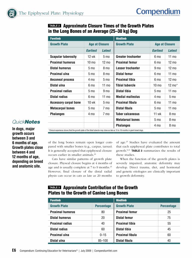
The Epiphyseal Plate: Physiology
E6 Compendium: Continuing Education for Veterinarians® | July 2009 | CompendiumVet.com
FREE
CE
of the long bones remain open longer compared with smaller bones (e.g., carpus, tarsus). It is generally accepted that epiphyseal closure occurs earlier in smaller animals.26 Cats have similar patterns of growth plate closure. Physeal closure begins at 4 months of age and is usually complete at 7 to 9 months.25 However, final closure of the distal radial physis can occur in cats as late as 20 months
of age.25 Studies have evaluated the amount that each epiphyseal plate contributes to total growth.8,31 Table 3 summarizes the results of these studies. When the function of the growth plates is severely impaired, anatomic deformity may develop. Direct trauma, diet, and hormonal and genetic etiologies are clinically important to growth deformity.
In dogs, major growth occurs between 3 and 6 months of age. Growth plates close between 4 and 12 months of age, depending on breed and anatomic site.
QuickNotes
Table 2 Approximate Closure Times of the Growth Plates in the Long Bones of an Average (25–30 kg) Dog
Forelimb Hindlimb
Growth Plate Age at Closure Growth Plate Age at Closure
Earliest Latest Earliest Latest
Scapular tuberosity 12 wk 5 mo Greater trochanter 6 mo 11 mo
Proximal humerus 10 mo 12 mo Proximal femur 6 mo 12 mo
Distal humerus 5 mo 8 mo Lesser trochanter 9 mo 12 mo
Proximal ulna 5 mo 8 mo Distal femur 6 mo 11 mo
Anconeal process 4 mo 5 mo Proximal tibia 6 mo 12 mo
Distal ulna 6 mo 11 mo Tibial tubercle 10 mo 12 mo*
Proximal radius 5 mo 9 mo Distal tibia 5 mo 11 mo
Distal radius 6 mo 11 mo Medial malleolus 4 mo 5 mo
Accessory carpal bone 10 wk 5 mo Proximal fibula 6 mo 11 mo
Metacarpal bones 5 mo 7 mo Distal fibula 5 mo 11 mo
Phalanges 4 mo 7 mo Tuber calcaneous 11 wk 8 mo
Metatarsal bones 5 mo 8 mo
Phalanges 4 mo 8 mo*Clinical experience shows that the growth plate of the tibial tubercle may close as late as 15 to 18 months in giant-breed dogs.
Table 3 Approximate Contribution of the Growth Plates to the Growth of Canine Long Bones
Forelimb Hindlimb
Growth Plate Percentage Growth Plate Percentage
Proximal humerus 80 Proximal femur 25
Distal humerus 20 Distal femur 75
Proximal radius 40 Proximal tibia 55
Distal radius 60 Distal tibia 45
Proximal ulna 0–15 Proximal fibula 60
Distal ulna 85–100 Distal fibula 40
The Epiphyseal Plate: Physiology
CompendiumVet.com | July 2009 | Compendium: Continuing Education for Veterinarians® E7
FREE
CE
Fractures of the Growth PlateAbout half (50% to 54%) of dogs with longbone fractures have been reported to be younger than 1 year.32,33 Among the reported cases, 30% had trauma to the growth plate
and 7% developed growth deformities. In a study of 92 dogs with growth deformity,34 75% showed a disturbance of the ulna or radius. The tibia accounted for 4% of the deformities reported, and the femur accounted for 8%.
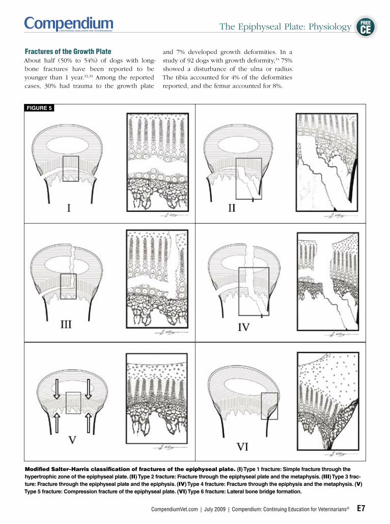
Figure 5
Modified Salter–Harris classification of fractures of the epiphyseal plate. (I) Type 1 fracture: Simple fracture through the hypertrophic zone of the epiphyseal plate. (II) Type 2 fracture: Fracture through the epiphyseal plate and the metaphysis. (III) Type 3 frac-ture: Fracture through the epiphyseal plate and the epiphysis. (IV) Type 4 fracture: Fracture through the epiphysis and the metaphysis. (V) Type 5 fracture: Compression fracture of the epiphyseal plate. (VI) Type 6 fracture: lateral bone bridge formation.
The Epiphyseal Plate: Physiology
E8 Compendium: Continuing Education for Veterinarians® | July 2009 | CompendiumVet.com
FREE
CE
Salter–Harris Classification SystemSalter and Harris created a prognostic classification system for fractures of the growth plate4 (Figure 5). Type I fractures involve complete epiphyseal separation at the level of the hypertrophic zone. The reserve zone (germinal layer of the growth plate) is usually intact. Type II fractures are partially through the growth plate and partially through the metaphysis. Types I and II are the most common, accounting for 65.5% of growth plate fractures in small animals.35
Type III and type IV fractures are intraarticular fractures. Type III fractures occur in the proximal humerus and distal femur, and the fracture line includes the hypertrophic and reserve zones of the epiphyseal plate. In type IV fractures, the fracture line extends into the metaphysis, thus completely crossing the growth plate. Type IV fractures are often condylar fractures and are most common in the distal humerus.35 Types III and IV make up 25.5% of all canine growth plate fractures.35
A type V fracture is characterized by partial or complete compression of the growth plate and cannot be diagnosed acutely based on radiographs. It may help to compare the width of the growth plate with the contralateral side and to repeat radiographic studies at 2week intervals to better assess damage, bone length, and curvature compared with the contralateral bone. Some investigators have suggested adding a sixth type of fracture to the traditional Salter–Harris classification system.36,37 A type VI fracture involves the groove of Ranvier and leads to eccentric premature closure of the physis. It commonly results from a local contusion or avulsion. Peripheral osseous bridge formation is common and may lead to peripherally localized epiphysiodesis and subsequent angular deformity.36,37
In their original article,4 Salter and Harris explained the results of an experimental study in rodents, demonstrating that fractures commonly developed through the hypertrophic zone, which is mechanically the weakest zone of the growth plate. They also investigated healing of the different fracture types and suggested that the higher the fracture grade, the worse the prognosis for normal growth.4 The prognostic reliability of the Salter–Harris classification system has since been questioned.35,37–41 Salter and Harris also postulated that interference with blood supply to the epiphysis is associated with a poor prognosis. This theory is now the basis for an additional classification system, described below.
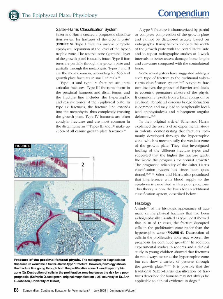
Histology A study42 of the histologic appearance of traumatic canine physeal fractures that had been radiographically classified as type I or II showed that in 10 of 13 cases, the fracture disrupted cells in the proliferative zone rather than the hypertrophic zone (Figure 6). Destruction of cells in the proliferative zone may worsen the prognosis for continued growth.42 In addition, experimental studies in rodents and a clinical study in young children showed that fractures do not always occur at the hypertrophic zone but can show a variety of patterns through the growth plate.38,43,44 It is possible that the traditional Salter–Harris classification of fractures described for humans may not always be applicable to clinical evidence in dogs.42
Fracture of the proximal femoral physis. The radiographic diagnosis for this fracture would be a Salter–Harris type 1 fracture. However, histology shows the fracture line going through both the proliferative zone (1) and hypertrophic zone (2). Destruction of cells in the proliferative zone increases the risk for a poor prognosis. (Safranin O, fast green; original magnification × 25; courtesy of Dr. ann l. Johnson, university of illinois)
Figure 6
The Epiphyseal Plate: Physiology
CompendiumVet.com | July 2009 | Compendium: Continuing Education for Veterinarians® E9
FREE
CE
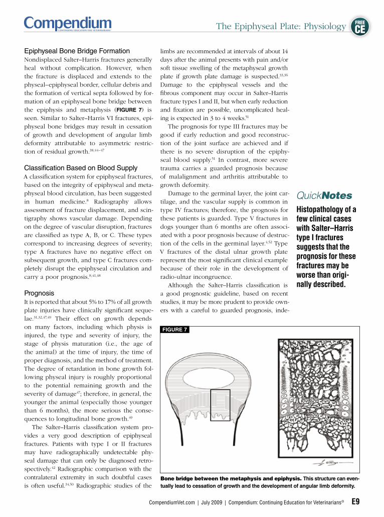
Epiphyseal Bone Bridge FormationNondisplaced Salter–Harris fractures generally heal without complication. However, when the fracture is displaced and extends to the physeal–epiphyseal border, cellular debris and the formation of vertical septa followed by formation of an epiphyseal bone bridge between the epiphysis and metaphysis (Figure 7) is seen. Similar to Salter–Harris VI fractures, epiphyseal bone bridges may result in cessation of growth and development of angular limb deformity attributable to asymmetric restriction of residual growth.38,44–47
Classification Based on Blood SupplyA classification system for epiphyseal fractures, based on the integrity of epiphyseal and metaphyseal blood circulation, has been suggested in human medicine.8 Radiography allows assessment of fracture displacement, and scintigraphy shows vascular damage. Depending on the degree of vascular disruption, fractures are classified as type A, B, or C. These types correspond to increasing degrees of severity; type A fractures have no negative effect on subsequent growth, and type C fractures completely disrupt the epiphyseal circulation and carry a poor prognosis.8,41,48
Prognosis It is reported that about 5% to 17% of all growth plate injuries have clinically significant sequelae.31,32,47,49 Their effect on growth depends on many factors, including which physis is injured, the type and severity of injury, the stage of physis maturation (i.e., the age of the animal) at the time of injury, the time of proper diagnosis, and the method of treatment. The degree of retardation in bone growth following physeal injury is roughly proportional to the potential remaining growth and the severity of damage47; therefore, in general, the younger the animal (especially those younger than 6 months), the more serious the consequences to longitudinal bone growth.49 The Salter–Harris classification system provides a very good description of epiphyseal fractures. Patients with type I or II fractures may have radiographically undetectable physeal damage that can only be diagnosed retrospectively.42 Radiographic comparison with the contralateral extremity in such doubtful cases is often useful.34,50 Radiographic studies of the
limbs are recommended at intervals of about 14 days after the animal presents with pain and/or soft tissue swelling of the metaphyseal growth plate if growth plate damage is suspected.33,36 Damage to the epiphyseal vessels and the fibrous component may occur in Salter–Harris fracture types I and II, but when early reduction and fixation are possible, uncomplicated healing is expected in 3 to 4 weeks.51 The prognosis for type III fractures may be good if early reduction and good reconstruction of the joint surface are achieved and if there is no severe disruption of the epiphyseal blood supply.51 In contrast, more severe trauma carries a guarded prognosis because of malalignment and arthritis attributable to growth deformity. Damage to the germinal layer, the joint cartilage, and the vascular supply is common in type IV fractures; therefore, the prognosis for these patients is guarded. Type V fractures in dogs younger than 6 months are often associated with a poor prognosis because of destruction of the cells in the germinal layer.4,52 Type V fractures of the distal ulnar growth plate represent the most significant clinical example because of their role in the development of radioulnar incongruence. Although the Salter–Harris classification is a good prognostic guideline, based on recent studies, it may be more prudent to provide owners with a careful to guarded prognosis, inde
Histopathology of a few clinical cases with Salter–Harris type I fractures suggests that the prognosis for these fractures may be worse than origi-nally described.
QuickNotes
Bone bridge between the metaphysis and epiphysis. This structure can even-tually lead to cessation of growth and the development of angular limb deformity.
Figure 7

The Epiphyseal Plate: Physiology
E10 Compendium: Continuing Education for Veterinarians® | July 2009 | CompendiumVet.com
FREE
CE
pendent of Salter–Harris fracture type.35,37,39,42,51 Partial or complete premature closure of the growth plate and development of bone deformity are frequently observed, and to minimize these, early diagnosis and treatment are necessary.
Osgood–Schlatter Disease Osgood–Schlatter disease (OSD) describes a benign, often selflimiting avulsion of cartilaginous or bony fragments from the insertion of the patellar tendon to the tibia in people.53–56 It is predominantly seen in rapidly growing, athletically active adolescents. The etiology is thought to be acute or chronic tensile trauma.53–56 Patients present with knee pain that worsens during exercise.53–56 Radiographically, swelling of the patellar tendon and joint is seen first, followed by ossified particles within the patellar tendon.53–56 The term OSD has also been reported in veterinary medicine, but the radiographic appearance differs from that described in humans.29 In dogs, mild separation of the entire tubercle from the proximal tibial metaphysis is seen29,57 (Figure 8), but
ossified particles within the distal patellar tendon have not been reported in the clinical setting.29,57 While the etiology for the canine disease process is the same as that in humans, these minimal but significant radiographic differences (Figure 9) may support using the term mild avulsion fracture for dogs.29 Depending on the severity of displacement and associated pain, treatment may be supportive care or surgical intervention.29,53–57
AcknowledgmentsTo Dr. Cheri Johnson, DVM, MS, DACVIM, for edi-torial work, F. Dennis Giddings, A.M.I., for artwork, Maggie Hofman for artwork, and Jörn Dittman, Dipl.Ing., for technical support.
Lateral radiograph of the proximal tibia of a dog with a low-grade avulsion of the tibial tubercle, resulting in a widened appearance of the apophyseal plate of the tibial tubercle. This injury has been called Osgood-Schlatter disease (OSD) of the dog, despite significant radiographic differences when compared with OSD in people (Figure 9).
Figure 8
Schematic illustration of the differences between radiographic findings in people and dogs with traction injuries to the tibial tuber-cle. in people, it is common to see small, avulsed, bony particles in the area of the patellar tendon insertion (black arrow). This appearance is called OSD. Dogs frequently present with a separation of the entire tibial tubercle from the tibial diaphysis (open arrow). This lesion may be more appropriately called an avulsion fracture of the tibial tubercle. a rare finding, and one that is often difficult to see, is a fracture line extending into the joint space (asterisk).
Figure 9
Osgood–Schlatter disease (OSD) is a traction injury to the insertion of the patellar tendon in people. The term OSD has also been used for dogs, although mild separation of the entire tibial tubercle is typical for this species and avul-sion fracture of the tibial tubercle may be a more accurate description.
QuickNotes
The Epiphyseal Plate: Physiology
CompendiumVet.com | July 2009 | Compendium: Continuing Education for Veterinarians® E11
FREE
CE
References1. Summerlee AJS. Bone formation and development. In: Sum-ner-Smith G, ed. Bone in Clinical Orthopedics. 2nd ed. Stuttgart, Germany: Thieme; 2002:1-20.2. Boskey AL. Connective tissues of the musculoskeletal system. In: Slatter D, ed. Textbook of Small Animal Surgery. 3rd ed. Phila-delphia: WB Saunders; 2002:1781-1782.3. Olsson SE, Ekman S. Morphology and physiology of the growth cartilage under normal and pathologic conditions. In: Sumner-Smith G, ed. Bone in Clinical Orthopedics. 2nd ed. Stuttgart, Ger-many: Thieme; 2002:117-150.4. Salter RB, Harris WR. Injuries involving the epiphyseal plate. J Bone Joint Surg [Am] 1963;277:7-71.5. Brighton CT. Morphology and biochemistry of the growth plate. Rheum Dis Clin North Am 1987;13(1):75-100.6. Braden TD. Histophysiology of the growth plate and growth plate injuries. In: Smeak DD, Bojrab JM, Bloomberg MS, eds. Dis-ease Mechanisms in Small Animal Surgery. 2nd ed. Philadelphia: Lippincott Williams & Wilkins; 1993:1027-1041.7. Tonna EA. The cellular complement of the skeletal system stud-ied autoradiographically with tritiated thymidine (H3TDR) during growth and aging. J Biophys Biochem Cytol 1961;9:813-824.8. Shapiro F. Epiphyseal disorders. New Engl J Med 1987;(31): 1702-1710.9. Deppermann F, Dallek M, Meenen N, et al. The biomechani-cal significance of the periosteum for the epiphyseal groove. Un-fallchirurgie 1989;15(4):165-173.10. Brookes M, Landon DN. The juxta-epiphyseal vessels in the long bones of foetal rats. J Bone Joint Surg [Br] 1964;46:336-345.11. DeMarneffe R. Recherches morphologiques et experimentales sur la vascularisation osseuese. Acta Chir Belg 1951;50:681.12. Kaderly RE, Anderson BG, Anderson WD. Intracapsular and in-traosseous vascular supply to the mature dog’s coxofemoral joint. Am J Vet Res 1983;44(10):1805-1812.13. Chung SM. The arterial supply of the developing proximal end of the human femur. J Bone Joint Surg [Am] 1976;58(7):961-970.14. Brighton CT, Lackman RD, Cuckler JM. Absence of the glycerol phosphate shuttle in the various zones of the growth plate. J Bone Joint Surg [Am] 1983;65(5):663-666.15. Orth MW. The regulation of growth plate cartilage turnover. J Anim Sci 1999;77(Suppl 2):183-189.16. Brighton CT, Sugioka Y, Hunt RM. Cytoplasmic structures of epiphyseal plate chondrocytes. Quantitative evaluation using elec-tron micrographs of rat costochondral junctions with special ref-erence to the fate of hypertrophic cells. J Bone Joint Surg [Am] 1973;55(4):771-784.17. Mendler M, Eich-Bender SG, Vaughan L, et al. Cartilage con-tains mixed fibrils of collagen types II, IX, and XI. J Cell Biol 1989;108(1):191-197.18. Wang J, Zhou J, Bondy CA. Igf1 promotes longitudinal bone growth by insulin-like actions augmenting chondrocyte hypertro-phy. FASEB J 1999;13(14):1985-1990.19. Cowell HR, Hunsiker EB, Rosenberg L. The role of hypertro-phic chondrocytes in endochondral ossification and in the develop-ment of secondary centers of ossification. J Bone Joint Surg Am 1987;69:159-161.20. Ali SY. Analysis of matrix vesicles and their role in the calcifica-tion of epiphyseal cartilage. Fed Proc 1976;35(2):135-142.21. Boskey AL. Current concepts of the physiology and biochemis-try of calcification. Clin Orthop Relat Res 1981;157:225-257.22. Schatzker J. Concepts of fracture stabilization. In: Sumner-Smith G, ed. Bone in Clinical Orthopedics. 2nd ed. Stuttgart, Ger-many: Thieme; 2002:327.23. Sullivan R, Klagsbrun M. Purification of cartilage-derived growth factor by heparin affinity chromatography. J Biol Chem 1985;25;260(4):2399-2403. 24. Carrig CB. Growth abnormalities of the canine radius and ulna. Vet Clin North Am Small Anim Pract 1983;13:91-115.25. Smith RN. The developing skeleton. J Am Vet Rad Soc 1963;9:30-36.26. Hare WCD. The age at which epiphyseal union takes place in the limb bones of the dog. Wien Tierärztl Monatsschr 1961;49:210-215.
27. Sumner-Smith G. Observations on epiphyseal fusion of the ca-nine appendicular skeleton. J Small Anim Pract 1966;7(4):303-311.28. Schebitz H, Wilkens H. Atlas der Röntgenanatomie bei Hund und Katze. Berlin: Parey; 1989:24.29. Ehrenborg G, Olsson SE. Avulsion of the tibial tuberosity in the dog. A comparative roentgenologic study with special reference to the Osgood-Schlatter lesion in man. Acta Chir Scand 1962;123:28-37.30. Coulsen A, Lewis N. Skeletal system; hindlimb-dog. In: Couls-en A, Lewis N, eds. An Atlas of Interpretative Radiographic Anato-my of the Dog and Cat. Oxford: Blackwell Science; 2002:125. 31. Conzemius MG, Smith GK, Brighton CT, et al. Analysis of phy-seal growth in dogs, using biplanar radiography. Am J Vet Res 1994;55(1):22-27.32. Phillips IR. A survey of bone fractures in the dog and cat. J Small Anim Pract 1979;20(11):661-674.33. Maretta SM, Schrader SC. Physeal injuries in the dog: a review of 135 cases. JAVMA 1983;182(7):708-710.34. Ramadan RO, Vaughan LC. Disturbance in the growth of the tibia and femur in dogs. Vet Rec 1979;104(19):433-435.35. Carmichael S. Fractures in skeletally immature animals. In: Coughlan A, Miller A, eds. BSAVA Manual of Small Animal Fracture Repair and Management. Shurdington, England: British Small Ani-mal Veterinary Association; 1998:103-109.36. Llewellyn HR. Growth plate injuries: diagnosis, prognosis and treatment. JAAHA 1976;12:77.37. Ogden JA. Injury to the growth mechanisms of the immature skeleton. Skeletal Radiol 1981;6(4):237-253.38. Gomes LS, Volpon JB, Goncalves RP. Traumatic separation of epiphyses. An experimental study in rats. Clin Orthop Relat Res 1988;236:286-295.39. Wattenbarger JM, Gruber HE, Phieffer LS. Physeal fractures, part I: histologic features of bone, cartilage, and bar formation in a small animal model. J Pediatr Orthop 2002;22(6):703-709.40. Lee BS, Esterhai JL Jr, Das M. Fracture of the distal radial epiphy-sis. Characteristics and surgical treatment of premature, post-trau-matic epiphyseal closure. Clin Orthop Relat Res 1984;5(185):90-96. 41. Shapiro F. Epiphyseal growth plate fracture-separations: a pathophysiologic approach. Orthopedics 1982;5:720-736.42. Johnson JM, Johnson AL, Eurell JA. Histological appear-ance of naturally occurring canine physeal fractures. Vet Surg 1994;23(2):81-86.43. Dale GG, Harris WR. Prognosis of epiphyseal separation. An experimental study. J Bone Joint Surg Br 1958;40:116-122. 44. Aitken AP. The end results of the fractured distal tibial epiphysis. J Bone Joint Surg 1936;18:685-691.45. Aitken AP, Agill HK. Fractures involving the distal epiphyseal cartilage. J Bone Joint Surg [Am] 1952;34:96-108.46. Brashear HR. Epiphyseal fractures. A microscopic study of the heal-ing process in rats. J Bone Joint Surg [Am] 1959;41-A:1055-1064.47. Campbell CJ, Grisolia A, Zanconato G. The effects produced in the cartilaginous epiphyseal plate of immature dogs by experi-mental surgical traumata. J Bone Joint Surg [Am] 1959;41-A:1221-1242.48. Neugebauer W, Kuper K, Flach A, et al. Value of scintigraphic examination methods with 99mTechnetium in injuries of the epi-physeal cartilage. Aktuelle Traumatol 1981;11(6):217-224.49. Berg RJ, Egger EL, Konde LJ, et al. Evaluation of prognostic factors for growth following distal femoral physeal injuries in 17 dogs. Vet Surg 1984;13:172-180.50. Kleine LJ. Radiographic diagnosis of epiphyseal plate trauma. JAAHA 1971;7:290-295.51. Prieur WD. Management of growth plate injuries in puppies and kittens. J Small Anim Pract 1989;30:631-638.52. Strobino LJ, Colonna PC, Brodey RS, et al. The effect of com-pression on the growth of epiphyseal bone. Surg Gynecol Obstet 1956;103(1):85-93.53. LaZerte GD, Rapp IH. Pathogenesis of Osgood-Schlatter’s dis-ease. J Pathol 1958;34:803-815.54. Mital MA, Matza RA, Cohen J. The so-called unresolved Os-good-Schlatter lesion: a concept based on fifteen surgically treated lesions. J Bone Joint Surg [Am] 1980;62(5):732-739.
The Epiphyseal Plate: Physiology
E12 Compendium: Continuing Education for Veterinarians® | July 2009 | CompendiumVet.com
FREE
CE
55. Willner P. Osgood-Schlatter’s disease: etiology and treatment. Clin Orthop 1969;62:178-179.56. Gholve PA, Scher DM, Khakharia S, et al. Osgood Schlatter syn-
drome. Curr Opin Pediatr 2007;19:44-50.57. Ehrenborg G, Engfeldt B, Olsson SE. On the etiology of the Os-good-Schlatter lesion. Acta Chir Scand 1961;122:445-457.
1. The __________ is the weakest part of the growth plate.
a. proliferative zone b. fibrous component c. hypertrophic zone d. bony component e. reserve zone
2. The growth plate consists mostly of a. growth factors. b. collagen bundles. c. osteoblasts and osteoclasts. d. hyaline cartilage. e. fibrous tissue.
3. Epiphyseal arteries supply blood up to which part of the growth plate?
a. proliferative zone b. fibrous component c. hypertrophic zone d. bony component e. reserve zone
4. Which zone is not part of the growth plate?
a. zone of fibrillation b. zone of maturation c. proliferative zone d. zone of provisional calcification e. zone of degeneration
5. The distal growth plate of the radius con-tributes ___ of total growth to this bone.
a. 15%
b. 40% c. 55% d. 60% e. 75%
6. A type IV Salter–Harris fracture is best described as a fracture involving the
a. hypertrophic zone. b. epiphyseal bone. c. metaphysis and articular surface. d. growth plate, metaphysis, and epiphysis. e. growth plate and metaphysis.
7. Which statement regarding the Salter–Harris classification system is correct?
a. Type IV fractures always involve the articular surface of the growth plate.
b. Type I fractures have a very poor prognosis.
c. Type I fractures may involve the prolifer-ative zone, which puts dogs with a large remaining growth potential at increased risk to develop angular limb deformities.
d. Type III fractures are always associated with severe disruption of the epiphyseal blood supply.
e. Type I and II fractures represent 25.5% of all growth plate fractures in dogs.
8. Epiphyseal bone bridge formation can a. increase the stability of the growth
plate, making the plate less susceptible to trauma.
b. prevent symmetric longitudinal growth.
c. increase growth because of excess growth factors in the bone bridge.
d. halt longitudinal growth 6 days after injury.
e. decrease the amount of collagen within the growth plate, thereby preventing extension of the growth plate.
9. The prognosis for growth plate fractures depends on the
a. affected growth plate. b. age of the animal. c. severity of trauma. d. method of treatment. e. all of the above
10. The term Osgood–Schlatter disease is best used to describe a(n)
a. traction lesion at the attachment of the patellar tendon to the proximal part of the tibial tubercle in young active adolescents.
b. traction injury to the apophysis of the tibial tubercle in young dogs.
c. avulsion of the greater trochanter in young German shepherds, most com-monly seen bilaterally.
d. radiolucent area, most commonly seen caudoproximal to the growth plate of the cranial tibial tubercle, that has been described for young adolescent boys, dogs, and cats.
e. cranioproximal displacement of the dis-tal femoral growth plate in cats.
3 CECREDITS Ce TeST This article qualifies for 3 contact hours of continuing education credit from the Auburn University College of Veterinary
Medicine. Subscribers may take individual CE tests online and get real-time scores at CompendiumVet.com. Those who wish to apply this credit to fulfill state relicensure requirements should consult their respective state authorities regarding the applicability of this program.
©Copyright 2009 Veterinary Learning Systems. This document is for internal purposes only. Reprinting or posting on an external website without written permission from VLS is a violation of copyright laws.


![2 [somatropin (rDNA origin) injection] · 2 [somatropin (rDNA origin) injection] ... Growth and metabolism of epiphyseal plate 52 cells are directly stimulated by GH and one of its](https://img.dokumen.tips/doc/110x75/5e33a9f22ddfbf3eca7e944d/2-somatropin-rdna-origin-injection-2-somatropin-rdna-origin-injection-.jpg)