Embed Size (px)
Citation preview

Bone Growth and
Articulation Notes
MMHS Science
Bone Formation
Known as “Ossification” or “Osteogenesis”
(=bone creation).
Two Types of Embryonic Ossification:
1. Intramembranous Ossification
2. Endochondral Ossification
Intramembranous Ossification
LOCATION: Occurs in flat bones like ribs and the plates of the skull. (=Epiphysis Formation)
1. Begins with the formation of connective tissue “sheets” in late embryonic development.
2. These sheets are highly vascularized and form osteoblasts on the interior.
3. The osteoblasts turn into osteocytes, thus forming the spongy bone.
4. The remaining CT “sheets” are layed down to form the Periosteum.
5. The newer osteoblasts accumulate on the edge of the spongy bone and then create the compact bone.

Order of Bone Formation
“CT” Sheets
Spongy Bone
Periosteum
Compact Bone

Intramembranous Growth

Endochondral Ossification
Location: Long, short & Irregular Bones (=Diaphysis Formation)
1. Chondrocytes swell up and begin to die.
2. Periosteum forms along the outside of the cartilage.
3. Osteoblasts invade the P.O.C (Primary Ossification Center) in the diaphysis turning into osteocytes.
4. Next, chondrocytes die in the epiphyses, osteoblasts invade the S.O.C (Secondary Ossification Center) turning into osteocytes.
5. The P.O.C and S.O.C never merge and are left with cartilage inbetween the 2 regions.
6. This remaining cartilage becomes the Epiphyseal Plate or “Growth Plate” where new cells are layed down.

Endochondral Growth

Bone Growth

Bone Growth
Two Types
1. Length-Wise (=Oppositional Growth)
2. Diameter/Width (=Appositional Growth)
Oppositional Growth
1. Chondrocytes in the epiphyseal plate divide
(via Mitosis).
2. They are repaced by bone on the diaphysis side
of the plate.
3. When growth stops, cartilage in the epiphyseal
plate is replaced by bone (osteocytes).
4. Osteocytes then lay down the calcified matrix
(=calcification)
Appositional Growth
1. Bone around medullary cavity is destroyed.
2. More yellow marrow moves into the void and
fills the space.
3. The periosteum adds new bone to the outside.
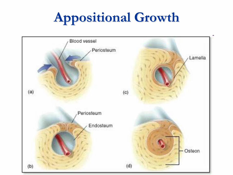
Appositional Growth

Articulations (=Joints)
4 Main Categories of Joints
1. Immovable
2. Fibrous
3. Cartilaginous
4. Synovial
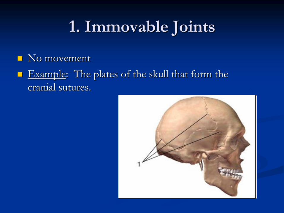
1. Immovable Joints
No movement
Example: The plates of the skull that form the
cranial sutures.
2. Fibrous Joints
Slight movement in the joint.
Dense connective tissue holds bones
together.
Forms Interosseus membrane.
Example: Ulna/Radius
Tibia/Fibula

Fibrous Joint

3. Cartilaginous Joint
Formed by Hyaline or Fibrocartilage.
Examples: Intervertebral disks (Vertebra)
Costal Cartilage (Ribs)
Symphysis Pubis (Pubic Bone)

3. Cartilaginous Joints

4. Synovial Joints
The most “movable” joints in the body.
Membrane secretes synovial fluid in the joint.
Fluid used for lubrication.
Fluid is produced by the bursa sack.
Bone ends have articular (hyaline) cartilage.

Synovial Joints

Types of Synovial Joints
Type of Synovial Joint Location Found in Body
1. Ball and Socket Hip; Shoulder
2. Condylloid Phallanges (fingers/toes)
3. Hinge Knee; Elbow
4. Pivot Radius/Ulna
5. Saddle Artic. b/w Thumb + Metacarpal.
6. Gliding Wrist or ankle
***Types of Synovial Joints (write chart underneath notes on ISN-43).***

Which Joint
Type?
ANS = Hinge

Which Joint
Type?
ANS = Saddle

Which Joint
Type?
ANS = Hinge
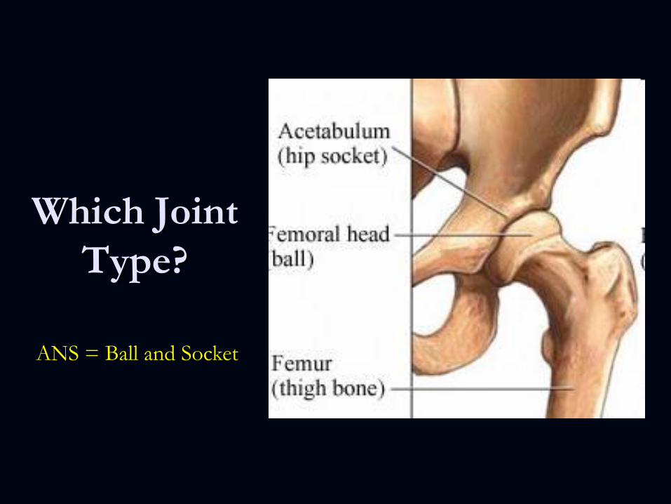
Which Joint
Type?
ANS = Ball and Socket

Which Joint
Type?
ANS = Gliding

Which Joint Type?
ANS = Gliding

Bone Terminology
1. Bumps on Bones
2. Depressions on Bone
3. Holes in Bones

1. Bumps on Bones
Process = projections
Condyle = rounded smooth projections
Epicondyle = above a condyle.
Spine = thorn-like, elevated projection.
Tubercle = knob-like process
Tuberosity = large, rough tubercle.
Trochanter = large, rough tuberosity.
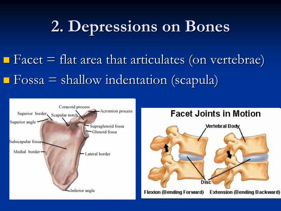
2. Depressions on Bones
Facet = flat area that articulates (on vertebrae)
Fossa = shallow indentation (scapula)

3. Holes in Bones
Foramen = holes for blood vessels, nerves, and
ligaments.
Meatus = bony tube (opening for ear)
Sinus Cavity = space filled with air

Sinus Meatus