Embed Size (px)
Citation preview

©Copyright 2009 Veterinary Learning Systems. This document is for internal purposes only. Reprinting or posting on an external website without written permission from VLS is a violation of copyright laws.
CompendiumVet.com | July 2009 | Compendium: Continuing Education for Veterinarians® E1
CE Article
The Epiphyseal Plate: Nutritional and Hormonal Influences; Hereditary and Other Disorders*
Nutritional, hormonal, and genetic factors play important roles in the growth of animals.1–15 For example,
unbalanced or incomplete diets can result in growth abnormalities.1–15 Hormonal fac-tors, such as prepubertal gonadectomy, have been associated with delayed closure of growth plates in cats, which increases the risk of slipped capital femoral epiphysiolysis (SCFE) and may also affect the distal radius and femur.1,2 Documented hereditary disor-ders affecting the epiphyseal plate include ocular-skeletal dysplasia, dwarfism, canine epiphyseal dysplasia, premature closure of the ulna in Skye terriers, multiple cartilagi-nous exostoses (MCE), and mucopolysaccha-ridosis (MPS).3–7 Osteochondrosis dissecans of the articular cartilage is a common dis-ease in growing dogs, but diseases of the epiphyseal cartilage, including hypertrophic osteodystrophy, retained cartilaginous core, and ununited anconeal process, can also be observed.8,9 The etiology of these conditions appears to be multifactorial,16,17 and further research is warranted to better understand, prevent, and treat these diseases.
NutritionMusculoskeletal disorders are common in young dogs but uncommon in cats. The
prevalence of musculoskeletal disorders is reported to be 22% in dogs younger than 1 year.12 Of these cases, 20% are thought to be nutrition related.12 Deficiencies in vita-min D or trace elements, excessive calcium or vitamin C, and high energy intake have been discussed as reasons for developmen-tal growth abnormalities.12–15 Calcium and energy are two of the most important fac-tors.16 Commercially prepared large-breed puppy diets that meet the nutritional guide-lines of the Association of American Feed Control Officials (AAFCO) and are labeled
“complete and balanced” should be fed to growing large-breed dogs until they reach approximately 80% of their mature size.18 Smaller dogs can be fed puppy diets until approximately 1 year of age and should then be switched to an adult diet.12,18–20 Recommended levels of key nutrients in a diet to prevent orthopedic disease in large-breed puppies are summarized in Table 1.
CalciumExcess feeding (>3% of dry matter) of calcium increases the risk of slowing chondrocyte maturation, which can lead to the development of retained cartilagi-nous cores and, in some cases, angular limb deformity.16 Osteochondrosis dissecans
❯❯ Dirsko J. F. von Pfeil, Dr.med.vet, DVM, DACVS, DECVS Veterinary Specialists of Alaska Anchorage, Alaska
❯❯ Charles E. DeCamp, DVM, MS, DACVS Sarah K. Abood, DVM, PhDa Michigan State University
3 CECREDITS
Nutrition Page E1
Hormonal Influences Page E3
Diseases With Uncertain Etiology
Page E4
Hereditary Disorders Affecting the Epiphyseal Plate
Page E6
At a Glance
Abstract: This article reviews nutritional and hormonal influences, diseases with uncertain etiol-ogy, and hereditary disorders affecting the growth and development of the long bones in dogs and cats.
*A companion article, “The Epiphyseal Plate: Physiology, Anatomy, and Trauma,” is also available on CompendiumVet.com.
aDr. Abood discloses that she has received financial support from Abbott Laboratories, Hill’s Pet Nutrition, and Nestlé Purina PetCare Company.

The Epiphyseal Plate: Influences and Disorders
E2 Compendium: Continuing Education for Veterinarians® | July 2009 | CompendiumVet.com
FREE
CE
Disorders of the growth plate and diseases of young bone and cartilage can be affected by unbalanced nutrition.
QuickNotes
may also develop with a calcium-rich diet.14,21 Growing giant-breed dogs fed high-calcium diets (>3% of dry matter as calcium) experi-ence greater development of osteochondrosis than control dogs.14,21,22 Even when dietary phosphorus is adjusted to maintain a physi-ologic calcium:phosphorus ratio, osteochon-drosis can still occur.14,21
PhosphorusImbalances of dietary phosphorus intake can affect calcium homeostasis and may influence bone metabolism.21 Low dietary phosphorus is uncommon but has been reported to increase calcium and phosphorus absorption in the gut.20,23 Clinical signs can include poor weight gain and reduced growth rate.20,23 By contrast, excessively high phosphorus levels can stim-ulate hormone secretion (secondary hyper-parathyroidism), which decreases calcium ab - sorption.20 The result is poorly calcified, soft bones that are predisposed to pathologic fractures.15–18 It is therefore recommended to maintain a dietary calcium:phosphorus ratio between 1.1:1 and 2:1.12
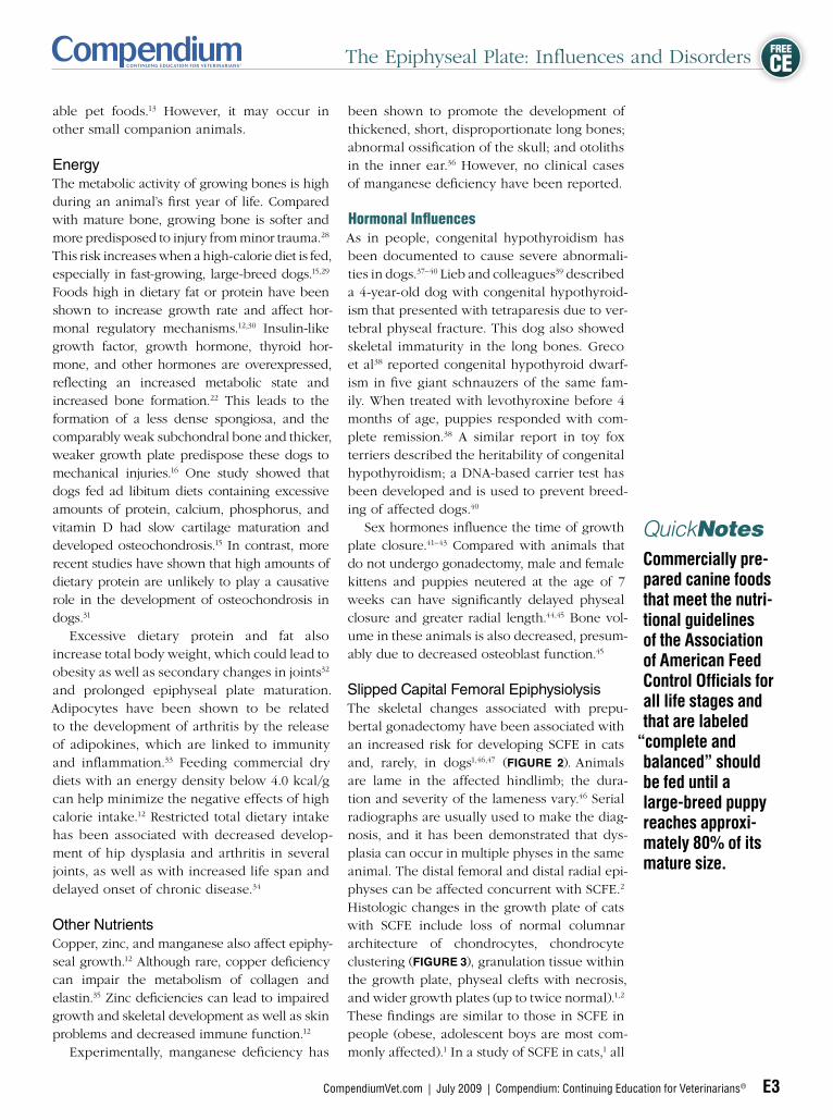
Vitamin DVitamin D and its metabolites are impor-tant in the regulatory mechanism of calcium metabolism and skeletal development in dogs and cats. Vitamin D deficiency leads to a decreased plasma concentration of calcium.24 Chronic hypocalcemia, especially during growth, causes inadequate mineralization of bone so that the cartilaginous matrix in the growth plate fails to calcify.16 The resultant disease, rickets, is characterized by soft bones, lameness, pain, angular limb deformation, and pathologic fractures.13,25 Radiographs typi-cally show thickened physeal cartilage plates and “cupping” of the metaphyseal bone with a dense sclerotic margin26 (Figure 1). Histology shows severe enlargement of the chondrocytes in the growth plate.27 Rickets is extremely rare in dogs and cats being fed commercially avail-
Table 1 Recommended Ranges of Key Nutrients and Energy Content in a Diet for Large-Breed Puppies18
Nutrient Percentage on Dry Matter Basis Amount Fed as Canned
Protein 29–34 6%–8%
Fat 11–16 2%–4%
Mean energy content 3.4–4.1 13.4–15.9 KJ/g DMB
Calcium 0.8–1.4 —
Phosphorus 0.7–1.2 —
Fiber 2.4–5.6 1%
KJ/g = kilojoule per gram; DMB = dry matter basis
Rickets in a 5-year-old chimpanzee. Note the extremely widened and radiolucent physes (arrows) of the pelvis, femora, and tibiae, typical of this disease. Excessive vitamin D deficiency may prevent the cal-cification process in the metaphyses. This disease is extremely rare in dogs and cats in developed countries because of the availability of well-balanced commer-cial veterinary diets. (Courtesy of Dr. Tobias Schwarz, University of Wisconsin-Madison)
Figure 1
The Epiphyseal Plate: Influences and Disorders
CompendiumVet.com | July 2009 | Compendium: Continuing Education for Veterinarians® E3
FREE
CE
Commercially pre-pared canine foods that meet the nutri-tional guidelines of the Association of American Feed Control Officials for all life stages and that are labeled
“complete and balanced” should be fed until a large-breed puppy reaches approxi-mately 80% of its mature size.
QuickNotes
able pet foods.13 However, it may occur in other small companion animals.
EnergyThe metabolic activity of growing bones is high during an animal’s first year of life. Compared with mature bone, growing bone is softer and more predisposed to injury from minor trauma.28 This risk increases when a high-calorie diet is fed, especially in fast-growing, large-breed dogs.15,29 Foods high in dietary fat or protein have been shown to increase growth rate and affect hor-monal regulatory mechanisms.12,30 Insulin-like growth factor, growth hormone, thyroid hor-mone, and other hormones are overexpressed, reflecting an increased metabolic state and increased bone formation.22 This leads to the formation of a less dense spongiosa, and the comparably weak subchondral bone and thicker, weaker growth plate predispose these dogs to mechanical injuries.16 One study showed that dogs fed ad libitum diets containing excessive amounts of protein, calcium, phosphorus, and vitamin D had slow cartilage maturation and developed osteochondrosis.15 In contrast, more recent studies have shown that high amounts of dietary protein are unlikely to play a causative role in the development of osteochondrosis in dogs.31 Excessive dietary protein and fat also increase total body weight, which could lead to obesity as well as secondary changes in joints32 and prolonged epiphyseal plate maturation. Adipocytes have been shown to be related to the development of arthritis by the release of adipokines, which are linked to immunity and inflammation.33 Feeding commercial dry diets with an energy density below 4.0 kcal/g can help minimize the negative effects of high calorie intake.12 Restricted total dietary intake has been associated with decreased develop-ment of hip dysplasia and arthritis in several joints, as well as with increased life span and delayed onset of chronic disease.34
Other NutrientsCopper, zinc, and manganese also affect epiphy-seal growth.12 Although rare, copper deficiency can impair the metabolism of collagen and elastin.35 Zinc deficiencies can lead to impaired growth and skeletal development as well as skin problems and decreased immune function.12
Experimentally, manganese deficiency has
been shown to promote the development of thickened, short, disproportionate long bones; abnormal ossification of the skull; and otoliths in the inner ear.36 However, no clinical cases of manganese deficiency have been reported.
Hormonal Influences As in people, congenital hypothyroidism has been documented to cause severe abnormali-ties in dogs.37–40 Lieb and colleagues39 described a 4-year-old dog with congenital hypothyroid-ism that presented with tetraparesis due to ver-tebral physeal fracture. This dog also showed skeletal immaturity in the long bones. Greco et al38 reported congenital hypothyroid dwarf-ism in five giant schnauzers of the same fam-ily. When treated with levothyroxine before 4 months of age, puppies responded with com-plete remission.38 A similar report in toy fox terriers described the heritability of congenital hypothyroidism; a DNA-based carrier test has been developed and is used to prevent breed-ing of affected dogs.40 Sex hormones influence the time of growth plate closure.41–43 Compared with animals that do not undergo gonadectomy, male and female kittens and puppies neutered at the age of 7 weeks can have significantly delayed physeal closure and greater radial length.44,45 Bone vol-ume in these animals is also decreased, presum-ably due to decreased osteoblast function.45
Slipped Capital Femoral EpiphysiolysisThe skeletal changes associated with prepu-bertal gonadectomy have been associated with an increased risk for developing SCFE in cats and, rarely, in dogs1,46,47 (Figure 2). Animals are lame in the affected hindlimb; the dura-tion and severity of the lameness vary.46 Serial radiographs are usually used to make the diag-nosis, and it has been demonstrated that dys-plasia can occur in multiple physes in the same animal. The distal femoral and distal radial epi-physes can be affected concurrent with SCFE.2 Histologic changes in the growth plate of cats with SCFE include loss of normal columnar architecture of chondrocytes, chondrocyte clustering (Figure 3), granulation tissue within the growth plate, physeal clefts with necrosis, and wider growth plates (up to twice normal).1,2 These findings are similar to those in SCFE in people (obese, adolescent boys are most com-monly affected).1 In a study of SCFE in cats,1 all

The Epiphyseal Plate: Influences and Disorders
E4 Compendium: Continuing Education for Veterinarians® | July 2009 | CompendiumVet.com
FREE
CE
affected animals were 5 to 24 months old, 85% were male, 23% were Siamese, and 90% were obese. These cats were neutered between 4 and 8 months of age.46 Depending on the clini-cal signs, conservative treatment or surgery (e.g., femoral head and neck ostectomy) may be indicated.
Diseases With Uncertain EtiologyFemoral Neck Metaphyseal Osteopathy in CatsCats with metaphyseal osteopathy develop necrosis in the proximal metaphysis, which eventually results in a secondary, pathologic fracture of the femoral neck48 (Figure 4). The condition may be unilateral or bilateral. Adult male cats (neutered and intact) are overrep-resented. Lameness originates from the coxo-femoral joint. Unlike SCFE, radiographs show characteristic chronic degenerative changes in the femoral neck region, including areas of radiolucency, bone resorption, an “apple-core” appearance, or an irregular radiolucent fracture line.48,49 Several etiologies have been proposed: traumatic fracture with secondary bone resorption, avascular necrosis, osteomy-elitis, feline herpesvirus, and changes second-ary to SCFE.48,49 Histology does not show the typical features of SCFE but may include bone necrosis and microfracture, fibrosis, metaplasia, osteomyelitis, and synovitis of the surrounding connective tissue.48,49 Excision of the femoral head and neck carries a good prognosis.48,49
Hypertrophic OsteodystrophyHypertrophic osteodystrophy is seen in grow-ing large- or giant-breed dogs with open
The recommended percentages, on a dry matter basis, of key nutrients in a diet for large-breed puppies to minimize the risk of orthopedic disease are as follows: pro-tein, 29% to 34%; fat, 11% to 16%; mean energy, 3.4% to 4.1%; calcium, 0.8% to 1.4%; phos-phorus, 0.7% to 1.2%; fiber, 2.4% to 5.6%.
QuickNotes
Slipped capital femoral epiphysiolysis (SCFE) in a 10-month-old castrated Siamese cat. Note the obvious injury on the left (arrow) and mild mediodistal displace-ment of the femoral capital epiphysis on the right (ellipse).
Irregular chondrocyte clusters (ellipse) attached to the metaphysis of a cat with SCFE secondary to dysplasia of the epiphyseal plate. (Hematoxylin–eosin stain; original magnification × 10; courtesy of Dr. Linden Craig, University of Tennessee)
Femoral neck metaphyseal osteopathy in a 2-year-old castrated cat. Note the severely irregular changes around the femoral necks. This radiographic appearance may represent changes secondary to SCFE.
Figure 2 Figure 3
Figure 4

The Epiphyseal Plate: Influences and Disorders
CompendiumVet.com | July 2009 | Compendium: Continuing Education for Veterinarians® E5
FREE
CE
epiphyseal plates.9 Affected animals present with intermit-tent lameness, fever, and extremely pain-ful, swollen distal metaphyses of the long bones. A radio-lucent line parallel to the growth plate (pseudophyseal line) is the hallmark of the disease. Possible causes of this line include increased leu-kocyte activity, bone lysis, and failure of ossification of the hypertrophic zone.50 Large, swollen, dense metaphyses and perio- steal exostosis com-plete the classic radio- graphic appearance (Figure 5). The etiology of hyper- trophic osteo dystrophy is unknown. Nutritional (vitamin C deficiency) and viral (distemper virus) factors have been suggested.50 However, these theories remain unproven. The disease is usually self-limiting,
but affected dogs can be extremely ill. Treatment consists of supportive care. Permanent bone changes, angular limb deformities second-ary to asymmetric or asynchronous growth or bridging of bone, and retardation of axial growth have been reported.9,50
Osteochondrosis Osteochondrosis is a multifocal disease with incomplete endochondral ossification of the articular-epiphyseal cartilage. It has also been reported to affect the cartilage of the epiphy-seal growth plate.51,52
The etiology of osteochondrosis is not clear. Early in an animal’s development, blood ves-sels, nerves, and lymphatics supply the epiphy-seal cartilage.53 As the animal ages and gains weight, the number of blood vessels decreases
until the cartilage becomes avascular. Studies of pigs and chickens in which ischemia was induced have demonstrated that, at some point during growth, the viability of cartilage cells depends on the integrity of cartilage canal vessels.11 Aside from ischemia, other etiologies, such as hereditary factors, rapid growth, and nutritional imbalances, have been suggested to cause osteochondrosis.27 Osteochondrosis results in failure of matrix calcification, with retention of cartilage rather than conversion to bone, leading to a thick-ened, weaker growth plate. Excessive caloric intake has been suggested to be a risk factor in the development of increased articular car-tilage thickness, but high dietary protein alone is unlikely to be the cause of osteochondrosis in dogs.22,54 Damage to the cartilage structures, retained cartilaginous cores, and asymmetric growth may be consequences of this process.27
Histologic examination of the lesions may reveal areas of necrotic chondrocytes in the reserve zone, close to necrotic vascular channels.55 When osteochondrosis affects the articular surface, lesions consist of necrotic cartilage with a cleft extending from the subchondral bone to the articular surface, resulting in synovitis, joint effusion, and clinical signs of lameness.11 This form of osteochondrosis is called osteochon-drosis dissecans (Figure 6). Treatment consists of removal of loose cartilage and debridement of subchondral bone. The prognosis varies depending on the affected joint and advance-ment of arthritis.
Retained Cartilaginous Core Retained cartilaginous cores are most com-monly seen in the distal ulna of growing large- and giant-breed dogs56 (Figure 7). These cores are a developmental disorder of endochon-dral ossification in which physeal calcifica-tion is disturbed, resulting in decreased bone growth of the ulna.57 The etiology is uncer-tain; however, dietary imbalances and a form of osteochondrosis have been suggested.10,57 Depending on the severity, affected dogs present with variable degrees of lameness and multiple deformities, including valgus defor-mity of the carpus and cranial bowing of the radius (with or without lameness). A radiolu-cent cartilage core in the center of the dis-tal ulnar physis can be seen radiographically (Figure 7). Treatments include return to a com-
Clients feeding a growing dog or cat a homemade diet (raw or cooked) should have the diet evaluated and their pet examined by a veterinarian at least twice yearly.
QuickNotesTypical radiographic appearance of the dis-tal radius and ulna of a dog with hypertro-phic osteodystrophy. Note the “double physeal line” (arrows), extra- and subperiosteal bone forma-tion at the metaphysis and diaphysis, and widening of the epiphyseal plate of the distal radius and ulna.
Figure 5

The Epiphyseal Plate: Influences and Disorders
E6 Compendium: Continuing Education for Veterinarians® | July 2009 | CompendiumVet.com
FREE
CE
plete, balanced diet and cessation of excessive dietary supplements in growing dogs. If clini-cally indicated, corrective osteotomy should be performed.10 The prognosis varies accord-
ing to the degree of deformity and lameness at initial presentation.
Ununited Anconeal ProcessUnlike small-breed dogs, in large-breed dogs, the anconeal process develops from a sepa-rate center of ossification.55 Failure of this cen-ter to fuse with the proximal ulna by 5 months of age is characteristic of ununited anconeal process (Figure 8). Disturbance of endochon-dral ossification, resulting in osteochondrosis, is presumed to be the underlying etiology.8 However, joint incongruity, growth plate trauma, excess calcium, excess weight, and genetic and hormonal factors have also been discussed.8,55 This disease is hereditary in some German shepherd lines, in which three dominant genes have been suggested for this trait.58 Treatment options are controversial and include medical management, excision of the anconeal process, attachment with a lag screw, and ulnar osteotomy.8,55,58 Combination of the latter two methods possibly provides a better prognosis than other treatment options.55
Hereditary Disorders Affecting the Epiphyseal PlateIncomplete Ossification of the Humeral CondyleIncomplete ossification of the humeral condyle (IOHC) was first described in 1990 by Drapé59
Gonadectomy at an early age can lead to significantly delayed physeal closure and slipped capital femoral epi-physiolysis (SCFE) in cats and, rarely, dogs. The distal femoral and distal radial epiphysis can be affected concur-rent with SCFE.
QuickNotes
Lateral radiograph of the shoulder joint showing osteochondrosis dissecans on the caudal aspect of the humeral head (arrow) in a 10-month-old rottweiler.
Ununited anconeal process (arrowhead) in a 7-month-old German shepherd. Normal closure time is approximately 5 months of age.
Lateral radiograph of the distal radius and ulna in a 5-month-old Newfoundland with a retained cartilaginous core. Note the “flame-like” radiolucent area in the center of the ulnar growth plate enclosed by two radiodense lines (box), which is diag-nostic for this condition.
Figure 6 Figure 8
Figure 7

The Epiphyseal Plate: Influences and Disorders
CompendiumVet.com | July 2009 | Compendium: Continuing Education for Veterinarians® E7
FREE
CE
(Figures 9 and 10). Fusion of the two centers of ossification normally occurs at around 10 weeks of age. With IOHC, minor trauma may lead to condylar fracture and acute onset of lameness. Although the disease occurs early in life, the age at presentation is commonly between 3 and 8 years. This condition occurs mainly in spaniels, for which an autosomal recessive mode of inheritance has been sug-gested.60 It is also described for rottweil-ers, English pointers, wachtelhunds, German wirehaired pointers, Bernese mountain dogs, Newfoundlands, giant schnauzers, Polish low-land sheepdogs, and Labrador retrievers.61–63 Radiographs should be obtained in 15° cran-iomedial to 15° caudolateral views.60 Both legs should be examined. The affected leg usu-ally shows a fracture of the humeral condyle (Figure 9). If radiographs are not sufficient, computed tomography or arthroscopy may help diagnose a bilateral condition (Figure 10). Lag screw fixation for the fractured leg or prophy-lactic transcondylar fixation for the contralateral leg is the best method of treatment; however,
Osteochondrosis, incomplete ossifica-tion of the humeral condyle, retained cartilaginous core, and ununited anconeal process should be on the dif-ferential diagnosis for young animals with orthopedic problems affecting the growth plate.
QuickNotesAnteroposterior radiograph showing a fracture of the lateral epicondyle of the humerus in a 6-year-old cocker spaniel. This breed is predisposed to incomplete ossification of the humeral condyle.
Images of the contralateral humeral condyle of the dog in Figure 9. The dog was not lame or painful in this leg. (A) Anteroposterior radiograph. (B) While difficult to see on radiographs, a radiolucent line separating the two condyles (ellipse) and minimal dis-placement is visible on computed tomography. To pre-vent fracture in the future, a prophylactic intercondylar screw was placed.
Figure 9 Figure 10
a
b

The Epiphyseal Plate: Influences and Disorders
E8 Compendium: Continuing Education for Veterinarians® | July 2009 | CompendiumVet.com
FREE
CE
nonunion is common with this disease.60,64 It has been suggested that dense cancellous bone, fibrous tissue, or undue motion may prevent appropriate or complete healing.60
Ocular-Skeletal DysplasiaThere are several reports of ocular-skeletal dys-plasia in dogs.65–67 This condition occurs mainly in Labrador retrievers, but it has also been described for Samoyeds and German shepherds. Barnett and colleagues68 reported the mode of inheri-tance as an autosomal recessive defect. Affected dogs present with a typical “downhill conforma-tion” (front limbs shorter than hindlimbs; Figure
11) and bony abnormalities, such as bone short-ening in the forelegs with malformation of the humeral condyles, fracture of the lateral portion of the humeral condyle, asynchronous growth of the radius and ulna, varus deformity and sec-ondary degenerative joint disease of the elbows, ununited and hypoplastic anconeal and/or coro-noid process, and carpus valgus (Figure 12). Hip dysplasia and retarded tibial growth may also occur.65–67 The degree of lameness varies with severity. Cortical thickness and density of the bones can appear to be reduced, and the epiphy-ses and cuboid bones are larger and misshapen compared with those of normal littermates.
Genetic disorders such as epiphyseal dysplasia, ocular-skeletal dysplasia, chondrodysplasia, multiple cartilagi-nous exostoses, and mucopolysaccha-ridosis should also be included in the differential diag-nosis for growth abnormalities.
QuickNotes
The ocular component of this disease may present as night blindness. Ocular pathology includes cataracts, retinal dysplasia, and retinal detachment. The degree of vision impairment depends on severity of the lesions.65–67 Carrig and colleagues66 established a breeding colony of Labrador retrievers to further investigate the mode of inheritance of this disorder. The heterozygotes were found to have a clinically normal skeleton with mild ocular abnormali-ties, while homozygotes showed clinical signs of both ocular and skeletal dysplasia. It was con-cluded that abnormalities resulted from a single gene with recessive effects on the skeleton but with incomplete dominant effects on the eyes.66
Depending on the degree of orthopedic and/or ophthalmologic disease, surgical treatment may be beneficial. The prognosis for complete restora-tion of normal orthopedic function is guarded to poor, and affected dogs should not be bred.67
ChondrodysplasiaChondrodysplasia, also commonly called dwarf-ism, has been described in Great Danes, Scottish deerhounds, Alaskan malamutes, Norwegian elk-hounds, and miniature poodles.5 Other breeds may be affected. In contrast to ocular-skeletal
One-year-old Labrador retriever with ocular-skeletal dysplasia, showing the typical downhill conformation with bone shortening in the forelegs and carpus valgus. (Courtesy of Dr. Ullrich Reif, Tierklinik Böbingen/Schwäbisch Gmünd, Germany)
Radiographs of the dog in Figure 11, showing malformation of the humeral condyles, asynchronous growth of the radius and ulna, and carpus valgus. (A) Lateral view. (B) Anteroposterior view.
Figure 11 Figure 12
a b

The Epiphyseal Plate: Influences and Disorders
CompendiumVet.com | July 2009 | Compendium: Continuing Education for Veterinarians® E9
FREE
CE
dysplasia, in which limb shortening is mild, chondrodysplasia is characterized by severely shortened limbs, normal body length, and nor-mally sized skull, leaving the impression of a dis-proportionate dwarf 5 (Figure 13). The disease is genetically transmitted as a simple autosomal recessive trait. In Alaskan malamutes, it is com-bined with a permanent macrocytic, hypochro-mic anemia.69,70 Affected animals are usually not lame. Chondrodysplasia appears radiographically similar to rickets and can easily be misin-terpreted. Radiographic changes include flar-ing of the distal metaphyseal borders of the radii and ulnae. Prominent curvature of the front limbs and carpus valgus are present. For Great Danes, flaring of all metaphyses, includ-ing a “trumpet-like” flaring of the distal tibia, has been described.6 Ossification of vertebral end plates and/or centers of ossification is delayed. Epiphyseal plates show delayed clo-sure with shortened, disorganized columns of chondrocyte proliferation, swollen chondro-cytes, and diminished endochondral ossifica-tion. Affected chondrocytes reveal a markedly irregular dilatation of cisternae of the rough endoplasmic reticulum.5 There may be a gen-eralized defect involving all hyaline cartilage throughout the body. If no severe abnormali-ties occur, the quality of life of affected dogs may be good. However, if animals show signs of osteopenia, kyphosis, joint laxity, reduced diameter of the tracheal lumen, or angular limb deformities, the prognosis is guarded.7
Epiphyseal DysplasiaEpiphyseal dysplasia is a hereditary condi-tion characterized by delayed and irregular ossification of the cartilage of the epiphysis.
Typical radiographic findings for epi-physeal dysplasia include shortening of long bones and widening of the metaphysis, lack of radiopacity, and ossification of cartilage.
QuickNotes
Affected animals may present with painless swelling, typically on the medial aspect of the joint, limited range of motion, and recurrent locking of the joints. Radiographic findings include shortening of long bones and widen-ing of the metaphysis, lack of radiopacity, and ossification of cartilage3 (Figure 14). At later stages in the disease, irregular bony enlarge-ments with secondary joint degeneration can be observed.71 Several human cases have been reported72–74; however, only one case has been reported in the veterinary literature.3 An unpublished feline case was seen at Michigan State University in 2006 (Figure 14).
Epiphyseal dysplasia develops secondary to an altered process of cell proliferation at the superficial zone of articular cartilage or a local-ized disturbance of the preaxial and postaxial part of the apical cap of the limb bud in early fetal development.75 The disease may also be a variant of MCE or osteochondroma arising within a joint.74 Histologically, there is intra-cellular accumulation of predominantly chon-droitin sulfate and glycoprotein, followed by liquefaction of these materials and formation
A 5-year-old golden retriever with chon-drodysplasia (dwarfism). Note the short legs compared with the large body and head.
Epiphyseal dysplasia in a 6-month-old cat with chronic hindlimb lameness. Radiographic changes are lack of radiopacity and ossification of cartilage (ellipse). Secondary joint degeneration can be expected at a later stage.
Figure 13 Figure 14

The Epiphyseal Plate: Influences and Disorders
E10 Compendium: Continuing Education for Veterinarians® | July 2009 | CompendiumVet.com
FREE
CE
of cysts, which finally calcify.71 Alterations of bone and cartilage with hyperplastic chondro-cytes and fibrous tissue interrupted by phases of normal endochondral ossification can also be seen.76 This condition may lead to pain and interference with function or to angular limb deformity. Corrective surgery may be required to restore function.75
Premature Closure of the Distal Ulnar Physis in Skye TerriersThirty years ago, Lau4 described premature closure of the distal ulnar physis in Skye terri-ers as a hereditary disease. Twenty-three dogs, all the offspring of two females and four males, presented with forelimb lameness between 3 and 5 months of age. Radiographic and physi-cal examination findings included carpus valgus, lateral subluxation of the radial head, circumduction of the elbows, and decreased range of motion of the elbow joint. Lau pro-vided evidence of a recessively inherited trait as the etiology for this disease. A similar syn-drome, although not well described in genetic terms, is seen in Welsh corgis, basset hounds, and other chondrodystrophic breeds.
Multiple Cartilaginous ExostosesMCE are “mini growth plates” found in ran-dom areas of metaphyseal bone50 (Figure
15). They are identified in cats and dogs of any age as bony proliferations on the body and spinous processes of the vertebrae; the
processes of the scapula, sternum, ribs, and ischii; and the petrous portion of the temporal bones.77,78 The disease is commonly an inci-dental finding because animals do not usually show clinical signs. This condition was for-merly called osteochondromatosis. Currently, a solitary lesion is described as osteochon-droma, whereas multiple lesions are referred to as MCE.79 These malformations are a result of disturbed endochondral ossification at the periphery of the growth plate with abnormal,
“benign” development of cartilage and fibrous connective tissue. MCE seem to be associated with FeLV infection in cats.80,81 MCE are reported to occur predominantly in Siamese cats, in which the temporal bone, vertebral bodies, and spinous processes of the scapula, vertebrae, sternum, ribs, and ischium can be affected.82 The disease is a heritable entity in dogs and has been identi-fied in the vertebrae, ribs, and long bones.83–85 Interestingly, lesions are not observed to affect long bone growth. Dogs and cats may have no clinical signs, unless the exostoses cause dysfunction of a joint or vital structure such as the trachea or spinal cord.86,87 Affected ani-mals may present with myelopathy, commonly at younger than 1 year.80 Depending on the severity of the lesion, progressive neurologic signs may be seen. Histologically, lesions are consistent with a site of endochondral ossifi-cation.85 Clinical presentation and radiographic and histologic findings help to differentiate MCE from other benign polyostotic exostoses such as tumoral calcinosis or canine dissemi-nated idiopathic skeletal hyperostosis.80 While the disease is often aggressive and carries a poor prognosis in cats, dogs usually have a good prognosis because the process ceases at maturity.88 However, in rare cases, transfor-mation into malignant neoplasia several years after initial diagnosis has been reported.80
MucopolysaccharidosisMPS is a rare storage disease in which dif-ferent lysosomal enzyme defects result in the inability to degrade glycosaminoglycans.5 Endochondral ossification is disturbed, result-ing in skeletal abnormalities. Six subtypes of MPS (I, II, IIIA, IIIB, VI, VII) have been described in dogs and cats.89,90 Mixed-breed dogs, German shepherds, Plott hounds, rottweilers, Labrador retrievers, wire-
Multiple cartilaginous exostoses in a dog. Note the bony changes on the spinous processes of the cranial thoracic vertebral bodies (ellipse) and at the sixth and seventh ribs (arrow).
Figure 15
Multiple cartilagi-nous exostoses are
“mini growth plates” in random areas of metaphyseal bone.
QuickNotes

The Epiphyseal Plate: Influences and Disorders
CompendiumVet.com | July 2009 | Compendium: Continuing Education for Veterinarians® E11
FREE
CE
haired dachshunds, New Zealand huntaways, miniature poodles, Chesapeake Bay retrievers, and miniature schnauzers have been reported to be affected by different types of MPS.89 The only MPS disorder with mostly neurologic signs is MPS IIIA, reported in wirehaired dachs-hunds and New Zealand huntaways.89 MPS I, MPS VI, and MPS VII have also been observed in cats.89,90 MPS VI occurs in Siamese cats and follows an autosomal recessive inheritance.90
Animals present with dwarfism, a dispro-portionately small and broad maxilla, small ears, large paws, hip dysplasia, crepitus and hypermotility in multiple joints, and fusion of vertebral bodies with subsequent neurologic deficiencies5 (Figures 16 and 17). Affected cats excrete high amounts of dermatan sulfate in their urine.90 Dermatan sulfate glycosamino-glycan (formerly called mucopolysaccharide) accumulates abnormally in several of the dif-ferent subtypes of MPS disorders and is found mostly in skin tissue but also in blood vessels,
heart valves, tendons, and the lungs.90 Breed-specific DNA tests have been established for the diagnosis of affected animals and for carrier detection.89 Treatment and prognosis depend on the type and severity of MPS. MPS III is usually incurable, and most animals are euthanized before 5 years of age.89
Typical phenotype of mucopolysaccharido-sis in a cat. Note the small ears and large head. This cat also had crepitus and hypermotility in several joints.
Figure 16
Mucopolysaccharidosis in a cat showing dysplastic hip joints with generalized radiolucency of the acetabulum, head, neck, and subtrochanteric area of the femur. Also note generalized radiolucency of the distal femoral condyles and tibiae and the mild varus deformity (medial bowing) at the level of the knee joints. (Courtesy of Drs. Van Knox, Susquehanna Veterinary Imaging, LLC, and Mark Haskins, University of Pennsylvania)
Figure 17
Mucopolysaccharidosis is associated with dwarfism, small ears, large paws, pathologic changes in multiple joints, and neurologic deficiencies.
QuickNotes
The Epiphyseal Plate: Influences and Disorders
E12 Compendium: Continuing Education for Veterinarians® | July 2009 | CompendiumVet.com
FREE
CE
ConclusionA thorough understanding of the anatomy and physiology of the growth plate is necessary to understand the effect of nutritional imbalances, hormonal influences, and hereditary disorders on developing long bones. Early correction of each specific condition is warranted to avert permanent damage. Dogs and cats that are
affected with a familial or potentially heritable problem should not be considered for breed-ing purposes.
AcknowledgmentsTo Dr. Cheri Johnson, DVM, MS, DACVIM, for edito-rial work, and Sandra Schallberger, Dr.med.vet., for technical support.
References1. Craig LE. Physeal dysplasia with slipped capital femoral epiphy-sis in 13 cats. Vet Pathol 2001;38:92-97.2. Newton AL, Craig LE. Multicentric physeal dysplasia in two cats. Brief communications and case reports. Vet Pathol 2006;43:388-390. 3. Vignoli M, Sarli G, Rossi F, et al. Dysplasia epiphysealis hemimel-ica in a Boxer puppy. Vet Radiol Ultrasound 2002;43(6):528-533.4. Lau R. Inherited premature closure of the distal ulnar physis. JAAHA 1977;13:609-612.5. Sande RD, Bingel SA. Animal models of dwarfism. Vet Clin North Am Small Anim Pract 1982;13(1):71-89.6. Bingel SA, Sande RD. Chondrodysplasia in five Great Pyrenees. JAVMA 1994;205(6):845-848.7. Breur GJ, Slocombe RF, Padgett GA, et al. Clinical, radiographic, pathologic and genetic features of osteochondroplasia in Scottish Deerhounds. JAVMA 1989;195(9):606-612.8. Piermattei DL, Flo GL, DeCamp CE. Osteochondrosis of the el-bow. In: Brinker, Piermattei, and Flo’s Handbook of Small Animal Orthopedics and Fracture Repair. 4th ed. St. Louis: Saunders El-sevier; 2006:339-344.9. Piermattei DL, Flo GL, DeCamp CE. Hypertrophic osteodystro-phy. In: Brinker, Piermattei, and Flo’s Handbook of Small Animal Or-thopedics and Fracture Repair. 4th ed. St. Louis: Saunders Elsevier; 2006:781-784. 10. Piermattei DL, Flo GL, DeCamp CE. Retained cartilaginous cores. In: Brinker, Piermattei, and Flo’s Handbook of Small Animal Orthopedics and Fracture Repair. 4th ed. St. Louis: Saunders El-sevier; 2006:780-781. 11. Ekman S, Carlson CS. The pathophysiology of osteochondrosis. Vet Clin North Am Small Anim Pract 1998;28(1):17-32.12. Richardson DC, Zentek J. Nutrition and osteochondrosis. Vet Clin North Am Small Anim Pract 1998;28(1):115-135.13. Kallfelz FA, Dzanis DA. Overnutrition: An epidemic prob-lem in pet animal practice? Vet Clin North Am Small Anim Pract 1989;19(3):433-446.14. Hazewinkel HAW, Goedengebuure SA, Poulos PW, et al. Influ-ences of chronic calcium excess on the skeletal development of growing Great Danes. JAAHA 1985;21:377-391.15. Hedhammar A, Wu FM, Krook L, et al. Overnutrition and skel-etal disease. An experimental study in growing Great Dane dogs. Cornell Vet 1974;64(2):Suppl 5:5-160.16. Fascetti AJ. Food for thought on canine developmental orthopedic disease.Vet Surg 2006;35(3):211-213.17. Lopez MJ, Quinn MM, Markel MD. Associations between ca-nine juvenile weight gain and coxofemoral joint laxity at 16 weeks of age. Vet Surg 2006;35(3):214-218.18. Remillard RL. Practical nutritional and dietary recommenda-tions: minimizing clinical aspects of orthopedic diseases. Proc Western Vet Conf 1995:25-28.19. Association of American Feed Control Officials (AAFCO). Offi-cial Publication Association of American Feed Control Officials In-corporated. Washington, DC: AAFCO; 2009:147.20. Lauten SD. Nutritional risks to large-breed dogs: from wean-ing to geriatric years. Vet Clin North Am Small Anim Pract 2006;36(6):1345-1359. 21. Hazewinkel HAW, Van den Brom WE, Van T Klooster AT, et al. Calcium metabolism in Great Dane dogs fed diets with various cal-cium and phosphorus levels. J Nutr 1991;121(11 suppl):99-106.22. Nap RC, Hazewinkel HA, Voorhout G, et al. Growth and skeletal
development in Great Dane pups fed different levels of protein in-take. J Nutr 1991;121(11 Suppl):107-113.23. Tanaka Y, DeLuca HF. The control of 25-hydroxyvitamin D metabolism by inorganic phosphorus. Arch Biochem Biophys 1977;154:566-574.24. Hall JE. Parathyroid hormone, calcitonin, calcium and phos-phate metabolism, Vitamin D, bone, and teeth. In: Guyton AC, Hall JE, eds. Textbook of Medical Physiology. 11th ed. Philadelphia: El-sevier Saunders; 2006:978-995.25. Braden TD. Histophysiology of the growth plate and growth plate injuries. In: Smeak DD, Bojrab JM, Bloomberg MS, eds. Dis-ease Mechanisms in Small Animal Surgery. 2nd ed. Philadelphia: Lippincott Williams & Wilkins; 1993:1027-1041.26. Morgan JP. Radiographic diagnosis of bone disease. In: Mor-gan JP, ed. Radiology of Veterinary Orthopedics: Features of Di-agnosis. 2nd ed. Iowa City: Iowa State University Press; 2000:116, 119.27. Olsson SE, Ekman S. Rickets and nutritional secondary hyper-parathyroidism. In: Sumner-Smith G, ed. Bone in Clinical Orthope-dics. 2nd ed. Stuttgart: Thieme; 2002:139, 142.28. Carter DR, Spengler DM. Biomechanics of fracture. In: Sum-ner-Smith G, ed. Bone in Clinical Orthopedics. 2nd ed. Stuttgart: Thieme; 2002:275.29. Kealy RD, Olsson SE, Monti KL, et al. Effects of limited food consumption on the incidence of hip dysplasia in growing dogs. JAVMA 1992;201(6):857-863.30. Glade MJ, Gupta S, Reimers TJ. Hormonal responses to high and low planes of nutrition in weanling thoroughbreds. J Anim Sci 1984;59(3):658-665.31. Nap RC, Hazewinkel HAW, Voorhout G, et al. The influence of the dietary protein content on growth in giant breed dogs. J Vet Comp Orthop Trauma 1993;6:1-8.32. Smith GK, Paster ER, Powers MY, et al. Lifelong diet restriction and radiographic evidence of osteoarthritis of the hip joint in dogs. JAVMA 2006;229:690-693.33. Trayhurn P, Bing C, Wood IS. Adipose tissue and adipok-ines—energy regulation from the human perspective. J Nutr 2006;136(7):1935S-1939S.34. Kealy RD, Lawler DF, Ballam JM, et al. Effects of diet re-striction on life span and age-related changes in dogs. JAVMA 2002;220(9):1315-1320.35. Danks DM. Copper deficiency in humans. In: Biological Roles of Copper. Proceedings of the CIBA Foundation Symposium 79. Amsterdam: Excerpta Medica; 1980:163-182.36. Hurley LS, Keen CL. Manganese. In: Mertz W, ed. Trace Ele-ments in Human and Animal Nutrition. Orlando, FL: Academic Press; 1986:185-223.37. Loder RT, Aronson DD, Bollinger RO. Seasonal variation of slipped capital femoral epiphysis. J Bone Joint Surg [Am] 1990;72(3):378-381.38. Greco DS, Feldman EC, Peterson ME, et al. Congenital hypo-thyroid dwarfism in a family of giant schnauzers. J Vet Intern Med 1991;5(5):306-307.39. Lieb AS, Grooters AM, Tyler JW, et al. Tetraparesis due to ver-tebral physeal fracture in an adult dog with congenital hypothyroid-ism. J Small Anim Pract 1997;38(8):364-367.40. Fyfe JC, Kampschmidt K, Dang V, et al. Congenital hypo-thyroidism with goiter in toy fox terriers. J Vet Intern Med 2003;17(1):50-57.41. Caruso-Nicoletti M, Cassorla F, Skerda M, et al. Short term, low

The Epiphyseal Plate: Influences and Disorders
CompendiumVet.com | July 2009 | Compendium: Continuing Education for Veterinarians® E13
FREE
CE
dose estradiol accelerates ulnar growth in boys. J Clin Endocrinol Metab 1985;61(5):896-898.42. Lloyd HM, Meares JD, Jacobi J, et al. Effects of stilboestrol on growth hormone secretion and pituitary cell proliferation in the male rat. J Endocrinol 1971;51(3):473-481.43. Cutler GB Jr, Cassorla FG, Ross JL, et al. Pubertal growth: physiology and pathophysiology. Recent Prog Horm Res 1986; 42:443-470.44. Salmeri KR, Bloomberg MS, Scruggs SL, et al. Gonadectomy in immature dogs: effects on skeletal, physical, and behavioral devel-opment. JAVMA 1991;198(7):1193-1203.45. Root MV, Johnston SD, Olson PN. The effect of prepuberal and postpuberal gonadectomy on radial physeal closure in male and female domestic cats. Vet Radiol Ultrasound 1997;38(1):42-47.46. McNicholas Jr WT, Wilkens BE, Blevins WE, et al. Sponta-neous femoral capital physeal fractures in adult cats: 26 cases (1996-2001). JAVMA 2002;221(12):1731-1736.47. Dupuis J, Breton L, Drolet R. Bilateral epiphysiolysis of the fem-oral heads in two dogs. JAVMA 1997;210(8):1162-1165.48. Queen J, Bennet D, Carmichael N. Femoral neck metaphyseal osteopathy in the cat. Vet Rec 1998;142:159-162.49. Ridge PA. What is your diagnosis? J Small Anim Pract 2006;47:291-293.50. Montgomery R. Miscellaneous orthopedic diseases. In: Slatter D, ed. Textbook of Small Animal Surgery. Philadelphia: Saunders; 2003:2251-2260. 51. Olsson SE, Reiland S. The nature of osteochondrosis in animals. Summary and conclusions with comparative aspects on osteochondritis dissecans in man. Acta Radiol Suppl 1978; 358:299-306.52. Reiland S, Stromberg B, Olsson SE, et al. Osteochondrosis in growing bulls. Pathology, frequency and severity on different feed-ings. Acta Radiol Suppl 1978;358:179-196.53. Gilmore RS, Palfrey AJ. A histological study of human femoral condylar articular cartilage. J Anat 1987;155:77-85.54. Schulz KS, Krotscheck U. Canine elbow dysplasia. In: Slatter D, ed. Textbook of Small Animal Surgery. Philadelphia: Saunders; 2003:1930-1935, 1936.55. Carlson CS, Meuten DJ, Richardson DC. Ischemic necrosis of cartilage in spontaneous and experimental lesions of osteochon-drosis. J Orthop Res 1991;9(3):317-329.56. Riser WH, Shirer JF. Normal and abnormal growth of the dis-tal foreleg in large and giant breed dogs. J Am Vet Radiol Soc 1965;VI:50-64.57. Corley EA, Sutherland TM, Carlson WD. Genetic aspects of ca-nine elbow dysplasia. JAVMA 1968;153(5):543-547.58. Johnson KA. Retardation of endochondral ossification at the distal ulnar growth plate in dogs. Aust Vet J 1981;57:474-478.59. Drapé J. Pathogénie des fractures du condyle latéral de l’humé-rus. Prat Méd Chir 1990;5:523-529.60. Marcellin-Little DJ, DeYoung DJ, Ferris KK, et al. Incom-plete ossification of the humeral condyle in spaniels. Vet Surg 1994;23(6):475-487.61. Rovesti GL, Fluckiger M, Margini A, et al. Fragmented coronoid process and incomplete ossification of the humeral condyle in a Rottweiler. Vet Surg 1998;27(4):354-7.62. Meyer-Lindenberg A, Heinen V, Fehr M, et al. Incomplete ossi-fication of the humeral condyle as the cause of lameness in dogs. Vet Comp Orthop Traumatol 2002;15(3):187-194.63. Gnudi G, Martini FM, Zanichelli S, et al. Incomplete humeral condylar fracture in two English pointer dogs. Vet Comp Orthop Traumatol 2005;18(4):243-245.64. Marcellin-Little DL, Roe SC, DeYoung DJ. What is your diagno-sis? Faint vertical condylar radiolucency, secondary to incomplete ossification of the humeral condyle. JAVMA 1996;209(4):727-728.65. Meyers VN, Jezyk PF, Aguirre GD, et al. Short-limbed dwarf- ism and ocular defects in the Samoyed dog. JAVMA 1983;183(9): 975-979.66. Carrig CB, Sponenberg DP, Schmidt GM, et al. Inheritance of associated ocular and skeletal dysplasia in Labrador retrievers. JAVMA 1988;193(10):1269-1272.
67. Cook JL, Jordan RC. What is your diagnosis: retinal dysplasia with concurrent developmental skeletal abnormalities in Labrador retrievers. JAAHA 1997;210(3):329-330.68. Barnett KC, Bjork GR, Kock E. Hereditary retinal dysplasia in the Labrador retriever in England and Sweden. J Small Anim Pract 1970;10:755-759.69. Fletch SM, Pinkerton PH, Brueckner PJ. The Alaskan malamute chondrodysplasia syndrome in review. JAAHA 1975;11:149-152.70. Fletch SM, Smart ME, Pennock PW, et al. Clinical and pathologi-cal features of chondrodysplasia (dwarfism-anemia) syndrome in review. JAVMA 1973;162:357-361.71. Rasmussen PG. Multiple epiphyseal dysplasia. Morphologi-cal and histochemical investigation of cartilage matrix, particu-larly in the pre-calcification stage. Acta Pathol Microbiol Scand 1975;83(5):493-502.72. Azouz EM, Slomic AM, Marton D, et al. The variable mani-festations of dysplasia epiphysealis hemimelica. Pediatr Radiol 1985;15(1):44-49.73. Acquaviva A, Municchi G, Marconcini S, et al. Dysplasia epiphy-sealis hemimelica in a young girl: role of MRI in the diagnosis and follow-up. Joint Bone Spine 2005;72(2):183-186.74. Hensinger RN, Cowell HR, Ramsey PL, et al. Familial dyspla-sia epiphysealis hemimelica, associated with chondromas and os-teochondromas. Report of a kindred with variable presentations. J Bone Joint Surg [Am] 1974;56(7):1513-1516.75. Kuo RS, Bellemore MC, Monsell FP, et al. Dysplasia epiphysea-lis hemimelica: clinical features and management. J Pediatr Orthop 1998;18(4):543-548.76. Martens M, Tanghe W, Mulier JC. Hemimelic epiphyseal dyspla-sia. Acta Orthop Belg 1968;34(4):625-644.77. Riddle WE, Leighton RL. Osteochondromatosis in a cat. JAVMA 1970;156:1428-1430.78. Pool RR, Carrig CB. Multiple cartilaginous exostoses in a cat. Vet Pathol 1972;9:350-359.79. Franch J, Font J, Ramis A, et al. Multiple cartilaginous exostosis in a golden retriever cross-bred puppy. Clinical, radiographic and backscattered scanning microscopy findings. Vet Comp Orthop Traumatol 2005;18(3):189-193.80. Jacobson LS, Kirberger RM. Canine multiple cartilaginous exostoses: unusual manifestations and a review of the literature. JAAHA 1996;32(1):45-51.81. Alexander JW. Selected skeletal dysplasia: craniomandibular osteopathy, multiple cartilaginous exostoses, and hypertrophic os-teodystrophy. Vet Clin North Am Small Anim Pract 1983;13:55.82. Brown JH, DeLuca SA. Growth plate injuries: Salter-Harris clas-sification. Am Fam Phys 1992;46(4):1180-1184.83. Goldschmidt MH, Thrall DE. Benign bone tumors in the dog. In: Newton CD, Nunamaker DM, eds. Textbook of Small Animal Ortho-paedics. Philadelphia: Lippincott; 1985:900.84. Chester DK. Multiple cartilaginous exostoses in two genera-tions of dogs. JAVMA 1971;159(7):895-897.85. Gambardella PC, Osborne CA, Stevens JB. Multiple cartilagi-nous exostoses in the dog. JAVMA 1975;166(8):761-768.86. Beck JA, Simpson DJ, Tisdall PL. Surgical management of os-teochondromatosis affecting the vertebrae and trachea in an Alas-kan malamute. Aust Vet J 1999;77(1):21-23.87. Prata RG, Stoll SG, Zaki FA. Spinal cord compression caused by osteocartilaginous exostoses of the spine in two dogs. JAVMA 1975;166(4):371-375.88. Dewey C, Coates JR. Cartilagenous exostoses. In: Slatter D, ed. Textbook of Small Animal Surgery. 3rd ed. Philadelphia: Saunders; 2003:1211.89. Wang P, Seng A, Huff A, et al. Mucopolysaccharidosis in dogs and cats: clinical signs to DNA tests. Proc Canine Feline Breeding Genetics Conf 2005. Accessed June 2009 at vin.com/proceedings/Proceedings.plx?CID=TUFTSBG2005 &PID=10662&Category=1485&O=Generic. 90. Haskins ME, Jezyk PF, Patterson DF. Mucopolysaccharide storage disease in three families of cats with arylsulfatase B de-ficiency: Leukocyte studies and carrier identification. Pediatr Res 1979;13(11):1203-1210.

The Epiphyseal Plate: Influences and Disorders
E14 Compendium: Continuing Education for Veterinarians® | July 2009 | CompendiumVet.com
FREE
CE
1. What is the recommended percentage of calcium on a dry matter basis in food for large-breed puppies?
a. 0.3% to 1.5% d. 7.4% to 8.1% b. 0.8% to 1.4% e. 25% to 30% c. 3.2% to 3.8%
2. What could be the effect of feeding ad libitum diets with high amounts of protein, calcium, phosphorus, and vitamin D?
a. retarded cartilage maturation and osteochondrosis
b. ocular-skeletal dysplasia c. rickets d. chondromalacia e. kidney failure
3. Prepubertal gonadectomy has been associated with which of the following?
a. diabetes mellitus b. MPS c. hypothyroidism d. SCFE e. hair loss
4. Epiphyseal dysplasia is characterized by which of the following?
a. a double physeal line b. lack of opacity and ossification of
cartilage
c. a circular defect in the articular cartilage d. flaring of the distal metaphyseal bor-
ders of the radius and ulna e. endosteal proliferation
5. Ocular-skeletal dysplasia occurs most commonly in
a. basset hounds. b. German shepherds. c. Welsh corgis. d. rottweilers. e. Labrador retrievers.
6. _______ is a finding in dogs with chondrodysplasia.
a. Osteopenia and angular limb deformity b. Kyphosis c. Joint laxity d. Reduced diameter of tracheal lumen e. all of the above
7. Which statement regarding hypertrophic osteodystrophy is false?
a. It is a self-limiting disease. b. The hallmark is a double physeal line. c. Swollen, extremely painful distal meta-
physes are common. d. It is common in chondrodystrophic
breeds. e. The etiology is unknown.
8. _______ has/have been implicated in the etiology of osteochondrosis.
a. Nutritional factors b. Hereditary factors c. Rapid growth d. Vascular impairment e. all of the above
9. _______ has/have been associated with MCE in cats.
a. FeLV b. Feline infectious viremia c. A single dominant autosomal gene d. Multiple genes e. A single gene with recessive effects on
the skeleton
10. Which is not a clinical sign of MPS? a. large ears b. dwarfism c. disproportionate and broad maxilla d. hip dysplasia e. fusion of vertebral bodies with subse-
quent neurologic signs
Ce TesT This article qualifies for 3 contact hours of continuing education credit from the Auburn University College of Veterinary Medicine. Subscribers may take individual CE tests online and get real-time scores at CompendiumVet.com. Those who wish to apply this
credit to fulfill state relicensure requirements should consult their respective state authorities regarding the applicability of this program.
©Copyright 2009 Veterinary Learning Systems. This document is for internal purposes only. Reprinting or posting on an external website without written permission from VLS is a violation of copyright laws.
3 CECREDITS