Embed Size (px)
Citation preview

The effects of periodontal therapyon serum antibody (IgG) levels toplaque microorganisms*
Ikramuddin Aukhil•^ Dennis E.Lopatin'̂ ', Saiam A. Syed^', EdithC. Morrison'"' and Charles J.Kowalski'"Departments of Pe^iodontios^ Oral BJology=,and the Dental Research Institute", TheUniversity of Michigan, Ann Arbor, Michigan48109-0402. USA
Aukhil I. Lopatin DE. Syed SA, Morrison EC and Kowalski CJ: The effects ofperiodontal therapy on serum antibody (IgG) levels to plaque microorganisms.J Clin Periodontol J988; ]5: 544-550.
Abstract. The influence of periodontal therapy on serum antibody titers to selectedperiodontal disease-associated microorganisms was assessed in 23 patientshaving chronic inflammatory periodontal disease (CIPD). The immunoglobulinG (IgG) titers were determined by the microELISA technique in serum samplesobtained prior to treatment; following a hygienic phase which included scaling,root planing, and oral hygiene instruction; following surgical treatment; and oneyear and two years following hygienic phase (maintenance phase). Considerableindividual variability existed in the magnitude of immune response to specificbacterial preparations. Significant reductions in the mean antibody titers wereseen to A. vi.scosus, S. .sanguis. F. nucleatum, S. sputigena. B. gingivalis. B. t'nterme-dius. B. melaninogenicm, T. vincentii, and T denticola by the end of the secondyear of maintenance. There was no consistent response to Capnocvtophaga. Whenindividual patient responses were examined. 6 of the 23 were found to haveelevated titers to at least one of the microorganisms in the interval betweenpretreatment and the end of the hygienic phase; however, in all but one case, thetiters at the end of the second year of maintenance were below pretreatment levels.Antibody levels to bacteria such as 5". sanguis were modified during therapy. Tliiswould indicate that immune responses to microbes not generally considered to be"periodontal pathogens" may be modified by adjuvant activity associated withsubgingival plaque or changes in the environment of the sulcus and that sub-sequent changes in titer do not necessarily rellect a role of that microorganismin the disease process.
Key words; periodontal therapy; antibodies;piaque microorganisms; immunogiobulins;longitudinal study.
Accepted for publication 28 November 1987
The host immunologic response to themicrobial flora of the ora! cavity hasbeen studied extensively for a potentialrole in the etiology of chronic inflamma-tory periodontat disease (CtPD) (Bakeret al. 1976, Baker et al. 197S, Gencoet al. 1974, Ivanyi et al. 1972, Page &Schroeder 1982). While its actual rolein the disease is still unclear, the hostresponse provides a sensitive indicatorof the flora that inhabils the oral cavity,especially the microorganisms that re-side in the gingivai sulcus (Tew et al.1985a, b, Vandesteen et al. 1984. Willi-
" Current address: Department of Periodon-tics, University of North Carolina al ChapelHill, School of Dentistry 209H, Chapel Hill.North Carolina 275M, USA.* This work was supported by United StalesPublic Health Service Grant DE 02731 fromthe National Institute for Dental Research,
ams et al. 1985). Recently, cross-sec-tional studies have indicated that levelsof certain microbial species are elevatedduring certain stages of CIPD and that[he titers of serum and gingivai crevicul-ar fluid antibodies to these microbes arealso elevated (Ebersole et al. 1986. Hof-stad 1984, Naito et al. 1985, Tew et al.1985a, b). Other studies have shownthat antibodies to many of these speciesappear normally in the sera duringmaturation from the neonate to theadolescent (Mouton et al. 1981). Thus,••preinfection" sera do not usually existand "normal" antibody titers to thesemicroorganisms vary considerably. As aresult, one must examine relativechanges in levels of antibodies whencomparing states of health and disea.se.An assessment of the success of a par-ticular stage of periodontal therapy isdifficult because it is cumbersome to
routinely monitor patients for the pres-ence of periodontal disease associatedfiora. Monitoring the flora by assessingthe host response to it may provide asimpie means of assessing patient statusduring maintenance phase of therapy.We report the results of a longitudinalstudy on the effects of periodontal ther-apy on the host antibody response tothe microbial fiora.
Material and MethodsSubjects
Sera were obtained from 23 patientsparticipating in an ongoing longitudinalclinical trial at The University of Michi-gan School of Dentistry (Hill et al.1981). There were nine males and four-teen females in the age range 24-60years. To be selected for the study, eachpatient had to fulfill the following cri-

Antibodies during periodontal therapy 545
teria; (1) have at least one periodontalpocket extending 4 mm or more apicallyto the cement-enamel junction, and (2)have at least twenty treatable teeth. Pa-tients with poor systemic health andthose in need of extensive restorativedentistry were excluded from the study.
Clinical procedures
Clinica! examinations were performedprior to treatment and foilowing differ-ent stages in therapy. The clinical par-ameters employed were Plaque Index(Silness & Loe 1964) gingival health ac-cording to the criteria of the PeriodontalDisease Index (Ramijord 1959), andpocket depth and attachmetit level inrelation lo the cement-enamel junction(Ramfjord 1967). AtYer the pretreat-ment examination (PT) the hygienicphase (PH) protocol was performed.This included scahng, root planing andoral hygiene instruction for all patients.Approximately 4-6 appointments overa period of one month (total of 5-8 h)were required to complete the hygienicphase for each subject. All initialmeasurements were repeated for eachsubject 4 weeks after the completion ofthe hygienic phase. Following the tatter,a periodontist performed surgery or sca-ling and root planing under local anes-thesia in each patient. The details ofVhe surgical and nonsurgical therapeuticprocedures have been described else-where (Hill et al. 1981). At the com-pletion of the surgical phase, patientswere placed on a three month mainten-ance prophylaxis recall. Patients wererescored annually from the post-hy-gienic phase evaluation prior to the re-call prophylaxis. Clinical parameterswere assessed at pretreatment (PT),post-hygienic phase (PH), post-surgicalphase (PS. 4 weeks after the removal ofthe last surgical dressing), maintenancephase 1 (MPl. one year after the end ofthe hygienic phase), and tnaintenancephase II (MP2, two years after the endof the hygienic phase). Whenever tbeclinical parameters were assessed, 50 mlof peripheral blood were obtained fromeach patient. Resultant sera were storedfrozen at — 7O'C until used.
Antigen preparation
Isolates of the following microorgan-isms were used: Actinomvces viseosus(AV), Streptococcus .sanguis (SS), Fusa-baciermm nucleatum (FN), Selenomonassputigena (SEL), Bacteroides gingivalis
(BG), Bacteroides intermedius (BI). Bac-teroides melaninogenicus (BM). Capno-cytophaga sp. (.CAP), Treponema vincen-(ii (TV) and Treponema denticola (TD).Unless otherwise noted, these isolateswere obtained from sites of naturallyoccurring gingivitis or periodontitis dur-ing previous clinical studies. Culturalmethods for isolation and characteriz-ation have been described elsewhere(Loesche et al. 1981. Mangan et al.1982). The cultures Were grown in 500ml batches and harvested by centrifuga-tion at 12,000 g for 30 minutes. The cellpellets were washed in sterile phosphatebuffered saline (PBS. 0.05 m PO4, 0.15M NaCl, pH 7.4) and suspended in ster-ile distilled water to give a fmal concen-tration of 20 mg (wet weight) per miUili-ter. The washed cells were subjected toa total of thirty minutes of ultrasonicdisruption, (model W185D, Heat Sys-tems Ultrasonics, Inc.. Plainview. NewYork) delivered in five minute intervalswith alternate periods of cooling in anice bath. The ultrasonicates were centri-fuged at 12.000 g for 30 min and thesupernatant fraction lyophilized.
Measurement of serum antibody titers
Ultrasonicate preparations of oralmicroorganisms were diluted (.5 //g/ml)in sodium carbonate coating buffer (pH9.6, containing O.OIVo NaN^) and then0,20 ml was added to the wells of roundbottom microtiter plates (Immulon, Dy-natek Laboratories, Inc., Alexandria,VA). This concentration was previouslydetermined to result in optimal sensiti-zation for the microorganisms tested.The plates were then sealed with cello-phane tape and incubated at room tem-perature (22-24'O overnight in aclosed chamber with high humidity. Thefollowing morning, the plates werewashed with PBS containing polyoxye-thylene(20)sorbitan (Tween 20'"'. Ma-theson, Coleman and Bel), Norwood.OH) (PBS-T).
Serutn san:iples were serially dilutedin PBS-T (beginning at 1/40). To assureconsistency, al) samples of a single pa-tient were always analyzed in the sameplate. The serutn dilutions were addedto the sensitized wed (0.20 ml/well). Inaddition, a reference serum, preparedby pooling 20 individual serum samplesfrom healthy donors having no perio-dontal disease (equal number of malesand females, ages 20 to 50 years) wasdiluted in a similar fashion and addedto a row of wells in each plate. This
reference serum served as an internalstandard with which to contro! for thedevelopment of the ELISA and for sub-sequent calculations of ELISA Units(see below). After a three hour incu-bation peTiod at room temperature, theplates were washed five times in PBS-T. Alkaline phosphatase (calf intestine,type VIL Sigma Chemical Co.. St.Louis. MO) was conjugated to heavychain-specific rabbit anti-human immu-noglobulin G CgG) Bio-Rad Labora-tories. Richmond, VA.) by glutaralde-hyde treatment. The conjugate was di-luted in PBS-T (1:2000: previouslydetermined to be optimal) and 0.20 mlwas added to each well. Following anovernight incubation at room tempera-lure, the plates were again washed 5times in PBS-T. Alkaline phosphatasesubstrate (Sigma 104'̂ '̂ . Sigma Chemi-cal Co., St. Louis. MO, I mg/nil in 0.05M NaCO, and 1 mM MgCl;, pH 9,8)was added to each welt (0,20 ml/well).After fifteen minutes, the reaction wasstopped with 0.025 ml of 6N sodiumhydroxide. The absorbancy (405 nm) ofeach well was then determined (TitertekMultiskan. Flow Laboratories. McLe-an, VA). Antibody activity in the serawas expressed as ELTSA units (EU).This value was defined by a linear re-gression analysis of the reference serumtitration, EU of all the samples werecalculated b^ relating optical densityvalues from each experimental sampleto the reference serum, which was assig-ned a value of 100 EU (Naito 1984),Only absorbance values occurring in thelinear portion of the titraiion curveswere used.
Statistical analysis
The significance of the changes in theantibody titers at successive timepointswere assessed by paired r-tests. Cross-correlation coefficients were computedto see whether or not the patterns ofchange of the various titers were similarwithin each of the patients (Kowalski &Guire 1974),
ResultsChanges in clinical parameters duDngtherapy
The clinical status of these patients andtheir response to treatment has beenpreviously reported (Lopatin et al.1983). Briefly, a significant reduction indental plaque (plaque index) and gin-gival inflammation (gingival index) was

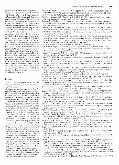
A B C 0 ES. sputlg«na
Fig. 1
A B C D E
. Intermedius
A B O D ET. denticola
BC
D
E
Prelreaimeni
Post nygenic phasePost surgical phase
Uaintanance pnase
Maintenance phase
Firsi year
Second year
A e C 0 EB. meianlnogenicus
B C 0 ET. vincentii
A B C D E
A. viseosus
A B O D EF. nucleatum
Fig. 2
A B C D ECapnocytophaga
Post hygenic phase
Post surgical phase
Uainienance phase • First year
Maintenance ^ s e - Second year
Figs. 1. 2. Longitudinal changes in antibacterial antibody titer following periodontal therapy. Each panel shows the mean ELISA unit values( + S.E.M.) calculated for the indicated microorganism at five time points during the treatment protoeol. Intervals: A. Pretreatment, B. Posthygienic phase. C Post surgical phase. D. Maintenance phase-firsl year, E. Maintenance phase-second year. Cross-hatched line indicateshealtUy pooled sera reference value (100 ELISA units).
observed at all examinations in the pa-tients group foUowing the hygienicphase. In addition, there was a decreasein probing depth in the 4^6 mm and >7 mm sites over the two years post surgi-cal phase and maintenance of level ofattachment in all sites during the sametime period.
Changes In antibody titers during therapy
As shown in Figs. I. 2. with the excep-tion of Capriocviophaga. periodontaltherapy pvoduced a reduction in themean antibacterial antibody titers,which was significant (/7<O.O5}. whenthe pretreatment (PT) measurementswere compared to those of the 2-yearpost surgical phase (MP2}, Since all ofthe antibody titrations were comparedto a reference serum pool (arbitrarilydefined as possessing a titev of 100 EU).obtained from an age-matched group ofhealthy individuals who had no evi-dence of inflammatory periodontal dis-
ease, the humoral responses of the pa-tients to these microorganisms could beclearly categorized by whether a de-crease in titer occurred from a supranor-mal value, i.e.. the pretreattrient ELISAtiter was significantly in excess of 100EU. or whether the periodontal therapycaused a decrease from a relatively nor-mal value, i.e., pretreatment titer lessl-han or equal to 100 EU. As shown inFigs. 1, 2. the mean antibody titers to 4oral microorganisms were clearly elev-ated over normal control values {indi-cated by cross-hatched bars at 100 EU).These included B. gingivalis (BG, 260EU), 5. sputtgena (SS. 175 EU). A. vi-seosus (AV. 240 EU), and fi. intermedius(BI. 140). There were significant(/j<0,05) reductions in the mean anti-body titers to the remaining microor-ganisms, however, those changes wereall relative to the values one would ex-pect to fmd in healthy, non-perio-dontally diseased control subjects.
The intervals during which the de-
creases in antibody titers occurred werenot the same for each microorganism.Table 1 indicates the % changes in theantibody titers (and significance of thechanges) between each treatment inter-val. In the interval between PT and PH.there is a significanl reduction in thetiter to SS (8.7%. /?<0.003), BG(13.5%. p^O.Ol), and BM (10.7%,;)<0.02). In the interval between PHand PS, there was another significantdecrease in the titer to BG (12.4%,;»<0.02). In no case was there a signifi-cant change in the antibody titer be-tween PS and MPl. In the final intervalexamined, MPl to MP2, decreases inthe mean titers occurred with all of themicroorganisms tested, however, onlythose decreases associated with AV(12.8%,/j<0.03),FN (15.4%./7< 0.02),SEL (18.1%. /j<0.01). and TV (13.9%.;?<0.02) were significant. In general, achange in the mean antibody titer lessthan 8% did not appear to be statisti-cally significant.

Amihodies during periodontal therapy 547
Table I. Relative changes in antibacterial antibody titers between treatment intervals*
Antigen
A. vLscosusE nuckalumS. sanguisS, sptiligenaB. gingivaJisB. inlermedimCapnocylophagaT. nmejitiiT. denticolaB. mefanmogenicus
PT^PH1.35.1
[877 (0.003) 18.5
ri3.5 (0.01)14.3066.4
10.7 (0.02)
PH^PS8.806.56.6
12.4 (0.02)1.5I.I2.87.9
-0.3
PS^MPI
00
1.600.7
-3.3- 4 . 3- 7 . 1- 3 . 6
MPl - MP2
12.8 (0.03)15.4(0.02)
7.8
n^^L^o.oi)129.8 (O.Ot) 1
6.57.5
13.9(0.02)4.93.9
PT - . MP2
24.8 (O.OI)121.2 (0.01) 1120.3 (0.02) 1135.4 (0.00()2) 1
7.9 (O.OI)3.4
120.2 (0.002) 115.5 (O.Oa10.8 (0.02)
* % decrease in ELISA Units. P-valui; indicated by parentheses. Boxed va\ue indicates a signil'icanl change with Bonferronl correction a(p<0.05).Abbreviations: PT, pretreatmeni; PH, post-hygienic phase; PS. post-surgical phase; MPl , maintenance phase h t year; MP2. maintiphase 2nd year.
ippVied
We might remark that since teveraltests were performed in Table 1 for eachmicroorganism, viz., one for each pairof successive timepoints for a total offive tests, some investigators might sug-gest the Bonferroni's approach (Glantz1981) to "correct" the individual F-values to ones more appropriate for thesituation in which multiple, related testsare perfotmed. This correction amountsto multiplying each of the P-valuesshown in Table 1 by 5 (the number oftests performed). Thus, the only con-trasts shown to be significant are thosefor which the uncorrected P-values are
If results of individual patients areexamined, however, a sJightly differentpicture of the change in the humoralimmune response to these microorgan-isms was seen. While it is difficult topresent such individual results in simpletabular form, lieveral obsevvations canbe made. There was no unique patternof humoral responses following treat-ment. Attempts to demonstrate re-lationships between antibody responsesto different microorganisms did not re-veal typical patterns. Wirh the exceptionof one patient, the trend was a generalreduction in antibody titers with treat-
Fig. 3. Changes in humoral antibody titersduring therapy to selected oraj microorgaH-isms in patient no. 10. Patient no. 10 repre-sented a unique pattern of humoral immuneresponse during periodontal therapy.
ment. In this patient and 5 others, therewere increases in antibody titers to se-lected microorganisms immediately fol-lowing the hygienic phase. In mostcases, these increases represented a re-sponse to a single microorganism andthis early increase was not predictive ofthe ultimate antibody titer at the end oftwo years. The responses of patient no.10 are shown in Fig, 3. This individual'sresponse to therapy was unique in thisgroup, since although a modest increasein titer was found to occur in five otherpatients (for 1-2 bacteria), which byMP2 was decreased below pretreatmentlevels in three subjects, this patient dem-onstrated significantly elevated anti-body titers to six of the ten bacteria,and he did not ultimately demonstratea significant decrease in titer over theentire course of treatment to any bac-teria. The case history and response toVherapy of this patient was unremark-able when compared to the other pa-tients in this study.
Discussion
Numerous studies have attempted tocorrelate patient systemic immunity tospecific oral microorganisms with theseverity of inflammatory periodontaldisease (Baker et al. 1976, Baker et al,1978. Genco et al. 1974, Ivanyi et al.1972, Page & Schroeder 1982).Lymphocyte blastogenic responses todisease-associated microorganisms havebeen examined in longitudinal studies(Lopatin et al. 1983. Osterberg et al.1983), however, their value in predictingor establishing the severity of disease isquestionable.
The humoral immune response hasalso been studied for its ability to corre-late with disease activity. High corre-
lations beiween antibody titers and dis-ease activity has been demonstrated forselected microorganisms in cross-sec-tional studies. For example, there is aparticularly high degree of associationbetween the presence of antibodies toAc/ifiohacHlux actlnomvceivtrtcomilansand the occurrence of localized juvenilepeTiodoniiiis (Ebersole et al. 1982). Incontrast, patients with adult or withrapidly progressive periodontitis fre-quently have increased levels of IgGantibodies to Bacteroides gingivalis(Mouton etal. 1981).
However, the predictive value ofsingle time point antibody measure-ments in assessing disease, or its utilityin assessing treatment is not clear be-cause, even with a high correlation be-tween disease severity and antibody ti-ter, numerous subjects possess antibodytiters that fall above and below theranges that identify health and disease,irrespective of their oral health status.This situation is 6MS. to fact that most ofthe members of the oral flora associatedwith chronic inflammatory periodontaldisease states can be identified inhealthy subjects (Loesche & Syed 1978,Loesche et al. 1985), and that antibodytiters to this flora appears in adoles-cence (Mouton et al. 1981) and can bedemonstrated in health (Doty et al.1982).
Since it is virtually impossible to ob-tain pre-disease sera from individualswho have developed CIPD, normalbaseline antibody levels lo the oral floraare not tisually available. We thereforemeasured the antibody levels to a selectgroup of oral microorganisms in alongitudinal fashion in order to assesstreatment effects. Presumably, a returnto health would result in a reductionof the oral immune sensitixation to the

548 Aukhil et al.
periodontal disease associated microor-ganisms.
The results suggest that scaling androot planing result in general reductionof antibody titers to the oral microor-ganisms, in many cases to levels belowthose of normal subjects. The humoralresponse appears to be elevated to sus-pected periodontal "pathogens" such asBG, as well as "innocuous" oral florasuch as SS, suggesting that there is ageneral sensitization to most of the flo-ra. 2 characteristic types of responseswere observed (Table 1). First, signifi-cant early (PT-PH) decreases in anti-body titers were associated with BG, SS,and BM. Second, progressive, cumula-tive changes that resulted in a significantdecrease in titers by the end of the main-tenance phase (MP2) were seen with theremaining microorganisms, with the ex-ception of CAP In this table. CAP is thesole species not resulting in a significantmean group change in response to treat-ment. CAP titers were found to be themost variable in response to treatment.
Presumably, the changes in antibodytiters seen during this study resuh fromreductions in the numbers of specificmembers of the microflora. Microbio-logical studies (reviewed by Page & Sch-roeder 1982) describe the presence ofhigh levels of BG in untreated lesionsoi adult periodontitis. Although themicrobiota of the gingival sulcus wasnot monitored in this study, the pro-gressive decrease in antibody titers toBG and other CIPD-associated micro-organisms probably reflects reductionsin levels of these microorganisms duringtherapy,
A recent report (Ebersole et al. 1985)also examined the humoral immune re-sponse to seiected oral microorganismsfoilowing therapy. In their study, pa-tients treated with scaling and root plan-ing demonstrated increased antibody ti-ters to certain microorganisms, whiletheir responses rarely decreased belowpretreatment values within the timefra-me examined. Their findings were ex-plained as a re-immunization to the oralflora resulting from the trauma of thetreatment protocol. Since the samplingintervals and methods of therapy dif-fered significantly between our studyand theirs, it is difficult to make a com-parison between the results. However.a comparison between the studies doessuggest that the antibody levels arequite sensitive to the therapy. In ourstudy, six patients demonstrated an in-crease in antibody titer to selected
plaque microorganisms during the PT-PH phase of treatment. Since the modeof therapy might be expected to resultin a certain level of additional sensitiza-tion, and bacteremias have been shownto accompany such procedures (Hock-ett et al. 1977), increasing levels of anti-bodies to the oral fiora would not beunexpected. However, three of the sixpatients ultimately demonstrated re-duced antibody titers by the end of thesecond maintenance phase. It is notclear at present what is unique about theremaining patients, especially patient 10who demonstrated increased titers to sixout of ten of the bacteria. Their (clin-ical) response to therapy did not differfrom that of the other patients.
Our findings do not shed light on therole of the periodontal disease associ-ated microorganisms in the etiology ofCIPD. In fact, since antibody levels tobacteria such as S. sanguis were alsomodified during therapy, one must con-sider the possibility that immune re-sponses to microbes not generally con-sidered to be "periodontai pathogens"are modified by adjuvant associatedwith subgingival plaque. Alternatively,the levels of these "non-pathogens" inthe sulcus may be influenced by the en-vironment created by the "pathogen".Thus, changes in antibody titers to amicrobiai antigen preparation wouldnot necessarily identify a role for thatmicroorganism in the disease etiology.The results of this study, however, tendto support the concept that periodontaltreatment reduces the antigenic stimuliin the periodontium. resulting in de-creased antibody titers to the plaque flo-ra, in general. In many cases, the anti-bacterial titers were quite resistant tochange, and the time elapsed betweeninitiation of therapy and occurrence ofsignificant changes in antibody levei ex-ceeded one year. This was also sug-gested by observations in edentuloussubjects where the length of time theindividuals were totally edentulous hasa significant influence upon the levels ofcirculating antibodies to oral microor-ganisms (Jacob et al. 1982). One mightexpect that once sensitized to a particu-lar antigen, maintenance of that im-
mune response would require minimallevels of antigen in the gingival sulcus,because of the sensitivity of the anamne-stic response. As a result, those re-sponses which are most easily eradi-cated might be thought to be to bacteriathat were most easily eliminated orwhich are the poorest immunogens. Themore persistent immune responsesmight, thus, be considered to be associ-ated with more difficult to eliminate flo-ra, or those associated with health. Themost significant use of this type ofanalysis might be the monitoring of themaintenance patient who has demon-strated reduced antibody titers as a re-sult of therapy. In a patient primed im-munologically to the CTPD-associatedmicroorganisms, one might expect avery rapid rise in the antibody titer ifthe crevicular fiora increases, possiblysuggesting that additional therapy is de-sirable. One cannot overstate, however.that because of individual variability inthe magnitude of immune responses tospecific bacterial preparations, pretreat-ment sera from each patient must serveas that individual's baseline reference.
Acknowledgements
We wish to thank Dr. Sigurd P.Ramfjord for allowing us to use his pa-tients, and Dr. Raul G. Caffesse for hishelpful comments.
Zusammenfassung
Die Auswirkung parodontaler Therapie auf dieSerum Aniikorper (IgG) - Niveatis vonPlaq ue-Mikro organ ismenDer EinfluB parodontaler Therapie aut" dieScrum-Anti korpcriiler von selckticrten, mitder Krankheit assoziierten parodontalen Mi-kroorganismen. wurde bei 23 Palienten milchronisch entzlindlicher Parodontalkrank-heit (CIPD) beurleilt. In Serum-Stichprobenwurde der Immunoglobulin G (IgG)-Titermit der Mikro-ELISA-Technik vor der Be-handlung beslimmt und zwar nach der Hy-gienephase mit Zahnsteinentfernung, Wurzel-glattung und Instruktion in oralcr Hygiene;nach der chirurgischen Behandlung. sowie 1Jahr und 2 Jahre nach der Hygienephase(Nachsorgephase). Die Immunantworten ge-geniiber speziellen bakteriellen Zubercitun-
References
Baker. J. J.. Chan, S. P., Socransky, S, S,. Oppenheim. J, J, & Mergenhagen. S, E, (1976)Importance of actinomyces and certain gram negative anaerobic organisms in the transmfor-mation of lymphocytes with periodontal disease. Infection and Immunity 13. 1363-1368,
Antibodies during periodonlal therapy 549
gen schwankten betrachtlich. Gegeniiber A.viscosu,^, S. sanguis. F. nucleatum, S. sputigenaund B. gingivalis, sowie B. intermedius, B.metaninogenicus, T, vincentii und T. denticotawurden gegen Ende des 2. Nachsorgejahressignifikanle Reduktionen der mittJereii Anti-korpcr-Titer beobachtet. Auf Capnocytopha-ga kam es zu keiner gleichbleibenden Niveau-Antwort. Bei naherer Untersuchung der Ant-worteii der einzelnen Palienten zeigle es sich.daB bei 6 Patienten in der Zeit zwischen Vor-behandlung und dem Ende der Hygienephaseein erhohter Titer gegen zumindest einen derMikroorganismen vorlag. Mit einer Ausnah-me lagen jedoch in alien Fallen die Titer ge-gen Ende des 2. Nachsorgejahres unterhalbder Niveaus, die vor der Behandlung konsta-tiert worden waren. Antikorpemiveaus ge-genuber Bakterien wie S. sanguis iindertensich wahrend der Behandlung. Das konntebedeuten, daB Immunantworten auf Kleinst-lebewesen. die generell nicht als "parodontalePathogene" angesprochen werden, durehStiilzmaBnahmen hinsichtlieh der subgingi-valen Plaque oder durch Veranderungen desSulkus-Milieus modifiziert werden konnenund daB die dann zu beobachtende Verande-rung des Titers nieht unbedingt die Rolle wi-dergibt. die dieser Mikroorganismus in demKrankheitsprozess spielt.
Resume
Effets du traitement parodontat sur les tauxd'anticorps seriques (IgGj aux microrganis-mes de la ptaqueL'influence du traitement parodontai sur lestaux d'anticoprs seriques a certains microrga-nismes associes a la maladie parodontale aete determinee chez 23 patients souffrant deparodontite chronique. Les taux d'IgG ontete determines par la technique microELISAd'echantilions de serum obtenus avant le trai-tement, apres une phase d'hygiene (detartra-ge, lissage radiculaire et instruction en hygie-ns buccale). apres la chirurgie parodontaleainsi qu'une et deux annees apres la phase demaintien. Une variatioh individuelle conside-rable existait dans I'ampleur des reponses im-munitaires aux preparations baeteriennesspecifiques. Des reductions significativesdans les taux d'anticorps moyens ont ete no-e'es pour t'A. viseosus, le S. sanguis, ie F.nucteatum, le S. sputigena, le B. gingivatis. leB, intermedius, le B. metaninogenicus, le T.vincentii et le T. denticota a la fin de la secondeannee de maintien. 11 n'y avait aucune repon-se coherente au Capnocytophaga, Lorsque lesreponses individuelles des patients etaient ex-aminees, six des 23 sujets possedaient destaux eleves a au moins un des microrganis-mes, dans I'intervalie enlre le pre-lraitementet la fin de la phase d'hygiene; cependant,dans tous les cas sauf un, les taux a la finde la seconde annee de maintien etaient endessous des taux pre-traitement. Des tauxd'anticorps aux baeteries teiies que !e S. san-guis ont ete modifies durant le traitement.Ced indiquerait que ies reponses immunilai-
Baker. J. .1.. Wright, W. E.. Chan. S. P. & Oppenheim, J. J. (1978) Longitudinal effects ofclinical iherap}' and the edemuJotis stale on the transformation of \ymphocyles with severeperiodontitis. Ctinicat and Experimentat Immunology 34, 199-205.
Doty. S. L.. Lopatin, D. E.. Syed. S, A. & Smith. F. N. (1982) Humoral immune response tooral microorganisms in periodontitis. Infection and Immunity 37, 499-505.
Ebersole, J. L., Taubmaji, M. A., Smith. D. J. & Frey, D. E. (1986) Human immune responsesto oral microorganisms: patterns of systemic antibody levels to Bacteroides species. Infectionand Immunity S\. 507-513.
Ebersole, J. L., Taubman, M. A., Smith. D. J., Genco, R. J. & Frey, D. E. (1982) Humanimmune response to oral microorganisms. 1. Association of local juvenile periodontitis(LJP) with serum antibody responses to Actinobacittus actinomycetemcomitans, Ctinicat andExperimentat Immunotogy 47, 43-53.
Ebersole. J. L., Taubman, M. A., D. J. & HaiTajee, A. D. (1985) EfTect of subgingival scalingon systemic antibody response to oral microorganisms. Infection and Immunity 48, 534-539.
Geneo, R. J., Mashimo. P. S., Krygier, G. & Ellison, S. A. (1974) Anti body-mediated effectson the periodonlium. Journat of Periodontotogy 45, 330-337.
Glantz, S. A. (1981) Primer of biostati.stivs. New York: McGraw-Hill.Hill, R. W., Ramfjord, S. P., Morrison, E. C , Appleberry. E. A., Caffesse. R. G.. Kerry, G.
J. & Nissle, R. R. (1981) Four types of periodontal treatment compared over two years.Journat of Periodontology 52, 655-662.
Hockett, R. N., Loesche, W. J. & Sodeman, T. M. (1977) Bacteracmia in asymptomatic human
subjects. Arctuves of Orat Biology 22, 91-98.Hofstad. T. (1984) Immune responses lo anaerobic gram-negalivc hacteria in health and
disease. Nahrung 28. 717-721.Ivanyi, L.. Wilton, J. M. A. & Lehner. T (1972) Cell-medialed immunity in periodonta!
disease; cytotoxidty, migration inhibition, and lymphocyte transfonnation studies. Immu-notogy 12, 141-145.
Jacob, E., Muller, T. F. & Nauman, R. K. (1982) Detection of elevated serum antibodies toTreponema denticola in humans with advanced periodontitis by an enzyme-linked immuno-sorhdnt as&ay. Jourmt of Periodontat Re.s-earch 17, 145-153.
Kowaiski, C. J. & Guire, K. E. (1974) Longitudinai dala analysis. Growth 38, 1.11-169.Loesche, W. J. & Syed. S. A. (1978) Bacteriology of human experimental gingivitis: effect of
plaque and gingivitis score. Infection and Immunity 21, 830-839.Loesche. W. J., Syed, S. A., Morrison. E. C . L a u ^ o n . B. & Grossman, N. S. (198!) The
trealmeni of periodonlai infections due to anaerobic bacieria wilh shorl-!erm meironidizole.Journat of Ctinicat Periodontotogy 8. 2 9 ^ 4 .
Loesehe, W. J., Syed, S. A.. Schmidt, E. & Morrison, E. C. (1985) Bacterial profiles ofsubgingival plaques in periodontitis. Journal of Periodontotogy 56, 447^56.
Lopatin, D. E., Smith, F. N., Syed, S. A. & Morrison, E. C. (1983) The effect of periodontaltherapy on lymphocyte blastogenesis to plaque associated microorganisms. Journat ofPeriodontat Research 18, 93-102.
Mangan, D. F., Laughon, B. E., Bower, B. & Lopatin, D. E. (1982) In vitro lymphocyteblaslogenic responses and titers of humoral antibodies from periodontitis patients to oralspirochete isolates. Infection and Immunity 37, 445^51 .
Mouton. C , Hammond, B. G., Slots, J. & Genco, R. J. (1981) Serum antibodies to oralBacteroides asaccharotyticus (Bacteroides gingivatis): relationship to age and periodontaldisease. Infection anti Immunity 31, 182-192.
Naito, Y., Okuda, K. & Takazoe, L (1984) Immunoglobulin G response to subgingival gram-negative bacteroides in human subjects. Infection and Immunity 45, 47-51.
Naito, Y., Okuda, K., Takazoe, L, Watanabe. H. & Ishikawa, L (1985) The relationshipbetween serum IgG levels to subgingival gram-negative bacteria and degree of periodontaldestruction. Journat of Dentat Research 64, 1306-1310.
Osterberg, S. K., Page, R. C , Sims, T. & Wilde, G. (1983) Blastogenic responsiveness ofperipheral blood mononuclear cells from individuals with various forms of periodontitisand effects of treatment. Joumat of Clinieat Periodontotogy 10, 72-88.
Page, R. C. & Schroeder. H. E. (1982) Periodontitis in man and other animals, pp. 5-57. Basel:
S. Karger.RamQord, S. P. (1959) Indices for prevalence and incidence of periodontal disease. Journat
of Periodontology 30, 51-59.Ramfjord, S. P (1967) The periodonlal disease index fPDl). Journal of Perio<tantology 38.
602-610.Silness, J. & Loe, H. (1964) Periodontal disease in pregnancy. IL Correlation between oral
hygiene and periodontal condition. Acta Odontotogica Seandinavica 22, 121-135.Tew. J. G., Marshall, D. R., Moore, W. E., Best. A. M., Palcanis, K. G. & Ranney, R. R.
(1985) Serum antibody reactive with predominant organisms in the subgingival flora ofyoung aduhs with generalized severe periodontitis. Infection and Immunity 48, 303-311.
Tew. J. G., Smibert, R. M., Scoti, E. A., Burmeister, J. A. & Ranney, R. R. (1985) Serumantibodies in young adult humans reactive wifh periodontitis associated treponemes. Journalof Periodontal Research 20, 580-590.
Vandesteen, G. E., Williams, B. L., Ebersoie, J. L., Altman, L. C. & Page, R. C. (1984)

550 Aukhil et al.
res aux microbes qui ne sont generalementpas consideres comme "pathogenes parodon-talement" peuvent etre modifiees par une ac-tivite supplementaire associee a la plaquesous-gingivale ou des changements dans l'en-vironnemenl du sillon et que les variationsulterieures dans les taux n'impiiquent pas ne-cessairement un role de ce microrganismedans tc processus de la maladie.
Clinical, microbiological and immunological studies of a family with a high prevalence ofearly-onset periodontitis. Journal of Periodontology 55, 159-169.
Williams, B. L., Ebersole, J. L., Spektor, M, D. & Page. R. C. (1985) Assessment of serumantibody paiterns and analysis of subgingival microflora of members of a family with ahigh prevalence of early-onset periodontitis. Infection and Immunity 49. 742-750,
Address:
Dennis E. Lopatin300 North Ingalls Building. Room 1192School of DentistryThe University of MichiganAnn Arbor. Michigan 48109-0402USA