Embed Size (px)
Citation preview
1
W I S S E N T E C H N I K L E I D E N S C H A F T
u www.tugraz.at
Protein Engineering
Antibodies
MOL.921 Molecular Biotechnology II

2 Antibody Engineering
Target: Specific Affinity
MOL.921 Molecular Biotechnology II
General structure of IgG antibodies IgG antibodies consist of two identical light and two identical heavy chains. These four chains are arranged in parallel and linked with different disulfide bonds to form a molecule. The antigen binding regions (VH = variable region of the heavy chain; VL = variable region of the light chain) are located at the N-terminus of each protein chain. In this region the antibodies can vary tremendously. “Framework regions” (FR1-FR4) are distinguished from “complementary determining regions” (CDR1-CDR4). This part of the antibody is named Fv-region. CL and OH are domains in the constant region of heavy and light chains. The region marked with “H” is the co-called “Hinge” region.
Variable region
(Fv fragment)
Constant
region
Light chain
(L)
Heavy chain
(L)
Murine IgG

3
J Allergy Clin Immunol. 2010 Feb; 125(2 0 2): S41–S52. http://dx.doi.org/10.1016/j.jaci.2009.09.046
IgG antibodies consist of 2 heavy (H) and 2 light (L) chains where the L chain can consist of either a or a chain. Each component chain contains one NH2-terminal variable (V) IgSF domain and 1 or more COOH-terminal constant (C) IgSF domains, each of which consists of 2 sandwiched b-pleated sheets pinned together by a disulfide bridge between 2 conserved cysteine residues. Papain digests IgG into 2 Fab fragments, each of which can bind antigen, and a single Fc fragment. Pepsin splits IgG into an Fc fragment and a single dimeric F(ab)2 that can cross-link, as well as bind, antigens. The Fab contains 1 complete L chain in its entirety and the V and CH1 portion of 1 H chain. The Fab can be further divided into a variable fragment (Fv) composed of the VH and VL domains, and a constant fragment composed of the CL and CH 1 domains. The primary sequence of the V domain is functionally divided into 3 hypervariable intervals termed complementarity-determining regions (CDRs) that are situated between 4 regions of stable sequence termed framework regions (FRs).
MOL.921 Molecular Biotechnology II

4
1. Fab region 2. Fc region 3. Heavy chain with one variable (VH) domain followed by a constant domain (CH1), a hinge region, and two more constant (CH2 and CH3) domains. 4. Light chain with one variable (VL) and one constant (CL) domain 5. Antigen binding site (paratope) 6. Hinge regions.
http://en.wikipedia.org
Antibody Engineering
MOL.921 Molecular Biotechnology II

5
Synthetic Antibody fragments
http://en.wikipedia.org
Antibody Engineering
MOL.921 Molecular Biotechnology II

6
Figure 1 Schematic representation of different antibody formats, showing intact ‘classic’ IgG molecules alongside camelid VhH-Ig and shark Ig-NAR immunoglobulins. Camelid VhH-Ig and shark Ig-NARs are unusual immunoglobulin -like structures comprising a homodimeric pair of two chains of V-like and C-like domains (neither has a light chain), in which the displayed V domains bind target independently. Shark Ig-NARs comprise a homodimer of one variable domain (V-NAR) and five C-like constant domains (C-NAR). A variety of antibody fragments are depicted, including Fab, scFv, single-domain VH, VhH and V-NAR and multimeric formats, such as minibodies, bis-scFv, diabodies, triabodies, tetrabodies and chemically conjugated Fab´ multimers (sizes given in kilodaltons are approximate).
Nature Biotechnology 23, 1126 - 1136 (2005); doi:10.1038/nbt1142
MOL.921 Molecular Biotechnology II

7
A cartoon representation of different antibody formats used for in vivo imaging, together with their respective molecular weights (kDa) and serum half-life ( phase).
Nature Biotechnology 23, 1126 - 1136 (2005); doi:10.1038/nbt1142
MOL.921 Molecular Biotechnology II

8
Antibody mimetic Framework Molar mass
Affibodies Staphylococcus ProteinA, Z domain 6 kDa
Affilins Gamma b crystallin Ubiquitin
20 kDa 10 kDa
Affimers Cystatin 12-14 kDa
Affitins Sac7d (from Sulfobolus acidocaldarius) 7 kDa
Alphabodies Triple helix coiled coil 10 kDa
Anticalins Human Lipocalins 20 kDa
Avimers A domains of various membrane receptors 9–18 kDa
DARPins Ankyrin repeat motif 12–19 kDa
Fynomers Fyn, SH3 domain 7 kDa
Kunitz domain peptides Kunitz domains of various protease inhibitors 6 kDa
Monobodies Fibronectin, 10th Type III domain 10 kDa
http://en.wikipedia.org
Antibody mimetics
MOL.921 Molecular Biotechnology II
9
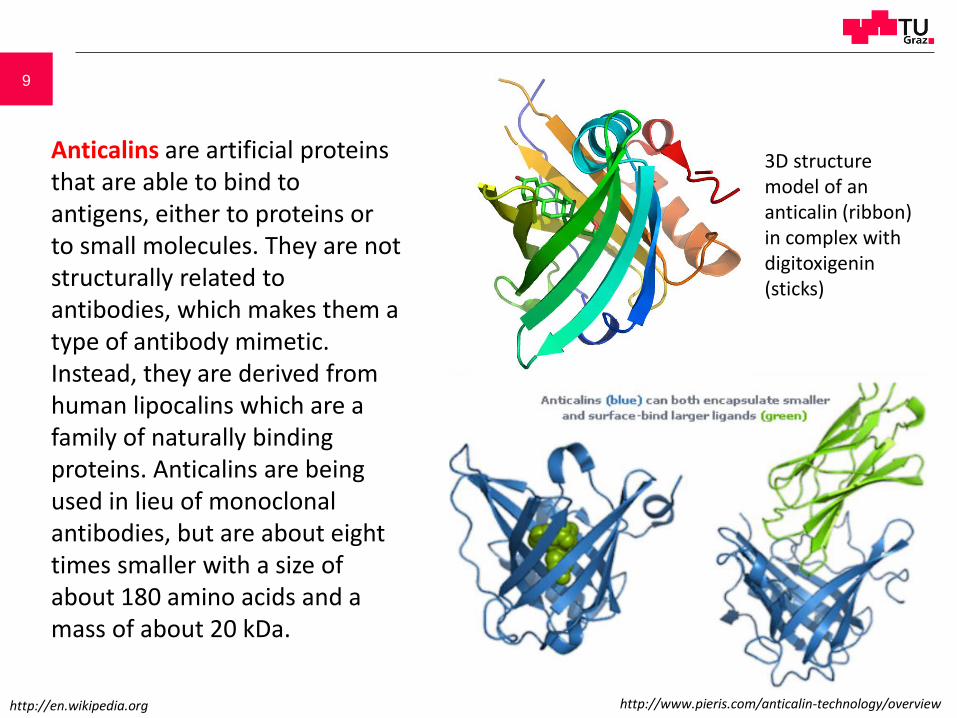
Anticalins are artificial proteins that are able to bind to antigens, either to proteins or to small molecules. They are not structurally related to antibodies, which makes them a type of antibody mimetic. Instead, they are derived from human lipocalins which are a family of naturally binding proteins. Anticalins are being used in lieu of monoclonal antibodies, but are about eight times smaller with a size of about 180 amino acids and a mass of about 20 kDa.
3D structure model of an anticalin (ribbon) in complex with digitoxigenin (sticks)
http://en.wikipedia.org http://www.pieris.com/anticalin-technology/overview

10
Monobodies consist of 94 amino acids and have a molecular mass of about 10 kDa, fifteen times smaller than an IgG type antibody and comparable to the size of a single variable domain of an antibody. They are based on the structure of human fibronectin, more specifically on its tenth extracellular type III domain. This domain has a structure similar to antibody variable domains, with seven beta sheets forming a barrel and three exposed loops on each side corresponding to the three complementarity determining regions. Monobodies lack binding sites for metal ions and the central disulfide bond. Monobodies with specificity for different proteins can be tailored by modifying the loops BC (between the second and third beta sheets) and FG (between the sixth and seventh sheets).
The tenth fibronectin type III domain (human, PDB 1TTG)
Variable domain of an antibody's lambda light chain (human, PDB 2RHE)
MOL.921 Molecular Biotechnology II

11
3D representations of non-Ig-based and Ig-like protein scaffolds Structures are extracted from the PDB protein database and are not drawn to scale. Arrows indicate the scaffold of interest if complexed. (a) Anticalin in complex with digitoxigenin (PDB ID: 1LNM); (b) Designed Ankyrin Repeat ProteIN (DARPin) in complex with aminoglycoside 3′ phosphotransferase (PDB ID: 2BKK); (c) adnectin in complex with interleukin-23 (IL-23, PDB ID: 3QWR); (d) prototypical A-domain structure, as part of the avimer scaffold (PDB ID: 1AJJ); (e) VHH nanobody in complex with lysozyme (PDB ID: 1MEL); (f) bivalent diabody [Bispecific T cell Engager (BiTE)-like structure, PDB ID: 1LMK]; (g) whole human IgG1 anti-HIV antibody (PDB ID: 1HZH); (h) schematic drawing of a whole IgG1, indicating the antigen-binding domain or variable domain.
Trends in Biotechnology,Volume 30, Issue 11, November 2012, Pages 575–582; doi:10.1016/j.tibtech.2012.07.006
MOL.921 Molecular Biotechnology II

12 Affibodies
Figure 1. Graphic representation of the wild-type Z domain (residues 5-58) based on the NMR structure (A). Main-chain trace ribbon diagram showing the three helix-bundle structure. (B) Space-filling representation of the domain showing the positions of the 13 amino acids located in helices 1 and 2 subjected to the randomization (red). The position of Ile31 (blue) is also shown.
Figure 2. Amino acid sequence corresponding to the wild-type Z domain aligned to deduced amino acid sequences of different phagemid clones selected against various target proteins. Residues Q9, Q10, N11, F13, Y14, L17, H18, E24, R27, N28, Q32 and K35 included in the randomization are indicated. Helices in the wild type Z domain are boxed. Horizontal bars indicate amino-acid identities. Two of the clones showed additional substitutions outside the variegated regions (-Taq 4.8 and -Apolipo 24:4).
Nat Biotechnol. 1997 Aug;15(8):772-7
MOL.921 Molecular Biotechnology II

13
Fig. 8.2.2 Three detailed sequences of variable regions of heavy (A) and light (B) chains of three antibodies. Amino acid sequences are shown in one-letter-code. FR = framework regions (black letters), CDR = complementarity determining regions (blue letters). Although differences within the FR-domains are visible, differences in the CDR-domains are substantially more distinctive.
Variable Regions of heavy and light chains Antibody Engineering
MOL.921 Molecular Biotechnology II

14
Fig. 8.2.3 Collection of diverse degenerate primers for the amplification of cDNA sequences of heavy (A) and Light (B) antibody chains. Primers bind at the N-terminal region and recognize the FR domain. These primers are named „backward-primer“. Degenerate positions are marked with orange letters.
Variability increase via degenerate oligos
Antibody Engineering
MOL.921 Molecular Biotechnology II

15
Antibody Engineering
Linker sequence length
Chemical
bond
Introduction of
cystein residues
via mutagenesis
Introduction of a
linker peptide
Fig. 8.2.5 Linkage of a VL and a VH domain A Both domains can be linked in different ways: firstly protein chains can be joined chemically with the help of a bifunctional reagent, secondly by the introduction of cysteine codons on gene level and connection of the chains via artificial disulfide bonds. The third and most common method is the use of a linker fragment which is introduced by PCR strategy. B Possible linker sequences for the chain connection
MOL.921 Molecular Biotechnology II

16
Fig. 8.2.12 Biosynthesis of a recombinant Fab-fragment. The pASK84 expression cassette is an “operator fusion”. The whole cassette is expressed under the control of the lacZ promoter. Two protein chains are made simultaneously based on the mRNA: one for the heavy and one for the light chain. The formed proteins are directed to the periplasm of the bacterial cell with the help of the signal peptides OpmA and PhoA. Signal peptides are hydrolysed while passing through the cell membrane. The reducing environment in the periplasm is sufficient for disulfide bond formation between heavy and light chains.
Transgenic bacterium
Expression of the operator fusion
Histidin
residues
Transcription, Translation
Antibody Engineering
MOL.921 Molecular Biotechnology II

17 Antibody Engineering
Fig. 8.2.6 Splicing by overlap extension. For the connection of DNA fragments for heavy and light chains, a linker is synthesized not only bearing the information for (Gly4Ser)3 but also being complementary to the 24 3´ terminal nucleotides of vH amplimer and the 24 5´-terminal nucleotides of vL amplimer, respectively. All three amplimers are put into the same PCR tube. In the next step the backward primer (heavy chain amplification) and forward primer (light chain amplification) are added. This experimental setup facilitates the combination of all three fragments and the amplification thereof. Another PCR step provides a terminal extension of the amplimers adding recognition sequences for the restriction endonucleases NotI and SfiI.
Denaturation and
new hybridisation
PCR with
PCR with
Restriction with NotI/SfiI;
cloning into expression plasmid
MOL.921 Molecular Biotechnology II

18
Abb. 8.2.8 Recombinant phage with a modified gpIII protein on the top
Antibody Engineering Phage display
Fig. 8.2.7 Composition of M13 phages. The capsid of a M13 phage is basically composed of five different proteins coded by the genes gpIII, gpVI, gpVII, gpVIII and gpIX. N-termini of two proteins protrude from the capsid the way that a fusion protein could be easily added without disrupting the composition of the phage capsid too much. This has already been realized for both proteins. Copy numbers of these two proteins differ considerably – a phage capsid has ca. 2500 gpVIII gene products, but only three to five gpIII.
Proximal
end
Distal
end
MOL.921 Molecular Biotechnology II

19
Antibody Engineering MOL.921 Molecular Biotechnology II

20
Antibody Engineering
Fig. 8.2.9 Expression of soluble scF or recombinant phages For expression of soluble scF-fragments or recombinant phages plasmid pHen1 is used. The plasmid consists of a colE1 replication origin, a phage replication origin, a ß-lactamase gene and an expression cassette under the control of a bacterial ß-galactosidase promoter (lacZ promoter) followed by a signal sequence for the transport of soluble antibody fragments to bacterial periplasm where disulfide bonds are formed. Subsequently gpIII gene sequence is located which is interrupted by a short sequence harboring SfiI and NotI restriction sites for in frame cloning of scF fragment coding sequence. The black dot stands for an amber stop codon termination of protein biosynthesis if the plasmid is transformed into a wild type E.coli strain. Soluble scF fragments are produced. However, if the plasmid is transformed into an E.coli strain, which harbors a tRNA for amber stop codon reading additionally to the general tRNPt setup, the rest of gpIII is translated. If those cells are infected with “helper phages” providing the residuary proteins for a correct phage assembly, modified gpIII gene products are incorporated into the phages resulting in recombinant phages with scfV fragment at the top.
MOL.921 Molecular Biotechnology II
Transformation in
suppressor strain
and helper phage
infection
Transformation in
non-suppressor
strain
pelB signal sequence
lacZ promoter
Gene III
Phage replication origin
colE1 ori (origin of replication)
ß-lactamase

21 Antibody Engineering
Fig. 8.2.9 Expression of soluble scF or recombinant phages For expression of soluble scF-fragments or recombinant phages plasmid pHen1 is used. The plasmid consists of a colE1 replication origin, a phage replication origin, a ß-lactamase gene and an expression cassette under the control of a bacterial ß-galactosidase promoter (lacZ promoter) followed by a signal sequence for the transport of soluble antibody fragments to bacterial periplasm where disulfide bonds are formed. Subsequently gpIII gene sequence is located which is interrupted by a short sequence harboring SfiI and NotI restriction sites for in frame cloning of scF fragment coding sequence. The black dot stands for an amber stop codon termination of protein biosynthesis if the plasmid is transformed into a wild type E.coli strain. Soluble scF fragments are produced. However, if the plasmid is transformed into an E.coli strain, which harbors a tRNA for amber stop codon reading additionally to the general tRNPt setup, the rest of gpIII is translated. If those cells are infected with “helper phages” providing the residuary proteins for a correct phage assembly, modified gpIII gene products are incorporated into the phages resulting in recombinant phages with scfV fragment at the top.
MOL.921 Molecular Biotechnology II
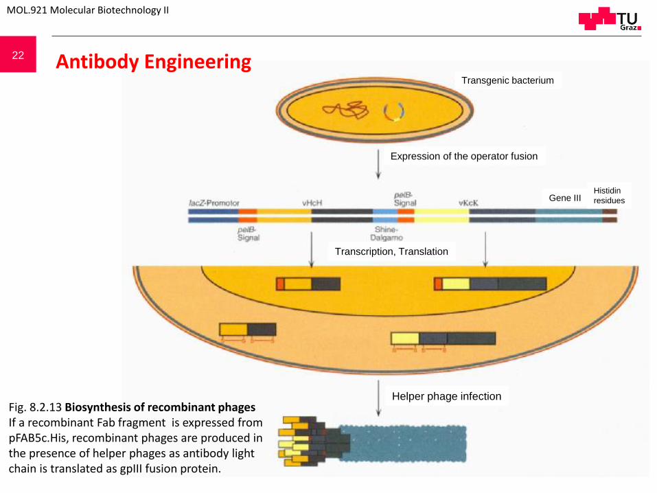
22
Transcription, Translation
Expression of the operator fusion
Transgenic bacterium
Histidin
residues Gene III
Helper phage infection
Antibody Engineering
Fig. 8.2.13 Biosynthesis of recombinant phages If a recombinant Fab fragment is expressed from pFAB5c.His, recombinant phages are produced in the presence of helper phages as antibody light chain is translated as gpIII fusion protein.
MOL.921 Molecular Biotechnology II

23 Antibody Engineering
Screening by panning
Fig. 8.2.16 „phage panning“ for antibody fragment selection For the identification and isolation of the antibody fragment that reacts best with the antigen, the antigen is fixed in a petri dish. After saturation of free valences with inert protein the phages are added. Stringent washing steps are performed to enrich the phages with highest affinity to the fixed antigen. E.coli cells, which enable the phages to replicate, are added for phage amplification. After several selection rounds, phage DNA is isolated and characterized.
MOL.921 Molecular Biotechnology II
unspecific
saturation
antigen
Phage pool
addition
Stringent
washing
Dissociation of
unbound phage
Infection of bacteria
Isolation and
characterization of
phage genome
mutagenesis
Phage pool

24
Schematic representation of the phage-display selection procedure: modified subtractive panning. Phage particles displaying the members of the library were produced. The specific binder (nmAb-KT) was allowed to bind with the target, and other variants were removed by washing. Molecular variants with specificity for the target were retrieved after multiple cycles of selection and were characterized in detail. Bound phages were eluted with HM-1 containing phosphate buffer (pH 7.0). HM-1 had high binding affinity to the immobilized nmAb-KT. This competition of binding favored quick dissociation of scFv-containing phages from the bound nmAb-KT and consequently increased the elution stringency of the infected phages.
Kabir et al. BMC Biotechnology 2009 9:99 doi:10.1186/1472-6750-9-99
Phage panning
MOL.921 Molecular Biotechnology II

25
P P
transcription mRNA
Library of mutated genes
P
P
P
Modification with
puromycin
In vitro translation
Screening for protein function
Gene Recovery by Protein – RNA Fusion
P
Reverse transcription with modified primer (tag)
Removal of tag
selected Gene
modified after Seelig and Szostak (2007) Nature 448,828
in vitro expression
MOL.921 Molecular Biotechnology II

26
transcription
mRNA
P
P
P
P
Modification with
puromycin
P Screening for protein function
Gene Recovery by Protein – RNA Fusion
P
Reverse transcription with modified primer (tag)
Removal of tag
selected Gene
Library of mutated genes
in vitro expression
MOL.921 Molecular Biotechnology II
modified after Seelig and Szostak (2007) Nature 448,828

27
21.5.15