Embed Size (px)
Citation preview

Hale Veterinary Clinic [email protected] www.toothvet.ca Local Calls: 519-822-8598 Fraser A. Hale, DVM, FAVD, Dipl AVDC Page 1 December 2010 Long Distance: 1-866-866-8483
What follows is going to contain generalizations and guidelines that may or may not apply to any specific case. It is not possible to cover all situations and scenarios in a newsletter and there will be some simplification of complex issues. Do not consider this the ‘last word’. As the attending veterinarian, it will be your job to gather credible information and make the clinical judgment about how it applies to your patient(s). Also keep in mind that what we “know” is constantly evolving. The following is based on my understanding of the situation at time of writing.
The Disease Formerly Known as
Lymphocytic/Plasmacytic Gingivo-stomatitis
I have been seeing a lot of enquiries about cats with refractory chronic oral inflammatory disease following extraction of all premolars and molars. It made me wonder where people were getting the idea that this condition can reasonably be treated by just removing the back teeth and then it was pointed out that one of my own old papers on this subject, available on my website, actually suggests caudal extraction as a reasonable option in some cases. So, time for an update on this most frustrating and painful of feline oral diseases.
The first thing I would like to address is nomenclature. The condition (or spectrum of conditions) I am referring to has been referred to as lymphocytic/plasmacytic gingivitis (LPS) or gingivostomatitis (LPGS), just feline stomatitis and a number of similar labels. Old habits die hard, but we are encouraged to abandon these terms in favour of more useful and accurate nomenclature. The Nomenclature Committee of the American Veterinary Dental College has been hard at work and has a web-page that covers this and much more. Go have a look - http://www.avdc.org/?q=node/29#OP.
That said, I still find it useful to have a quick label to use as a broad category, not a specific diagnosis. I have been in the habit of calling this Chronic Oral Inflammatory Disease or COID but a more widely accepted ‘nickname’ for this condition is Feline
Chronic Gingivostomatitis or FCGS, which is the acronym I will use for the rest of this article.
Be Specific When describing oropharyngeal inflammation, it is important to be geographically specific. You want to record specifically where the inflammation is occurring. What follows is copied directly from the AVDC Nomenclature page.
Gingivitis: inflammation of gingiva
Periodontitis: inflammation of non-gingival periodontal tissues (i.e., the periodontal ligament and alveolar bone)
Alveolar mucositis: inflammation of alveolar mucosa (i.e., mucosa overlying the alveolar process and extending from the mucogingival junction without obvious demarcation to the vestibular sulcus and to the floor of the mouth)
Sublingual mucositis: inflammation of mucosa on the floor of the mouth
Labial/buccal mucositis: inflammation of lip/cheek mucosa
Caudal mucositis: inflammation of mucosa of the caudal oral cavity, bordered medially by the palatoglossal folds and fauces, dorsally by the hard and soft palate, and rostrally by alveolar and buccal mucosa
Contact mucositis and contact mucosal ulceration: lesions in susceptible individuals that are secondary to mucosal contact with a tooth surface bearing the responsible irritant, allergen, or antigen. They have also been called “contact ulcers” and “kissing ulcers”.
Palatitis: inflammation of mucosa covering the hard and/or soft palate
Glossitis: inflammation of mucosa of the dorsal and/or ventral tongue surface
Cheilitis: inflammation of the lip (including the mucocutaneous junction area and skin of the lip)
Osteomyelitis: inflammation of the bone and bone marrow
Stomatitis: inflammation of the mucous lining of any of the structures in the mouth; in clinical use the term should be reserved to describe wide-spread oral inflammation (beyond gingivitis and periodontitis) that may also extend into submucosal tissues (e.g., marked caudal mucositis extending into submucosal tissues may be termed caudal stomatitis)

Hale Veterinary Clinic [email protected] www.toothvet.ca Local Calls: 519-822-8598 Fraser A. Hale, DVM, FAVD, Dipl AVDC Page 2 December 2010 Long Distance: 1-866-866-8483
Tonsillitis: inflammation of the palatine tonsil
Pharyngitis:inflammation of the pharynx
Photographic examples of each of these are also on the AVDC web page. Go have a look.
It is very important to not only be specific in the written records but also to maintain photographic or pictorial records of the distribution and severity of inflammation so that response to therapy and the passage of time can be accurately assessed.
Etiology The short answer here is that we do not know the cause(s) of this condition. If anyone claims to know the cause, ask to see some real evidence to back their claim. At time of writing, there is none.
Back in 2000, Frank Verstraete of UCDavis presented a paper at the Veterinary Dental Forum. After an extensive literature search he concluded that the only statement he could make was that the condition is an atypical immune response of unknown etiology. Since that time, various theories have been proposed but none have held up to scrutiny.
A draft position statement from the European Veterinary Dental College states that the condition can “be thought of as an individual inappropriate immunologic response from the cat to a variety of antigenic triggers rather than in terms of specific causal agents.” The statement then cautions against focusing on the immunologic response as this often leads to inappropriate use of immunosuppressive agents. Rather it is felt that “in order to achieve good control of the syndrome, it is essential to identify these antigenic trigger factors so that it can be appropriately managed.”
There have been some statistical associations with various infectious agents reported but that means nothing with respect to cause-and-effect. Every cat I see with this condition has two ears so there is a very strong statistical association there, but it is totally irrelevant. Also, many of the supposed statistical associations have since been debunked as unrepeatable or simply not there when larger numbers and better inclusion criteria were used.
Bacteria
Every cat mouth is full of hundreds of species of bacteria (as is your own) and teasing out which are causing problems and which are present without harm is very difficult. While there is no evidence that any particular bacteria is the ‘cause’ it is felt that bacterial plaque (the same dirty slime that causes garden-
variety gingivitis and periodontitis) plays a significant role and so must be managed scrupulously.
Bartonella
Some years ago, a rumour was started that FCGS was, at least in some cases, associated with/caused by Bartonella. As we are all desperate to find the ‘cause’ and the ‘cure’, this news spread wide and far and now it is a common misconception in the profession and elsewhere.
In the years since this idea first found the light of day, there has been no credible evidence that there is any connection between Bartonella and FCGS. Therefore, testing for Bartonella is only indicated if you live in an endemic area and/or if your patient or any members of their family are showing clinical signs of Bartonella or ‘cat scratch fever’. If your patient has concurrent Bartonella infection, treatment may be indicated. Since treatment is azythromycin (an antibiotic), and cats with FCGS often do show temporary improvement with antibiotics, their oral disease may improve while on treatment. However, as soon as drugs are finished the problem comes roaring back. Getting the patient Bartonella-free has no lasting effect on oral health.
There is no credible justification for testing for or treating empirically for Bartonella simply because the patient has FCGS.
Viruses
Current thinking is that FeLV and FIV may exacerbate symptoms of FCGS but are not causative agents. One question is, is it the virus itself or the immunosuppression that it causes that results in the anecdotal observation that FIV and FeLV positive cats are more severely affected?
There does seem to be evidence that calicivirus is a player in FCGS but more as a modifying agent than as a causative one. I see this as academic, since a cat that has calicivirus always will have it. You cannot do anything about this. Knowing that the cat is calicivirus positive does not really change the treatment plan.
Herpes virus has been suggested as a player in FCGS but there is no evidence that carriers or shedders are more common in the FCGS population than in the general cat population. Also, anti-herpetic therapies such as nucleoside analogs and lysine seem to have no beneficial effect in the management of FCGS.
Food Antigens
While food antigen may or may not play a role, most feel it is more important to feed the cat what it is

Hale Veterinary Clinic [email protected] www.toothvet.ca Local Calls: 519-822-8598 Fraser A. Hale, DVM, FAVD, Dipl AVDC Page 3 December 2010 Long Distance: 1-866-866-8483
willing to eat than to institute a hypoallergenic feeding trial. Maintaining an adequate plane of nutrition is essential and often these cats are dysphagic to some degree.
Diagnosis With respect to periodontal health, each tooth in each patient (dog or cat) will lie somewhere on the spectrum from perfectly healthy gingiva and periodontal tissues to end-stage periodontal disease (which can include various combinations of gingivitis, gingival recession, alveolar bone loss and pocket formation). Each tooth may also be affected by varying types and stages of tooth resorption. See these papers for more on that:
tooth resorption in cats
tooth resorption in dogs
Cats that have gingivitis and periodontitis that is in proportion to the degree of plaque and calculus accumulation and duration of dental neglect, especially if the disease is localized to some teeth rather than affecting all regions of the mouth, would be considered to have ‘garden-variety’ periodontitis and would be treated as you would treat any periodontal patient. That is, a full COHAT followed by appropriate daily plaque control measures and then annual professional assessments and maintenance therapy for as long as the patient has teeth.
Cats with tooth resorption will certainly have inflammation around the affected teeth but this is an expected reaction to the problem, not an overblown reaction. Again, a COHAT is in order to document the situation, extract affected teeth and perform other appropriate treatments based on findings and discussion with the owner to obtain informed consent.
So how does one decide that a patient has crossed over the line from ‘garden-variety’ disease to the more challenging and frustrating FCGS?
There are no tests or specific signs that conclusively state that a cat has FCGS. Even the old name LPGS is vague and non-specific. It stands for lymphocytic/plasmacytic gingivostomatitis. These are merely histologic descriptions of the effects of the disease. Any inflammation of the gingiva is gingivitis and with the other oral tissues involved it can be termed a stomatitis regardless of the cause. Any chronic inflammatory response, anywhere in the body, will result in the accumulation of lymphocytes and plasmacytes. So any time you see chronically inflamed oral tissues, you can call it LPGS regardless of the cause(s).
Step Away from the Formalin
FCGS is a difficult condition to understand. When we don’t understand something, the reasonable course of action is to find out more about it. Therefore, there is a temptation to biopsy these patients. I am a big fan of biopsying when there is any question, but this is one instance in which I feel there is no need or value. When there is wide-spread, generalized combinations of gingivitis, periodontitis, mucositis, palatitis and/or pharyngitis taking a biopsy has a low probability of giving you any useful information. The pathologist will be able to tell you what you can already see with your eyes (chronic inflammation with variable amounts of ulceration and proliferation) but they will not be able to give you any idea as to the etiology. In fact, the pathologist I use (Dr. Brian Wilcock of Histovet Surgical Pathology in Guelph) adds the following comment to feline gingival biopsy reports:
The problem is that about 90% of feline gingival biopsies contain identical histologic lesions: epithelial hyperplasia alternating with ulceration, and an intense subepithelial band of plasma cells intermingled with smaller numbers of neutrophils. I see this lesion day after day in cases of stubborn, refractory stomatitis/gingivitis. I have no confidence at all that this represents a single disease entity. Either there is one specific disease of the oral mucosa of cats that is remarkably prevalent, or (more likely) the oral mucosa of cats responds in the same histologic fashion regardless of the nature of the injury. On the basis of these biopsies, I cannot give you any specific insight into etiology, pathogenesis, or rational therapy.
There are exceptions to my ‘don’t waste your time with a biopsy’ guideline as in the following bit from a previous issue of The CUSP. However, for the most part, I see biopsying these cats to be an ineffective use of time and financial resources.
LPGS to SCC? Funny how things happen sometimes. I was recently talking to a group of feline practitioners on the subject of chronic oral inflammatory disease (aka lymphocytic/plasmacytic gingivostomatitis and other aliases) when someone asked if I had ever seen this condition lead to malignant transformation and the development of oral squamous cell carcinoma. I answered that I could not remember having seen this happen. Not 48 hours later I saw this:

Hale Veterinary Clinic [email protected] www.toothvet.ca Local Calls: 519-822-8598 Fraser A. Hale, DVM, FAVD, Dipl AVDC Page 4 December 2010 Long Distance: 1-866-866-8483
This cat had been a stray taken in by a kind soul some years ago. He had several problems at the time, including his mouth, which had severe ulcerative inflammation all around. With good professional care and vastly improved living conditions, he improved dramatically in many ways but the mouth never really got any better despite several attempts at medical management.
While whole-mouth extraction was recommended early on, the owner got the message (from a variety of sources, including the internet) that extraction would be considered a last resort. Therefore a more conservative treatment plan was maintained for three or more years. Finally, when the swelling in the left buccal pouch was detected, the cat was referred for extraction.
When I saw the patient, it was evident that he needed whole mouth extraction if there was any chance of offering him a future with a mouth free from pain and infection. Most teeth had one or more of dramatic gingival recession, periodontal bone loss and tooth resorption. However, the presence of the large mass in the left upper lip likely meant that he had no future at all. Therefore, rather than doing the extraction/wound closure surgery, I did core biopsies of the mass and sent the cat home on analgesics pending results of the biopsy.
The title of this piece gives away the punch line – and a real punch to the guts it is – “poorly differentiated squamous cell carcinoma…looking even more dangerous than most”. The mass’s size and location made it impossible to remove it surgically with reasonable margins and so this was an inoperable, terminal cancer and a few days after receiving the histology report, the cat was euthanized.
No one can say for sure that the chronic inflammation of LPGS had anything to do with the development of SCC or not but it is known that chronically ulcerated and inflamed epithelium is fertile ground for the development of tumors. While there is no definite link, there is a theoretical one that gives us one more reason to be aggressive in treating LPGS.
If you are seeing uniform, generalized, symmetrical oral inflammation a biopsy is likely of no value so forget it. If you see an area that looks different from the rest of the mouth, even in a case where there is uniform, generalized, symmetrical oral inflammation
elsewhere, it would be prudent to biopsy the unique lesion to make sure that there is not also some other coincidental and more sinister problem going on.
If you do decide to biopsy, please ensure that you get a deep enough, large enough sample that you can be confident that the results are truly indicative of the disease. So many of the path reports sent to me by rDVMs indicate equivocal results because of inadequate sampling technique.
Let’s drop the “LP”
The use of the adjectives “lymphocytic” and ‘plasmacytic” add nothing to our understanding of the condition and should be dropped from the diagnostic vernacular. These cats have chronic inflammation. Any chronic inflammation will have lymphyocytes and plasmacytes and so including the names of these cells in the name of the condition is redundant. It would be like referring to an erythrocytic/leukocytic blood sample. Of course blood has erythrocytes and leuhkcytes and of course chronically inflamed oral tissues will have a preponderance of lymphocytes and leukocytes. There is no need to mention them when describing the cat’s condition.
Back to Diagnosis
So how do you decide if your feline patient has FCGS when there is no specific test? And what other tests might be appropriate in assessing these patients?
Because I see patients by referral, I see the worst of the worst and by the time a cat gets to me, it is often painfully obvious that the cat is suffering from FCGS. But earlier on in the course of the condition, it may be much less clear. Signs that you are dealing with FCGS would include:
-severe, ulcerative gingivitis in the face of what seems to be relatively minor plaque and calculus accumulation
-inflammation/ulceration of the oral mucosa (not just the gingiva)
-inflammation in tooth-less areas such as the caudal buccal pouches and into the pharynx or sublingual areas and areas of previous (complete) extraction (ie no retained root remnants)
-proliferative gingivitis/mucositis/pharyngitis
-return of signs within weeks of a COHAT
I would not consider the following cat to have FCGS. He had loads of tooth resorption and periodontal disease but all the inflammation was around the diseased teeth. It is somewhat academic what you call this, as the treatment plan is the same – complete and

Hale Veterinary Clinic [email protected] www.toothvet.ca Local Calls: 519-822-8598 Fraser A. Hale, DVM, FAVD, Dipl AVDC Page 5 December 2010 Long Distance: 1-866-866-8483
total whole mouth extraction (removal of all teeth, all roots, smoothing of bone, removal of inflamed soft tissue and closure of all the wounds). However, the prognosis for a mouth free of pain and infection would be very good.

Hale Veterinary Clinic [email protected] www.toothvet.ca Local Calls: 519-822-8598 Fraser A. Hale, DVM, FAVD, Dipl AVDC Page 6 December 2010 Long Distance: 1-866-866-8483
Here is another cat with severe periodontal disease and tooth resorption…
…but I would not call this FCGS – just really terrible periodontal disease with tooth resorption. Again, whole mouth extraction was the only rational option.

Hale Veterinary Clinic [email protected] www.toothvet.ca Local Calls: 519-822-8598 Fraser A. Hale, DVM, FAVD, Dipl AVDC Page 7 December 2010 Long Distance: 1-866-866-8483
On the other hand, I would say that this cat did have FCGS.
You can see that there are some differences, but they are not eye-popping. Certainly this third cat also has many teeth with periodontal disease (gingival recession, bone loss, furcation exposure) as well as tooth resorption affecting many. What made me call

Hale Veterinary Clinic [email protected] www.toothvet.ca Local Calls: 519-822-8598 Fraser A. Hale, DVM, FAVD, Dipl AVDC Page 8 December 2010 Long Distance: 1-866-866-8483
this FCGS is the degree of inflammation persisting in the right caudal buccal pouch and right caudal maxillary region where there were no teeth or retained roots and the ulcerative nature of the gingivitis around the right mandibular teeth (these gums bled without provocation). I only photographed the right side as the left side looked just the same.
Blood Test
Biochemical profiles of these cats rarely show anything of great value unless there is some concurrent systemic disease. The common finding for them is that they often have a hypergammaglobulinemia. As with biopsy, this is neither a surprise nor very helpful. This is just an indication of chronic inflammatory reaction. If the cat has become debilitated, it may also show hematologic and biochemical signs of dehydration and/or malnutrition.
Biochemistry and hematology are indicated to look for concurrent disease that may impact anesthetic management and post-operative analgesia (especially NSAIDs) but you will not be able to either confirm or deny the presence of FCGS based on blood tests.
More information is always better than less information and so testing for FIV, FeLV and Calicivirus is worth doing. Again, the results will neither support nor refute the diagnosis of FCGS and may have no effect on case management, but results may offer useful prognostic information. Cats with concomitant caudal mucositis/pharyngitis and calcivirus infection seem to be more inclined to have refractory disease.
VERY IMPORTANT
One diagnostic step that is totally essential and must be done at the onset of signs is a complete dental/oral examination with probing and exploration of all teeth and collection and interpretation of diagnostic whole-mouth intra-oral dental radiographs.
All findings, including degree and distribution of inflammation must be accurately recorded (either diagrammatically or photographically) for comparison and tracking of response to therapy/progression of disease. This is especially important in multi-doctor practices where different people will evaluate the patient at different times.
Diagnostic intra-oral dental radiographs are needed to look for signs of tooth resorption and to document areas of and degrees of bone loss.
Management of FCGS Our objective for these cats is the same as it is for all of our dental patients. We want them all to have a mouth free of pain and infection. Domestic dogs and cats do not need teeth to live long and happy lives and they will all do far better with no teeth than they do with bad teeth. Accept this and help your clients see the logic of it as well. Pet dogs and cats do not need to hunt and kill or chew raw meat from a carcass but they do deserve a mouth free from pain and infection and if that means a mouth with no teeth, then that is what you must give them.
Step one with this condition is no different than that for every other condition. You must first do an accurate and detailed assessment of the patient and the condition. Diagnosis first – then treatment. This means far more than just flipping the lip, seeing some inflammation and reaching for the pill bottles.
First, what NOT to do.
Do NOT dispense antibiotics as a first-line treatment. Antibiotics may provide some symptomatic relief but will have no lasting effect, will delay proper evaluation and treatment while consuming owner resources. For more on this in general terms, see this paper - Antimicrobials.pdf.
Do NOT dispense or administer steroids as a first line treatment. Steroids may provide some symptomatic relief but will have no lasting effect, will delay proper evaluation and treatment while consuming owner resources. Also, drugs such as repositol corticosteroids can induce iatrogenic diabetes mellitus.
Do NOT recommend a plaque-retardant diet or tooth brushing as a fist line treatment. These home-care measures are intended to help maintain good oral hygiene when used in a clean and healthy mouth. They will NOT treat established disease and will very likely cause pain if attempted with a patient that already has dental disease.
So then let’s look at a logical approach to these cases, keeping in mind that this cannot possibly cover every presentation and scenario. I would consider these cases NFCO (not for the casual operator).
I must say it again. The evaluation of these cats (and all dental patients) MUST include diagnostic whole-mouth intra-oral dental radiographs. Without these images you simply cannot make an accurate assessment

Hale Veterinary Clinic [email protected] www.toothvet.ca Local Calls: 519-822-8598 Fraser A. Hale, DVM, FAVD, Dipl AVDC Page 9 December 2010 Long Distance: 1-866-866-8483
of your patient’s pathology and without that assessment, you cannot make appropriate treatment decisions. Please do not attempt to manage these patients without the benefit of radiographs.
Young Cats
Some young cats will have significant eruption gingivitis as the primary teeth are exfoliating and the adult teeth are erupting. In some cases this is associated with what appears to be gingival hyperplasia, but I suspect that it is actually inadequate apoptosis (programmed cell death). As the adult teeth erupt, soft tissue over the crowns must die, opening a hole through which the teeth can erupt. As the teeth are erupting, the gingiva is very loosely attached to the tooth so that the tooth can erupt. Until the tooth has erupted completely and the gingiva has died back to an appropriate height, there will be excess gingiva and deeper-than-normal probing depths around the tooth. These false pockets can be plaque-traps and triggers for gingivitis.
See these papers for more about false pockets and inflammation around under-erupted teeth - gingival_hyperplasia.pdf and pericoronitis.pdf.
If you are seeing gingivitis as the primary teeth are exfoliating and adult teeth are erupting, do not panic. This may all be transient and resolve spontaneously once all the primaries are gone, the adults are all in fully and the gingiva has adjusted to proper height and contour and has attached properly to the tooth.
If the gingivitis persists after the teeth are fully erupted (say beyond seven months of age) and especially if there appears to be excess gingival tissue around the teeth then it is time for a COHAT. Whole mouth radiographs are required to evaluate bone and root structure and to look for developmental abnormalities below the gum line. Excess gingiva should be carefully contoured away, being sure to leave an appropriate amount of gingiva around each tooth. Teeth with significant bone loss or deep true pockets (not just false pockets) should be removed. Remaining teeth are then scaled and polished above and below the gum line.
I hear you thinking, but these brand new teeth have no tartar so why clean them? Tartar is not the problem. Plaque, particularly subgingival plaque, is the issue and it is invisible to the naked eye and hiding below the gum line. So yes, you do need to do an oral hygiene procedure to get back to a clean oral environment. How quickly does plaque accumulate? Well, if you brush your teeth before going to bed, they are clean and smooth. When you wake up in the
morning, your teeth are wearing fuzzy little sweaters of plaque that formed overnight as you slept. That is how long it takes.
Following this COHAT, the owner must institute a program of daily plaque control (dental home care). Plaque retardant diets are indicated but many are not appropriate for growing kittens as they are adult formulations. I strongly recommend products that have the VOHC seal of acceptance for plaque control (way more important than tartar control) and you can find a list of these products on the VOHC website. However, these diets are not intended to support the growth and development of a young cat. Therefore, feeding a growth diet in combination with the plaque-retardant diet until a year of age would seem appropriate.
The mainstay of daily plaque control is tooth brushing and this should be started as soon as the mouth has healed from any COHAT-related surgery. It is not nearly enough to just give your client a tooth brush and tell them to get at it. They need detailed instructions on how to go about training their cat how to enjoy having its teeth brushed daily. I have some guidelines on my website (dentalcare.html) and there are some helpful videos such as this - Cornell brushing video - on the internet. Every clinic should have a training program that they can outline in detail for their clients. Without this crucial knowledge, I can guarantee that every home care program you recommend will fail.
A third strategy is to incorporate a legitimate water-additive to the cat’s life. At time of writing, the only such product with VOHC acceptance is the dog version of healthymouth™. There is a feline version which is very similar to the dog recipe. It is quite acceptable to give the dog recipe to cats and vice versa. For more about this product, have a look at this from the August, 2010 edition of The CUSP - HealthyMouth.pdf.
If the owners can institute an effective plaque control program after your appropriate treatment, then some of these juvenile cases will settle down to have relatively normal feline dental health. They should still have annual COHATs to look for and manage any issues and to try to stay ahead of dental disease.
For cats/owners who will not/cannot institute an effective plaque control program, the problem is likely to persist and escalate and so warn them early that whole-mouth extraction may not be far off.
Mature Cats.
For mature cats with FCGS, the prognosis is far less rosy, though early and aggressive intervention can be

Hale Veterinary Clinic [email protected] www.toothvet.ca Local Calls: 519-822-8598 Fraser A. Hale, DVM, FAVD, Dipl AVDC Page 10 December 2010 Long Distance: 1-866-866-8483
very rewarding. How you proceed with any given case is going to depend on a great many factors.
Scenario 1: Life-Long patient – first dental treatment.
Consider a cat that has been your patient all of its life, has had an uneventful dental history, but you are starting to see some significant gingival inflammation (with or without noticeable calculus). In this example, the inflammation is restricted to the tissues immediately surrounding the teeth (gingivitis +/- periodontitis). The first step is a COHAT. You need to thoroughly evaluate each tooth above and below the gum line visually, with probing and whole-mouth intra-oral dental radiographs. Teeth with tooth resorption and/or significant periodontal disease need to be extracted. If there are extractions to be done you need to then look at how the mouth will function following those extractions. For example:
• If a cat loses its upper canine tooth there is a real possibility that the upper lip will sag in and then the lower canine will constantly and annoyingly/painfully bite the upper lip. Therefore, when a cat requires extraction of an upper canine tooth, it is often necessary to also remove the lower canine tooth or do crown reduction and root canal treatment on it.
Upper canine gone: lower canine causing traumatic ulcer on margin of upper lip. Ouch!
• If a cat loses its lower molar, there is a real possibility that the upper 4th premolar and upper molar will traumatize the soft tissues where that lower molar used to be. These upper teeth will be functionally useless with the lower molar gone but may cause a persistent and painful traumatic lesion on the mandible. Therefore, if the lower molar in a cat requires extraction due to tooth resorption or periodontal disease, it is often best to also remove the upper 4th premolar and molar, even if they are healthy.
• If the cat has lost all the lower premolars and the molar, the upper premolars and molar are now functionally useless but if left in the mouth have the potential to become problems in the future, so it makes sense to remove them proactively. If they are not going to be an asset, don’t leave them there to be a liability.
• On the other hand, if a cat loses the upper chewing teeth, the lower premolars and molar can still be of some value acting as knives to cut food against the hard palate, so if the lower premolars and molars are healthy, I consider leaving them in.
After cataloguing all the teeth that have to go because of their disease state and then adding all the teeth that should likely go as they are now useless and may cause trauma, ask yourself if there are enough healthy and functional teeth left to make it worth leaving them in the mouth. Be ruthless here, because every single tooth you leave in has the potential to become a problem in the future. There has to be sufficient benefit to the cat in retaining the tooth to balance against the need for future care and treatment of that tooth.
If the cat only requires a few extractions, then fine, do those extractions, clean up the rest and get the cat on a good daily plaque control program at home followed by annual COHATs. Each year, examine and radiograph all remaining teeth, asking the same questions each time. At each visit, every tooth must prove to you that it should stay in the mouth. Do this until the threshold is reached that there just are not enough useful teeth left to warrant anesthetizing this cat again next year. When you reach that point, remove all remaining teeth and be finished. Remember, cats do not need teeth, but they deserve a mouth free from pain and infection. This paper discusses this a bit further - CleaningHouse.pdf.
Scenario 2: New-to-you patient with “bad-mouth”
Now let us consider the mature cat that has recently moved to your practice and at first visit, you notice significant oral disease that either has not been managed at all or has been woefully under managed. The first step is the same though the sell may be tougher. If your client has never been told that their cat’s mouth needed attention, it can be difficult to deliver the message that now it needs a lot of work. Never-the-less, no matter what their previous veterinarian has told them, you are now the person responsible for the medical care of the animal and so you have a responsibility to advocate for the animal’s well-being. You can say, “I do not know what this mouth looked like last year as I did not see it then. All I can say is that what I am seeing today tells me that

Hale Veterinary Clinic [email protected] www.toothvet.ca Local Calls: 519-822-8598 Fraser A. Hale, DVM, FAVD, Dipl AVDC Page 11 December 2010 Long Distance: 1-866-866-8483
there is significant and painful disease here and it needs immediate attention.”
Start with a COHAT to get an accurate assessment of the situation and then devise an appropriate treatment plan. The thought processes are just like in the last example, but this time you are likely dealing with more extensive and wide-spread problems. Therefore, anticipate more extractions to deal with years of accumulated pathology. Also acknowledge that the cat has likely had significant dental pain for some time, may be very mouth shy and so establishing a tooth brushing program may be a real challenge. This would lower your expectation that a daily plaque control program is going to be really effective and also lowers your threshold to extract questionable teeth.
In the first two photo essays (pages 7 & 8), neither cat had FCGS but both had such wide-spread and advanced periodontal disease and tooth resorption that their first dental treatments were also their last.
Scenario 3: More severe and widespread inflammation
What about a mature cat who has severe ulcerative gingivitis and the inflammation is extending to the caudal buccal pouch, pharynx and/or the sublingual tissues? Start with a COHAT (starting to see a theme here?). If you are recognizing this problem early in its course, you may see no significant periodontal disease or tooth resorption, so there may be no immediate indication for extractions. You can start with a thorough oral hygiene procedure to remove all plaque and calculus from above and below the gum line and then work on establishing that daily plaque control program at home but be blunt with the owners. This is not likely going to work.
If we are seeing true FCGS, these cats are very sensitive to whatever antigen they are reacting to (bacterial plaque, food, the dental tissues themselves…) and their oral tissues are sore. So brushing all of the teeth every day to keep them very clean can be really difficult. Adding a plaque-retardant diet may help but only if the cat has sufficient healthy chewing hardware to be able to manage the diet. So in many cases, the owners simply cannot maintain the level of oral hygiene required to keep the mouth healthy and comfortable.
If the case is mild and the owners can effectively control plaque on a daily basis, then the cat may do reasonably well with annual COHATs and daily home care. There must be cats out there like that, but I don’t see them.
If the owners are unable to provide the necessary home care and the severe gingivitis returns shortly after professional treatment, then the writing is on the wall. Extractions are going to be required.
This is where it gets a bit tricky – deciding how many teeth to extract. It is frequently suggested that in the early going, it is appropriate to extract just the premolars and molars, leaving the canines and incisors in place. However, from my perspective (seeing the worst of the worst) that is hardly ever going to resolve the problem.
If and only if ALL of the following conditions are met might you reasonably consider extraction of just the premolars and molars:
• The canines and incisors and their surrounding tissues are very healthy clinically and radiographically (no tooth resorption, no periodontal disease, only minor gingivitis)
• There is no dramatic caudal buccal, pharyngeal or sublingual ulceroproliferative mucositis
• The owner is willing and able to brush the canines and incisors daily
• The owners are willing to and you trust that they will return the patient annually (or more frequently) for COHATs of the remaining teeth
• The patient is a good candidate for annual COHATs
• The owner accepts that in the majority of these cats, the canines and incisors will need to be removed sooner than later but they would still like to try.
If even one of those conditions is not met, you will be doing everyone, most particularly the patient, a greater service by removing ALL teeth right away.
Scenario 4: Cat with chronic history of oral inflammation with many attempts at medical management (anti-inflammatories and/or antibiotics)
While there is a lot we still do not understand about this most frustrating feline malady, one thing keeps coming up again and again. As long as these cats have even a hint of dental tissue remaining in their heads, they need surgery not medicine. This problem cannot be medicated away as long as there are teeth/roots present and any attempts to manage medically will only result in failure and frustration. These cats need whole-mouth extraction! Do not even consider trying to leave the canines and incisors in place. Remove everything!

Hale Veterinary Clinic [email protected] www.toothvet.ca Local Calls: 519-822-8598 Fraser A. Hale, DVM, FAVD, Dipl AVDC Page 12 December 2010 Long Distance: 1-866-866-8483
Extraction for FCGS Cats
With many of our dental patients, things heal well even if our technique is a little off. With the average dental patient, there is some margin for error. Cats with FCGS need meticulous surgical care. There is no room for error or lazy technique with FCGS cats.
Broken-Record Alert – you are going to need diagnostic intra-oral dental radiographs before proceeding as well as intra-operative and post-op images.
Many cats with FCGS also have tooth resorption. While I am not comfortable with this, some feel that teeth in advanced stages of type-2 resorption can be managed with intentional root remnant retention (incomplete extraction). This is NOT acceptable for FCGS cats. Regardless of the type or stage of tooth resorption, the entire socket needs to be cleaned out.
As well as removing the entire root, some veterinary dentists feel it is also important to remove the periodontal ligament. This is achieved by taking a diamond or carbide dental bur in a high speed hand piece and running it around on the inside of the socket. This must be done delicately. There is not much bone between the socket and the nasal passage or mandibular canal. Drill too far and you will have complications.
After the teeth are out, the top of the sockets should be smoothed down (alveoloplasty). This is so that when the wound is closed, the flaps are lying down on a smooth bed, with no spikes poking at them from the underside.
Now the wounds need to be closed. FCGS cats have inflamed, friable gingiva that should be removed or in many cases just falls away as the flap is elevated. With this tissue gone, it can be a time-consuming and bloody challenge to elevate flaps of sufficient mobility to allow tension-free closure. This is another imperative. If there is tension across the sutures, the flaps will pull open, so it is important to get tension-free closure.
For wound closure, I use 5-0 Monocryl™ or Monocryl Plus™ and will use the Ford interlocking pattern. This pattern is much faster to place than a simple interrupted closure and leaves far fewer knots to trap food, hair and plaque.
As well as excising the most diseased gingival tissue, I typically like to debride/debulk the ulceroproliferative sublingual and pharyngeal tissue. Typically I accomplish this with a periosteal elevator, curetting/scraping the friable superficial tissue away.
In removing this deeply convoluted tissue I decrease the surface area and hiding places for food debris and bacterial accumulation.
Post-Operative Care and Expectations.
Any cat undergoing dental extractions is going to require post-operative analgesia as well as some delicate handling. As with many issues, you will find differences of opinion on some of this, but here are my thoughts.
I do not send home any antibiotics for a few reasons.
• In most cases, there just is no benefit or indication.
• For owners to give oral antibiotics, they need to handle/manipulate the cat’s mouth and in doing this, there is a real risk that they will put tension/pressure on the suture lines and pull the wounds open.
• If the medication is hidden in the food, it may give the cat further excuse to not eat. They may already be reluctant to eat because of pain and putting something strange in their food might make them less likely to eat.
I do not send home any oral antiseptics for the same reasons. I do not want the owners handling the mouth at all for 14 days post-op as I do not want them to cause the patient pain or disrupt healing.
I do send home pain relief, but again, I do not send home oral medications. Instead, we have transdermal codeine compounded by a local pharmacy (Chiron) and dose it at 1mg/kg BID for five days, adjusting based on response. Owners simply rub a small volume of the paste into the skin on the inside of the pinnae (the blue areas in this photo).
My experience so far is that the cats that undergo whole-mouth extraction in my hands are eating by the next day. Some take a few days to get back up to speed, but I have not yet experienced a cat that needed tube feeding post-op. Having said that, this

Hale Veterinary Clinic [email protected] www.toothvet.ca Local Calls: 519-822-8598 Fraser A. Hale, DVM, FAVD, Dipl AVDC Page 13 December 2010 Long Distance: 1-866-866-8483
can be an issue and some veterinary dentists advocate placing an esophegostomy tube at the time of extraction so that if the patient is dysphagic post-op, it can be fed. It is so much easier to remove an unneeded tube than to place one two days after extractions if the cat refuses to eat.
I want someone to evaluate healing at two-weeks post-operatively. As many of my patients are from a distance, this is often done by the rDVM.
I tell everyone to expect that there will be inflammation at the two week recheck as there will be reaction to the plaque that accumulates on the suture material. I hope the mouth will look some better at two weeks, but not as good as we expect it to get. I warn people that inflammation will persist for at least a few weeks after all of the suture material is gone and this can take a month or more.
My main hope for the two week recheck is that the owners can report that the cat is eating well and seems happier overall. As long as the patient is doing better clinically, I am not too concerned if some inflammation persists for some time post-op.
Scenario 5: All the teeth have been removed but there is still trouble
While whole-mouth extraction continues to be the most reliably predictable way of giving these cats lasting and meaningful relief, there are some who will be refractory even after whole-mouth extraction. Sometimes there is an explanation and sometimes there is not.
The First Question to Address
The first question to ask of a refractory case is, how long has it been since the whole-mouth extraction? Following extraction, wounds need to be sutured closed (I use 5-0 Monocryl™) and these sutures will trap plaque. I always tell clients that they can expect inflammation to persist for at least two weeks after the suture material is all gone and this could be five weeks after surgery. So if we are at the two-week post-op recheck, I expect things to look better than pre-op but certainly not as good as it should eventually get.
An anecdotal observation that many of us have made is that the longer the inflammation has been in place before whole-mouth extraction, the longer it will take to resolve after surgery. So adjust your expectations accordingly. If the mouth has been on fire for a year, it could take months for the tissues to be reprogrammed to be happy.
An example of this is illustrated by Mandie. I saw this cat for whole-mouth extraction in May of 2006. I did
radiographs then, but did not take photos. In July of 2006, her mouth looked like this:
That still looks pretty nasty but Mandie was eating well, was grooming, her coat looked great and she was, by all accounts, a much happier cat. We decided to try human alpha interferon (Roferon A) but it had only minor (if any) effect. When I saw Mandie again September of 2006 she looked like this:
The area of inflammation was a bit smaller and a little less angry looking but still not great. Mandie was, however, still doing very well clinically. Next recheck was in January of 2007 and she looked like this:
After this visit, I lost touch with Mandie, as her owner lived a distance away. Then her owner brought another cat to see me in September of 2009 and brought Mandie along for the ride and she looked like this:
Hale Veterinary Clinic [email protected] www.toothvet.ca Local Calls: 519-822-8598 Fraser A. Hale, DVM, FAVD, Dipl AVDC Page 14 December 2010 Long Distance: 1-866-866-8483
To me, that looks like 100% resolution with no medications for over two years. I do not know at what point the inflammation finally resolved completely but it was somewhere between January 2007 and September of 2009.
The point I want to get across here is that sometimes the best medicine is tincture of time. Mandie was eating, grooming, purring and playing like she had not done for a long time pre-op. I could have gotten aggressive trying to medicate all the inflammation away and in so doing, might have caused all sorts of other problems. Accepting some residual inflammation (as long as the cat is doing clinically well) and giving them a drug holiday is a good idea.
The Other First Question to Address
The other first question to ask in these cases is, how confident are we that EVERY vestige of every root has been completely removed? Do not ask me to explain why this would be, but it seems that if even a tiny fragment of root tissue is left anywhere in these cats, there is a real chance of persistent, refractory inflammation, even in areas of the mouth/pharynx far removed from that retained root remnant. If there is any doubt whatever that there might be some root remnants in place, then it is time for another set of diagnostic intra-oral dental radiographs. The problem there is that while radiographs are a very valuable diagnostic tool, they are not perfect and depending on a variety of factors, small root remnants simply may not show up radiographically.
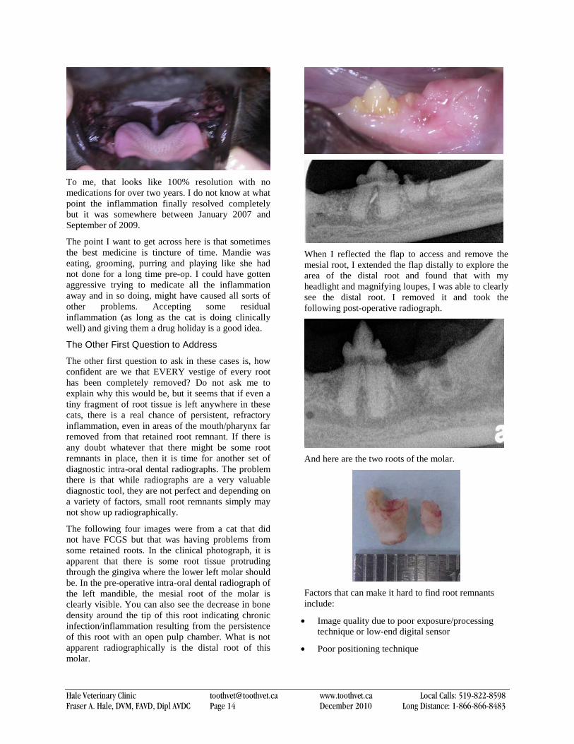
The following four images were from a cat that did not have FCGS but that was having problems from some retained roots. In the clinical photograph, it is apparent that there is some root tissue protruding through the gingiva where the lower left molar should be. In the pre-operative intra-oral dental radiograph of the left mandible, the mesial root of the molar is clearly visible. You can also see the decrease in bone density around the tip of this root indicating chronic infection/inflammation resulting from the persistence of this root with an open pulp chamber. What is not apparent radiographically is the distal root of this molar.
When I reflected the flap to access and remove the mesial root, I extended the flap distally to explore the area of the distal root and found that with my headlight and magnifying loupes, I was able to clearly see the distal root. I removed it and took the following post-operative radiograph.
And here are the two roots of the molar.
Factors that can make it hard to find root remnants include:
• Image quality due to poor exposure/processing technique or low-end digital sensor
• Poor positioning technique

Hale Veterinary Clinic [email protected] www.toothvet.ca Local Calls: 519-822-8598 Fraser A. Hale, DVM, FAVD, Dipl AVDC Page 15 December 2010 Long Distance: 1-866-866-8483
• Roots that are undergoing resorption or have calcified periodontal ligaments
• Tiny fragments that are too small to show up when surrounded on all sides by bone
Some of these can be over-come by using a good digital sensor or ensuring your exposure and processing technique are good and by taking multiple images from a variety of angles. Even with that, it is possible for root remnants to go undetected if it has been a while since the extractions were done and bone has grown around them.
The best way to ensure that there are no retained root remnants is if the person doing the extractions obtains both pre-operative and immediately post-operative intra-oral dental radiographs to clearly demonstrate that every alveolus is empty (no root remnants, no fragments of calculus or bone…).
Here are some pre- and post-op radiographs showing total extraction. It’s so much easier to document total extraction at the time of the extraction than months later after some healing.
In the absence of these immediate post-op images, it might be necessary to surgically open the soft tissues over the alveoli to explore and examine visually to rule out retained root remnants. This would allow for smoothing off any rough or irregular bone that might be poking at the underside of the soft tissue. It would allow for surgical removal of some of the inflamed tissue and closure of the wounds with healthier tissue. It would also allow for debridement/excision of any cul-de-sacs in the soft tissue. Sometimes, during healing, the soft tissue will dip down (or up) into the sockets, creating a blind-ended depression that can trap food and bacterial antigen, resulting in persistent inflammation.
Back to Management of the Refractory Patient
In a cat who has had all teeth and all roots completely removed (for sure) but that still has significant inflammation and is not doing well clinically, further treatment is needed. Some of the treatment options that have been used/recommended include:
Laser/Radiosurgical Rastering:
Using laser or radiosurgery, the ulceroproliferative tissues at the back of the mouth and into the pharynx can be debulked and scorched. The theory holds that this sterilizes the tissue, reduces the surface area and convolutions that would trap antigen and results in the production of scar tissue, which is more resistant to inflammation than normal epithelium. I have not had a lot of experience doing this, but those that have say they plan on doing at least three treatments at roughly monthly intervals.
Drugs
Now that all of the surgical options have been exhausted, it is time to consider medical management. A great many things have been tried over the years but we still do not have anything reliable or predictable. The other sad news is that none of the more effective medical options are without serious down-sides.

Hale Veterinary Clinic [email protected] www.toothvet.ca Local Calls: 519-822-8598 Fraser A. Hale, DVM, FAVD, Dipl AVDC Page 16 December 2010 Long Distance: 1-866-866-8483
Steroids:
In some cases, as a salvage/rescue treatment, short-term corticosteroids may be appropriate. Be careful with repositol injections such as DepoMedrol™ as there have been many reports of cats becoming diabetic after such treatment.
Interferon:
Human alpha interferon (Roferon A) has been used in the management of refractory cases of FCGS for some time. I have tried it on occasion and my experience has mirrored that of others – spotty at best. I no longer consider it much of an option.
On the other hand, feline omega interferon (Virbagen™ from Virbac™) has been getting some very good reviews. The problem with it is that it is not currently licensed or commercially available in North American. It can be obtained from England but that requires some paperwork. Also, the stuff needs to be kept cold, so it needs to be packed on ice and sent overnight with customs pre-clearance so there is no hold-up at the port of entry. I have not done this yet, but I am told it can be accomplished with the right letters to the right people.
The other draw-back for Virbagen™ is the price. A minimum order is about $1500. I believe this includes five vials which is enough for several cases and the extra can be kept frozen for a time. However, you have no guarantee that you will find other cases to use it on before it expires, so sharing your supply with local colleagues may make this all more practical.
I won`t bother with dosing regimens here but I will say the drug is best given orally. Reports are that there seems to be some topical effect as well as systemic and when the diluted solution is placed in the mouth to bathe the inflamed tissues, results are more impressive than when the drug is given by injection.
Cyclosporin:
While cyclosporin has proven to be helpful in a number of refractory cases, it too is somewhat problematic on a number of fronts.
Atopica is only licensed for use in dogs, so you would be going “off-label”. Therefore it is important to obtain informed owner consent before proceeding.
Cyclosporin is intensely and non-selectively immunosuppressive. It should not be given to cats that have positive results for FIV, FeLV, FIP or toxoplasmosis. It has also been suggested that it should not be used in cats that are allowed to go outside.
Cyclosporin is available in a number of different branded and generic forms. Not all of these forms are absorbed well or consistently in all patients. The recommendation is to stick with Atopica™ and to test for trough blood levels on a regular basis and adjust the dose based on this and on clinical results looking to find the lowest dose that does the trick.
Cyclosporin should not be used in diabetic patients as it can interfere with insulin regulation.
Cyclosporin can cause hematologic and biochemical problems and so CBC and profiles should be monitored regularly.
The Future It is always risky to try to predict the future but there are some interesting ideas under development.
If one accepts that FCGS is an imbalance of the immune response then re-establishing the balance could be the key. Several chronic inflammatory diseases in pets and people are thought to be the result of a dissociation between T-helper-1 and T-helper-2 cells. Targeted immune-therapy (immunomodulation rather than blanket immunosuppresion) is currently an active area of research. Watch for developments in this area over the next decade.
Of course, while this line of research may lead to treatment options for very early cases and for refractory edentulous cases, many patients are still going to need loads of extractions. By the time FCGS has been going on long enough to be called FCGS, there is usually more than enough permanent tissue destruction (periodontal disease and tooth resorption) to necessitate extractions.
Conclusion As you can see, there really are no simple or straight-forward answers for cats suffering from FCGS. However, if I had to boil it down to a few key points for you to get across to your clients they would be as follows:
Cats do NOT need teeth to live long and happy lives.
Cats DO deserve a mouth free of pain and infection.
No dental disease, let alone one of this magnitude, can be properly assessed or treated without diagnostic intra-oral dental radiographs.
As long as these cats have ANY dental tissues in their head, they need surgery, not medicine.
The sooner whole mouth extraction is accomplished, the better the prognosis for lasting and meaningful relief.