Embed Size (px)
Citation preview

RESEARCH ARTICLE
The clinical and microbiological characteristics
of enteric fever in Cambodia, 2008-2015
Laura M. F. Kuijpers1,2*, Thong Phe3, Chhun H. Veng3, Kruy Lim3, Sovann Ieng3,
Chun Kham3, Nizar Fawal4, Laetitia Fabre4, Simon Le Hello4, Erika Vlieghe1,5, Francois-
Xavier Weill4, Jan Jacobs1,2, Willy E. Peetermans6
1 Department of Clinical Sciences, Institute of Tropical Medicine, Antwerp, Belgium, 2 Department of
Microbiology & Immunology, KU Leuven, Leuven, Belgium, 3 Sihanouk Hospital Center of HOPE, Phnom
Penh, Cambodia, 4 Unite des Bacteries Pathogènes Enteriques, Centre National de Reference des E. coli,
Shigella et Salmonella, Institut Pasteur, Paris, France, 5 Department of General Internal Medicine, Infectious
diseases and Tropical Medicine, University Hospital Antwerp, Antwerp, Belgium, 6 Department of Internal
Medicine, University Hospital Leuven, Leuven, Belgium
Abstract
Background
Enteric fever remains a major public health problem in low resource settings and antibiotic
resistance is increasing. In Asia, an increasing proportion of infections is caused by Salmo-
nella enterica serovar Paratyphi A, which for a long time was assumed to cause a milder
clinical syndrome compared to Salmonella enterica serovar Typhi.
Methodology
A retrospective chart review study was conducted of 254 unique cases of blood culture con-
firmed enteric fever who presented at a referral adult hospital in Phnom Penh, Cambodia
between 2008 and 2015. Demographic, clinical and laboratory data were collected from clin-
ical charts and antibiotic susceptibility testing was performed. Whole genome sequence
analysis was performed on a subset of 121 isolates.
Results
One-hundred-and-ninety unique patients were diagnosed with Salmonella Paratyphi A and
64 with Salmonella Typhi. In the period 2008–2012, Salmonella Paratyphi A comprised
25.5% of 47 enteric fever cases compared to 86.0% of 207 cases during 2013–2015. Pre-
senting symptoms were identical for both serovars but higher median leukocyte counts (6.8
x 109/L vs. 6.3 x 10⁹/L; p = 0.035) and C-reactive protein (CRP) values (47.0 mg/L vs. 36
mg/L; p = 0.034) were observed for Salmonella Typhi infections. All but one of the Salmo-
nella Typhi isolates belonged to haplotype H58 associated with multidrug resistance (MDR)
(i.e. resistance to ampicillin, chloramphenicol and co-trimoxazole).;42.9% actually displayed
MDR compared to none of the Salmonella Paratyphi A isolates. Decreased ciprofloxacin
susceptibility (DCS) was observed in 96.9% (62/64) of Salmonella Typhi isolates versus
PLOS Neglected Tropical Diseases | https://doi.org/10.1371/journal.pntd.0005964 September 20, 2017 1 / 17
a1111111111
a1111111111
a1111111111
a1111111111
a1111111111
OPENACCESS
Citation: Kuijpers LMF, Phe T, Veng CH, Lim K,
Ieng S, Kham C, et al. (2017) The clinical and
microbiological characteristics of enteric fever in
Cambodia, 2008-2015. PLoS Negl Trop Dis 11(9):
e0005964. https://doi.org/10.1371/journal.
pntd.0005964
Editor: Thomas C Darton, Oxford University Clinical
Research Unit Vietnam, VIET NAM
Received: April 21, 2017
Accepted: September 14, 2017
Published: September 20, 2017
Copyright: © 2017 Kuijpers et al. This is an open
access article distributed under the terms of the
Creative Commons Attribution License, which
permits unrestricted use, distribution, and
reproduction in any medium, provided the original
author and source are credited.
Data Availability Statement: All relevant data are
within the paper and its Supporting Information
files. Short-read sequences have been deposited to
the European Nucleotide Archive (ENA) (http://
www.ebi.ac.uk/ena), under study accession
number PRJEB19906 (http://www.ebi.ac.uk/ena/
data/view/PRJEB19906).
Funding: The surveillance of bloodstream
infections at the Sihanouk Hospital Center of
HOPE is supported by the Belgian Directorate of
Development Cooperation (DGD)
11.5% (21/183) of Salmonella Paratyphi A isolates (all but one from 2015). All isolates were
susceptible to azithromycin and ceftriaxone.
Conclusions
In Phnom Penh, Cambodia, Salmonella Paratyphi A now causes the majority of enteric
fever cases and decreased susceptibility against ciprofloxacin is increasing. Overall, Salmo-
nella Typhi was significantly more associated with MDR and DCS compared to Salmonella
Paratyphi A.
Author summary
Enteric fever is a bloodstream infection caused by the bacteria Salmonella Typhi or Salmo-nella Paratyphi A, B, or C. It is common in low resource settings and linked to poor water
quality and sanitation. The disease is also endemic in Cambodia and since 2013 there has
been a sharp increase in the number of Salmonella Paratyphi A infections. We sought to
compare the clinical phenotypes and antibiotic susceptibilities of Salmonella Paratyphi A
infections with those of Salmonella Typhi infections in this setting. We retrospectively col-
lected demographic, clinical and laboratory data from clinical charts of 254 patients with a
blood culture positive for enteric fever. We also assessed antibiotic susceptibility patterns
and sequenced the genome of a subset of isolates. We found that since 2013 the majority
of enteric fever cases are caused by Salmonella Paratyphi A which increasingly shows
decreased susceptibility to the antibiotic ciprofloxacin, the current first line treatment. In
contrast, in Salmonella Typhi a re-emergence of susceptibility for the former first line anti-
biotics of ampicillin, co-trimoxazole and chloramphenicol was observed. Presenting
symptoms of Salmonella Typhi and Salmonella Paratyphi A were identical, minor differ-
ences were observed in laboratory parameters.
Introduction
Salmonella enterica serovar Typhi (Salmonella Typhi) and Salmonella enterica serovars Paraty-
phi (Salmonella Paratyphi) A, B, and C are Gram-negative bacteria which can invade the
bloodstream and cause typhoid and paratyphoid fever respectively (also jointly known as
‘enteric fever’). They are confined to the human host and are transmitted via the fecal-oral
route. Enteric fever poses a serious disease burden in low resource settings where the infection
is linked to poor sanitation and limited access to safe drinking water [1]. Although enteric
fever has become rare in Western countries it continues to affect international travelers return-
ing from endemic countries [2].
Patients with enteric fever typically present with acute fever and non-specific symptoms.
For a long time, Salmonella Paratyphi A was thought to cause milder disease than SalmonellaTyphi but several studies have contradicted this [1–4].
For both serovars, antibiotic resistance is increasingly reported and there is now widespread
presence of co-resistance against the former first line treatment options of ampicillin, co-tri-
moxazole and chloramphenicol (known as ‘multidrug resistance’) and decreased susceptibility
to ciprofloxacin, the current first line drug [5, 6]. Resistance to ciprofloxacin is also increas-
ingly reported [7, 8]. Worrisome are recent reports on emerging resistance against third-gen-
eration cephalosporins and azithromycin, the current alternative treatment options [9, 10].
Enteric fever in Phnom Penh, Cambodia
PLOS Neglected Tropical Diseases | https://doi.org/10.1371/journal.pntd.0005964 September 20, 2017 2 / 17
(https://diplomatie.belgium.be/en/policy/
development_cooperation/who_we_are/our_
organisation/dgd) through project 2.08 of the
Third Framework Agreement between the Belgian
DGD (Ministry of Development Cooperation) and
the Institute of Tropical Medicine (ITM). This
study was further supported by the Strategic
Network Laboratory Quality Management (LQM)
project (http://www.labquality.be/) (Belgian
Development Cooperation), the Institut Pasteur
and the French government’s Investissement
d’Avenir programme, Laboratoire d’Excellence
‘Integrative Biology of Emerging Infectious
Diseases’ (http://www.agence-nationale-
recherche.fr/?ProjetIA=10-LABX-0062) (grant
number ANR-10-LABX-62-IBEID). LMFK is
supported by the Flemish Ministry of Sciences
(EWI, SOFI project IDIS) (http://www.ewi-
vlaanderen.be/en) and received additional travel
grants (grant numbers K2.060.16N and
K2.065.17N) from the Fund for Scientific
Research Flanders (F.W.O.-Vlaanderen, Belgium)
(http://www.fwo.be/). The funders had no role in
study design, data collection and analysis,
decision to publish, or preparation of the
manuscript.
Competing interests: The authors have declared
that no competing interests exist.
Although historically the majority of enteric fever cases were caused by Salmonella Typhi,
the proportion of Salmonella Paratyphi A infections has been increasing steadily since the turn
of the century, in particular on the Asian continent [11].
In 2013, a significant increase in Salmonella Paratyphi A infections was also observed in
Cambodia, a country where enteric fever remains one of the most common clinical and blood
culture-confirmed diseases [12]. The increase was described in local residents as well as in trav-
elers returning from Cambodia to Europe, New Zealand, Japan and the United States [13–16].
Surprisingly, little is known about the clinical and microbiological characteristics of Salmo-nella Typhi and Salmonella Paratyphi A infections in Cambodian adults.
This study therefore aims to assess the clinical and microbiological aspects of enteric fever
in patients attending an adult hospital in Phnom Penh, Cambodia, during 2008–2015. More
specifically it aims to assess differences between infections caused by Salmonella Typhi as com-
pared to Salmonella Paratyphi A.
Materials and methods
Study setting & population
Sihanouk Hospital Center of HOPE (SHCH) is a 40-bed non-governmental referral hospital
for adults in Phnom Penh, Cambodia. Since July 2007, SHCH and the Institute of Tropical
Medicine (ITM) in Antwerp, Belgium, have been jointly organising the surveillance of blood-
stream infections at this hospital and its associated clinics. For this study, all data collected
between 2008–2015 were analyzed. Blood cultures were systematically sampled in all patients
presenting at SHCH who were suspected of having sepsis according to the Systemic Inflamma-
tory Response Syndrome (SIRS) criteria [17]. Recently, new definitions and criteria for sepsis
have been proposed such as the Sequential [Sepsis-related] Organ Failure Assessment (SOFA)
score [18]. Over the 8-year period, 18,927 blood cultures were sampled from mostly, but not
exclusively, adults [19]. Of these cultures 1,654 (8,7%) yielded clinically significant organisms.
Clinical review
From all patients whose blood was drawn for culture, basic demographic and clinical data
were registered in a surveillance logbook. A medical doctor verified missing data with patients
during a routine phone call one week after discharge from the hospital which was part of stan-
dard care. In addition, for this study, the available medical charts of all patients with blood cul-
ture-confirmed enteric fever were reviewed retrospectively by a second medical doctor for
additional symptoms and signs.
Laboratory methods
Hematology parameters were analyzed using a Sysmex KX-21 and T-1800i analyzer (Sysmex
Corporation, Kobe, Japan) and CRP values were measured using a TEMIS Linear Analyzer
(Linear Chemicals sl, Montgat, Spain).
Blood cultures were sampled and worked-up as previously described [20]. Isolates bio-
chemically identified as Salmonella spp. were stored at -70˚C on porous beads in cryopreserva-
tive (Microbank, Pro-Lab Diagnostics, Richmond Hill, Canada) and eventually shipped to the
ITM in Belgium. At ITM, the isolates were serotyped using commercial antisera (Sifin, Berlin,
Germany) following the White-Kauffmann-Le Minor scheme. A selection of 91 isolates were
sent to the Institut Pasteur in Paris for confirmation and whole genome sequencing.
At the ITM, antibiotic susceptibility was determined for all available isolates by disk diffu-
sion on Mueller-Hinton II agar in accordance with the CLSI 2016 guidelines [21]. The
Enteric fever in Phnom Penh, Cambodia
PLOS Neglected Tropical Diseases | https://doi.org/10.1371/journal.pntd.0005964 September 20, 2017 3 / 17
following antimicrobial drugs (Neo-Sensitabs, Rosco, Taastrup, Denmark) were tested: ampi-
cillin, sulfamethoxazole-trimethoprim, chloramphenicol, nalidixic acid, pefloxacin, gentami-
cin, tetracycline, ceftriaxone, ceftazidime, meropenem and ertapenem. Nalidixic acid and
pefloxacin served as predictors for ciprofloxacin non-susceptibility.
In addition, for all available isolates, minimal inhibitory concentration (MIC) values for
ciprofloxacin and azithromycin were determined by the E-test macro method (bioMerieux,
Marcy L’Etoile, France).
Quality control was performed using Escherichia coli (ATCC 25922) and Staphylococcusaureus (ATCC 29213).
Multidrug resistance (MDR) was defined as co-resistance to ampicillin, chloramphenicol
and trimethoprim-sulfamethoxazole [22]. For comparison with previously published litera-
ture, we used the superseded term ‘decreased ciprofloxacin susceptibility (DCS)’, defined as
MIC-values of�0.12 mg/L and�0.5 mg/L, i.e. currently classified as ‘intermediate susceptibil-
ity’ but associated with treatment failures or delayed treatment response [21].
Assessment of antimicrobial resistance genes and Salmonella Typhi
H58 typing
Whole genome sequencing was carried out on all 65 Salmonella Typhi isolates and a selection
of 26 Salmonella Paratyphi A isolates at the Plateforme de microbiologie mutualisee (P2M)
from the Pasteur International Bioresources network (PIBnet, Institut Pasteur, Paris, France).
Short-read sequences from 30 previously published Salmonella Paratyphi A genomes were
also included [23]. The run accession numbers and related metadata are detailed in S1 Table.
Short-read sequences have been deposited to the European Nucleotide Archive (ENA) (http://
www.ebi.ac.uk/ena) (accession number PRJEB19906).
The MagNAPure 96 system (Roche Diagnostics, Indianopolis, IN, USA) was used for DNA
extraction, libraries were prepared using the Nextera XT kit (Illumina, San Diego, CA, USA)
and sequencing was done with the NextSeq 500 system (Illumina). Read alignment, single
nucleotide polymorphism (SNP) detection and maximum-likelihood phylogeny were carried
out as described previously [23]. Sequence assembly was performed using SPAdes v. 3.6.0 [24].
Salmonella Typhi isolates were categorized as belonging to haplotype H58 based on the
presence of the H58 specific single SNP (T at nucleotide 252 on the gene glpA corresponding
to STY2513 from GenBank accession no. AL513382, Salmonella Typhi CT18) [25]. Genotyphi
(https://github.com/katholt/genotyphi) was also used to classify Salmonella Typhi [26]. Salmo-nella Paratyphi A isolates were categorized as belonging to clade C5 (the dominant clade in
Cambodia) based on the presence of the C5-specific SNP (G to A at position 2 381 607 within
the SPA_RS11495 gene) [23]. The presence of antibiotic resistance genes was determined with
ResFinder version 2.1 (https://cge.cbs.dtu.dk/services/ResFinder/) [27] and plasmids with
PlasmidFinder version 1.3 (https://cge.cbs.dtu.dk/services/PlasmidFinder/) and pMLST ver-
sion 1.4 (https://cge.cbs.dtu.dk/services/pMLST-1.4/) [27, 28]. The presence of mutations in
the quinolone-resistance determining region of the DNA gyrase and topoisomerase IV genes
(gyrA, gyrB, parC and parE) was assessed by the visual examination of sequences.
Data registration, statistical analysis
Demographic, clinical and microbiological data were entered encoded into an Excel database
that was created for this study (Microsoft, Redmond, WA, USA). The code referring to the
patient identity was only known by two medical doctors. Access to the database was restricted
to these two medical doctors and patient identifiers were removed prior to analysis.
Enteric fever in Phnom Penh, Cambodia
PLOS Neglected Tropical Diseases | https://doi.org/10.1371/journal.pntd.0005964 September 20, 2017 4 / 17

Only the first isolate and associated clinical data for each unique patient was considered.
Isolates recovered from a second blood culture drawn within two weeks after the initial one
were considered as duplicates, whereas isolates recovered from a repeat blood culture more
than two weeks after the initial one were considered as recurrences (either relapse or repeat
infections). Statistical analysis was done with Stata 12 (Stata Corp., College Station, TX, USA).
Continuous variables are described by a median and interquartile range (IQR). Comparisons
between Salmonella Typhi and Salmonella Paratyphi were performed using a Mann-Whitney
U test for continuous values and a Chi square test or Fisher exact test for proportions. A
p-value of< 0.05 was considered significant.
Ethics
The study was conducted according to the principles expressed in the Declaration of Helsinki
and involves use of information that was previously collected in the course of routine care.
Ethical approval for the Microbiological Surveillance Study was granted by the Institutional
Review Board of the ITM, the Ethics Committee of Antwerp University, and the National Eth-
ics Committee for Health Research in Cambodia. This study and approval includes retrospec-
tive review of demographic and clinical data which are part of routine clinical history taking as
recorded in the clinical chart.
Results
Surveillance of invasive Salmonella infections
Between 1 January 2008 and 31 December 2015 193 Salmonella Paratyphi A isolates were
retrieved from 190 patients and 65 Salmonella Typhi isolates from 64 patients. There were
no Salmonella Paratyphi B or C isolates; sixty-two non-typhoidal Salmonella isolates were
retrieved from 49 patients. The combined annual proportion of Salmonella Typhi and Salmo-nella Paratyphi A among all clinically significant organisms varied between 2.8% (2008) and
31.7% (2014).
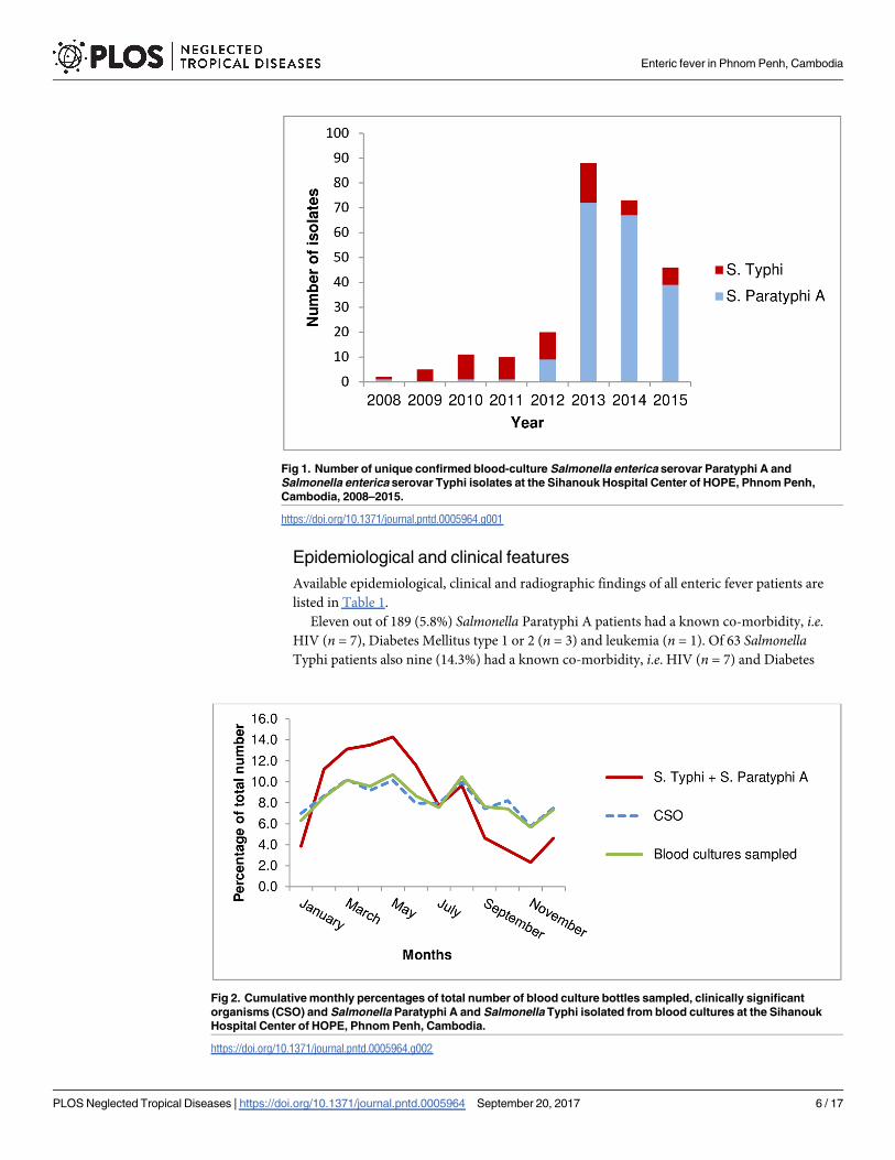
During 2008–2012, enteric fever was caused mostly by Salmonella Typhi (35 cases) and
only 12 cases of Salmonella Paratyphi A infection were identified (Fig 1). In 2013 however, a
sharp increase in the number of Salmonella Paratyphi A cases was observed with a total of 72
unique cases. In 2014 and 2015, the absolute annual number of Salmonella Paratyphi A cases
decreased, but remained higher than for the period preceding 2013. During this period, the
number of Salmonella Typhi cases remained relatively stable.
The majority of Salmonella Paratyphi A (64.7%; 123/190) and Salmonella Typhi infections
(59.4%; 38/64) cases occurred during the dry season (months November—April) while there
was an overall decreasing trend during the rainy season (months June-October) (Fig 2). Com-
pared to the monthly percentage of total blood cultures sampled and clinically significant
organisms found, the monthly combined percentage of Salmonella Typhi and Salmonella Para-
typhi A was higher during the hot and dry season (March—May) and lower during the rainy
season (June-October).
Recurrent infections
There were four cases of recurrent infections (37–48 days interval between first and recurrent
infection), three with Salmonella Paratyphi A and one with Salmonella Typhi; whole genome
sequence data was available for three of the four pairs. SNP analysis of the paired isolates
revealed that they differed by only two or three SNPs and the isolate pairs formed discrete clus-
ters within the trees (S1 and S2 Figs).
Enteric fever in Phnom Penh, Cambodia
PLOS Neglected Tropical Diseases | https://doi.org/10.1371/journal.pntd.0005964 September 20, 2017 5 / 17
Epidemiological and clinical features
Available epidemiological, clinical and radiographic findings of all enteric fever patients are
listed in Table 1.
Eleven out of 189 (5.8%) Salmonella Paratyphi A patients had a known co-morbidity, i.e.HIV (n = 7), Diabetes Mellitus type 1 or 2 (n = 3) and leukemia (n = 1). Of 63 SalmonellaTyphi patients also nine (14.3%) had a known co-morbidity, i.e. HIV (n = 7) and Diabetes
Fig 1. Number of unique confirmed blood-culture Salmonella enterica serovar Paratyphi A and
Salmonella enterica serovar Typhi isolates at the Sihanouk Hospital Center of HOPE, Phnom Penh,
Cambodia, 2008–2015.
https://doi.org/10.1371/journal.pntd.0005964.g001
Fig 2. Cumulative monthly percentages of total number of blood culture bottles sampled, clinically significant
organisms (CSO) and Salmonella Paratyphi A and Salmonella Typhi isolated from blood cultures at the Sihanouk
Hospital Center of HOPE, Phnom Penh, Cambodia.
https://doi.org/10.1371/journal.pntd.0005964.g002
Enteric fever in Phnom Penh, Cambodia
PLOS Neglected Tropical Diseases | https://doi.org/10.1371/journal.pntd.0005964 September 20, 2017 6 / 17
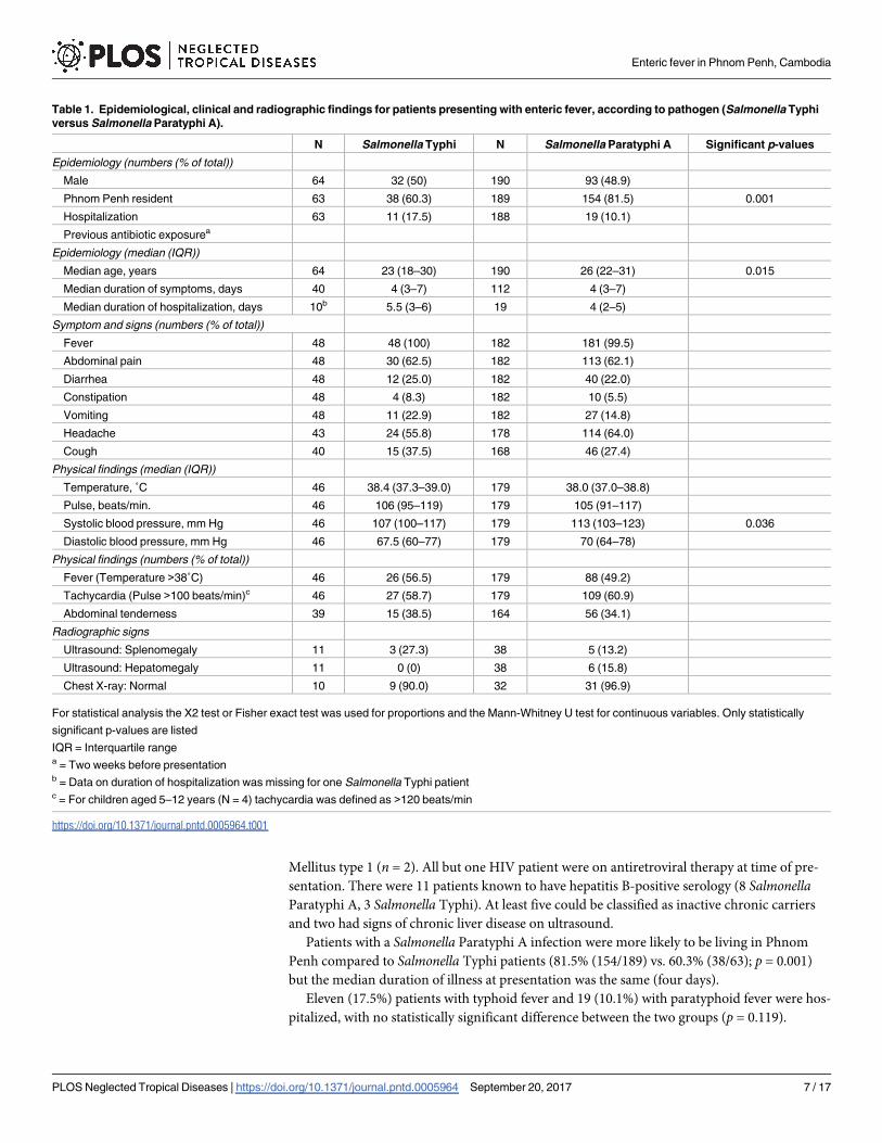
Mellitus type 1 (n = 2). All but one HIV patient were on antiretroviral therapy at time of pre-
sentation. There were 11 patients known to have hepatitis B-positive serology (8 SalmonellaParatyphi A, 3 Salmonella Typhi). At least five could be classified as inactive chronic carriers
and two had signs of chronic liver disease on ultrasound.
Patients with a Salmonella Paratyphi A infection were more likely to be living in Phnom
Penh compared to Salmonella Typhi patients (81.5% (154/189) vs. 60.3% (38/63); p = 0.001)
but the median duration of illness at presentation was the same (four days).
Eleven (17.5%) patients with typhoid fever and 19 (10.1%) with paratyphoid fever were hos-
pitalized, with no statistically significant difference between the two groups (p = 0.119).
Table 1. Epidemiological, clinical and radiographic findings for patients presenting with enteric fever, according to pathogen (Salmonella Typhi
versus Salmonella Paratyphi A).
N Salmonella Typhi N Salmonella Paratyphi A Significant p-values
Epidemiology (numbers (% of total))
Male 64 32 (50) 190 93 (48.9)
Phnom Penh resident 63 38 (60.3) 189 154 (81.5) 0.001
Hospitalization 63 11 (17.5) 188 19 (10.1)
Previous antibiotic exposurea
Epidemiology (median (IQR))
Median age, years 64 23 (18–30) 190 26 (22–31) 0.015
Median duration of symptoms, days 40 4 (3–7) 112 4 (3–7)
Median duration of hospitalization, days 10b 5.5 (3–6) 19 4 (2–5)
Symptom and signs (numbers (% of total))
Fever 48 48 (100) 182 181 (99.5)
Abdominal pain 48 30 (62.5) 182 113 (62.1)
Diarrhea 48 12 (25.0) 182 40 (22.0)
Constipation 48 4 (8.3) 182 10 (5.5)
Vomiting 48 11 (22.9) 182 27 (14.8)
Headache 43 24 (55.8) 178 114 (64.0)
Cough 40 15 (37.5) 168 46 (27.4)
Physical findings (median (IQR))
Temperature, ˚C 46 38.4 (37.3–39.0) 179 38.0 (37.0–38.8)
Pulse, beats/min. 46 106 (95–119) 179 105 (91–117)
Systolic blood pressure, mm Hg 46 107 (100–117) 179 113 (103–123) 0.036
Diastolic blood pressure, mm Hg 46 67.5 (60–77) 179 70 (64–78)
Physical findings (numbers (% of total))
Fever (Temperature >38˚C) 46 26 (56.5) 179 88 (49.2)
Tachycardia (Pulse >100 beats/min)c 46 27 (58.7) 179 109 (60.9)
Abdominal tenderness 39 15 (38.5) 164 56 (34.1)
Radiographic signs
Ultrasound: Splenomegaly 11 3 (27.3) 38 5 (13.2)
Ultrasound: Hepatomegaly 11 0 (0) 38 6 (15.8)
Chest X-ray: Normal 10 9 (90.0) 32 31 (96.9)
For statistical analysis the X2 test or Fisher exact test was used for proportions and the Mann-Whitney U test for continuous variables. Only statistically
significant p-values are listed
IQR = Interquartile rangea = Two weeks before presentationb = Data on duration of hospitalization was missing for one Salmonella Typhi patientc = For children aged 5–12 years (N = 4) tachycardia was defined as >120 beats/min
https://doi.org/10.1371/journal.pntd.0005964.t001
Enteric fever in Phnom Penh, Cambodia
PLOS Neglected Tropical Diseases | https://doi.org/10.1371/journal.pntd.0005964 September 20, 2017 7 / 17
Reasons for hospitalization included sepsis, persistent fever despite antibiotic therapy, dizzi-
ness due to low blood pressure, suspicion of dengue hemorrhagic fever (thrombocytopenia)
and dysregulated diabetes mellitus. There were no deaths nor complications noted.
The most frequently reported symptoms in all enteric fever patients together were fever in
229 patients (99.6%), headache in 138 (62.4%) and abdominal pain in 143 (62,2%). Presence or
absence of classic enteric fever signs such as a coated tongue and rose spots were infrequently
mentioned in clinical files and therefore not evaluated.
Despite the non-specific symptoms, physicians noted typhoid fever in their differential
diagnosis upon admission in 67.7% (136/201) of the cases.
There were no statistically significant differences in individual symptoms between typhoid
and paratyphoid fever patients, but patients infected with Salmonella Typhi had a slightly but
significantly lower median systolic blood pressure (107 mm Hg vs. 113 mm Hg; p = 0.036).
Treatment was not systematically recorded for all patients as many were lost to follow-up.
Various antimicrobial regimens were used, but ceftriaxone (2g I.V., once daily) was given
most frequently as empirical treatment and as monotherapy, normally for 10–14 days. In case
of de-escalation to oral antibiotics, this concerned mostly ciprofloxacin (500 mg, twice daily)
and next amoxicillin/clavulanate (625 mg, three times a day). In case of persistent fever while
awaiting blood culture results, amikacin was occasionally added to ceftriaxone.
Laboratory parameters
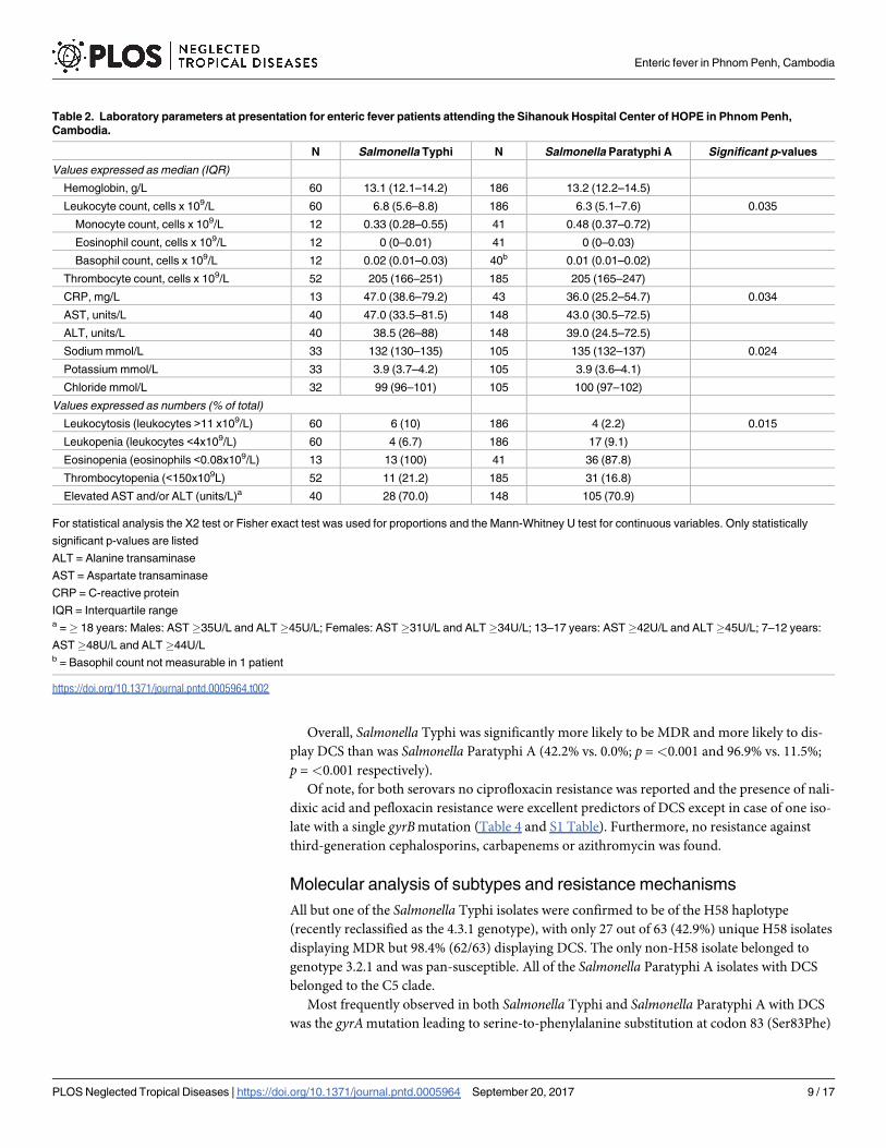
The laboratory parameters of enteric fever patients on admission are summarized in Table 2.
Common laboratory abnormalities for enteric fever patients included moderately risen trans-
aminase levels in 133 patients (70.7%), an elevated CRP in 53 patients (94.6%) and eosinopenia
in 49 patients (90.7%). Hematological abnormalities were uncommon; the leukocyte count
was normal in 88.1% of all patients. Compared to Salmonella Paratyphi A infected patients,
Salmonella Typhi patients had slightly but significantly higher median values for leukocytes
(6.8 x 10⁹/L vs. 6.3 x 10⁹/L; p = 0.035) and C-reactive protein (CRP) (47.0 mg/L vs. 36 mg/L;
p = 0.034), with more presence of leukocytosis (10.0% vs. 2.2% p = 0.015). Salmonella Paratyphi
A infection was associated with a higher monocyte count compared to Salmonella Typhi (0.48
x 109/L vs. 0.33 x 10⁹/L), but this difference did not reach statistical significance (p = 0.069).
Microbiological features
Both anaerobic and aerobic blood cultures showed signs of growth after a median of two days
(IQR 2–3) for all enteric fever patients. In 221 enteric fever patients a pair of one aerobic bottle
and one anaerobic bottle was sampled, and in 180 of those cases (81.4%) both bottles grew. In
the other cases (growth in only a single bottle), it was the aerobic bottle which grew in nearly
two-thirds (65.9%; 27/41) of pairs.
Reported antibiotic exposure in the two weeks before blood culture sampling was not asso-
ciated with a difference in the median days to growth for both aerobic bottles and anaerobic
bottles.
In total, 183 out of 190 (96.3%) unique Salmonella Paratyphi A isolates and all 64 unique
Salmonella Typhi isolates recovered during the study period were available for antibiotic sus-
ceptibility testing (Table 3).
For Salmonella Typhi, there was a significant decrease (p =<0.001) in the proportion of iso-
lates that were MDR over the 8-year period (62.9% vs. 17.2%) while decreased susceptibility to
ciprofloxacin remained at nearly 100% (96.9%; 62/64) during the entire period. For SalmonellaParatyphi A the emergence of DCS was noted as of 2015 (S1 Table). In this year 19 out of 36
unique isolates (52.8%) showed DCS.
Enteric fever in Phnom Penh, Cambodia
PLOS Neglected Tropical Diseases | https://doi.org/10.1371/journal.pntd.0005964 September 20, 2017 8 / 17
Overall, Salmonella Typhi was significantly more likely to be MDR and more likely to dis-
play DCS than was Salmonella Paratyphi A (42.2% vs. 0.0%; p =<0.001 and 96.9% vs. 11.5%;
p = <0.001 respectively).
Of note, for both serovars no ciprofloxacin resistance was reported and the presence of nali-
dixic acid and pefloxacin resistance were excellent predictors of DCS except in case of one iso-
late with a single gyrBmutation (Table 4 and S1 Table). Furthermore, no resistance against
third-generation cephalosporins, carbapenems or azithromycin was found.
Molecular analysis of subtypes and resistance mechanisms
All but one of the Salmonella Typhi isolates were confirmed to be of the H58 haplotype
(recently reclassified as the 4.3.1 genotype), with only 27 out of 63 (42.9%) unique H58 isolates
displaying MDR but 98.4% (62/63) displaying DCS. The only non-H58 isolate belonged to
genotype 3.2.1 and was pan-susceptible. All of the Salmonella Paratyphi A isolates with DCS
belonged to the C5 clade.
Most frequently observed in both Salmonella Typhi and Salmonella Paratyphi A with DCS
was the gyrAmutation leading to serine-to-phenylalanine substitution at codon 83 (Ser83Phe)
Table 2. Laboratory parameters at presentation for enteric fever patients attending the Sihanouk Hospital Center of HOPE in Phnom Penh,
Cambodia.
N Salmonella Typhi N Salmonella Paratyphi A Significant p-values
Values expressed as median (IQR)
Hemoglobin, g/L 60 13.1 (12.1–14.2) 186 13.2 (12.2–14.5)
Leukocyte count, cells x 109/L 60 6.8 (5.6–8.8) 186 6.3 (5.1–7.6) 0.035
Monocyte count, cells x 109/L 12 0.33 (0.28–0.55) 41 0.48 (0.37–0.72)
Eosinophil count, cells x 109/L 12 0 (0–0.01) 41 0 (0–0.03)
Basophil count, cells x 109/L 12 0.02 (0.01–0.03) 40b 0.01 (0.01–0.02)
Thrombocyte count, cells x 109/L 52 205 (166–251) 185 205 (165–247)
CRP, mg/L 13 47.0 (38.6–79.2) 43 36.0 (25.2–54.7) 0.034
AST, units/L 40 47.0 (33.5–81.5) 148 43.0 (30.5–72.5)
ALT, units/L 40 38.5 (26–88) 148 39.0 (24.5–72.5)
Sodium mmol/L 33 132 (130–135) 105 135 (132–137) 0.024
Potassium mmol/L 33 3.9 (3.7–4.2) 105 3.9 (3.6–4.1)
Chloride mmol/L 32 99 (96–101) 105 100 (97–102)
Values expressed as numbers (% of total)
Leukocytosis (leukocytes >11 x109/L) 60 6 (10) 186 4 (2.2) 0.015
Leukopenia (leukocytes <4x10⁹/L) 60 4 (6.7) 186 17 (9.1)
Eosinopenia (eosinophils <0.08x10⁹/L) 13 13 (100) 41 36 (87.8)
Thrombocytopenia (<150x109L) 52 11 (21.2) 185 31 (16.8)
Elevated AST and/or ALT (units/L)a 40 28 (70.0) 148 105 (70.9)
For statistical analysis the X2 test or Fisher exact test was used for proportions and the Mann-Whitney U test for continuous variables. Only statistically
significant p-values are listed
ALT = Alanine transaminase
AST = Aspartate transaminase
CRP = C-reactive protein
IQR = Interquartile rangea =� 18 years: Males: AST�35U/L and ALT�45U/L; Females: AST�31U/L and ALT�34U/L; 13–17 years: AST�42U/L and ALT�45U/L; 7–12 years:
AST�48U/L and ALT�44U/Lb = Basophil count not measurable in 1 patient
https://doi.org/10.1371/journal.pntd.0005964.t002
Enteric fever in Phnom Penh, Cambodia
PLOS Neglected Tropical Diseases | https://doi.org/10.1371/journal.pntd.0005964 September 20, 2017 9 / 17
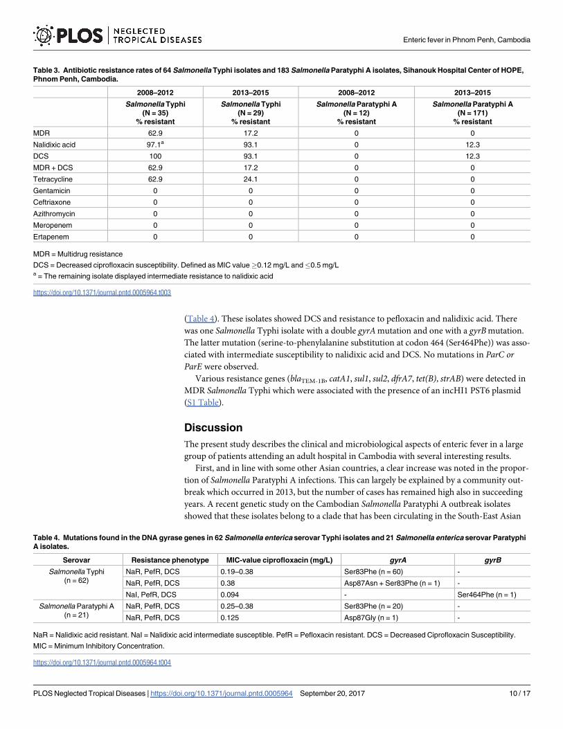
(Table 4). These isolates showed DCS and resistance to pefloxacin and nalidixic acid. There
was one Salmonella Typhi isolate with a double gyrAmutation and one with a gyrB mutation.
The latter mutation (serine-to-phenylalanine substitution at codon 464 (Ser464Phe)) was asso-
ciated with intermediate susceptibility to nalidixic acid and DCS. No mutations in ParC orParE were observed.
Various resistance genes (blaTEM-1B, catA1, sul1, sul2, dfrA7, tet(B), strAB) were detected in
MDR Salmonella Typhi which were associated with the presence of an incHI1 PST6 plasmid
(S1 Table).
Discussion
The present study describes the clinical and microbiological aspects of enteric fever in a large
group of patients attending an adult hospital in Cambodia with several interesting results.
First, and in line with some other Asian countries, a clear increase was noted in the propor-
tion of Salmonella Paratyphi A infections. This can largely be explained by a community out-
break which occurred in 2013, but the number of cases has remained high also in succeeding
years. A recent genetic study on the Cambodian Salmonella Paratyphi A outbreak isolates
showed that these isolates belong to a clade that has been circulating in the South-East Asian
Table 3. Antibiotic resistance rates of 64 Salmonella Typhi isolates and 183 Salmonella Paratyphi A isolates, Sihanouk Hospital Center of HOPE,
Phnom Penh, Cambodia.
2008–2012 2013–2015 2008–2012 2013–2015
Salmonella Typhi
(N = 35)
% resistant
Salmonella Typhi
(N = 29)
% resistant
Salmonella Paratyphi A
(N = 12)
% resistant
Salmonella Paratyphi A
(N = 171)
% resistant
MDR 62.9 17.2 0 0
Nalidixic acid 97.1a 93.1 0 12.3
DCS 100 93.1 0 12.3
MDR + DCS 62.9 17.2 0 0
Tetracycline 62.9 24.1 0 0
Gentamicin 0 0 0 0
Ceftriaxone 0 0 0 0
Azithromycin 0 0 0 0
Meropenem 0 0 0 0
Ertapenem 0 0 0 0
MDR = Multidrug resistance
DCS = Decreased ciprofloxacin susceptibility. Defined as MIC value�0.12 mg/L and�0.5 mg/La = The remaining isolate displayed intermediate resistance to nalidixic acid
https://doi.org/10.1371/journal.pntd.0005964.t003
Table 4. Mutations found in the DNA gyrase genes in 62 Salmonella enterica serovar Typhi isolates and 21 Salmonella enterica serovar Paratyphi
A isolates.
Serovar Resistance phenotype MIC-value ciprofloxacin (mg/L) gyrA gyrB
Salmonella Typhi
(n = 62)
NaR, PefR, DCS 0.19–0.38 Ser83Phe (n = 60) -
NaR, PefR, DCS 0.38 Asp87Asn + Ser83Phe (n = 1) -
NaI, PefR, DCS 0.094 - Ser464Phe (n = 1)
Salmonella Paratyphi A
(n = 21)
NaR, PefR, DCS 0.25–0.38 Ser83Phe (n = 20) -
NaR, PefR, DCS 0.125 Asp87Gly (n = 1) -
NaR = Nalidixic acid resistant. NaI = Nalidixic acid intermediate susceptible. PefR = Pefloxacin resistant. DCS = Decreased Ciprofloxacin Susceptibility.
MIC = Minimum Inhibitory Concentration.
https://doi.org/10.1371/journal.pntd.0005964.t004
Enteric fever in Phnom Penh, Cambodia
PLOS Neglected Tropical Diseases | https://doi.org/10.1371/journal.pntd.0005964 September 20, 2017 10 / 17
region already for decades [23]. Further, no indications were found for significant genetic
changes within the Cambodian isolates suggesting that environmental and/or behavioral fac-
tors are more likely to play a role.
Patients with paratyphoid fever were significantly more likely than typhoid fever patients to
be residents of Phnom Penh, which suggests that exposure to the bacterium is more common
in the city. Previous studies from Nepal and Indonesia have linked paratyphoid fever to recent
immigration into the capital and consumption of street food [29, 30]. Increased dependency
on street food has been linked to urbanization, and Phnom Penh is rapidly expanding.
As part of urban expansion, some of the city’s peri-urban lakes have been filled with sand to
reclaim land [31]. These lakes are estimated to receive 80% of the city’s (untreated) waste
water and act as a natural sewage treatment through aquatic cultivation of vegetables of which
some are consumed raw [32]. Reductions in the size of these lakes could have led to higher
concentrations of fecal sludge and bacteria in the remaining water and increased flooding in
the city [33].
The majority of enteric fever cases occurred in the dry season. During this season more veg-
etables are harvested and it is also known for an increased availability of snails and clams
(bivalve shellfish), due to low water levels in rivers. Shellfish are known to be able to concen-
trate micro-organisms from water. They are popular snacks which are dried outside rather
than boiled during the dry season. In addition, this season coincides with the two most impor-
tant festivities of the year, the Chinese and Khmer New Year which are associated with
increased migration in and out of the city and longer storage duration of food. Last, high daily
temperatures may lead to more indiscriminate intake of water and ice cubes. These factors are
currently being explored more in-depth.
Second, based on individual symptoms at presentation, infections caused by SalmonellaTyphi vs. Salmonella Paratyphi A were similar and indistinguishable, which is in line with
other studies from Asia [3, 4]. For both serovars, the median pulse rates at presentation (106
and 105 beats/minute) were high. This has been noted before in children with enteric fever
[34].
No complications or deaths occurred which could be ascribed to a prompt start of antibiotic
therapy and an early presentation. The latter can also explain the absence of relative bradycar-
dia and the low rate of diarrhea observed which are typically seen in later stages of the disease
[35].
In general, laboratory abnormalities were non-specific and leukocyte counts were normal
in 87.4% of all enteric fever patients which was also found by others [36]. Higher leukocyte
counts and CRP values were found among Salmonella Typhi-infected patients, suggesting a
more severe infection. This is in line with a recent human challenge study, which found that a
challenge with Salmonella Paratyphi A in healthy volunteers resulted in a milder disease profile
(high rates of afebrile bacteremia) than that observed following typhoid challenge [37].
Some differences in presentation were noted when comparing these results to travelers
infected with the same Salmonella Paratyphi A C5 strain returning from Cambodia to France.
In the latter study, the majority of patients did have diarrhea (70.6%) and were hospitalized
(86%) [14]. This difference may be due to other waterborne or oral-fecal infections travelers
frequently contract and/or less financial constrains related to hospital admission [2]. Clinical
presentation in the present study also differed from typhoid fever patients in African countries
where higher rates of severe complications and mortality are observed [38]. As the same H58
haplotype of Salmonella Typhi is dominant in Asia and in eastern Africa, differences could per-
haps be explained by timely access to health care and adequate treatment as well as to host-
related factors including underlying co-morbidities like malnutrition.
Enteric fever in Phnom Penh, Cambodia
PLOS Neglected Tropical Diseases | https://doi.org/10.1371/journal.pntd.0005964 September 20, 2017 11 / 17
It has been suggested that isolates of the H58 lineage and MDR strains in general are associ-
ated with increased virulence and pathogenicity [39–41]. Therefore the results regarding the
clinical presentation and severity of cases as described here, might not be applicable to areas
where other lineages dominate.
As a third observation, antibiotic resistance trends were very different for the two serovars.
While 42.2% of the unique Salmonella Typhi isolates displayed MDR, none of the SalmonellaParatyphi A isolates did. DCS was present in nearly all Salmonella Typhi isolates, but only
emerged in Salmonella Paratyphi A from 2015. The rapid increase of DCS in Salmonella Para-
typhi during that year is of concern as ciprofloxacin is the treatment of choice for uncompli-
cated enteric fever.
Fourth, although all but one Salmonella Typhi isolates were found to belong to the globally
dominant H58 haplotype, more than half were not associated with MDR and the proportion
of (plasmid-mediated) MDR Salmonella Typhi significantly decreased during the study period.
This trend has previously been noted in India, Nepal and neighboring country Vietnam [42–
44], but contrasts with a recent study on Salmonella Typhi isolates from rural Cambodia where
89% of the H58 isolates displayed the MDR phenotype [45]. The re-emergence of susceptibility
might result from a lack of antibiotic pressure since fluoroquinolones have become the pre-
ferred treatment both in community and hospital settings in Phnom Penh.
No resistance against ceftriaxone nor against azithromycin was observed. However, reports
on extended spectrum beta-lactamases (ESBL) positive and azithromycin resistant Salmonellaspp. isolates are emerging globally including one from the same hospital on Salmonella entericaserovar Choleraesuis [20, 46, 47] underlining the importance of continued microbiological
surveillance.
Last, molecular analysis of isolates from three patients with a recurrent infection showed
relapse was more likely than re-infection with isolate pairs differing only 2–3 SNPs which can
occur during the period of persistence within the human body and suggests relapse rather
than re-infection [48]. Relapse is estimated to occur in around 5–10% of enteric fever cases
usually two to three weeks after the resolution of fever [5]. In our study, a blood culture con-
firmed recurrence was witnessed only in 4 out of 254 cases (1.6%). It is likely that other recur-
rent infections have been missed, partly due to the different medical systems that co-exist in
Cambodia in which patients readily switch from one healthcare provider to another, especially
if symptoms persist.
This study has several limitations. First, the hospital-based setting precluded generalization
to patients whose symptoms were not severe enough to seek medical care in a hospital or
clinic. Second, the study concerned mostly adults and therefore findings might not be equally
applicable to a pediatric population. Third, the study was retrospective in nature; not all clini-
cal charts were available for review and clinical record keeping was variable among different
clinicians. It was not possible to reliably estimate time to defervescence.
Despite these limitations, this is one of the largest and most comprehensive descriptive
studies on Salmonella Paratyphi A infections so far which is relevant given the global increase
in Salmonella Paratyphi A infections. The data do not represent one single hospital, but several
clinics located in different districts of the city. Some of these clinics have reduced rates for the
poor and during the study period all blood cultures were provided for free. This helped to
overcome some of the bias associated with a hospital based study.
The high proportion of Salmonella Typhi and Paratyphi A recovered from blood cultures
indicated that enteric fever is a very frequent disease in Phnom Penh. While efforts are made
to increase the microbiological diagnostic capacity in the country, a rapid test for invasive Sal-monella infections would be a welcome tool for fast and reliable diagnosis. It could increase
Enteric fever in Phnom Penh, Cambodia
PLOS Neglected Tropical Diseases | https://doi.org/10.1371/journal.pntd.0005964 September 20, 2017 12 / 17
knowledge on the burden of disease in the community and could replace the flawed Widal test
that is still frequently used.
As the current Salmonella Typhi vaccine provides no to very little protection against Salmo-nella Paratyphi A, the development of an effective Salmonella Paratyphi A vaccine should be
promoted, pending improved water quality and sanitation [49].
Conclusion
Enteric fever is frequent in Phnom Penh and the proportion of cases due to Salmonella Paraty-
phi A has increased. Studies to investigate risk factors and possible transmission routes are
urgently needed to advise public health interventions. No MDR was observed for SalmonellaParatyphi A but DCS increased rapidly. DCS remained highly prevalent in Salmonella Typhi
while MDR rates have declined. Ceftriaxone and azithromycin remain highly active in vitro
but continued surveillance is imperative to monitor resistance.
Supporting information
S1 Table. Strain list and accession numbers for organisms used in this study.
(XLSX)
S1 Fig. Maximum Likelihood (ML) tree of 185 Salmonella Paratyphi A genomes. Maximum
Likelihood (ML) tree of 185 Salmonella Paratyphi A genomes including 159 previously pub-
lished genomes (Kuijpers & Le Hello et al., 2016 [23] and Zhou et al., 2014 [50]). Fifty-six
genomes represent isolates collected at the Sihanouk Hospital Center of HOPE, Phnom Penh,
Cambodia between 2008–2015. For readability, only the position of the reference genome (Sal-monella Paratyphi A ATCC 9150) and the paired isolates are shown. Only clades C1-C5 are
indicated. The big arrow indicates the paired isolates (ID 6778 and 6748, 2 SNPs difference; ID
6610 and 6670; 3 SNPs difference).
(PPTX)
S2 Fig. Maximum Likelihood (ML) tree of 66 Salmonella Typhi genomes. Maximum Likeli-
hood (ML) tree of 66 Salmonella Typhi genomes including the Salmonella Typhi CT18 refer-
ence genome (AL513382) and 65 genomes of Salmonella Typhi isolates collected at the
Sihanouk Hospital Center of HOPE, Phnom Penh, Cambodia between 2008–2015. For read-
ability, only the position of the reference genome (Salmonella Typhi CT18) and of the paired
isolates (ID 4764 and 4855, 3 SNPs difference) are indicated (with a big arrow).
(PPTX)
S1 Checklist. STROBE checklist.
(PDF)
Acknowledgments
We would like to thank Barbara Barbe, Marleen Verlinden and Marjan Peeters for technical
assistance at ITM. We would also like to thank all colleagues involved in blood culture surveil-
lance and clinical care at the Sihanouk Hospital Center of HOPE and Laura Brinas, Andrea
Alexandru, Maud Vanpeene, Sobhy Wilhame, and Vincent Enouf (Institut Pasteur, Pasteur
International Bioresources network (PIBnet), Plateforme de microbiologie mutualisee (P2M),
Paris, France, for performing the sequencing experiments.
Enteric fever in Phnom Penh, Cambodia
PLOS Neglected Tropical Diseases | https://doi.org/10.1371/journal.pntd.0005964 September 20, 2017 13 / 17
Author Contributions
Conceptualization: Laura M. F. Kuijpers, Simon Le Hello, Erika Vlieghe, Francois-Xavier
Weill, Jan Jacobs.
Data curation: Laura M. F. Kuijpers, Thong Phe, Chhun H. Veng, Nizar Fawal.
Formal analysis: Laura M. F. Kuijpers, Nizar Fawal, Simon Le Hello, Francois-Xavier Weill,
Willy E. Peetermans.
Funding acquisition: Jan Jacobs.
Investigation: Laura M. F. Kuijpers, Thong Phe, Chhun H. Veng, Kruy Lim, Sovann Ieng,
Chun Kham, Nizar Fawal, Laetitia Fabre, Erika Vlieghe.
Methodology: Laura M. F. Kuijpers, Nizar Fawal, Laetitia Fabre, Erika Vlieghe, Francois-
Xavier Weill, Jan Jacobs.
Project administration: Thong Phe, Chhun H. Veng, Chun Kham, Simon Le Hello, Jan
Jacobs.
Resources: Thong Phe, Chun Kham, Simon Le Hello, Francois-Xavier Weill, Jan Jacobs.
Software: Nizar Fawal, Laetitia Fabre, Francois-Xavier Weill.
Supervision: Thong Phe, Chun Kham, Simon Le Hello, Erika Vlieghe, Francois-Xavier Weill,
Jan Jacobs, Willy E. Peetermans.
Validation: Laura M. F. Kuijpers, Francois-Xavier Weill, Jan Jacobs, Willy E. Peetermans.
Writing – original draft: Laura M. F. Kuijpers.
Writing – review & editing: Laura M. F. Kuijpers, Thong Phe, Chhun H. Veng, Kruy Lim,
Sovann Ieng, Chun Kham, Nizar Fawal, Laetitia Fabre, Simon Le Hello, Erika Vlieghe,
Francois-Xavier Weill, Jan Jacobs, Willy E. Peetermans.
References
1. Bhan MK, Bahl R, Bhatnagar S. Typhoid and paratyphoid fever. Lancet. 2005; 366(9487):749–62.
https://doi.org/10.1016/S0140-6736(05)67181-4 PMID: 16125594
2. Meltzer E, Sadik C, Schwartz E. Enteric fever in Israeli travelers: a nationwide study. J Travel Med.
2005; 12(5):275–81. PMID: 16256052
3. Maskey AP, Day JN, Phung QT, Thwaites GE, Campbell JI, Zimmerman M, et al. Salmonella enterica
serovar Paratyphi A and S. enterica serovar Typhi cause indistinguishable clinical syndromes in Kath-
mandu, Nepal. Clin Infect Dis. 2006; 42(9):1247–53. https://doi.org/10.1086/503033 PMID: 16586383
4. Vollaard AM, Ali S, Widjaja S, Asten HA, Visser LG, Surjadi C, et al. Identification of typhoid fever and
paratyphoid fever cases at presentation in outpatient clinics in Jakarta, Indonesia. Trans R Soc Trop
Med Hyg. 2005; 99(6):440–50. https://doi.org/10.1016/j.trstmh.2004.09.012 PMID: 15837356
5. Parry CM, Hien TT, Dougan G, White NJ, Farrar JJ. Typhoid fever. N Engl J Med. 2002; 347(22):1770–
82. https://doi.org/10.1056/NEJMra020201 PMID: 12456854
6. Wain J, Kidgell C. The emergence of multidrug resistance to antimicrobial agents for the treatment of
typhoid fever. Trans R Soc Trop Med Hyg. 2004; 98(7):423–30. https://doi.org/10.1016/j.trstmh.2003.
10.015 PMID: 15138079
7. Medalla F, Sjolund-Karlsson M, Shin S, Harvey E, Joyce K, Theobald L, et al. Ciprofloxacin-resistant
Salmonella enterica Serotype Typhi, United States, 1999–2008. Emerg Infect Dis. 2011; 17(6):1095–8.
https://doi.org/10.3201/eid/1706.100594 PMID: 21749779
8. Chiou CS, Lauderdale TL, Phung DC, Watanabe H, Kuo JC, Wang PJ, et al. Antimicrobial resistance in
Salmonella enterica Serovar Typhi isolates from Bangladesh, Indonesia, Taiwan, and Vietnam. Antimi-
crob Agents Chemother. 2014; 58(11):6501–7. https://doi.org/10.1128/AAC.03608-14 PMID: 25136011
9. Pokharel BM, Koirala J, Dahal RK, Mishra SK, Khadga PK, Tuladhar NR. Multidrug-resistant and
extended-spectrum beta-lactamase (ESBL)-producing Salmonella enterica (serotypes Typhi and
Enteric fever in Phnom Penh, Cambodia
PLOS Neglected Tropical Diseases | https://doi.org/10.1371/journal.pntd.0005964 September 20, 2017 14 / 17
Paratyphi A) from blood isolates in Nepal: surveillance of resistance and a search for newer alternatives.
Int J Infect Dis. 2006; 10(6):434–8. https://doi.org/10.1016/j.ijid.2006.07.001 PMID: 16978898
10. Hassing RJ, Goessens WH, van Pelt W, Mevius DJ, Stricker BH, Molhoek N, et al. Salmonella subtypes
with increased MICs for azithromycin in travelers returned to The Netherlands. Emerg Infect Dis. 2014;
20(4):705–8. https://doi.org/10.3201/eid2004.131536 PMID: 24655478
11. Ochiai RL, Wang X, von Seidlein L, Yang J, Bhutta ZA, Bhattacharya SK, et al. Salmonella paratyphi A
rates, Asia. Emerg Infect Dis. 2005; 11(11):1764–6. https://doi.org/10.3201/eid1111.050168 PMID:
16318734
12. Vlieghe ER, Phe T, De Smet B, Veng HC, Kham C, Lim K, et al. Bloodstream infection among adults in
Phnom Penh, Cambodia: key pathogens and resistance patterns. PLoS One. 2013; 8(3):e59775.
https://doi.org/10.1371/journal.pone.0059775 PMID: 23555777
13. Vlieghe E, Phe T, De Smet B, Veng CH, Kham C, Sar D, et al. Increase in Salmonella enterica serovar
Paratyphi A infections in Phnom Penh, Cambodia, January 2011 to August 2013. Euro Surveill. 2013;
18(39).
14. Tourdjman M, Le Hello S, Gossner C, Delmas G, Tubiana S, Fabre L, et al. Unusual increase in
reported cases of paratyphoid A fever among travellers returning from Cambodia, January to Septem-
ber 2013. Euro Surveill. 2013; 18(39).
15. Saitoh T, Morita M, Shimada T, Izumiya H, Kanayama A, Oishi K, et al. Increase in paratyphoid fever
cases in Japanese travellers returning from Cambodia in 2013. Epidemiol Infect. 2016; 144(3):602–6.
https://doi.org/10.1017/S0950268815001648 PMID: 26169980
16. Judd MC, Grass JE, Mintz ED, Bicknese A, Mahon BE. Salmonella enterica Paratyphi A Infections in
Travelers Returning from Cambodia, United States. Emerg Infect Dis. 2015; 21(6):1089–91. https://doi.
org/10.3201/eid2106.150088 PMID: 25988984
17. Bone RC, Balk RA, Cerra FB, Dellinger RP, Fein AM, Knaus WA, et al. Definitions for sepsis and organ
failure and guidelines for the use of innovative therapies in sepsis. The ACCP/SCCM Consensus Con-
ference Committee. American College of Chest Physicians/Society of Critical Care Medicine. Chest.
1992; 101(6):1644–55. PMID: 1303622
18. Seymour CW, Liu VX, Iwashyna TJ, Brunkhorst FM, Rea TD, Scherag A, et al. Assessment of Clinical
Criteria for Sepsis: For the Third International Consensus Definitions for Sepsis and Septic Shock (Sep-
sis-3). JAMA. 2016; 315(8):762–74. https://doi.org/10.1001/jama.2016.0288 PMID: 26903335
19. Phe T VE, Lim K, Veng CH, Thai S, Leng L, Kham C, Jacobs J. Surveillance of bloodstream infection
and antibiotic resistance in Phnom Penh, Cambodia (2007–2014). Poster presented at: 17th Interna-
tional Congress on Infectious Diseases; 2016 March 2–5; Hyderabad, India.
20. Vlieghe ER, Phe T, De Smet B, Veng CH, Kham C, Bertrand S, et al. Azithromycin and ciprofloxacin
resistance in Salmonella bloodstream infections in Cambodian adults. PLoS Negl Trop Dis. 2012; 6
(12):e1933. https://doi.org/10.1371/journal.pntd.0001933 PMID: 23272255
21. Clinical and Laboratory Standards Institute (CLSI). Performance standards for antimicrobial susceptibil-
ity testing. 26th informational supplement. CLSI Document M100-S26. Wayne, PA: CLSI 2016.
22. World Health Organization (WHO). Background document: the diagnosis, treatment and prevention of
typhoid fever. 2003.
23. Kuijpers LMF, Le Hello S, Fawal N, Fabre L, Tourdjman M, Dufour M, Sar D, Kham C, Phe T, Vlieghe E,
Bouchier C, Jacobs J, Weill FX. Genomic analysis of Salmonella enterica serotype Paratyphi A during
an outbreak in Cambodia, 2013–2015. Microbial Genomics. 2016; https://doi.org/10.1099/mgen.0.
000092 PMID: 28348832
24. Bankevich A, Nurk S, Antipov D, Gurevich AA, Dvorkin M, Kulikov AS, et al. SPAdes: a new genome
assembly algorithm and its applications to single-cell sequencing. J Comput Biol. 2012; 19(5):455–77.
https://doi.org/10.1089/cmb.2012.0021 PMID: 22506599
25. Roumagnac P, Weill FX, Dolecek C, Baker S, Brisse S, Chinh NT, et al. Evolutionary history of Salmo-
nella typhi. Science. 2006; 314(5803):1301–4. https://doi.org/10.1126/science.1134933 PMID:
17124322
26. Wong VK, Baker S, Connor TR, Pickard D, Page AJ, Dave J, et al. An extended genotyping framework
for Salmonella enterica serovar Typhi, the cause of human typhoid. Nat Commun. 2016; 7:12827.
https://doi.org/10.1038/ncomms12827 PMID: 27703135
27. Zankari E, Hasman H, Cosentino S, Vestergaard M, Rasmussen S, Lund O, et al. Identification of
acquired antimicrobial resistance genes. J Antimicrob Chemother. 2012; 67(11):2640–4. https://doi.org/
10.1093/jac/dks261 PMID: 22782487
28. Carattoli A, Zankari E, Garcia-Fernandez A, Voldby Larsen M, Lund O, Villa L, et al. In silico detection
and typing of plasmids using PlasmidFinder and plasmid multilocus sequence typing. Antimicrob
Agents Chemother. 2014; 58(7):3895–903. https://doi.org/10.1128/AAC.02412-14 PMID: 24777092
Enteric fever in Phnom Penh, Cambodia
PLOS Neglected Tropical Diseases | https://doi.org/10.1371/journal.pntd.0005964 September 20, 2017 15 / 17
29. Vollaard AM, Ali S, van Asten HA, Widjaja S, Visser LG, Surjadi C, et al. Risk factors for typhoid and
paratyphoid fever in Jakarta, Indonesia. JAMA. 2004; 291(21):2607–15. https://doi.org/10.1001/jama.
291.21.2607 PMID: 15173152
30. Karkey A, Thompson CN, Tran Vu Thieu N, Dongol S, Le Thi Phuong T, Voong Vinh P, et al. Differential
epidemiology of Salmonella Typhi and Paratyphi A in Kathmandu, Nepal: a matched case control inves-
tigation in a highly endemic enteric fever setting. PLoS Negl Trop Dis. 2013; 7(8):e2391. https://doi.org/
10.1371/journal.pntd.0002391 PMID: 23991240
31. Halim H, Muong, V. Capital’s remaining freshwater lake a sinking ship. Phnom Penh Post. 2017 Feb 23.
http://www.phnompenhpost.com/post-property/capitals-remaining-freshwater-lake-sinking-ship
32. Sar S, Chervier C, Lim P, Warrender C, Warrender GW, Gilbert RG. Seasonal Direct-Use Value of
Cheung Ek Peri-Urban Lake, Phnom Penh, Cambodia. Int J Environmental & Rural Development.
2010; 1:113–8
33. Yeap C. Faecal build-up a threat: study. The Phnom Penh Post. 2012 Apr 25. http://www.
phnompenhpost.com/national/faecal-build-threat-study
34. Davis TM, Makepeace AE, Dallimore EA, Choo KE. Relative bradycardia is not a feature of enteric
fever in children. Clin Infect Dis. 1999; 28(3):582–6. https://doi.org/10.1086/515143 PMID: 10194082
35. Cunha BA. Osler on typhoid fever: differentiating typhoid from typhus and malaria. Infect Dis Clin North
Am. 2004; 18(1):111–25. https://doi.org/10.1016/S0891-5520(03)00094-1 PMID: 15081508
36. Caumes E, Ehya N, Nguyen J, Bricaire F. Typhoid and paratyphoid fever: a 10-year retrospective study
of 41 cases in a Parisian hospital. J Travel Med. 2001; 8(6):293–7. PMID: 11726293
37. Dobinson HC, Gibani MM, Jones C, Thomaides-Brears HB, Voysey M, Darton TC, et al. Evaluation of
the Clinical and Microbiological Response to Salmonella Paratyphi A Infection in the First Paratyphoid
Human Challenge Model. Clin Infect Dis. 2017; 64(8):1066–73. https://doi.org/10.1093/cid/cix042
PMID: 28158395
38. Otegbayo JA, Daramola OO, Onyegbutulem HC, Balogun WF, Oguntoye OO. Retrospective analysis
of typhoid fever in a tropical tertiary health facility. Trop Gastroenterol. 2002; 23(1):9–12. PMID:
12170927
39. Wong VK, Baker S, Pickard DJ, Parkhill J, Page AJ, Feasey NA, et al. Phylogeographical analysis of
the dominant multidrug-resistant H58 clade of Salmonella Typhi identifies inter- and intracontinental
transmission events. Nat Genet. 2015; 47(6):632–9. https://doi.org/10.1038/ng.3281 PMID: 25961941
40. Bhutta ZA. Impact of age and drug resistance on mortality in typhoid fever. Arch Dis Child. 1996; 75
(3):214–7. PMID: 8976660
41. Wain J, Pham VB, Ha V, Nguyen NM, To SD, Walsh AL, et al. Quantitation of bacteria in bone marrow
from patients with typhoid fever: relationship between counts and clinical features. J Clin Microbiol.
2001; 39(4):1571–6. https://doi.org/10.1128/JCM.39.4.1571-1576.2001 PMID: 11283089
42. Shrestha KL, Pant ND, Bhandari R, Khatri S, Shrestha B, Lekhak B. Re-emergence of the susceptibility
of the Salmonella spp. isolated from blood samples to conventional first line antibiotics. Antimicrob
Resist Infect Control. 2016; 5:22. https://doi.org/10.1186/s13756-016-0121-8 PMID: 27231547
43. Menezes GA, Harish BN, Khan MA, Goessens WH, Hays JP. Antimicrobial resistance trends in blood
culture positive Salmonella Typhi isolates from Pondicherry, India, 2005–2009. Clin Microbiol Infect.
2012; 18(3):239–45. https://doi.org/10.1111/j.1469-0691.2011.03546.x PMID: 21714829
44. Le TA, Fabre L, Roumagnac P, Grimont PA, Scavizzi MR, Weill FX. Clonal expansion and microevolu-
tion of quinolone-resistant Salmonella enterica serotype typhi in Vietnam from 1996 to 2004. J Clin
Microbiol. 2007; 45(11):3485–92. https://doi.org/10.1128/JCM.00948-07 PMID: 17728470
45. Pham Thanh D, Thompson CN, Rabaa MA, Sona S, Sopheary S, Kumar V, et al. The Molecular and
Spatial Epidemiology of Typhoid Fever in Rural Cambodia. PLoS Negl Trop Dis. 2016; 10(6):e0004785.
https://doi.org/10.1371/journal.pntd.0004785 PMID: 27331909
46. Nair S, Ashton P, Doumith M, Connell S, Painset A, Mwaigwisya S, et al. WGS for surveillance of antimi-
crobial resistance: a pilot study to detect the prevalence and mechanism of resistance to azithromycin
in a UK population of non-typhoidal Salmonella. J Antimicrob Chemother. 2016; 71(12):3400–3408.
https://doi.org/10.1093/jac/dkw318 PMID: 27585964
47. Kalonji LM, Post A, Phoba MF, Falay D, Ngbonda D, Muyembe JJ, et al. Invasive Salmonella Infections
at Multiple Surveillance Sites in the Democratic Republic of the Congo, 2011–2014. Clin Infect Dis.
2015; 61 Suppl 4:S346–53.
48. Okoro CK, Kingsley RA, Quail MA, Kankwatira AM, Feasey NA, Parkhill J, et al. High-resolution single
nucleotide polymorphism analysis distinguishes recrudescence and reinfection in recurrent invasive
nontyphoidal Salmonella typhimurium disease. Clin Infect Dis. 2012; 54(7):955–63. https://doi.org/10.
1093/cid/cir1032 PMID: 22318974
Enteric fever in Phnom Penh, Cambodia
PLOS Neglected Tropical Diseases | https://doi.org/10.1371/journal.pntd.0005964 September 20, 2017 16 / 17
49. Simanjuntak CH, Paleologo FP, Punjabi NH, Darmowigoto R, Soeprawoto, Totosudirjo H, et al. Oral
immunisation against typhoid fever in Indonesia with Ty21a vaccine. Lancet. 1991; 338(8774):1055–9.
PMID: 1681365
50. Zhou Z, McCann A, Weill FX, Blin C, Nair S, Wain J, et al. Transient Darwinian selection in Salmonella
enterica serovar Paratyphi A during 450 years of global spread of enteric fever. Proc Natl Acad Sci U S
A. 2014; 111(33):12199–204. https://doi.org/10.1073/pnas.1411012111 PMID: 25092320
Enteric fever in Phnom Penh, Cambodia
PLOS Neglected Tropical Diseases | https://doi.org/10.1371/journal.pntd.0005964 September 20, 2017 17 / 17
![Epigenetic profiling of Italian patients identified methylation ......De Lillo et al. Clin Epigenet Page 2 of 12and in non-endemic countries [10, 11]. hATTR phe3, - notypic heterogeneity](https://img.dokumen.tips/doc/110x75/6140b5782e263e64232a3d29/epigenetic-profiling-of-italian-patients-identified-methylation-de-lillo.jpg)