Embed Size (px)
Citation preview
A.S.A. Melo et al. 664
Genetics and Molecular Research 5 (4): 664-687 (2006) www.funpecrp.com.br
The Candida albicans AAA ATPase homologueof Saccharomyces cerevisiae Rix7p (YLL034c)is essential for proper morphology, biofilmformation and activity of secreted aspartylproteinases
A.S.A. Melo1, A.C.B. Padovan1, R.C. Serafim1, L. Puzer2,A.K. Carmona2, L. Juliano Neto2, A. Brunstein1 and M.R.S. Briones1
1Departamento de Microbiologia, Imunologia e Parasitologia,2Departamento de Bioquímica, Universidade Federal de São Paulo,São Paulo, SP, BrasilCorresponding author: M.R.S. BrionesE-mail: [email protected]
Genet. Mol. Res. 5 (4): 664-687 (2006)Received June 26, 2006Accepted September 4, 2006Published November 9, 2006
ABSTRACT. Proper morphology is essential for the ability of Can-dida albicans to switch between yeast and hyphae and thereby sustainits virulence. Here we identified, by differential screening, a novel C.albicans AAA ATPase encoding gene, CaYLL34 (RIX7), with enhancedexpression in hyphae. Phylogenetic analysis suggests that CaYLL34belongs to a “VCP-like” subgroup of AAA ATPases essential for yeastviability and contains a bipartite nuclear localization signal. Inactivationof one copy of CaYLL34, by the URA-Blaster method, generated theheterozygous mutant strain M61. This strain has severe phenotypic al-terations, such as a highly increased vacuole, abnormal cell shape andreduced growth in different conditions. Also, major pathogenicity fac-tors are affected in M61, for instance, a significant decrease of hyphaformation (>90%), surface biofilm adhesion (86%) and secreted aspar-tyl proteinase activity (76.5%). Our results show that the partial impair-
Genetics and Molecular Research 5 (4): 664-687 (2006) FUNPEC-RP www.funpecrp.com.br

Candida albicans Rix7/YLL34 gene 665
Genetics and Molecular Research 5 (4): 664-687 (2006) www.funpecrp.com.br
ment of CaYll34p cellular levels is sufficient to affect the proper cellularmorphology and pathogenicity factors and suggest that this protein isrequired for biogenesis of ribosomal subunits. Accordingly, we proposethat the product of CaYLL34 could be tested as a novel target for anti-fungal drugs.
Key words: AAA ATPases, Candida albicans, CottonPrep, YLL34,RIX7, Differential screening, Insertional mutagenesis
INTRODUCTION
The yeast Candida albicans is commensal in humans, where it predominantly colo-nizes the mucosal surfaces of the gastrointestinal tract. However, especially in immunocompro-mised patients, C. albicans develops into an opportunistic pathogen that can cause superficialas well as life-threatening disseminated infections (Odds, 1988; Fidel and Sobel, 1996). Theopportunistic infections in immunocompromised hosts represent an increasingly common causeof mortality and morbidity (Fisher-Hoch and Hutwagner, 1995; Groll et al., 1998). C. albicanscan switch its morphology from budding yeast to pseudohyphae (chains of elongated cells withvisible constrictions at the sites of septa) and hyphae (linear filaments without visible constric-tions at the septa) (Mitchell, 1998). The pathogenicity of C. albicans seems to be dependent onthis capacity for yeast to hypha transition and also, the formation of biofilms and secretion ofaspartyl proteinases.
Hyphae are considered the more invasive forms because they are frequently identifiedin infected tissues and because mutant strains defective in hyphal growth are avirulent (Lebereret al., 1996; Lo et al., 1997; Stoldt et al., 1997). Yeast to hyphae transition depends on environ-mental conditions and could be controlled by transcriptional regulators such as the products ofgenes TUP1, CPH1 and EFG1 (Braun and Johnson, 2000). TUP1 represses genes responsiblefor carrying out filamentous growth and EFG1 and CPH1 (an orthologue of Saccharomycescerevisiae STE12) are putative transcriptional activators of hyphae (Braun and Johnson, 2000).Under different growth conditions, a network of signalling pathways is employed to simulta-neously assess the multiple nutrients available, cell density and other growth conditions. Theintegrated output of these pathways determines gene expression and dimorphic transition (Liu,2001). Different signalling pathways and transcriptional factors seem to converge to regulatethe transcription of a common set of hyphal-specific genes (Liu, 2001). Several genes havebeen identified whose expression is induced during hyphal growth. Such genes include HYR1,ALS3, HWP1, and ECE1, most of which encode cell wall proteins. HYR1 is activated in re-sponse to hyphal development (Bailey et al., 1996). ECE1 is associated with cell elongation(Birse et al., 1993). ALS3 is a hyphal-specific gene and its feature suggests that Als3p is a cellsurface glycoprotein (Hoyer et al., 1998). HWP1 encodes a hyphal-specific surface protein thatis implicated in adhesion to human oral epithelial cells (Staab et al., 1999).
Biofilm formation by Candida spp is a major pathogenicity factor and has gained con-siderable interest recently (Calderone and Gow, 2002). Infections by Candida involve biofilmformation on implanted devices such as indwelling catheters and prosthetic heart valves (Haw-
A.S.A. Melo et al. 666
Genetics and Molecular Research 5 (4): 664-687 (2006) www.funpecrp.com.br
ser and Douglas, 1994; Douglas, 2003). Biofilms of C. albicans formed in vitro on cathetermaterial consist of matrix-enclosed microcolonies of yeasts and hyphae, arranged in a bilayerstructure. These biofilms seem to contribute to multiple resistance to drugs in current clinicaluse, including amphotericin B and fluconazole (Baillie and Douglas, 1999). The extent of resis-tance varies according to the Candida species and the nature of the material on which thebiofilm is formed, and apparently, the bloodstream is constantly seeded with cells from withinthe biofilm formed on implanted devices.
Another important pathogenicity factor of C. albicans is the extracellular proteolylicactivity of the secreted aspartyl proteinases (SAPs) which are encoded by at least 10 geneswhose expression varies with morphogenesis and environment (Hube et al., 1994; Monod et al.,1998; Naglik et al., 1999). These SAPs have different pH optima and specificity for distinctsubstrates and participate in host protein digestion for nutritional supplement, escape from im-munity, adhesion and tissue degradation during invasion (Hube et al., 1998; Koelsch et al., 2000).Inactivation of SAP1 to SAP3 genes influences the course of superficial infections, while SAP4to SAP6 are important for systemic infections (Sanglard et al., 1997; De Bernardis et al., 1999;Schaller et al., 1999; Felk et al., 2002). These proteinases are expressed in distinct stages ofsystemic infections and are responsible for the damage of different tissues and organs (Felk etal., 2002). Although all factors that regulate the expression of each specific proteinase have notyet been identified, these authors propose that impaired proteinase expression is responsible forthe decreased virulence of hypha defective mutants.
Differential screening has been used to search for differentially expressed genes indistinct phases of organismal development (Van De Loo et al., 1995; Thipyapong et al., 1997).Also, antifungal drugs currently used are based on a relatively small number of targets, andtherefore, the characterization of novel, fungal-specific functions, preferentially related to fun-gal pathogenicity, could contribute to the discovery of new drug targets (Whiteway, 2000).Here, to discover novel genes differentially expressed in C. albicans hyphae, we used a differ-ential screening method and characterized CaYLL34, a gene with enhanced expression in hy-phae of C. albicans and a putative member of the AAA ATPase (ATPase associated withvarious cellular activities) family. Insertional inactivation of this gene leads to impairment ofseveral cellular functions related to morphology and pathogenicity.
MATERIAL AND METHODS
Candida albicans genomic library construction
Candida albicans ATCC 90029 strain was grown on a rotary shaker at 30°C in YPDmedium (1% yeast extract, 2% dextrose, 2% peptone) overnight. Total DNA was extractedusing the small scale and quick protocol (Wash et al., 1994) and fragmented by sonication.Fragments from 1 to 4 kb were isolated by preparative agarose electrophoresis, ligated in pUC18vector and used to transform E. coli DH5-α. DNA from 552 clones was extracted using a“CottonPrep” method, a modification of the MiniPrep protocol (Sambrook et al., 1989) adaptedfor 96-well plates. Briefly, after growth for 16 h at 37ºC, lysis in a 200-µL solution II (0.2 NNaOH, 1% SDS) and neutralization in a 200-µL solution III (3 M potassium, 5 M potassiumacetate), the plates were incubated for 30 min on ice. A small amount of hydrophobic cottonwas added to each well and the plates were centrifuged for 45 min at 4,000 rpm at 10°C and 250

Candida albicans Rix7/YLL34 gene 667
Genetics and Molecular Research 5 (4): 664-687 (2006) www.funpecrp.com.br
µL of the supernatant was collected, transferred to other 96-well plates (1 mL capacity) con-taining 200 µL cold isopropanol and briefly homogenized. After plate centrifugation at 4,000 rpmfor 20 min at 10°C, the supernatant was discarded and the plate inverted on absorbent paper.Pellets were washed with 200 µL cold ethanol, centrifuged for 15 min at 4,000 rpm at 10°C anddried at 60°C for 15 min, and plasmids were suspended in 20 µL sterile water and homogenizedby vortexing for 3 min.
Differential screening
Plasmids from individual clones of the genomic library described above were spotted onsix Hybond-N+ membranes using a dot-blot vacuum system. These membranes contained ascontrols an actin gene clone, a plasmid with no inserts (pUC18) and a dot with no plasmid.Radiolabeled yeast and hyphal cDNA probes were prepared from total RNA by RT-PCR (de-scribed below) in which dCTP was substituted by [α-32P]-dCTP. The six library membraneswere incubated with 20 mL hybridization solution (6X SSC, 50% formamide, 5X Denhardt’ssolution, 1% SDS, and 1.5 mg salmon sperm DNA) at 42°C for 2 h. Yeast cDNA probe wasadded and the membranes were hybridized overnight. Membranes were washed three times for10 min at room temperature with 1X SSC/1% SDS and exposed to X-ray film for 48 h. Thesecond hybridization, using hyphal cDNA probe, was performed after the removal of yeastprobe checked by autoradiography. Autoradiograms were scanned and analyzed by theImageQuant program (Molecular Dynamics) and data processed using Excel (Microsoft).
Probes for library screening were prepared from yeast and hyphal cDNA. For this, C.albicans ATCC 90029 was grown on YPD medium overnight at 30ºC with constant agitation toproduce yeast cells. To obtain hyphae, yeasts were diluted to 1.0 x 10⁷ cells/mL in fetal bovineserum containing 5 mg/mL dextrose and incubated at 37°C with agitation for 3 h. Hyphal induc-tion was 80% (germ tube formation). RNA was extracted from 1.5 mL of yeast or hyphalculture where cells were treated with 200 µL 1 M sorbitol/Zymoliase (20,000 IU) and incubatedat 37°C for 120 min with mild agitation. After centrifugation the pellets were resuspended in 1mL TrizolTM (Life technologies) and the RNA extracted according to the manufacturer. Afterchecking integrity by agarose gel electrophoresis, RNA samples were treated with DNAse-RNAse free (Promega).
Yeast and hyphal cDNA were produced using SuperscriptTM II enzyme (Life Technolo-gies) following the manufacturer’s protocol. Each reaction contained 1 µg of total RNA, 1 µLoligo dT
12-18, 50 mM and 1 µL 10 mM dNTP in 10 µL final volume. Reactions were incubated at
65°C for 5 min and cooled on ice. To each reaction tube, 10 µL of the following mixture wasadded: 4 µL of 5X first-strand buffer, 2 µL 10 mM MgCl
2, 2 µL 0.1 M DTT, 1.4 µL RNAguardTM
(Amersham Pharmacia Biotech), and 1 µL SuperscripTM II. Reactions were incubated at 42°Cfor 50 min and then at 70°C for 15 min, and the resulting cDNAs stored at -20°C.
Reverse transcriptase-polymerase chain reaction
Primers for amplification of CaYLL34 3’ region were YLL34F (5’-ACG CCG TGTATT ATT TTC TTT G) and YLL34R (5’-ATT TCT TGT ATT TTT GGG TTT GG), and foramplification of ACT1 (C. albicans actin gene) primers ACT1F (5’-AGA ATT GAT TTG GCTGGT AGA GAC) and ACT2R (5’-AGA AGA TGG AGC CAA AGC AGT AAT) were used.

A.S.A. Melo et al. 668
Genetics and Molecular Research 5 (4): 664-687 (2006) www.funpecrp.com.br
RT-PCRs were performed in a total volume of 25 µL with 1 µL cDNA (approximately 2.5 ng forradiolabeling and 0.06 ng for expression analysis), 1 U Taq DNA pol (Gibco), 25 ρmol of eachprimer, 1X Taq buffer, 0.8 mM each dNTP and 4 mM MgCl
2. Cycling conditions were as
followed: 94°C for 5 min; 35 cycles of 94°C for 1 min, 50°C for 1 min, 72°C for 1:30 min, andfinal extension at 72°C for 5 min. Products of these reactions were separated by ethidiumbromide/agarose gel electrophoresis and gel images were scanned. Amplicons were quantifiedusing the ImageQuant (Molecular Dynamics) and Excel (Microsoft) programs and band inten-sities normalized using actin amplification as control.
DNA sequencing and contig assembly
For sequencing, clones of interest were subcloned because the average insert sizeswere above 2,000 bp. Clone plasmids were extracted and fragmented by sonication. Fragmentsbetween 300 and 1,000 bp were isolated from preparative 1% agarose gels, ligated into pUC18and cloned in E. coli DH5-α. Inserts were sequenced using dideoxy-nucleotide BigDye Termi-nators (Applied Biosystems) in an ABI377/96 automated sequencer. Contigs were assembledusing Phred-Phrap-Consed programs (Ewing and Green, 1998; Ewing et al., 1998; Gordon etal., 1998).
Computational sequence analysis
Nucleotide sequences were submitted to BLAST analysis (Blastn and Blastx) at theNCBI - National Center for Biotechnology Information (http://www.ncbi.nlm.nih.gov) and Can-dida Stanford Genome - Stanford Genome Technology Center (http://sequence-www.stanford.edu/group/candida/search.html) sites. To search for conserved functional motifs, protein se-quences were analyzed at the Pfam website, a database of protein families and HMMs, Wash-ington University in St. Louis (http://pfam.wustl.edu/hmmsearch.shtml), and to search for trans-membrane domains, sequences were submitted to CBS, Center for Biological Sequence Anal-ysis, BioCentrum-DTU, Technical University of Denmark (http://www.cbs.dtu.dk/services/TMHMM). The amino acid sequence of the CaYLL34 protein was obtained using EditSeqprogram (DNAstar, Madison, WI, USA) and the standard codon usage except for CTG, whichencodes serine instead of leucine in C. albicans (Santos and Tuite, 1995). Sequences werealigned using MEGALIGN program (DNAstar) and the Clustal method (Higgins and Sharp,1989). The alignments and the corresponding accession number tables are available upon re-quest to: [email protected].
Phylogenetic inference
The amino acid sequence of CaYLL34p (GenBank accession No. AAR84642) wasused as query on TBLASTN search. The amino acid sequences of the 100 best hits wereselected and aligned by eye using SEAVIEW (Galtier et al., 1996). Removal of sequences withlarge number of gaps resulted in the alignment of 505 positions from 75 sequences, excludinggaps. Phylogenetic tree was inferred using MrBayes program (Huelsenbeck and Ronquist,2001) with the JTT model (Jones et al., 1992) for amino acid substitution with gamma distribu-tion (Yang, 1994), alpha shape parameter = 0.899821 and 95% confidence interval of 0.794562-
Candida albicans Rix7/YLL34 gene 669
Genetics and Molecular Research 5 (4): 664-687 (2006) www.funpecrp.com.br
1.020755. The chain length was 100,000 and four simultaneous chains were run and sampledevery 100 generations. A consensus tree was built from 600 trees corresponding to 60,000generations for which the likelihood scores converged to a stable value. Bootstrap values wereobtained from 100 pseudo-replicates generated by the neighbor-joining method in PAUP 4.0.(Swofford, 1998).
Candida albicans transformation and isolation and characterization of mutants
Transformation was performed using a slight modification of the lithium acetate methoddescribed elsewhere (Fonzi and Irwin, 1993). Briefly, C. albicans CAI4 (ura3∆::imm434/ura3∆::imm434) was incubated in YPD-uridine at 30°C for 48 h. A single colony was isolatedand used to inoculate 2 mL YPD-uridine. After growth overnight at 30°C, 0.5 mL of this suspen-sion were transferred to 50 mL YPD-uridine and incubated at 30°C for 6 h. When the absorb-ance (600 nm) increased 4-fold, cells were pelleted by centrifugation, washed in sterile waterand resuspended in 0.5 mL TE/LiOAc. For transformation, 5 to 10 µL cassette DNA and 10 µgsalmon sperm DNA were added to 0.1 mL of competent cells which were prepared as de-scribed above, and then incubated for 30 min at room temperature. After addition of 0.7 mLPLATE mix the suspension was incubated for 16 h at room temperature. The suspension wasincubated at 42°C for 1 h and cells pelleted (30 s) were resuspended in 0.1 mL sterile water,plated on SD-agar and incubated for 5 days. After replating for 48 h, the DNA of transformantswas extracted.
Southern blot analysis was performed using genomic DNA of ATCC 90029, CAI4 andM61 that was digested with BamHI and BglII. After separation by agarose gel electrophoresis,fragments of the digestion products were transferred to a nylon membrane as described else-where (Sambrook et al., 1989). A sequence of CaYLL34 3’ region labeled with [α-32P]-dCTPusing Random Primers DNA Labeling System (Life Technologies) was used as a probe (555bp) (Figure 6A). This sequence was also present in cassette construction to CaYLL34 inactiva-tion, and therefore hybridizes to the cassette and the wild-type gene.
Growth curves of the ATCC 90029, CAI4, L296, and M61 strains were determined forcultures in YPD agar at 30°C for 72 h and then incubated in 2X YPD overnight in a shaker at200 rpm at 37°C. The cultures were diluted with fresh medium and the cell suspensions wereset to 0.5 optical density (OD) at 600 nm. An inoculum of 5 mL of each strain was added to 45mL of 2X YPD, in 2 replicates. Afterward, one replicate was incubated at 30°C and the other at37°C in a shaker at 200 rpm. To estimate the doubling time of strains in both growth conditions,the cultures were measured (OD
600) every 3 h in the first 30 h. After that, the cultures were
measured at 36 and 48 h.Counter-selection was used to select Ura3– auxotrophs as described elsewhere [Fonzi
and Irwin, 1993 #82], on minimal medium containing 5-fluoroorotic acid (5-FOA, 625 µg/mL)and uridine (100 µg/mL). Prior to selection, M61 mutant was plated on YPD medium supple-mented with uridine and incubated for 48 h at 30°C. One individual colony was taken from theplate and suspended in H
2O (1 mL). Dilutions of the suspension were spread on SD with uridine
to determine the number of colony forming units present and portions were spread on 5-FOAmedium to select Ura3– cells. The 5-FOA plates were scored after 3-4 days of incubation at30°C. DNA of some colonies was extracted and submitted to PCR with primers URA3-F andURA3-R to confirm URA3 split.
A.S.A. Melo et al. 670
Genetics and Molecular Research 5 (4): 664-687 (2006) www.funpecrp.com.br
Microscopy analysis and phenotypic tests for species identification
To analyze the phenotypic changes of M61 mutant strain, CAI4 (parental strain) andM61 were grown in YPD, SD 50 (minimum medium containing 50 mM glucose), and fetalbovine serum (FBS), overnight at 37°C in a shaker. The morphology was examined by phase-contrast microscopy (BX 60 System Microscope, Olympus Optical Co., Tokyo, Japan) andphotographed (PM 30 Automatic Photomicrographic System, Olympus Optical Co.). Micro-morphology analysis was used to confirm species identification, ATCC 90029, CAI4 and M61strains were cultured for 48 h in Sabouraud-Dextrose agar (Difco Laboratories, Detroit, MI,USA). One colony of each strain was striated on cornmeal agar Tween-80, covered with ster-ilized coverslips and incubated at 25-28°C for 48 to 96 h. The cultures were examined daily withan optical microscope (phase-contrast) (BX 60 System Microscope, Olympus Optical Co.)using 10X and 40X lenses to observe blastoconidia, artroconidia, hyphae, pseudohyphae, andchlamydoconidia. The presence of chlamydoconidia in all three cultures was confirmatory forC. albicans identification. Species identification was further confirmed for these three strainsby culturing on CHROMagar Candida medium (CHROMagar, Paris, France), and all of themproduced green colonies, which is diagnostic of C. albicans. Classical biochemical analysis ofsugar assimilation and fermentation also confirmed the C. albicans profile in all three strains.
Biofilm production measurement by crystal violet staining and XTT reduction assays
The growth conditions and biofilm formation for crystal violet staining and XTT (tetra-zolium salt 2,3-bis(2-methoxy-4-nitro-5-sulfophenyl)-5-[(phenylamino) carbonyl]-2H-tetrazoliumhydroxide) reduction assay used in this study were adapted from that described elsewhere (Jinet al., 2003). The ATCC 90029, CAI4, L296, and M61 strains were cultured in YPD agar at30°C for 48 h and then incubated in SD 110 mM glucose broth overnight on a shaker at 200 rpmfor 18 h at 37°C. The cultures were harvested and washed twice with PBS, the cell suspensionswere set to an absorbance of 0.4 at 520 nm, and 100 µL of these suspensions were tranferred toa 96-well cell culture plate. No cells were added to one row to be used as control. The plate wasincubated at 37°C for 1 h 30 min at 75 rpm (adhesion period). The wells were washed twicewith PBS and 100 µL of SD 110 mM glucose was added. The plate was incubated at 37°C for66 h at 75 rpm (biofilm formation).
For the crystal violet staining, each well was washed twice with PBS and dried for 1 hat room temperature. Next, each of the washed wells was stained with 110 µL of 0.4% aqueouscrystal violet solution for 45 min. Afterwards, the wells were washed four times with 200 µL MilliQsterile water and destained with 200 µL of 95% ethanol. After 45 min, 100 µL of destaining solutionof each sample was transferred to a new plate that was measured with a microtiter plate reader(Multiskan MS, Labsystems, Finland) at 540 nm. The absorbance values of the controls weresubtracted from the values of the test wells to minimize background interference. Two inde-pendent experiments were performed with 7 replicates for each strain (a total of 14 replicates).
For the XTT reduction assay, an XTT solution was prepared (1 mg/mL in PBS) andfilter-sterilized through a 0.22-µm pore size filter. Menadione solution (0.4 mM in acetone) wasfiltered and mixed with XTT solution at a ratio of 1 to 5 by volume before the assay. Afterbiofilm formation, the wells were washed three times with 200 µL PBS. To each well, 200 µL ofPBS and 12 µL of the XTT-menadione solution were added. The culture plate was incubated in
Candida albicans Rix7/YLL34 gene 671
Genetics and Molecular Research 5 (4): 664-687 (2006) www.funpecrp.com.br
the dark for 2 h at 37°C, and then, 100 µL of solution of each sample was transferred to a newplate and measured with a microtiter plate reader at 492 nm. The absorbance values of thecontrols were subtracted from the values of the test. The experiment was performed using 7replicates for each strain.
Aspartyl proteinase assays
The growth conditions for biofilm induction was modified from that described by Haw-ser and Douglas (1994). The ATCC 90029, CAI4, L296, and M61 strains were cultured in SD50 mM (YNB medium supplemented with 50 mM glucose) plates at 30°C for 72 h. A loopfulwas inoculated in 20 mL of SD 50 mM and cultured on a shaker at 37°C for 24 h at 70 rpm. Thecells were washed twice with PBS and suspended to an absorbance of 0.8 at 520 nm. Onemilliliter of each cell suspension was added to the wells of 24-well tissue culture plates andincubated for 1 h 30 min on a shaker at 37°C (adhesion period) at 70 rpm. The wells werewashed once with 2.5 mL PBS and 1 mL of SD 50 mM was added. For the CAI4 strain, themedium was supplemented with 0.1 M uridine. The plates were incubated in a shaker at 37°Cfor 48 h at 70 rpm to allow biofilm formation. The medium was changed after 24 h. The mediumwas removed and 100 µL PBS was added to resuspend the biofilms. The biofilms of 8 wells ofeach stain were transferred to an Eppendorf tube and centrifuged briefly. Fifty microliters of thesupernatant was used in the aspartyl proteinase assays.
The internally quenched fluorogenic peptide Abz-AIKFFARQ-EDDnp flanked by ortho-aminobenzoic acid (Abz) and N-[2,4-dinitrophenyl] ethylenediamine (EDDnp) was used to testthe aspartyl proteinase activity following the method previously described (Pimenta et al., 2001). Thepeptide was synthesized by solid-phase in an automated bench-top simultaneous multiple solid-phasepeptide synthesizer (PSSM 8 system, Shimadzu, Japan) as described elsewhere (Hirata et al.,1994). The molecular weight and purity of the synthesized peptide was checked by amino acidanalysis and MALDI-TOF mass spectrometry, using a TofSpec-E from Micromass, Manches-ter, UK. Stock solutions of the substrate were prepared in DMSO, and the concentration wasmeasured spectrophotometrically using the molar extinction coefficient of 17,300 M-1 cm-1 at365 nm. Abz-AIKFFARQ-EDDnp (5 µM) was assayed in 50 mM sodium citrate buffer, pH 4.0,at 37°C. The reaction was followed by measuring the fluorescence at λ
ex = 320 nm and λ
em =
420 nm continuously in a Hitachi F-2500 spectrofluorometer. The slope was converted intonmols of substrate hydrolyzed per minute based on a calibration curve obtained from the com-plete hydrolysis of peptide. For inhibition studies, 2 µM of pepstatin was added to the reaction.
RESULTS
Differential screening and clone selection
To identify genes that are differentially expressed in yeast-to-hypha transition, we useda differential screening method where six Hybond-N+ membranes were spotted with 540 clonesof C. albicans genomic library and screened sequentially with radiolabeled yeast and hyphalcDNA probes as previously described (Melo et al., 2003). Hybridization signals were quantifiedby scanning of the autoradiogram and analysis by the ImageQuant program (Molecular Dynam-ics) with signal normalization using actin gene sequences. Putative differentially expressed clones

A.S.A. Melo et al. 672
Genetics and Molecular Research 5 (4): 664-687 (2006) www.funpecrp.com.br
were blotted in another membrane for confirmatory hybridization which revealed that threeclones, G3, D4 and E4, had 1.7-, 1.5- and 3.1-fold increased signals with the hyphal probe,respectively (Figure 1). Clone E4 was selected for further analysis because it was the onlyclone of our set to show an estimated hyphal expression level above 2-fold compared to yeastforms. The full-length sequence of clone E4 insert is 2274 bp and was determined by shotgunsequencing. The contig of clone E4 was assembled using 208 reads ranging between 500 and600 bases and yielding a high-quality contig sequence (phred score >40 as calculated by Phred-Phrap-Consed programs) (Ewing and Green, 1998; Ewing et al., 1998; Gordon et al., 1998).
Figure 1. Differential gene expression of Candida albicans calculated from membrane VII used for differential screening.The intensity of the two images produced by membrane VII (A and B) was analyzed by ImageQuant (Molecular Dynamics)program, using actin gene expression as reference. Positive values on scale indicate 2- and 4-fold increases in geneexpression in hyphae and negative values indicate increased gene expression in yeast forms. Clones G3, D4 and E4 containgenes with increased expression in hyphae.
To identify genes contained in clone E4, potential open-reading frames (ORF) weresought using the “Find ORF” tool in the NCBI website. Two ORFs were found in clone E4(frame -3). The first ORF, E4.1, corresponded to 507 bp in the central region (positions 1142 to1648) and another ORF, E4.2’, was in the contig end, positions 1 to 376 (Figure 2). Blastx
Candida albicans Rix7/YLL34 gene 673
Genetics and Molecular Research 5 (4): 664-687 (2006) www.funpecrp.com.br
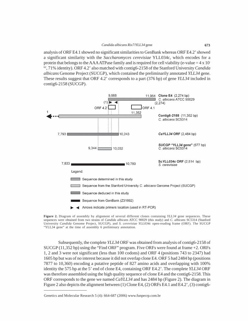
analysis of ORF E4.1 showed no significant similarities to GenBank whereas ORF E4.2’ showeda significant similarity with the Saccharomyces cerevisiae YLL034c, which encodes for aprotein that belongs to the AAA ATPase family and is required for cell viability (e-value = 4 x 10-
62, 71% identity). ORF 4.2’ also matched with contig6-2158 of the Stanford University Candidaalbicans Genome Project (SUCGP), which contained the preliminarily annotated YLL34 gene.These results suggest that ORF 4.2’ corresponds to a part (376 bp) of gene YLL34 included incontig6-2158 (SUCGP).
Figure 2. Diagram of assembly by alignment of several different clones containing YLL34 gene sequences. Thesesequences were obtained from two strains of Candida albicans ATCC 90029 (this study) and C. albicans SC5314 (StanfordUniversity Candida Genome Project, SUCGP), and S. cerevisiae YLL034c open-reading frame (ORF). The SUCGP“YLL34 gene” at the time of assembly 6 preliminary annotation.
Subsequently, the complete YLL34 ORF was obtained from analysis of contig6-2158 ofSUCGP (11,352 bp) using the “Find ORF” program. Five ORFs were found at frame +2. ORFs1, 2 and 3 were not significant (less than 100 codons) and ORF 4 (positions 743 to 2347) had1605 bp but was of no interest because it did not overlap clone E4. ORF 5 had 2484 bp (positions7877 to 10,360) encoding a putative peptide of 827 amino acids and overlapping with 100%identity the 575 bp at the 5’ end of clone E4, containing ORF E4.2’. The complete YLL34 ORFwas therefore assembled using the high quality sequence of clone E4 and the contig6-2158. ThisORF corresponds to the gene we named CaYLL34 and has 2484 bp (Figure 2). The diagram inFigure 2 also depicts the alignment between (1) Clone E4, (2) ORFs E4.1 and E4.2’, (3) contig6-

A.S.A. Melo et al. 674
Genetics and Molecular Research 5 (4): 664-687 (2006) www.funpecrp.com.br
2158 (SUCGP), (4) our inferred CaYLL34 ORF, (5) the YLL34 gene annotated by SUCGP, and(6) ScYLL034c ORF (GenBank Z31692). This result shows that the CaYLL34 ORF is fullylocated in contig6-2158.
Characterization of the putative CaYLL34 gene product
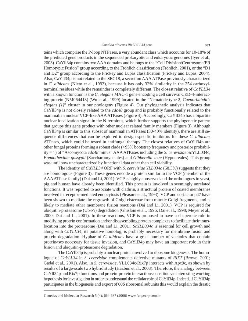
The CaYLL34 ORF (GenBank accession No. AY493662) encodes a putative proteinof 827 amino acids and 91.8 kDa (Figure 3A) and was compared to similar proteins that belongto the AAA ATPase family, namely, protein ScYLL034cp (837 amino acids, from GenBankZ731339 and NP013066), S. cerevisiae valosin-containing protein (VCP) (NP010157), Homosapiens VCP (NP009057) and Schizosaccharomyces pombe AAA ATPase (NP596710) (Figure3B). The pairwise identities between CaYll34p and these proteins were: 58.3% to ScYLL034cp,37.2% to S. cerevisiae VCP, 36.7% to Homo sapiens VCP, and 29.3% to S. pombe AAAATPase. The alignment depicted in Figure 3B also suggests that the AAA ATPase family has atleast two groups of members containing two AAA motifs: one group containing the CDC48domain (proteins 4 and 5, Figure 3B) and the other without a CDC48 domain (proteins 1, 2 and3, Figure 3B). This analysis suggests that CaYll34p and ScYLL034cp are encoded by ortholo-gous genes and that CaYll34p does not belong to the CDC48 plus, valosin group.
Analysis of CaYll34p using Pfam indicates the presence of two domains common tothe AAA ATPase family (P-loop) (Iyer et al., 2003). The scores of each domain were E = 2.7x 10-85 and E = 5.5 x 10-89, respectively. These domains were localized between residues 229-420 and 558-746 (Figure 3A). Other proteins similar to CaYLL34p were also submitted to Pfamanalysis. The VCP proteins contain two AAA ATPase domains and one CDC48 (cell divisionprotein 48) domain (Figure 3A). These results indicate that all of these proteins belong to theAAA ATPase family.
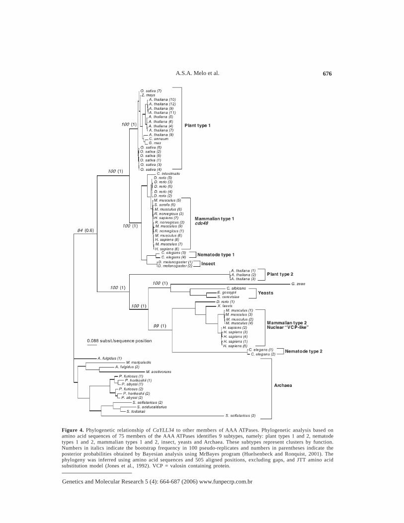
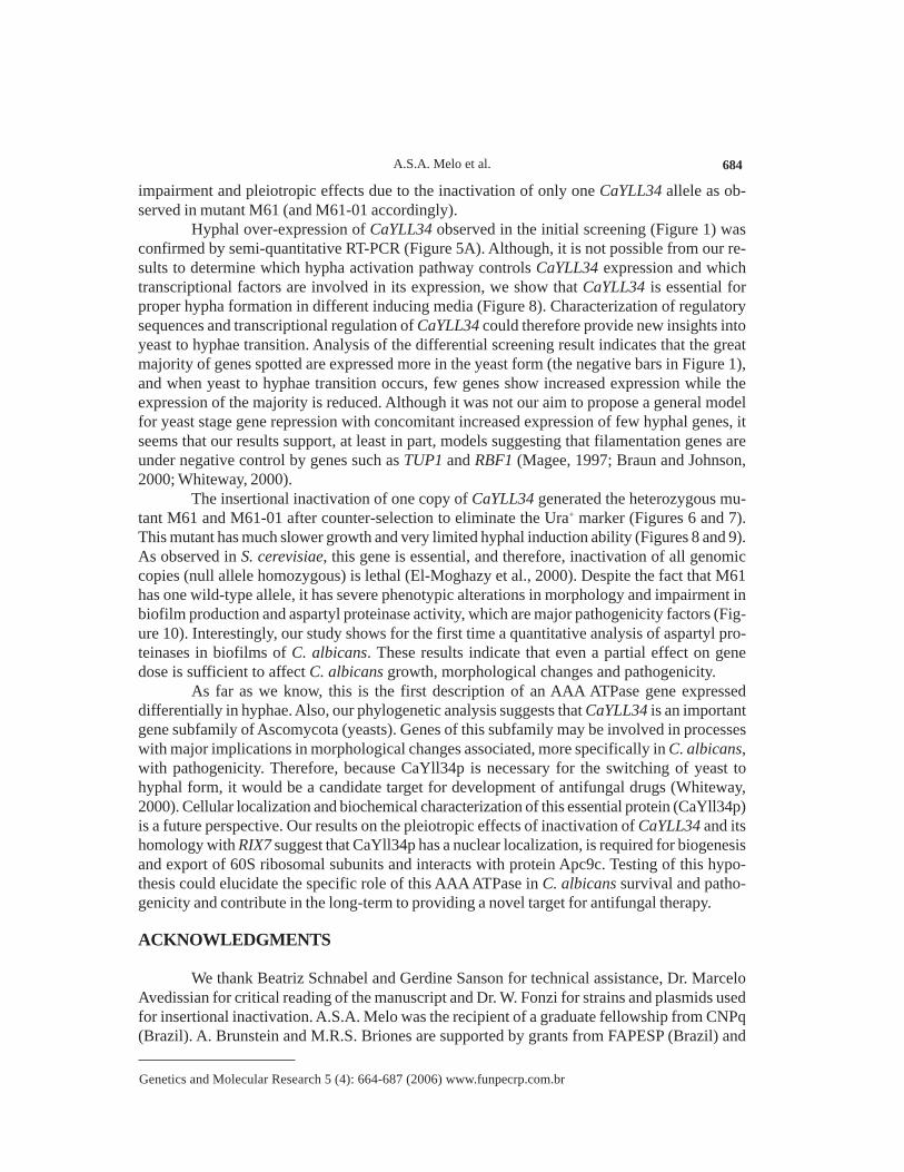
Because the AAA ATPases are a vast family of very ancient and related proteins weinvestigated whether CaYLL34p belongs to a subgroup within this family with a more specificor defined cell role. One hundred members of AAA ATPases were considered in the analysisfrom which 75 provided high-quality alignment with CaYll34p (available upon request [email protected]). This dataset was used as input for phylogeny inference using a Baye-sian method. In Figure 4, the resulting phylogenetic tree depicts 9 major groups clustered byfunction. At least two types, or groups, of plant, mammalian and nematode AAA ATPase areobserved while only one group of yeasts is present. The yeast group is most closely related tothe mammalian nuclear group, thus suggesting that CaYll34p might be located in the nucleus. Sofar, no functions or properties have been attributed to this group of AAA ATPases in C. albi-cans although the deletion of the S. cerevisiae homolog ScYLL034cp encoding gene is lethal inhaploid yeasts (El-Moghazy et al., 2000). In addition, the S. cerevisiae mutant rix7-1 is comple-mented by YLL034c, and therefore, YLL34 and RIX7 are synonymous (http://db.yeastgenome.org/cgi-bin/locus.pl?locus=Rix7, accessed December 10, 2004) (Cherry et al., 1998). Gene RIX7encodes a member of the AAA ATPase family, which contains a bipartite nuclear localizationsignal and which is required for biogenesis and nuclear export of 60S ribosomal subunits (Gadalet al., 2001). Accordingly, CaYll34p has a potential bipartite nuclear localization signal (http://bo.expasy.org/prosite/) in residues 165-182 at the N-terminus (Prosite sequence PS00015) (Huloet al., 2004) (Figure 3B). Our results suggest that CaYLL34 is an essential gene encoding anAAA ATPase functionally related to “Mammalian type 2” and “Nematode type 2” (Figure 4)

Candida albicans Rix7/YLL34 gene 675
Genetics and Molecular Research 5 (4): 664-687 (2006) www.funpecrp.com.br
that may have a function related to RIX7 in ribosome biogenesis. Because CaYll34p has only30-40% identity to human VCP-like AAA ATPases, it may be possible in the long run to finddrugs that will interfere with CaYll34p function but not with the human homologs.
Figure 3. Sequence analysis of the putative CaYll34p protein. In A the two AAA ATPase domains of CaYll34p (216amino acids each) are underlined. In B depiction of Pfam HMM search result of 1) CaYll34p, 2) ScYLL034cp, 3) S.pombe AAA ATPase (gi|19113502|ref|NP_596710.1), 4) Homo sapiens valosin containing protein (VCP) (gi|6005942|ref|NP_009057.1) and 5) S. cerevisiae VCP (gi|6320077||NP_010157.1). In all of these proteins, two AAA ATPase motifsof 216 amino acids were found. The e-value of these matches were e<1.1 x 10-82. CDC48 motif matched only to VCPproteins. Bipartite nuclear localization signals are present only in CaYll34p and ScYLL034cp (RIX7) as determined usingProsite search (http://bo.expasy.org/prosite/). The sequences of CaYll34p nuclear localization signals (KRKAKGLAKTQLKKQKR) and the corresponding ScYLL034cp (RIX7) (KKSKKRSKEGTCKVKRQKIK) are indicated in the corre-sponding domains.
A.S.A. Melo et al. 676
Genetics and Molecular Research 5 (4): 664-687 (2006) www.funpecrp.com.br
Figure 4. Phylogenetic relationship of CaYLL34 to other members of AAA ATPases. Phylogenetic analysis based onamino acid sequences of 75 members of the AAA ATPases identifies 9 subtypes, namely: plant types 1 and 2, nematodetypes 1 and 2, mammalian types 1 and 2, insect, yeasts and Archaea. These subtypes represent clusters by function.Numbers in italics indicate the bootstrap frequency in 100 pseudo-replicates and numbers in parentheses indicate theposterior probabilities obtained by Bayesian analysis using MrBayes program (Huelsenbeck and Ronquist, 2001). Thephylogeny was inferred using amino acid sequences and 505 aligned positions, excluding gaps, and JTT amino acidsubstitution model (Jones et al., 1992). VCP = valosin containing protein.
Candida albicans Rix7/YLL34 gene 677
Genetics and Molecular Research 5 (4): 664-687 (2006) www.funpecrp.com.br
Differential expression of CaYLL34
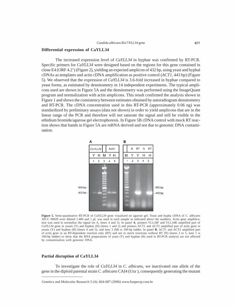
The increased expression level of CaYLL34 in hyphae was confirmed by RT-PCR.Specific primers for CaYLL34 were designed based on the regions for this gene contained inclone E4 (ORF 4.2’) (Figure 2), yielding an expected amplicon of 432 bp, using yeast and hyphalcDNAs as templates and actin cDNA amplification as positive control (ACT1, 443 bp) (Figure5). We observed that the expression of CaYLL34 is 3.6-fold increased in hyphae compared toyeast forms, as estimated by densitometry in 14 independent experiments. The typical ampli-cons used are shown in Figure 5A and the densitometry was performed using the ImageQuantprogram and normalization with actin amplicons. This result confirmed the analysis shown inFigure 1 and shows the consistency between estimates obtained by autoradiogram densitometryand RT-PCR. The cDNA concentration used in this RT-PCR (approximately 0.06 ng) wasstandardized by preliminary assays (data not shown) in order to yield amplicons that are in thelinear range of the PCR and therefore will not saturate the signal and still be visible in theethidium bromide/agarose gel electrophoresis. In Figure 5B cDNA control with mock RT reac-tion shows that bands in Figure 5A are mRNA derived and not due to genomic DNA contami-nation.
Figure 5. Semi-quantitative RT-PCR of CaYLL34 gene visualized on agarose gel. Yeast and hypha cDNA of C. albicansATCC 90029 were diluted 1:400 and 1 µL was used in each sample as indicated above the numbers. Actin gene amplifica-tion was used to normalize the signal (in A, lanes 4 and 5). In panel A, primers YLL34F and YLL34R amplified part ofCaYLL34 gene in yeasts (Y) and hyphae (H) (lanes 1 and 2) and primers ACT1 and ACT2 amplified part of actin gene inyeasts (Y) and hyphae (H) (lanes 4 and 5), and lane 3 (M) is 100-bp ladder. In panel B, ACT1 and ACT2 amplified partof actin gene in an RT-dependent reaction only (RT) and not in mock reactions without RT (N) (lanes 2 to 5, lane 1 is100-bp ladder) to show that the RNA preparations of yeast (Y) and hyphae (H) used in RT-PCR analysis are not affectedby contamination with genomic DNA.
Partial disruption of CaYLL34
To investigate the role of CaYLL34 in C. albicans, we inactivated one allele of thegene in the diploid parental strain C. albicans CAI4 (Ura–), consequently generating the mutant
A.S.A. Melo et al. 678
Genetics and Molecular Research 5 (4): 664-687 (2006) www.funpecrp.com.br
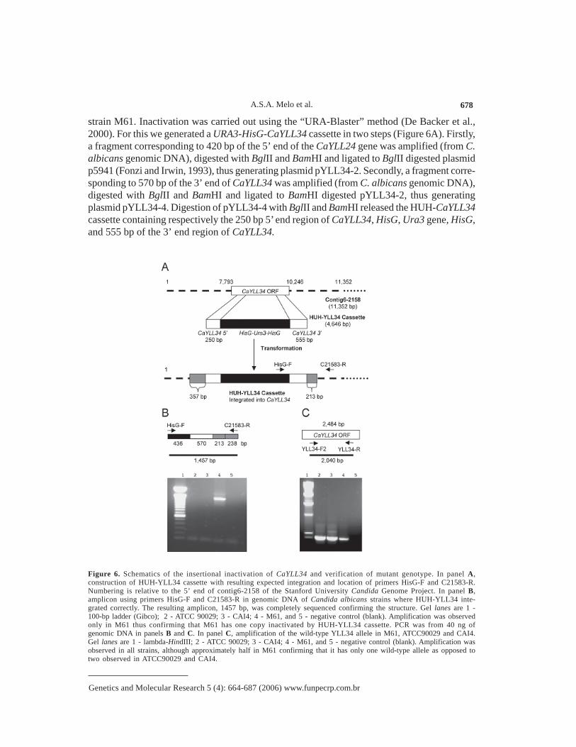
strain M61. Inactivation was carried out using the “URA-Blaster” method (De Backer et al.,2000). For this we generated a URA3-HisG-CaYLL34 cassette in two steps (Figure 6A). Firstly,a fragment corresponding to 420 bp of the 5’ end of the CaYLL24 gene was amplified (from C.albicans genomic DNA), digested with BglII and BamHI and ligated to BglII digested plasmidp5941 (Fonzi and Irwin, 1993), thus generating plasmid pYLL34-2. Secondly, a fragment corre-sponding to 570 bp of the 3’ end of CaYLL34 was amplified (from C. albicans genomic DNA),digested with BglII and BamHI and ligated to BamHI digested pYLL34-2, thus generatingplasmid pYLL34-4. Digestion of pYLL34-4 with BglII and BamHI released the HUH-CaYLL34cassette containing respectively the 250 bp 5’ end region of CaYLL34, HisG, Ura3 gene, HisG,and 555 bp of the 3’ end region of CaYLL34.
Figure 6. Schematics of the insertional inactivation of CaYLL34 and verification of mutant genotype. In panel A,construction of HUH-YLL34 cassette with resulting expected integration and location of primers HisG-F and C21583-R.Numbering is relative to the 5’ end of contig6-2158 of the Stanford University Candida Genome Project. In panel B,amplicon using primers HisG-F and C21583-R in genomic DNA of Candida albicans strains where HUH-YLL34 inte-grated correctly. The resulting amplicon, 1457 bp, was completely sequenced confirming the structure. Gel lanes are 1 -100-bp ladder (Gibco); 2 - ATCC 90029; 3 - CAI4; 4 - M61, and 5 - negative control (blank). Amplification was observedonly in M61 thus confirming that M61 has one copy inactivated by HUH-YLL34 cassette. PCR was from 40 ng ofgenomic DNA in panels B and C. In panel C, amplification of the wild-type YLL34 allele in M61, ATCC90029 and CAI4.Gel lanes are 1 - lambda-HindIII; 2 - ATCC 90029; 3 - CAI4; 4 - M61, and 5 - negative control (blank). Amplification wasobserved in all strains, although approximately half in M61 confirming that it has only one wild-type allele as opposed totwo observed in ATCC90029 and CAI4.

Candida albicans Rix7/YLL34 gene 679
Genetics and Molecular Research 5 (4): 664-687 (2006) www.funpecrp.com.br
The HUH-YLL34 cassette was used to transform the C. albicans CAI4 (Ura–) strainusing the lithium acetate method and three different concentrations of DNA (18, 10 and 5 µg).After five days of incubation, 98 Ura+ colonies were observed. Transformant colonies wereanalyzed by PCR using primers HisG-F and C21583-R, which amplified a fragment of 1457 bpin the mutant containing the correctly integrated cassette (Figure 6B). The 1457-bp ampliconfrom mutant M61 (yll34∆::hisG-CaURA3-hisG/YLL34/ura3∆::imm434/ura3∆::imm434) wassequenced and the expected structure, generated by homologous recombination, was confirmed.Amplification of CaYLL34 shows that strain M61 is a heterozygote. Specific primers forCaYLL34 were used for PCR from M61 strain genomic DNA, generating the 2040-bp amplicon(Figure 6C). The amplification signal in M61 as compared to CAI4 suggests that the number ofcopies of CaYLL34 per diploid genome is approximately half in M61. Also, Southern blot anal-ysis shows that the HUH-YLL34 cassette integrated only one the CaYLL34 allele (Figure 7A)and in no other part of the genome, thereby confirming the results obtained by PCR (Figure 6C)and showing that no other genes were disrupted. The 6.1-kb band in the BamHI/BglII digest ofM61 corresponds to the CaYLL34 allele with the HUH-YLL34 integrated and the 2.4-kb bandin CAI4, M61 and ATCC90029 represents the wild-type allele (Figure 7A).
Figure 7. Genotype analysis on mutant M61 and M61-01 by Southern blot and PCR. In A Southern blot of digested DNAof CAI4, M61 and ATCC 90029 with BamHI and BglII. The probe was CaYLL34 3’ (555 bp) region labeled with [α-32P]-dCTP. This probe hybridized with CaYLL34 (~2000 bp) present in all strains. In M61 the ~6500-bp fragment correspondsto the cassette inserted in CaYLL34 gene. The cassette size is 4648 bp. PCR of genomic DNA of CAI4, M61 and M61-01(URA3–). In B amplicon of 213 bp of URA3 gene using primers URA3-F and URA3-R showing that URA3 was not presentin M61-01 mutant. In C amplicon of 1457 bp using primers HisG-F and C21583-R showing that HisG sequence interruptsCaYLL34 in M61-01 mutant.
M61 was then used to generate another mutant, M61-01, by counter-selection with 5-FOA rendering M61-01 Ura minus but still having one allele of CaYLL34 inactivated by inter-vening HisG sequence (Figure 7B) (Fonzi and Irwin, 1993). In all the phenotypic analysis dis-cussed below we did not observe differences between M61 and M61-01, and therefore, thephenotypic alterations in M61 do not seem to be a consequence of the “Ura effect” (Lay et al.,1998; Brand et al., 2004).

A.S.A. Melo et al. 680
Genetics and Molecular Research 5 (4): 664-687 (2006) www.funpecrp.com.br
Phenotypic analysis of M61 mutant strain
To analyze the morphologic changes that occurred in M61-01 due to insertional inacti-vation of CaYLL34, this strain was examined by phase-contrast microscopy and compared toits parental strain CAI4 under different growth conditions (Figure 8). The comparison betweenCAI4 (Ura–) and M61-01 (Ura–) in SD medium at 37ºC is striking and shows that failure toinduce filamentation is not caused by any effects involving the Ura+ marker and strongly sug-gests that inactivation of only one allele of CaYLL34 is sufficient to impair hyphal formation(Figure 8). There was no difference in morphology when M61-01 (Ura–) and M61 (Ura+) werecompared. In YPD and SD50 media (no hyphal inducers) CAI4 exhibited blastoconidia andminor hyphal formation while M61 just showed blastoconidia and no hyphae at all. In FBSmedium (hyphal inducer) CAI4 showed abundant hypha formation and few blastoconidia whileM61 formed very short hyphae, pseudohyphae and many blastoconidia. Also, M61 blastoconidiahave a very large central vacuole. These cells showed a significant increase relative to theparental phenotype suggesting an abnormal vacuolar accumulation. These enlarged cells havebuds with identical morphology in all growth conditions tested.
Figure 8. Micromorphology of CAI4 (parental strain) and M61-01 (URA3– mutant) cultured in SD 50 (50 mM glucose)and FBS (fetal bovine serum), after incubation at 37°C, 70 rpm for 24 h. Photographs with phase-contrast microscopy at400X magnification.
Strains ATCC90029, CAI4 and M61 were subjected to species identification tests suchas micromorphologic analysis, culture on CHROMagar Candida, sugar assimilation and fer-mentation, and RAPD which readily identified them as C. albicans (Melo et al., 1998), thusruling out any possible contamination with other fungal species (data not shown).
Growth curves of the mutant M61, the parental strain CAI4, the C. albicans referencestrain ATCC 90029, and the L296 strain (clinical isolate from hemoculture) were also compared(data not shown). The exponential phase of the curves at 30°C suggests that M61 growth is
Candida albicans Rix7/YLL34 gene 681
Genetics and Molecular Research 5 (4): 664-687 (2006) www.funpecrp.com.br
slower than in CAI4, ATCC and L296. The exponential growth phase at 37°C indicates thatCAI4 and M61 grow at approximately the same rates and much more slowly than ATCC andL296.
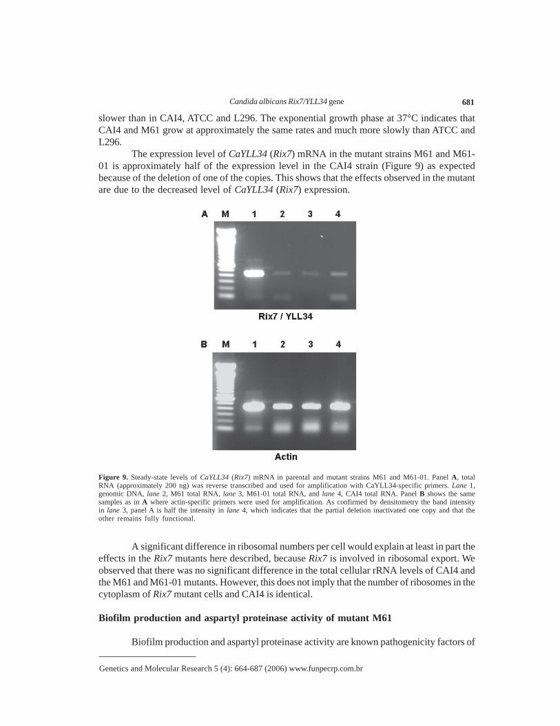
The expression level of CaYLL34 (Rix7) mRNA in the mutant strains M61 and M61-01 is approximately half of the expression level in the CAI4 strain (Figure 9) as expectedbecause of the deletion of one of the copies. This shows that the effects observed in the mutantare due to the decreased level of CaYLL34 (Rix7) expression.
Figure 9. Steady-state levels of CaYLL34 (Rix7) mRNA in parental and mutant strains M61 and M61-01. Panel A, totalRNA (approximately 200 ng) was reverse transcribed and used for amplification with CaYLL34-specific primers. Lane 1,genomic DNA, lane 2, M61 total RNA, lane 3, M61-01 total RNA, and lane 4, CAI4 total RNA. Panel B shows the samesamples as in A where actin-specific primers were used for amplification. As confirmed by densitometry the band intensityin lane 3, panel A is half the intensity in lane 4, which indicates that the partial deletion inactivated one copy and that theother remains fully functional.
A significant difference in ribosomal numbers per cell would explain at least in part theeffects in the Rix7 mutants here described, because Rix7 is involved in ribosomal export. Weobserved that there was no significant difference in the total cellular rRNA levels of CAI4 andthe M61 and M61-01 mutants. However, this does not imply that the number of ribosomes in thecytoplasm of Rix7 mutant cells and CAI4 is identical.
Biofilm production and aspartyl proteinase activity of mutant M61
Biofilm production and aspartyl proteinase activity are known pathogenicity factors of
A.S.A. Melo et al. 682
Genetics and Molecular Research 5 (4): 664-687 (2006) www.funpecrp.com.br
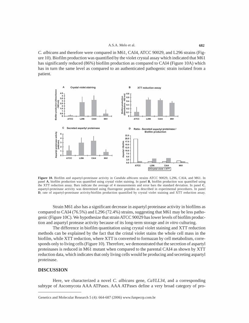
C. albicans and therefore were compared in M61, CAI4, ATCC 90029, and L296 strains (Fig-ure 10). Biofilm production was quantified by the violet crystal assay which indicated that M61has significantly reduced (86%) biofilm production as compared to CAI4 (Figure 10A) whichhas in turn the same level as compared to an authenticated pathogenic strain isolated from apatient.
Figure 10. Biofilm and aspartyl-proteinase activity in Candida albicans strains ATCC 90029, L296, CAI4, and M61. Inpanel A, biofilm production was quantified using crystal violet staining. In panel B, biofilm production was quantified usingthe XTT reduction assay. Bars indicate the average of 4 measurements and error bars the standard deviation. In panel C,aspartyl-proteinase activity was determined using fluorogenic peptides as described in experimental procedures. In panelD, rate of aspartyl-proteinase activity/biofilm production quantified by crystal violet staining and XTT reduction assay.
Strain M61 also has a significant decrease in aspartyl proteinase activity in biofilms ascompared to CAI4 (76.5%) and L296 (72.4%) strains, suggesting that M61 may be less patho-genic (Figure 10C). We hypothesize that strain ATCC 90029 has lower levels of biofilm produc-tion and aspartyl protease activity because of its long-term storage and in vitro culturing.
The difference in biofilm quantitation using crystal violet staining and XTT reductionmethods can be explained by the fact that the cristal violet stains the whole cell mass in thebiofilm, while XTT reduction, where XTT is converted to formazan by cell metabolism, corre-sponds only to living cells (Figure 10). Therefore, we demonstrated that the secretion of aspartylproteinases is reduced in M61 mutant when compared to the parental CAI4 as shown by XTTreduction data, which indicates that only living cells would be producing and secreting aspartylproteinase.
DISCUSSION
Here, we characterized a novel C. albicans gene, CaYLL34, and a correspondingsubtype of Ascomycota AAA ATPases. AAA ATPases define a very broad category of pro-
Candida albicans Rix7/YLL34 gene 683
Genetics and Molecular Research 5 (4): 664-687 (2006) www.funpecrp.com.br
teins which comprise the P-loop NTPases, a very abundant class which accounts for 10-18% ofthe predicted gene products in the sequenced prokaryotic and eukaryotic genomes (Iyer et al.,2003). CaYll34p contains two AAA domains and belongs to the “Cell Division/Centrosome/ERHomotypic Fusion” group according to the Fröhlich classification (Fröhlich, 2001), or the “D1and D2” group according to the Frickey and Lupas classification (Frickey and Lupas, 2004).Also, CaYll34p is not related to the SEC18, a secretion AAA ATPase previously characterizedin C. albicans (Nieto et al., 1993), because it has only 32% similarity in the 254 carboxyl-terminal residues while the remainder is completely different. The closest relative of CaYLL34with a known function is the C. elegans MAC-1 gene encoding a cell survival CED-4-interact-ing protein (NM064413) (Wu et al., 1999) located in the “Nematode type 2, Caenorhabditiselegans (1)” cluster in our phylogeny (Figure 4). Our phylogenetic analysis indicates thatCaYll34p is not closely related to the cdc48 group and is probably functionally related to themammalian nuclear VCP-like AAA ATPases (Figure 4). Accordingly, CaYll34p has a bipartitenuclear localization signal in the N-terminus, which further supports the phylogenetic patternthat groups this gene product with other nuclear related family members (Figure 3). AlthoughCaYll34p is similar to this subset of mammalian ATPases (30-40% identity), there are still se-quence differences that can be explored to design specific inhibitors for these C. albicansATPases, which could be tested in antifungal therapy. The closest relatives of CaYll34p areother fungal proteins forming a robust clade (>95% bootstrap frequency and posterior probabil-ity = 1) of “Ascomycota cdc48 minus” AAA ATPases including the S. cerevisiae ScYLL034c,Eremothecium gossypii (Saccharomycetales) and Gibberella zeae (Hypocreales). This groupwas until now uncharacterized by functional data other than cell viability.
The identity of CaYLL34 ORF with S. cerevisiae YLL034c (58.3%) suggests that theyare homologous (Figure 3). These genes encode a protein similar to the VCP (member of theAAA ATPase family) (Dai and Li, 2001). VCP is highly conserved and the orthologues in yeast,pig and human have already been identified. This protein is involved in seemingly unrelatedfunctions. It was reported to associate with clathrin, a structural protein of coated membranesinvolved in receptor-mediated endocytosis (Pleasure et al., 1993). VCP and co-factor p47 havebeen shown to mediate the regrowth of Golgi cisternae from mitotic Golgi fragments, and islikely to mediate other membrane fusion reactions (Dai and Li, 2001). VCP is required forubiquitin-proteasome (Ub-Pr) degradation (Ghislain et al., 1996; Dai et al., 1998; Meyer et al.,2000; Dai and Li, 2001). In these reactions, VCP is proposed to have a chaperone role inmodifying protein conformation and/or disassembling protein complexes to facilitate their trans-location into the proteasome (Dai and Li, 2001). ScYLL034c is essential for cell growth andalong with CaYLL34, its putative homolog, is probably necessary for membrane fusion andprotein degradation. Hyphae of C. albicans have a great number of vacuoles that containproteinases necessary for tissue invasion, and CaYll34p may have an important role in theirfusion and ubiquitin-proteasome degradation.
The CaYll34p is probably a nuclear protein involved in ribosome biogenesis. The homo-logue of CaYLL34 in S. cerevisiae complements defective mutants of RIX7 (Brown, 2001;Gadal et al., 2001). Also, in S. cerevisiae, YLL034c/Rix7p interacts with Apc9c, as shown byresults of a large-scale two hybrid study (Hazbun et al., 2003). Therefore, the analogy betweenCaYll34p and Rix7p functions and protein-protein interactions constitute an interesting workinghypothesis for investigation in order to understand the cellular role of CaYll34p. Indeed, if CaYll34pparticipates in the biogenesis and export of 60S ribosomal subunits this would explain the drastic
A.S.A. Melo et al. 684
Genetics and Molecular Research 5 (4): 664-687 (2006) www.funpecrp.com.br
impairment and pleiotropic effects due to the inactivation of only one CaYLL34 allele as ob-served in mutant M61 (and M61-01 accordingly).
Hyphal over-expression of CaYLL34 observed in the initial screening (Figure 1) wasconfirmed by semi-quantitative RT-PCR (Figure 5A). Although, it is not possible from our re-sults to determine which hypha activation pathway controls CaYLL34 expression and whichtranscriptional factors are involved in its expression, we show that CaYLL34 is essential forproper hypha formation in different inducing media (Figure 8). Characterization of regulatorysequences and transcriptional regulation of CaYLL34 could therefore provide new insights intoyeast to hyphae transition. Analysis of the differential screening result indicates that the greatmajority of genes spotted are expressed more in the yeast form (the negative bars in Figure 1),and when yeast to hyphae transition occurs, few genes show increased expression while theexpression of the majority is reduced. Although it was not our aim to propose a general modelfor yeast stage gene repression with concomitant increased expression of few hyphal genes, itseems that our results support, at least in part, models suggesting that filamentation genes areunder negative control by genes such as TUP1 and RBF1 (Magee, 1997; Braun and Johnson,2000; Whiteway, 2000).
The insertional inactivation of one copy of CaYLL34 generated the heterozygous mu-tant M61 and M61-01 after counter-selection to eliminate the Ura+ marker (Figures 6 and 7).This mutant has much slower growth and very limited hyphal induction ability (Figures 8 and 9).As observed in S. cerevisiae, this gene is essential, and therefore, inactivation of all genomiccopies (null allele homozygous) is lethal (El-Moghazy et al., 2000). Despite the fact that M61has one wild-type allele, it has severe phenotypic alterations in morphology and impairment inbiofilm production and aspartyl proteinase activity, which are major pathogenicity factors (Fig-ure 10). Interestingly, our study shows for the first time a quantitative analysis of aspartyl pro-teinases in biofilms of C. albicans. These results indicate that even a partial effect on genedose is sufficient to affect C. albicans growth, morphological changes and pathogenicity.
As far as we know, this is the first description of an AAA ATPase gene expresseddifferentially in hyphae. Also, our phylogenetic analysis suggests that CaYLL34 is an importantgene subfamily of Ascomycota (yeasts). Genes of this subfamily may be involved in processeswith major implications in morphological changes associated, more specifically in C. albicans,with pathogenicity. Therefore, because CaYll34p is necessary for the switching of yeast tohyphal form, it would be a candidate target for development of antifungal drugs (Whiteway,2000). Cellular localization and biochemical characterization of this essential protein (CaYll34p)is a future perspective. Our results on the pleiotropic effects of inactivation of CaYLL34 and itshomology with RIX7 suggest that CaYll34p has a nuclear localization, is required for biogenesisand export of 60S ribosomal subunits and interacts with protein Apc9c. Testing of this hypo-thesis could elucidate the specific role of this AAA ATPase in C. albicans survival and patho-genicity and contribute in the long-term to providing a novel target for antifungal therapy.
ACKNOWLEDGMENTS
We thank Beatriz Schnabel and Gerdine Sanson for technical assistance, Dr. MarceloAvedissian for critical reading of the manuscript and Dr. W. Fonzi for strains and plasmids usedfor insertional inactivation. A.S.A. Melo was the recipient of a graduate fellowship from CNPq(Brazil). A. Brunstein and M.R.S. Briones are supported by grants from FAPESP (Brazil) and
Candida albicans Rix7/YLL34 gene 685
Genetics and Molecular Research 5 (4): 664-687 (2006) www.funpecrp.com.br
M.R.S. Briones is a recipient of an International Research Scholar grant from the HowardHughes Medical Institute (USA).
REFERENCES
Bailey DA, Feldmann PJ, Bovey M, Gow NA, et al. (1996). The Candida albicans HYR1 gene, which isactivated in response to hyphal development, belongs to a gene family encoding yeast cell wallproteins. J. Bacteriol. 178: 5353-5360.
Baillie GS and Douglas LJ (1999). Candida biofilms and their susceptibility to antifungal agents. MethodsEnzymol. 310: 644-656.
Birse CE, Irwin MY, Fonzi WA and Sypherd PS (1993). Cloning and characterization of ECE1, a geneexpressed in association with cell elongation of the dimorphic pathogen Candida albicans. Infect.Immun. 61: 3648-3655.
Brand A, Maccallum DM, Brown AJ, Gow NA, et al. (2004). Ectopic expression of URA3 can influence thevirulence phenotypes and proteome of Candida albicans but can be overcome by targeted reinte-gration of URA3 at the RPS10 locus. Eukaryotic Cell 3: 900-909.
Braun BR and Johnson AD (2000). TUP1, CPH1 and EFG1 make independent contributions to filamentationin Candida albicans. Genetics 155: 57-67.
Brown JD (2001). Ribosome biogenesis: stripping for AAAction? Curr. Biol. 11: R710-R712.Calderone RA and Gow N (2002). Host recognition by Candida species. In: Candida and candidiasis
(Calderone RA, ed.). ASM Press, Washington, 67-86.Cherry JM, Adler C, Ball C, Chervitz SA, et al. (1998). SGD: Saccharomyces Genome Database. Nucleic
Acids Res. 26: 73-79.Dai RM and Li CC (2001). Valosin-containing protein is a multi-ubiquitin chain-targeting factor required in
ubiquitin-proteasome degradation. Nat. Cell Biol. 3: 740-744.Dai RM, Chen E, Longo DL, Gorbea CM, et al. (1998). Involvement of valosin-containing protein, an
ATPase co-purified with IkappaBalpha and 26S proteasome, in ubiquitin-proteasome-mediated deg-radation of IkappaBalpha. J. Biol. Chem. 273: 3562-3573.
De Backer MD, Magee PT and Pla J (2000). Recent developments in molecular genetics of Candidaalbicans. Annu. Rev. Microbiol. 54: 463-498.
De Bernardis F, Arancia S, Morelli L, Hube B, et al. (1999). Evidence that members of the secretory aspartylproteinase gene family, in particular SAP2, are virulence factors for Candida vaginitis. J. Infect. Dis.179: 201-208.
Douglas LJ (2003). Candida biofilms and their role in infection. Trends Microbiol. 11: 30-36.El-Moghazy AN, Zhang N, Ismail T, Wu J, et al. (2000). Functional analysis of six novel ORFs on the left
arm of chromosome XII in Saccharomyces cerevisiae reveals two essential genes, one of which isunder cell-cycle control. Yeast 16: 277-288.
Ewing B and Green P (1998). Base-calling of automated sequencer traces using phred. II. Error probabili-ties. Genome Res. 8: 186-194.
Ewing B, Hillier L, Wendl MC and Green P (1998). Base-calling of automated sequencer traces using phred.I. Accuracy assessment. Genome Res. 8: 175-185.
Felk A, Kretschmar M, Albrecht A, Schaller M, et al. (2002). Candida albicans hyphal formation and theexpression of the Efg1-regulated proteinases Sap4 to Sap6 are required for the invasion of parenchy-mal organs. Infect. Immun. 70: 3689-3700.
Fidel PL Jr and Sobel JD (1996). Immunopathogenesis of recurrent vulvovaginal candidiasis. Clin. Microbiol.Rev. 9: 335-348.
Fisher-Hoch SP and Hutwagner L (1995). Opportunistic candidiasis: an epidemic of the 1980s. Clin. Infect.Dis. 21: 897-904.
Fonzi WA and Irwin MY (1993). Isogenic strain construction and gene mapping in Candida albicans.Genetics 134: 717-728.
Frickey T and Lupas AN (2004). Phylogenetic analysis of AAA proteins. J. Struct. Biol. 146: 2-10.Fröhlich K (2001). An AAA family tree. J. Cell Sci. 114: 1601-1602.Gadal O, Strauss D, Braspenning J, Hoepfner D, et al. (2001). A nuclear AAA-type ATPase (Rix7p) is
required for biogenesis and nuclear export of 60S ribosomal subunits. EMBO J. 20: 3695-3704.Galtier N, Gouy M and Gautier C (1996). SEAVIEW and PHYLO_WIN: two graphic tools for sequence
alignment and molecular phylogeny. Comput. Appl. Biosci. 12: 543-548.Ghislain M, Dohmen RJ, Levy F and Varshavsky A (1996). Cdc48p interacts with Ufd3p, a WD repeat
A.S.A. Melo et al. 686
Genetics and Molecular Research 5 (4): 664-687 (2006) www.funpecrp.com.br
protein required for ubiquitin-mediated proteolysis in Saccharomyces cerevisiae. EMBO J. 15: 4884-4899.
Gordon D, Abajian C and Green P (1998). Consed: a graphical tool for sequence finishing. Genome Res. 8:195-202.
Groll AH, Piscitelli SC and Walsh TJ (1998). Clinical pharmacology of systemic antifungal agents: acomprehensive review of agents in clinical use, current investigational compounds, and putativetargets for antifungal drug development. Adv. Pharmacol. 44: 343-500.
Hawser SP and Douglas LJ (1994). Biofilm formation by Candida species on the surface of cathetermaterials in vitro. Infect. Immun. 62: 915-921.
Hazbun TR, Malmstrom L, Anderson S, Graczyk BJ, et al. (2003). Assigning function to yeast proteins byintegration of technologies. Mol. Cell 12: 1353-1365.
Higgins DG and Sharp PM (1989). Fast and sensitive multiple sequence alignments on a microcomputer.Comput. Appl. Biosci. 5: 151-153.
Hirata IY, Cezari MH, Nakaie CR and Boshcov P (1994). Internally quenched fluorogenic protease sub-strates: solid-phase synthesis and fluorescent spectroscopy of peptides containing ortho-amino-benzoyl-dinitrophenyl groups as donor-acceptor pairs. Lett. Peptide Sci. 1: 299-308.
Hoyer LL, Payne TL, Bell M, Myers AM, et al. (1998). Candida albicans ALS3 and insights into the natureof the ALS gene family. Curr. Genet. 33: 451-459.
Hube B, Monod M, Schofield DA, Brown AJ, et al. (1994). Expression of seven members of the gene familyencoding secretory aspartyl proteinases in Candida albicans. Mol. Microbiol. 14: 87-99.
Hube B, Sanglard D, Schaller M, Ibrahim A, et al. (1998). What functions do six different genes forsecretory proteinases have in Candida albicans? Mycoses 41: 47-50.
Huelsenbeck JP and Ronquist F (2001). MRBAYES: Bayesian inference of phylogenetic trees. Bioinfor-matics 17: 754-755.
Hulo N, Sigrist CJ, Le Saux V, Langendijk-Genevaux PS, et al. (2004). Recent improvements to the PROSITEdatabase. Nucleic Acids Res. 32: D134-D137.
Iyer LM, Leipe DD, Koonin EV and Aravind L (2003). Evolutionary history and higher order classificationof AAA+ ATPase. J. Struct. Biol. 146: 11-31.
Jin Y, Yip HK, Samaranayake YH, Yau JY, et al. (2003). Biofilm-forming ability of Candida albicans isunlikely to contribute to high levels of oral yeast carriage in cases of human immunodeficiency virusinfection. J. Clin. Microbiol. 41: 2961-2967.
Jones DT, Taylor WR and Thornton JM (1992). The rapid generation of mutation data matrices fromprotein sequences. Comput. Appl. Biosci. 8: 275-282.
Koelsch G, Tang J, Loy JA, Monod M, et al. (2000). Enzymic characteristics of secreted aspartic proteasesof Candida albicans. Biochim. Biophys. Acta 1480: 117-131.
Lay J, Henry LK, Clifford J, Koltin Y, et al. (1998). Altered expression of selectable marker URA3 in gene-disrupted Candida albicans strains complicates interpretation of virulence studies. Infect. Immun.66: 5301-5306.
Leberer E, Harcus D, Broadbent ID, Clark KL, et al. (1996). Signal transduction through homologs of theSte20p and Ste7p protein kinases can trigger hyphal formation in the pathogenic fungus Candidaalbicans. Proc. Natl. Acad. Sci. USA 93: 13217-13222.
Liu H (2001). Transcriptional control of dimorphism in Candida albicans. Curr. Opin. Microbiol. 4: 728-735.
Lo HJ, Kohler JR, DiDomenico B, Loebenberg D, et al. (1997). Nonfilamentous C. albicans mutants areavirulent. Cell 90: 939-949.
Magee PT (1997). Which came first, the hypha or the yeast? Science 277: 52-53.Melo AS, de Almeida LP, Colombo AL and Briones MR (1998). Evolutionary distances and identification
of Candida species in clinical isolates by randomly amplified polymorphic DNA (RAPD). Mycopa-thologia 142: 57-66.
Melo AS, Serafim RC and Briones MR (2003). Identification of genes differentially expressed in hyphae ofCandida albicans. Braz. J. Microbiol. 34: 135-137.
Meyer HH, Shorter JG, Seemann J, Pappin D, et al. (2000). A complex of mammalian ufd1 and npl4 links theAAA-ATPase, p97, to ubiquitin and nuclear transport pathways. EMBO J. 19: 2181-2192.
Mitchell AP (1998). Dimorphism and virulence in Candida albicans. Curr. Opin. Microbiol. 1: 687-692.Monod M, Hube B, Hess D and Sanglard D (1998). Differential regulation of SAP8 and SAP9, which
encode two new members of the secreted aspartic proteinase family in Candida albicans. Microbi-ology 144: 2731-2737.
Naglik JR, Newport G, White TC, Fernandes-Naglik LL, et al. (1999). In vivo analysis of secreted aspartyl
Candida albicans Rix7/YLL34 gene 687
Genetics and Molecular Research 5 (4): 664-687 (2006) www.funpecrp.com.br
proteinase expression in human oral candidiasis. Infect. Immun. 67: 2482-2490.Nieto A, Sanz P, Sentandreu R and del Castillo AL (1993). Cloning and characterization of the SEC18 gene
from Candida albicans. Yeast 9: 875-887.Odds FC (1988). Candida and candidosis. Bailliere Tindall Ltd., London.Pimenta DC, Oliveira A, Juliano MA and Juliano L (2001). Substrate specificity of human cathepsin D
using internally quenched fluorescent peptides derived from reactive site loop of kallistatin. Biochim.Biophys. Acta 1544: 113-122.
Pleasure IT, Black MM and Keen JH (1993). Valosin-containing protein, VCP, is a ubiquitous clathrin-binding protein. Nature 365: 459-462.
Sambrook J, Fritsch EF and Maniatis T (1989). Molecular cloning: a laboratory manual. Cold Spring HarborLaboratory, Cold Spring Harbor.
Sanglard D, Hube B, Monod M, Odds FC, et al. (1997). A triple deletion of the secreted aspartyl proteinasegenes SAP4, SAP5, and SAP6 of Candida albicans causes attenuated virulence. Infect. Immun. 65:3539-3546.
Santos MA and Tuite MF (1995). The CUG codon is decoded in vivo as serine and not leucine in Candidaalbicans. Nucleic Acids Res. 23: 1481-1486.
Schaller M, Korting HC, Schäfer W, Bastert J, et al. (1999). Secreted aspartic proteinase (Sap) activitycontributes to tissue damage in a model of human oral candidosis. Mol. Microbiol. 34: 169-180.
Staab JF, Bradway SD, Fidel PL and Sundstrom P (1999). Adhesive and mammalian transglutaminasesubstrate properties of Candida albicans Hwp1. Science 283: 1535-1538.
Stoldt VR, Sonneborn A, Leuker CE and Ernst JF (1997). Efg1p, an essential regulator of morphogenesis ofthe human pathogen Candida albicans, is a member of a conserved class of bHLH proteins regulat-ing morphogenetic processes in fungi. EMBO J. 16: 1982-1991.
Swofford DL (1998). PAUP*. Phylogenetic analysis using Parsimony (* and other methods). Version 4b6.Sinauer Associates Inc., Sunderland.
Thipyapong P, Joel DM and Steffens JC (1997). Differential expression and turnover of the tomato polyphe-nol oxidase gene family during vegetative and reproductive development. Plant Physiol. 113: 707-718.
Van de Loo FJ, Turner S and Somerville C (1995). Expressed sequence tags from developing castor seeds.Plant Physiol. 108: 1141-1150.
Wash A, Pick H and Philppsen P (1994). Procedures for isolating yeast DNA for different purposes. In:Molecular genetics of yeast (Johnston JR, ed.). Oxford University Press, Eynsham, Oxon, 10-11.
Whiteway M (2000). Transcriptional control of cell type and morphogenesis in Candida albicans. Curr.Opin. Microbiol. 3: 582-588.
Wu D, Chen PJ, Chen S, Hu Y, et al. (1999). C. elegans MAC-1, an essential member of the AAA family ofATPases, can bind CED-4 and prevent cell death. Development 126: 2021-2031.
Yang Z (1994). Maximum likelihood phylogenetic estimation from DNA sequences with variable rates oversites: approximate methods. J. Mol. Evol. 39: 306-314.