Embed Size (px)
Citation preview

THE AETIOLOGY, PATHOLOGY, DIAGNOSISAND TREATMENT OF ACUTE PANCREATITIS
A Review of 110 CasesHunterian Lecture delivered at the Royal College of Surgeons of England
on28th February, 1949
byR. A. Russell Taylor, F.R.C.S., M.R.C.O.G.
Medical Superintendent and Surgeon, Pinderfields General Hospital
IN THE PREPARATION of this lecture, only cases which were proved (1) atoperation, (2) at post mortem, or (3) clinically, with a raised urinarydiastase or serum amylase have been included.
In order to appreciate thoroughly the difficulties which face the surgeonin the diagnosis and treatment of acute pancreatitis, it is essential to givea resume of the xtiology and pathology of this still obscure condition.
AETIOLOGYUntil recently, very few observations on definite cases of acute pancrea-
titis regarding the anatomical variations in, the arrangements of thecommon bile duct and the pancreatic ducts, had been made.
Popper et al. (1948) state that there is a common channel in 89 per cent.of cases of pancreatic cedema, acute pancreatitis and pancreatic necrosis.Howard and Jones (1947) found that where there was an obstruction atthe Ampulla of Vater, fluid injected into the common bile duct refluxedinto the duct of Wirsung in 54 per cent. of specimens, and in cases wherethe duct of Santorini was present, the incidence of reflux rose to 82 percent. It is therefore justifiable to suggest that, other things being equal,there might be a greater incidence of acute pancreatitis in patients with apatent duct of Santorini, and I commend this fact to you for furtherinvestigation. To pursue this point further, it is also reasonable to assumethat when there is an obstruction at the Ampulla of Vater, the pressurein the pancreatic ducts is less than that in the biliary passages when thereis a patent duct of Santorini, thus allowing a reflux of bile along theduct of Wirsung. That this is not the whole story is evident from the factthat cases of acute pancreatitis have also been described in which the ductshave opened separately into the duodenum and rare cases in which thenecrosis has been restricted to the region drained by the duct of Santorini.
In 50 per cent. of cases, it is said that the obstruction is caused by agall-stone, but in less than 5 per cent. of cases is this stone found, as itmay have been passed into the intestine after producing cedema or necrosisof the pancreas. Other causes of obstruction may be pancreatic calculi,round worms, tumours of the head of the pancreas, aneurysm of theneighbouring vessels, and in a case described by Forty (1939) a barleycorn.
Obstruction which would allow reflux of bile along the pancreatic ductmay also be produced by spasm of the sphincter of Oddi ; the spasm being
213
R. A. RUSSELL TAYLOR
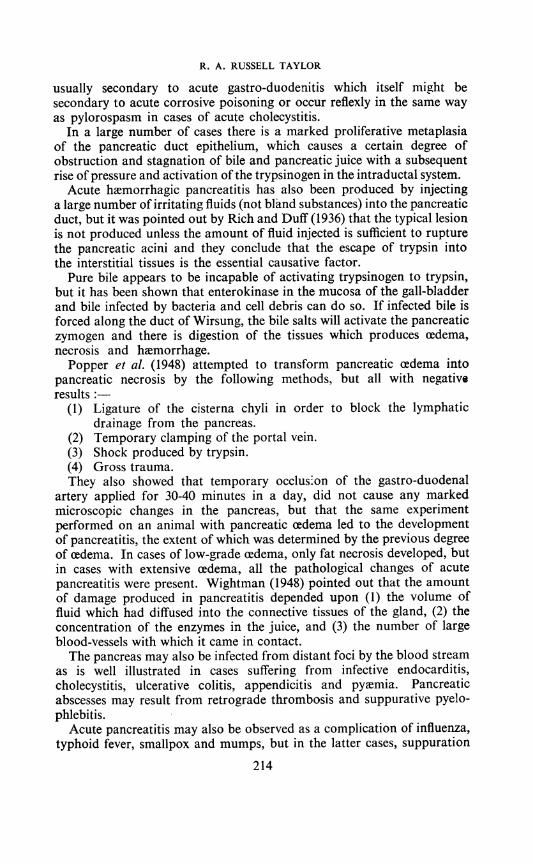
usually secondary to acute gastro-duodenitis which itself might besecondary to acute corrosive poisoning or occur reflexly in the same wayas pylorospasm in cases of acute cholecystitis.
In a large number of cases there is a marked proliferative metaplasiaof the pancreatic duct epithelium, which causes a certain degree ofobstruction and stagnation of bile and pancreatic juice with a subsequentrise of pressure and activation of the trypsinogen in the intraductal system.
Acute haemorrhagic pancreatitis has also been produced by injectinga large number of irritating fluids (not bland substances) into the pancreaticduct, but it was pointed out by Rich and Duff (1936) that the typical lesionis not produced unless the amount of fluid injected is sufficient to rupturethe pancreatic acini and they conclude that the escape of trypsin intothe interstitial tissues is the essential causative factor.
Pure bile appears to be incapable of activating trypsinogen to trypsin,but it has been shown that enterokinase in the mucosa of the gall-bladderand bile infected by bacteria and cell debris can do so. If infected bile isforced along the duct of Wirsung, the bile salts will activate the pancreaticzymogen and there is digestion of the tissues which produces cedema,necrosis and hxmorrhage.Popper et al. (1948) attempted to transform pancreatic (edema into
pancreatic necrosis by the following methods, but all with negativeresults
(1) Ligature of the cisterna chyli in order to block the lymphaticdrainage from the pancreas.
(2) Temporary clamping of the portal vein.(3) Shock produced by trypsin.(4) Gross trauma.They also showed that temporary occlusion of the gastro-duodenal
artery applied for 30-40 minutes in a day, did not cause any markedmicroscopic changes in the pancreas, but that the same experimentperformed on an animal with pancreatic cedema led to the developmentof pancreatitis, the extent of which was determined by the previous degreeof cedema. In cases of low-grade cedema, only fat necrosis developed, butin cases with extensive cedema, all the pathological changes of acutepancreatitis were present. Wightman (1948) pointed out that the amountof damage produced in pancreatitis depended upon (1) the volume offluid which had diffused into the connective tissues of the gland, (2) theconcentration of the enzymes in the juice, and (3) the number of largeblood-vessels with which it came in contact.The pancreas may also be infected from distant foci by the blood stream
as is well illustrated in cases suffering from infective endocarditis,cholecystitis, ulcerative colitis, appendicitis and pyemia. Pancreaticabscesses may result from retrograde thrombosis and suppurative pyelo-phlebitis.
Acute pancreatitis may also be observed as a complication of influenza,typhoid fever, smallpox and mumps, but in the latter cases, suppuration
214

ACUTE PANCREATITIS
or necrosis never occur. Very rarely it has been attributed to tuberculosisand syphilis.
Paxton and Payne (1948) found that 18 per cent. of their cases wereadmitted to hospital in an intoxicated condition and that in 25 per cent.of cases the pain came on immediately after a heavy meal. Cole (1938)however, found that the interval between a meal and the onset of pain wasusually 2-3 hours.
Cases of acute pancreatitis have followed penetrating and non-penetrat-ing abdominal injuries and also operations on the stomach, duodenum orlower end of the common bile duct when the pancreas has been injured.
Ackerman (1942) reported on a case of acute pancreatitis which followedtransfusion with incompatible blood, which at autopsy showed thrombosisof the pancreatic veins.The frequent association of acute pancreatitis with infection of the
biliary tract suggests that the lymphatic route is a possible connexionbetween the inflamed gall-bladder and the pancreas, but the experimentsof Kaufmann (1927) on rabbits practically discounted this.
PATHOLOGYAcute pancreatitis occurs most commonly about 40-60 years of age
and with about equal frequency in the two sexes. There also seems to bea definite association with obesity, cholecystitis and cholelithiasis.
Pratt (1940) maintains that acute pancreatitis is not an infection butan intoxication by the pancreatic ferments. Nevertheless, the intensityof the primary destructive changes determines the extent of the pathologicalchanges because this condition of auto-digestion may be self-perpetuatingand progressive even although the original stimulus has been removed.The progress of the disease may be continuous or intermittent and itmay become arrested at any stage.
Appearances at Post MortemThe body is sometimes very obese and in approximately 50 per cent.
of cases the patient is overweight. There may be local discoloration ofthe abdominal wall around the umbilicus (Cullen's sign) and in the loins(Grey Turner's sign). This discoloration is only seen in severe caseswhere the patient has lived for 2-3 days after the acute onset, and mustnot be confused with post-mortem staining.On opening the abdomen it must be remembered that aeute necrosis
may be present and yet macroscopically the pancreas may appear normal.The mildest type of acute pancreatitis is that known clinically as " transientpancreatitis" and recovery is common. Here we get cedema of thepancreas in which the intrapancreatic and peripancreatic cedema consistsof pancreatic fluid which has escaped into the pancreatic interstitial tissues.This cedema does little or no harm and will disappear soon after thesecretory stimulus has been discontinued. It is usually accompanied bycatarrhal changes in the duodenum which extends up the pancreatic duct.The pancreas itself may be enlarged to 2-3 times its normal size and is of
215

R. A. RUSSELL TAYLOR
varying consistence. It is indurated and small areas of fat necrosis maybe present, but hamorrhage is slight or absent. The changes may involvethe whole organ or be localised to the head, body or tail, the usualpercentage being, whole of pancreas, 73-1 per cent., body, 19-2 per cent.and tail, 7-7 per cent. (Fallis, 1939). The retroperitoneal1tissues are alsousually cedematous or infiltrated with blood.The next stage, that of hlmorrhagic pancreatitis, is seen if the arterial
blood supply has been interrupted. The resistance of the acinar andinterstitial cells has been weakened by this temporary ischxmia and theyare attacked by the enzymatic action of the cedematous fluid in which thetrypsinogen has been activated to trypsin by the bacteria and cellular debris.
In the most severe cases, the pancreas is seen as a large, dark, purplish,soft and friable mass on the posterior abdominal wall, shining through theperitoneum of the lesser sac. The peritoneal cavity may contain sanguinousor sero-sanguinous fluid which is present in the greatest amount in thelesser sac. This fluid may, however, have a yellowish-green colour dueto bile staining. It is invariably sterile, but later it may become infected,resulting in a localised or generalised peritonitis.
Gangrenous pancreatitis is generally regarded as a late stage ofhxemorrhagic pancreatitis and therefore oLcurs in cases which havesurvived the initial stages. The pancreas becomes softened, breaking upof the tissues with subsequent infection occurs, and a local or generalperitonitis results.
In suppurative pancreatitis there may be a considerable destructionof the gland due to (1) one or more abscesses in the pancreas itself, (2)retroperitoneal abscess, or (3) an abscess in the lesser sac. The infectionmay spread along the pancreatic ducts or directly from contact with theinfected pancreatic tissues. The abscesses are usually sterile and containthin pus or watery turbid fluid. Grey sloughs of pancreatic tissue ornecrotic fatty tissue may also be present and the necrotic tissue maysubsequently liquefy and give rise to pseudocysts of the pancreas.
In patients who survive, fibrosis occurs, which, if extensive, results ina reduction of the glandular tissue, distortion of the ducts and theformation of pancreatic cysts, i.e., a similar picture to that found incases of chronic pancreatitis.
In addition to the above, other pathological conditions are usuallypresent :-
TABLE 1
Associated Pathology Lewison (1940) Fallis & Plain (1939)
Chronic Cholecystitis .. .. 700% 57.70%Acute Cholecystitis .. .. 20% 15.40%Acute Cholelithiasis .. .. 800% 800%Choledocholithiasis .. .. 9% 8%Ampullary Stones .. .. 3 %Normal Gall-Bladder 200% 26 9%
216

ACUTE PANCREATITIS
In these cases the gall-stones are invariably small and the bile is usuallyinfected, dark in colour and may be blood-stained.
Microscopic AppearancesThree main pictures may be seen. In the first, the pancreas is seen
to be uniformly hamorrhagic; secondly there is marked necrosis of theglandular and interstitial tissues with a varying amount of hrmorrhagearound the necrotic areas and in the areolar tissues, and thirdly thepancreas is mainly necrotic.When hxmorrhage is the outstanding feature, the whole glandular
tissue is infiltrated with blood. The activated enzymes digest the blood-vessel walls, and the severity of the secondary hemorrhage, in and aroundthe pancreas, depends to a certain extent on the size of the blood-vesselsinvolved. Marked thrombosis of the blood-vessels, which have hyalinedegenerative changes in their walls, can also be seen.
Diffuse necrosis of acini may occur or the necrosis may affect theglandular and interstitial tissue with accompanying leucocytic infiltration.It is interesting to note that the inflammatory reaction is greatest in theless acute cases. The appearance of the parenchyma and the hemorrhagestrongly suggests that the condition is due to some toxic agent, but,although bacteria, e.g., B.Coli and Streptococci have been found, insome cases in large numbers, there is not enough evidence to support thetheory that this condition is the result of bacterial invasion.
It must not be forgotten, however, that haemorrhage into the pancreasmay also occur in blood diseases, sepsis and poisoning, especially infat people.
Fat NecrosisThis is the most distinctive feature of acute pancreatitis, and is seen
as dull opaque, yellowish-white areas suggestive of drops of tallow, butthey are not raised above the surface. Their size varies from that of apin-head to about '-inch in diameter and they are most abundant in thevicinity of the pancreas, but the retroperitoneal tissues, omentum,mesentery, mediastinum, pericardium, pleura and anterior abdominal wall,may also be involved. It is invariably attributed to the action of lipasefollowing its liberation due to damage to the gland tissue by infection,abscess formation or mechanical injury. The lipase has travelled alongthe lymphatics or by the blood stream and this theory explains its patchynature and also the occurrence of distant foci in the bone marrow, etc.Pancreatic lipase splits up the fatty molecule into glycerine and fatty acidand the latter combines with calcium to form an insoluble soap. If thepatient survives, these deposits are absorbed in a matter of weeks.
Microscopically the necrosed fat cells are seen to be wholly or partlyopaque and the whole area is surrounded by a ring of leucocytes.
Fat necrosis is not, however, pathognomonic of acute pancreatitis as itmay occur with perforation of an ulcer of the second part of the duodenum.
217

R. A. RUSSELL TAYLOR
Also it is necessary to distinguish between fat necrosis in acute pancreatitisand post-mortem fat necrosis. In the latter condition, only scattered,white spots in and around the pancreas itself are seen, with no hemorrhage,vascular congestion or leucocytic infiltration.
SIGNS AND SYMPTOMSIn preparing this paper I have been greatly impressed by the marked
diversity of the signs and symptoms occurring in this disease, but bypresenting them in a series of tables, it is hoped to demonstrate more
clearly the predominant features. Critical and careful study of these will,it is hoped, enable a correct diagnosis to be made in the majority of cases,
thus resulting in the appropriate treatment being instituted at the earliestpossible moment.
Age.-In the series under review, the largest age groups lie between40 and 70 years of age, the youngest being a girl of 10 years and theoldest being 85 years of age.
TABLE 2
Age Number of Patients Percentage
2 -2 .. i . . .3210-19 .. .. .. .. 1 .9
40-49 .. .. .. .. 22 20050-59 .. .. .. .. 29 26 460-69 .. .. .. .. 31 28 270-79 .. .. .. .. 16 14 580-89 .. .. .. .. 1 .9
TOTAL .. .. 110 100-0
Sex.-McWhorter -(1932) and Abell (1938) found that the sexes wereequally affected, but my own figures show a large preponderance offemales over males.
TABLE 3
Sex Number of Cases Percentage
Males .. .. .. .. 37 33-6Females .. .. .. .. 73 66-4
TOTAL .. .. 110 100-0
Obesity.-In a number of cases obesity has been pronounced but inonly 16 cases (14 5 per cent.) was it considered sufficient to be commentedupon. It is, however, considered significant to mention that approximately50 per cent. of cases were overweight.
218
ACUTE PANCREATITIS
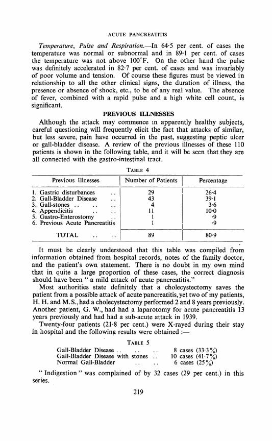
Temperature, Pulse and Respiration.-In 64-5 per cent. of cases thetemperature was normal or subnormal and in 89 1 per cent. of casesthe temperature was not above 100°F. On the other hand the pulsewas definitely accelerated in 82-7 per cent. of cases and was invariablyof poor volume and tension. Of course these figures must be viewed inrelationship to all the other clinical signs, the duration of illness, thepresence or absence of shock, etc., to be of any real value. The absenceof fever, combined with a rapid pulse and a high white cell count, issignificant.
PREVIOUS ILLNESSESAlthough the attack may commence in apparently healthy subjects,
careful questioning will frequently elicit the fact that attacks of similar,but less severe, pain have occurred in the past, suggesting peptic ulceror gall-bladder disease. A review of the previous illnesses of these 110patients is shown in the following table, and it will be seen that they areall connected with the gastro-intestinal tract.
TABLE 4
Previous Illnesses Number of Patients Percentage
1. Gastric disturbances .. 29 26-42. Gall-Bladder Disease .. 43 39-13. Gall-stones .. .. .. 4 3-64. Appendicitis .. .. 11 1005. Gastro-Enterostomy . 1 .96. Previous Acute Pancreatitis 1 .9
TOTAL .. .. 89 80-9
It must be clearly understood that this table was compiled frominformation obtained from hospital records, notes of the family doctor,and the patient's own statement. There is no doubt in my own mindthat in quite a large proportion of these cases, the correct diagnosisshould have been " a mild attack of acute pancreatitis."Most authorities state definitely that a cholecystectomy saves the
patient from a possible attack of acute pancreatitis, yet two of my patients,H. H. and M. S.,had a cholecystectomy performed 2 and 8 years previously.Another patient, G. W., had had a laparotomy for acute pancreatitis 13years previously and had had a sub-acute attack in 1939.
Twenty-four patients (21 8 per cent.) were X-rayed during their stayin hospital and the following results were obtained
TABLE 5Gall-Bladder Disease .. .. .. 8 cases (33 3%)Gall-Bladder Disease with stones .. 10 cases (417%)Normal Gall-Bladder .. .. 6 cases (25%)
"Indigestion " was complained of by 32 cases (29 per cent.) in thisseries.
219

R. A. RUSSELL TAYLOR
PAINThe onset of the pain is invariably sudden and this initial pain mav
be due to one or more of the following causes(1) Associated biliary pathology, e.g., Acute Cholecystitis.(2) Rapid inflammatory swelling of the pancreas with stimulation of
the nerves in the coeliac plexus and post-parietal peritoneum.(3) Early and copious toxic exudate into the peritoneal cavity pro-
ducing marked irritation of the parietal peritoneum.(4) Raised intraductal pressure.(5) Trauma.(a) Situation.-The site of the pain varies greatly, and the varying
figures quoted in the literature are quite understandable when the follow-ing facts are considered:
(1) The time between the onset of the disease and admission to hospital,e.g., pain may be primarily epigastric, but soon becomes generalised.
(2) The extent of involvement of the pancreas, e.g., whole, head, bodyor tail.
(3) Presence and amount of free fluid in peritoneal cavity.(4) Pain in the right iliac fossa due to secretions leaking through the
foramen of Winslow down the right paracolic gutter.(5) Patient being nursed in Fowler's position may result in lower
abdominal pain but with marked epigastric tenderness.In this series of cases the pain was localised- to the upper abdomen in
91 cases (82 7 per cent.), being primarily epigastric in 60 cases (54 5 percent.) and becoming generalised in 48 cases (43-6 per cent.) by the time ofadmission. In 15 cases (13 6 per cent.) the pain was generalised from theonset and the patient was unable to demonstrate the point of maximumintensity. In only 18 cases (16 4 per cent.) did the patient complain ofpain over the gall-bladder, whilst in two cases (18 per cent.) pain waslocalised to the left hypochondriac region. Umbilical pain was primarilypresent in five cases (4 5 per cent.), in the right iliac fossa in three cases(2-7 per cent.) and in the left iliac fossa in one case (0 9 per cent.).Praecordial pain was also complained of in one case (0.9 per cent.).Another very important feature of this pain is its tendency to radiate.Broadly speaking radiation occurs in over 60 per cent. of all the cases andthe usual sites are the costo-vertebral angles, and the shoulder tips. Severebackache in the lower thoracic region was a marked feature in only38 cases (34 5 per cent.). This backache may be in the midline or in oneor other costo-vertebral angle; the left being by far the most significant.In 12 cases (10 9 per cent.) there was pain in the shoulder tips, beingthree times commoner in the right than in the left.
Pain radiating across the epigastric region from right to left is a mostimportant sign and is practically pathognomonic of this disease.The next question is this: " Is the site of the pain determined by the
portion of the pancreas affected'7" A categorical answer cannot be
220

ACUTE PANCREATITIS
given because of the numerous factors involved, but this can almostcertainly be said, " that where the whole pancreas is involved, the painusually extends right across the abdomen, but if the head or tail is chieflyaffected, other things being equal, the pain is situated in the right hypo-chondriac and right costo-vertebral regions or the left hypochondriac andleft costo-vertebral regions respectively." The referred pains are merely anindication of the irritation of the peritoneal and diaphragmatic areas bythe serous exudate.
(b) Severity.-In most of the literature, pain which is present in100 per cent. of cases, is described as sudden, agonising, persistent, colickyor stabbing, but it would be most misleading to expect this in every case.In this series, the pain was of intense severity in 24 cases (21 8 per cent.)and in three of these, it was so severe as to awaken the patients fromsleep. In the remaining 86 cases (78-2 per cent.), the pain, althoughsevere, did not appear to be unbearable and in a few of these cases wasactually of a gnawing and burning character. In the majority of caseshowever, it is usually of such severity that it warrants the use of morphinewhich may only partially relieve it. I personally have abandoned the useof morphine in this disease as it either did not control the pain or it hadto be given in very large doses.From a close study of this series, I have come to the conclusion that
the severity of the pain depends upon one or more of the followingfactors:-
(1) the degree of obstruction at the ampulla of Vater;(2) the extent of the pancreatic involvement;(3) the amount of pancreatic hlmorrhage;(4) the amount of the serous exudate;(5) the extent of the involvement of the retro-peritoneal tissues;(6) the concomitant biliary pathology especially if " acute";(7) presence of local or general peritonitis.
(c) Type.-On careful questioning the patient frequently admits tosimilar but less severe attacks, in some cases over a period of years. Inmy youngest case (a female aged 10 years) she had had four severe attacksover a period of five months, which lasted for half an hour and madethe child scream with pain. Frequently there is also a previous historyof biliary colic which may be accompanied by intermittent jaundice.
In 99 cases (90 per cent.) the pain, whether epigastric, upper abdominalor generalised, continued without intermission, but in 11 cases (10 percent.) paroxysms of still more severe pain were felt. Of these 11 cases,eight definitely had gall-stones, whilst the remaining three showed evidenceof gall-bladder disease. In one case which complained of severeparoxysmal pain, a stone was found in the ampulla of Vater at postmortem. These paroxysms of pain may well be due to an attempt to passa gall-stone from the ampulla of Vater into the duodenum and shouldthis be successful, the paroxysms will cease and the stone will therefore
221
R. A. RUSSELL TAYLOR
not be found at operation or post-mortem. Another cause of thisparoxysmal pain may be further hxmorrhage into the pancreas andperipancreatic tissues from the blood vessels whose walls have beendigested.IMPORTANCE OF THE, TYPE OF PAIN IN DIFFERENTIAL DIAGNOSIS
(1) Acute Cholecystitis.-In this disease, the pain is usually confinedto the right hypochondriac area and frequently radiates to the rightscapula or to the right shoulder tip. Also, we frequently get a history ofrecurrent biliary colic with intermittent jaundice.
(2) Acute Intestinal Obstruction.-The spasmodic pain of acuteintestinal obstruction does not occur in acute pancreatitis, but the painmay be continuous and be referred to the region of the umbilicus. As thetoxaemia of intestinal obstruction progresses, the pain diminishes, but thevomiting continues. Pain in the back is felt only very rarely in intestinalobstruction.
(3) Perforated Peptic Ulcer.-The striking features of the painin perforated peptic ulcer are:-
(i) the severity, which doubles up the patient and is increased bymovement;'
(ii) pain referred to the supraspinous fossa or summit of the shoulder;(iii) partial relief of pain in the " second stage."(4) Aneurysm.-In differentiating the pain due to abdominal aneurysm
or dissecting aneurysm of the abdominal aorta, it is to be noted that thispain is neuralgic in character or may simulate renal colic.
(5) Acute Coronzary Artery Occlusion.-Substernal pain or painradiating to the neck or left arm is strongly suggestive of this condition.
SHOCKIt is generally accepted that the severe pain is almost invariably
accompanied by shock or collapse which may be so prbfound as to provefatal in a matter of hours. The cause of this rapid collapse is not clear,but it may be due to (1) the pressure of blood on the semilunar gangliaand cceliac plexus, (2) the absorption of toxins derived from the proteindigestion in the abnormal pancreas, (3) reflex disturbance mediatedthrough the nerves of the region, (4) the severity of the hemorrhage, or (5)the stripping of the parietal peritoneum off the posterior abdominal wall.
Fifteen cases (13.6 per cent.) of this series were suffering from profoundshock when admitted to hospital. It must however, be observed that inthose patients suffering from repeated paroxysms of pain, spontaneousrecovery from the primary shock is unlikely to occur and may even beincreased.
NAUSEA AND VOMITINGVomiting occurs early and is usually unaccompanied by nausea. It is
repeated at frequent intervals and may be as often as half-hourly. Thevomiting is also forcible in character, but the amount varies greatly. It
222

ACUTE PANCREATITIS
consists at first of gastric contents, later of bile and occasionally it containsa trace of blood, but it is never fical.
It has been pointed out that the absence of bile in the vomit may bean additional finding of great value in the diagnosis of the presence ofampullar obstruction. If the patient has not vomited, aspiration of theduodenal contents is a relatively easy procedure and may serve as a meansof determining the patency of the common bile duct.Vomiting was a pronounced feature in 89 cases (80-9 per.cent.) but blood
was present in only one case (0 9 per cent.). This blood in the vomitcould be due to a co-existing peptic ulcer, blood passing from the pancrea-tic duct into the duodenum or from an intensely inflamed stomach orduodenum.
Another important point is that the vomiting gradually tends to subsidein contrast to the vomiting of acute intestinal obstruction which becomesmore marked and eventually stercoraceous. In contrast to this again,the patient with a perforated peptic ulcer may vomit once or twice butno more. In thrombosis and embolism of the superior mesenteric artery,vomiting is early and severe, the vomitus sometimes containing blood, andit is followed by melkna.
FLATULENCEFlatulence was present in 22 cases (20 per cent.) and flatus was expelled
both by the mouth and per rectum. It was a very distressing symptomand caused the patients most acute discomfort.
BOWELSConstipation was a recent symptom in 24 cases (218 per cent.) but it
was never absolute, which is the rule in acute intestinal obstruction.In four cases (3 6 per cent.) the patient complained of diarrhoea, but melknawas not observed in any case of this series, although it has been mentionedby other observers.
JAUNDICEJaundice may be as high as 43 per cent. (Lewison (1940)) or as low as
10 per cent. (Abell (1938)); the number in this series being 13, giving apercentage of 11 8. The jaundice is usually attributed to an obstructionat the ampulla of Vater, or pressure of the swollen pancreas on thecommon bile duct.
CYANOSIS AND SKIN DISCOLORATIONThere may be only slight cyanosis of the lips and ears or it may be widely
distributed over the abdomen and limbs. This has been attributed toshallow breathing because of the painful abdominal lesion, to markedshock and cardiac failure, or to toxemia. Occasionally there is a localdiscoloration of the abdominal wall around the umbilicus (Cullen's sign)and in the loins (Grey Turner's sign). It is only seen in cases of some2-3 days' standing and the patches have the appearance of the skin incases with late extravasation of urine, gas gangrene, or virulent influenzalpneumonia. The discoloured areas are slightly cedematous and the
22317

R. A. RUSSELL TAYLOR
cedema fades into the surrounding tissues. Their size varies greatly andin one case, described by Blauvelt (1946), it was 5 cms. in diameter. Theyare usually attributed to the direct action of the pancreatic fermentswhich escape via the retroperitoneal tissues and pass by the most directroute to the surface, or to the action of the pancreatic lipase carried bythe blood stream.
Cyanosis of the lips and ears was present in 6 cases (5 5 per cent.) anddiscoloration of the flanks in 1 case (0.9 per cent.). As the latter signonly occurs in very acute cases the prognosis is bad, Eliason (1930) puttingthe mortality at 85 per cent.
In acute coronary occlusion, cyanosis is usually present, but the maindistinguishing features are an irregular pulse and precordial distress.
ABDOMEN(a) Inspection.-Examination of the abdomen shows that the move-
ments are definitely limited and thei efore the breathing is mainly thoracic.In cases with severe epigastric pain the abdomen may be immobile abovethe umbilicus. In the early stages the contour of the abdomen is that whichis normal for the individual, but it soon becomes distended. Thisabdominal distension was a feature in 29 cases (26-4 per cent.) and wasmost evident in the epigastric region. It is due to the transverse colonbeing paralysed and distended with gas, and also to an incomplete ileus.In acute intestinal obstruction the distension is usually more marked andgeneralised, but it is not present until the very late stages of perforatedpeptic ulcer.
(b) Palpation.-Abdominal palpation demonstrated the presence ofextreme local tenderness in 35 cases (318 per cent.) and generalisedtenderness in 47 cases (42-7 per cent.). A milder type of tenderness waspresent in the remaining 28 cases (25-5 per cent.) but there is no directrelationship between the degree of tenderness and the type of the pancreaticlesion.
Very careful palpation may determine that there is deep tendernessover the whole of the pancreas, whilst in other cases it may be moremarked on the left side of the epigastrium. If the tenderness is chieflyon the right side, then the possibility of co-existing gall-bladder diseasemust be considered. The most important sign, however, is tenderness inthe costo-vertebral angles, especially if this is on the left side. Recoiltenderness is invariably present and is often very marked, especially inthe region of the upper abdomen.
In the early stages, the abdominal wall is flaccid. Muscular rigidity iseither absent, or only occurs to a very mild degree. This mild degree ofrigidity was present in 59 cases (53-6 per cent.) being generalised in 33 cases(30 per cent.) and localised to the epigastrium in 26 cases (23-6 per cent.).In approximately half the cases, the point of maximum tendernesscorresponded to the point of maximum muscle spasm.
In the later stages of this disease, the rigidity may be generalised and
224

ACUTE PANCREATITIS
severe, but obviously the extent and degree of rigidity must be correlatedwith the whole of the clinical picture in order to be of value. It is thecombination of extreme tenderness and the lack of definite muscularrigidity which is so characteristic; the rigidity which may develop lateris due to peritonitis secondary to pancreatic infection.
In acute cholecystitis, muscular rigidity is usually marked in the upperhalf of the right rectus muscle and in both upper quadrants or evengeneralised in perforated peptic ulcer.No tumour is likely to be felt in the epigastric region until the third day
(Kr6te's sign) and even then the pancreas may not be palpable. This massmay be felt either in the epigastrium or left loin and was present in 17 cases(15-4 per cent.) in this series. The swelling moves little on respiration andoften transmits a non-expansile pulsation from the underlying aorta.It may be separated from the liver and spleen by areas of resonance.
In acute cholecystitis, the gall-bladder may be palpable. A smallrounded swelling with an expansile pulsation situated usually to the leftof the mid-line is present in abdominal aneurysm and a vague mass indissecting aneurysm of the abdominal aorta. Dinsmore and Nosik (1939)suggest that areas of hyper-esthesia on the left side corresponding to thesegments of T 8-10, possibly even higher, would be found, if sought for,but although I have been unable to confirm this in more than 3 cases(2-7 per cent.) it is of definite significance.
(c) Percussion.-There is no alteration in the area of hepatic dullnessin acute pancreatitis, but it may be diminished or absent in perforatedpeptic ulcer. The presence of free fluid may be demonstrated in theperitoneal cavity and may be of such an amount as to give rise to" shifting " dullness. This was present in 8 cases (7-2 per cent.). Morton(1940) and Fallis (1939) have withdrawn this fluid by abdominal para-centesis, the former reporting on a characteristic prune juice fluid andthe latter finding blood-stained fluid. Personally I am of the opinion thatthis method of investigation is quite unjustified.
AuscultationIntestinal sounds in acute pancreatitis disappear almost immediately
but in acute intestinal obstruction the sounds are easily heard and onlydisappear at a late stage.
Rectal examinationThis may yield no definite information, butl is extremely tender when
there is free irritant fluid in the peritoneal cavity and when peritonitishas developed in the later stages.
URINEChanges in the urine are not constant but nevertheless may be important
when taken with the other signs and symptoms. The following tableshows the abnormalities which occurred in this series
225
17-2

R. A. RUSSELL TAYLOR
TABLE 6Urine
Number of Cases Percentage
Albumen .. .. .. 27 24-5Blood .. .. .. .. 4 3-6Bile .. .. .. .. 12 10-8Sugar .. .. .. .. 14 12-7Acetone Bodies .. .. 6 5-4Increased Urinary Diastase 15 13-6Dysuria .. .. .. .. 7 6-3Oliguria .. .. 1 .9
Albuminuria was present in 25 per cent. of cases and in the vast majoritywas only transient. It could be due to one of the following causes, namely,cardiac failure, shock, toxvmia, fever, renal damage or simply frompressure of the cedematous pancreas on the renal veins. Hematuria onlyoccurred in 3-6 per cent. of cases and is probably due to an accompanyingnephritis or to severe renal congestion, again due to pressure on therenal vens.As mentioned above, jaundice was present in 13 cases and in 12 of
these, bile was detected in the urine.If glycosuria is found, it tends to confirm the diagnosis, but this finding
is said to be uncommon, probably because death occurs too rapidly.(Proof: in animals, even total removal of the pancreas is not followed byglycosuria for several days.) Its presence undoubtedly demonstratesdestruction or temporary non-functioning of the Islets of Langerhans,and as mentioned later, the patient may develop diabetes mellitus, but nosuch case occurred in this series.
Urinary Diastase.-In the healthy subject the concentration of diastasein the urine is usually between 2 and 50 units, but there are markedvariations in the figures obtained at different times of the day, owing topolyuria or oliguria.The daily output of diastase is estimated by multiplying the volume in
cubic centimetres of a 24-hour specimen of urine by the diastase index.The figure normally lies between 8,000-30,000 units.
In acute pancreatitis, the first specimen of urine available is taken andin the majority of definite cases, it usually contains 100 or more units percubic centimetre; lesser 0ncentrations being of little or no significance.The highest figures are usually obtained if the test is performed within afew hours of the acute onset of the disease, but the urinary diastase onlyrises from 6-24 hours after the rise in blood amylase.
In some definitely proved cases of acute pancreatitis, there is no increasein urinary diastase, especially if the test is performed 14 days after theacute onset. This is almost invariably found in the following types ofcases
226

ACUTE PANCREATITIS
(1) Pancreatic aedema and mild cases of acute hemorrhagicpancreatitis.
(2) Cases seen late and in which there is a certain recovery of functicn.(3) Widespread and apparently total glandular destruction.In 10 out of 25 established cases in which this test was employed, the
results were within normal limits.In one of these cases in which it was increased, the diastase index was
600 units per cc. representing a daily output of diastase of 336,000 units.An increased urinary diastase is not, however, pathognomonic of acutepancreatitis because, although it never reaches very high figures, it mayrise to 200 units per cc. in carcinoma of the head of the pancreas due toobstruction of the ducts, and toxemia of pregnancy due to increasedpermeability of the kidneys.
Oliguria occurs only in very severe cases and may even amount to adefinite anuria. It is usually attributed to low blood pressure causingimpaired renal circulation or pressure of the pancreas on the renal veinscausing marked renal congestion.
BLOOD CHANGES(a) Blood Diastase.-The range of normal levels is usually given as
60-200 units of diastase per cc. of serum and there is now no doubt that arise of serum amylase is almost invariably associated with pancreaticdisease and if it is over 1,000 units per cc. the prognosis is said to be bad.I do not support this view regarding the prognosis unless the level remainspersistently high over a period of 24-72 hours. The highest figures areusually found within 12 hours of the acute attack and the vast majorityhave returned to normal by the 6th-lOth day. It must not be forgotten,however, that a definite case of acute pancreatitis may have a normalblood diastase. This usually indicates, if the clinical condition is satis-factory, that the pancreatitis is subsiding or has subsided.
In this series, the blood diastase was estimated in 23 cases (20-9 percent.) at times varying from 4 hours to 8 days after the onset of symptoms.In 14 cases (12.7 per cent.) figures over 1,000 units per cc. all occurredwithin four days of the onset and all except one had returned to normalwithin five days. In five cases (4 5 per cent.) there was no rise in theserum amylase.
Other conditions which may give rise to increased blood diastase are(1) Parotitis.(2) Renal disease.(3) Trauma to pancreatic gland.(4) Pancreatic cysts.(5) Carcinoma of head of pancreas.(6) Chronic venous congestion.(7) Diabetes Mellitus (rarely).It will be noted that only two acute surgical conditions, namely, acute
pancreatitis and trauma to the pancreatic gland are accompanied by an
227

R,- A. RUSSELL TAYLOR
increased blood or urinary diastase and therefore this factor, in conjunc-tion with the foregoing signs and symptoms, is of definite diagnosticvalue, but a normal diastase index does not exclude pancreatitis.
(b) Hyperglycemia.-If the blood sugar rises to over 300 milligramsper cent. the outcome is invariably fatal. In 11 cases (10 per cent.) ofthis series, no increase in the blood sugar was detected. Shumacker (1940)concluded that at least 2 per cent. of all cases with severe acutepancreatitis acquired diabetes mellitus, and, of those surviving the acuteillness, from 3-10 per cent. became subjects of diabetes mellitus.
(c) Blood Count.-In this series the maximum recorded white cellcount was 38,000 per cubic millimetre and this was in a case of markedpancreatic necrosis. The majority of cases had a count ranging from6,000-15,000 per cu. mm.
(d) Blood Calcium.-Edmondson and Berne (1944) reported that in72 per cent. of cases the serum calcium was below 9 milligrams per 100 cc.between the 2nd and 15th day of the disease and that the average serumcalcium value was the lowest on the 6th day. If the serum calcium levelwas below 7 milligrams per 100 cc., the prognosis was invariably fatal.At these low levels it is necessary to keep a careful watch for the onset oftetany and to treat it vigorously.
(e) Plasma, Protein and Prothrombin.-Lowering of the protein andprothrombin content of the blood during an attack of acute pancreatitisis said to occur, but further investigation is necessary before any definiteconclusions can be drawn.
SPECIAL TESTSLoewi's Test
In this series the test was negative in nine out of 14 definitely establishedcases of acute pancreatitis, whilst being positive in a case of gangrenouscholecystitis.
Cammidge's Pancreatic ReactionThis is not pathognomonic of acute pancreatitis and is now regarded
as so unreliable as to be useless.LATE CASES
If the patient is seen at a later stage, the clinical picture is slightlydifferent. Dittler and McGavack (1938) reported on a case of acutepancreatitis complicated by impure auricular fibrillation and flutter, butat autopsy no organic lesion could be found. They therefore concludedthat the cardiac condition was due to reflexes from the abdomen. Loefflerand Esseluer (1946) state that acute pericarditis has also been found inseveral cases. Dyspncea occasionally occurs and may even amount to airhunger. Hiccup may be present and if persistent, generally indicates agrave prognosis. Two patients (18 per cent.) in this series found this amost distressing symptom and both died.
Shifting dullness, uncommon in the early.stages, can usually be elicited
228

ACUTE PANCREATITIS
at this juncture. In some cases, after the acute onset, the symptomssubside, but the pulse rate remains high and a mass develops in theepigastric region. This is followed by localised suppuration or sloughingof the gland. In the suppurative form, the pus may (1) collect in the sub-stance of the pancreas, (2) fill up the lesser sac and bulge beneath theleft vault of the diaphragm, or (3) present in the left lumbar region andsimulate perinephric abscess.
In the later stages, when there is no localisation, there are signs ofdiffuse peritonitis with free fluid or retroperitoneal cellulitis.The symptoms of sepsis usually develop after 7-10 days and ultimate
recovery after prolonged illness may follow rupture of an abscess into thebowel. Perforation into the peritoneal cavity, stomach or duodenummay occur. Temporary improvement may end in late death in 4-6 weeksafter operation from pancreatic insufficiency. The symptoms then aremalnutrition, wasting, profound weakness, hypotension, hypoproteinxmia,glossitis, vitamin D and vitamin K deficiency, fatty diarrhoea and un-controllable vomiting of small amounts. Death may also be due tohemorrhage from neighbouring vessels or to septic absorption.
Gangrenous pancreatitis may follow (1) haemorrhagic or suppurativeinfiltration of the pancreas, (2) trauma, or (3) perforated gastric ulcer.The symptoms of hemorrhagic pancreatitis may precede or be associatedwith it, and death usually occurs in 10-20 days.
DIFFERENTIAL DIAGNOSISTABLE 7
Diagnosis by General Practitioner Number of Cases Percentage
(1) Acute Abdominal Pain .. .. 43 39-1(2) Acute Cholecystitis .. .. .. 23 20.9(3) Acute Intestinal Obstruction .. 14 12 7(4) Perforated Peptic Ulcer .. .. 8 7-3(5) Peritonitis .. .. .. .. 6 5 5(6) Coronary Thrombosis .. .. 5 4.5(7) Acute Appendicitis .. .. .. 5 4.5(8) Acute Gastritis .. .. .. 2 1-8(9) Acute Pancreatitis 4.. .. .. 3-6
A diagnosis of acute cholecystitis in 23 cases (20-9 per cent.) wasprobably due to the fact that these cases exhibited a history of gall-bladderdisease whilst 13 cases (11 8 per cent.) showed some degree of jaundice.A history of recent constipation and the presence of repeated vomiting
has undoubtedly played a part in arriving at a diagnosis of acute intestinalobstruction which was given in 14 cases (12.7 per cent.) of this series.A correct diagnosis was made in only 4 of the series (3-6 per cent.).
On admission to hospital the surgeon made a correct diagnosis in only46 cases (41 8 per cent.), whilst the other possible diagnoses are tabulatedon the following page.
229

R. A. RUSSELL TAYLOR
TABLE 8
Surgeon's Diagnosis Number of Cases Percentage
(1) Acute Pancreatitis .. .. 46 41 8(2) Acute Cholecystitis .. .. .. 32 29-1(3) Acute Intestinal Obstruction .. 13 11 8(4) Perforated Peptic Ulcer .. .. 16 14 5(5) Acute Appendicitis with Peritonitis 2 18(6) Coronary Thrombosis .. .. 1 .9
TOTAL.. .. .. .. 110 99 9
Other observers have made the following observationsTABLE 9
Author A.P. G.B.D. App. P.P.U. A.I.O. M.T. C.T.0/ 0/ ~~0/ /0/ 0 0
Morton and Widger (1940) 17 - - - - - -Lewison (1940) .. .. 13 70 10Morton (1940) .. .. 17 43 5 27 8 5 5Fallis (1939) .. .. 308 - -
Abell (1938) .. .. 12 -
A.P.G.B.D.App.
Key= Acute Pancreatitis P.P.U. = Perforated Peptic Ulcer= Gall-Bladder Disease A.I.O. = Acute Intestinal Obstruction= Appendicitis M.T. = Mesenteric Thrombosis
C.T. = Coronary Thrombosis
Other conditions which must also be considered are :-(1) pneumonia,(2) acute nephritis, (3) ruptured ectopic pregnancy, and (4) spontaneousrupture of the common bile duct.
TREATMENT(1) Prophylactic.-As Cole (1938) pointed out, gall-bladder disease isfar too common, and acute pancreatitis too uncommon, to justifycholecystectomy merely in the endeavour to prevent acute pancreatitis.Chronic alcoholism, obesity and any disease of the biliary tract should betreated.(2) Conservative.-Modern treatment tends to be conservative, andcertainly the figures published of cases so treated show a definite decreasein the mortality rate. Evidence of old fat necrosis discovered at a lateroperation as well as the biochemical tests mentioned above, definitelyprove that conservative treatment has been successful. In acute pancreatitisdue to mumps, operation is never indicated and recovery is usuallycomplete within a week. If conservative treatment is decided upon,frequent and detailed clinical examination of the patient is essential withhourly recordings of the pulse rate and blood pressure. Forty-one of thisseries (37 3 per cent.) were treated by conservative methods. Of these,30 cases (73-2 per cent.) made an uninterrupted recovery, whilst 11 cases
230

ACUTE PANCREATITIS
(26 8 per cent.) died. Post-mortem examination was performed on eachof these 1 patients and the diagnosis was confirmed. Of these 11 patients,3 died within 24 hours of admission, whilst 4 more died within 5 days.The post-mortem findings were interesting and are detailed as follows:in 3 cases the gall-bladder and bile ducts were found to be perfectlyhealthy, whilst in 8 cases chronic cholecystitis was present, 4 of thesealso having gall stones. In 2 cases, with cholelithiasis, a stone wasfound in the ampulla of Vater. In one case the peritoneal cavity containedone pint of straw-coloured fluid and the upper part of the duodenumwas markedly dilated. Five cases showed fat necrosis and 2 cases hadthrombosis of the splenic vein.
ShockThis is not the appropriate occasion to describe the classical signs and
treatment of shock, but a few points on its treatment as related to thisdisease are worth recording.
(i) Transfusion and Infusion.-As pointed out by Jensenn (1946) theplasma given by transfusion may pass into the serous cavities irritatedby the pancreatic secretions and may therefore account for the cyanosisand shock with hxmoconcentration. In certain cases transfusion ofwhole blood is indicated to counteract that lost during the hemorrhagicphase.
If the patient is dehydrated from repeated vomiting, intravenous salinewith 5 per cent. glucose should,be administered. A word of warningmust be given here because large doses of intravenous glucose may bedangerous. Experimentally it has been shown that an elevation of theblood sugar level causes an increased flow of pancreatic juices rich inferments. This may be counteracted in humans to a certain extent bygiving one unit of insulin for each four grammes of glucose.
(ii) Drugs.-As stated previously, I do not advocate the use of morphiafor the relief of pain in acute pancreatitis because although it will do soin full doses by its action on the central nervous system, at the sametime it causes further spasm, or at least increases the tone of the smoothmuscle. This would cause a contraction of the sphincter of Oddi with aresulting rise in the biliary and pancreatic pressures and possibly furtherdamage to the liver and pancreas. I therefore advise the administrationof anti-spasmodics because not only is the pain diminished or abolished,but a stone or plug of mucus in the ampulla of Vater, if present, can bepassed into the duodenum by the relaxation of the sphincter whilst italso produces a fall in the intra-ductal pressures.
Immediate, although only very temporary, relief can be obtained bygiving inhalations of amyl nitrate, but a better method is to give tablets ofnitroglycerine to suck or chew. Papaverine or Eupaverin give moreprolonged effects, and ephedrine, either alone or in combination withatropin and papaverine, is excellent. Papaverine may also be of value forits vaso-dilator effect because the extent of local vasoconstriction may
231

R. A. RUSSELL TAYLOR
determine the type and degree of pancreatitis. Atropin Sulphate, given inas large doses as --L gr. 6-hourly for 24 hours, acts as an excellent anti-spasmodic and proportionately smaller doses may be given later. This drugis also extremely useful if there is excessive sweating. According toSmead (1940) small doses of ephedrine 4-hourly are also useful if vascularcollapse is present. Popper (1933) described 3 cases in which he relievedpain by the paravertebral injection of the 8th-lOth dorsal nerves withnovocaine, and partly ascribed these good results to the local vasodilatationproduced by blocking the sympathetic innervation to the pancreas.
Considerable quantities of calcium may be present in the lesions incases of acute pancreatic necrosis and it is logical to assume that a plentifulsupply of available calcium is desirable in order to facilitate the formationof calcium soap in situ without undue depletion of serum calcium and thepossible onset of tetany. This may be given in the form of a 10 per cent.solution of calcium gluconate intravenously in 10 cc. doses. Lastly,chemotherapy and penicillin therapy should be. employed in full doses toprevent local infection of the necrotic pancreatic tissues and in an attemptto prevent infection spreading into the peritoneal cavity.
DIETIt is essential that intravenous therapy should be continued for 3-4 days
and that nothing be given by mouth in an attempt to inhibit the activityof the pancreas. The intake of food must be resumed very carefullyand easily assimilable carbohydrates should be given, e.g., milk, milkpuddings, orange juice, glucose, honey, carrots, white of egg, etc. Fatis not permissible for several weeks and meat is forbidden as the proteinintake must be limited.
LENGTH OF STAY IN HOSPITALTABLE 10
Time Number of Patients Percentage
Less than 1 week .. .. 2 6.61-2 weeks .. .. .. 13 43-32-3 weeks .. .. .. 9 30.03-4 weeks .. .. .. 4 13-34-8 weeks .. .. .. 1 3-38-12 weeks .. .. .. 1 3-3
TOTAL .. .. 30 99 8
It will be noted that with conservative treatment 24 (80 per cent.) ofthe 30 cases which survived were discharged from hospital withinthree weeks.
Before turning to the discussion of operative treatment, it should beemphasised that it is unwise to give or to proceed with conservativetreatment if the following conditions are present, and early operation
232

ACUTE PANCREATITIS
should be advised :-(l) persistent fever or development of fever alongwith other abdominal signs, (2) spreading peritonitis, (3) pancreaticnecrosis, (4) distension of lesser peritoneal cavity, (5) enlargement of thegall-bladder, (6) jaundice, or (7) no response to conservative treatment.
OPERATIVE TREATMENTOperation was previously advised in cases of acute pancreatitis for the
following reasons:(1) To remove the cause, e.g., gall-stones and to drain the bile passages.(2) To relieve the tension about the pancreas by incising its peritoneal
covering.(3) To remove the fluid from the greater and lesser sacs.(4) To provide drainage from neighbourhood of gland.As will be seen from the etiology, the cause of acute pancreatitis is not
definitely known, and even if gall-stones are present they may not be thedetermining factor. Provided that there is no stone in the ampulla ofVater, drainage of the biliary tract and pancreatic ducts can be stimulatedby duodenal suction.
Incision of the peritoneal covering of the pancreas does not relieve thetension completely because the acini have their own fibrous capsule andit is impossible to operate on them individually. The removal of thehaemorrhagic fluid from the peritoneal cavity also seems to be unnecessary.Ireneus (1941) found that the hxmorrhagic exudate in animals with acutehxmorrhagic pancreatitis was not toxic on the intra-peritoneal injectionof 2-3 cc. into white mice or on intravenous injection into dogs.
Laparotomy as a Diagnostic ProcedureIf a definite diagnosis cannot be made from any acute abdominal
condition requiring an emergency laparotomy, this must be performed.If cedema of the pancreas oran acute hemorrhagic pancreatitis is observed,then the abdomen should be quickly but very carefully closed andconservative treatment instituted as detailed above.
Details of Operative ProceduresIn 69 cases (62-7 per cent.) operation was performed, and of these
28 died, giving a mortality rate of 406 per cent.
Anesthetic UsedTABLE 11
Aniesthetic No. of Percentage Deaths
Gas, Oxygen and Ether 51 73-9 21Open Ether .. .. 14 20-3 5Spinal .. .. .. 4 58 2
TOTAL . 69 100-0 28
233

R. A. RUSSELL TAYLOR
This table, however, gives no indication of the clinical condition ofthe patient when submitted to operation. There is, however, no doubt inmy mind that these cases should have their anesthetic administered by aconsultant anxsthetist and that the practice of House Surgeons givingthe anmsthetic to emergency cases should be whole-heartedly condemned.
Various types of operations have been performed in acute pancreatitisand those employed in this series are detailed in the following table:
TABLE 12
Type of Operation Cases Deaths Mortality
Laparotomy .. .. .. .. 28 8 28.60%Laparotomy with suprapubic drainage .. 4 1 2500%Laparotomy with drainage of lesser sac .. 8 5 62 5 %Laparotomy with drainage of lesser sacand suprapubic drainage .. 2 1 5000%
Laparotomy with cholecystostomy .. 7 4 56-3 %Laparotomy with cholecystostomy and
suprapubic drainage .. .. .. 2 1 5000%Laparotomy with cholecystostomy and
drainage of lesser sac .. .. .. 10 6 6000%Laparotomy with cholecystostomy and
drainage of Rutherford Morrison'spouch .. .. .. .. .. 2 Nil Nil
Laparotomy with drainage of posteriorabdominal wall .. .. .. 1 1 10000%
Laparotomy with cholecystectomy .. 4 1 25 %Laparotomy with cholecystectomy andcholedochostomy I. .. .. 1 Nil Nil
N.B.-It will be noted that laparotomy and laparotomy with supra-pubic drainage have the lowest mortality, namely, 28 per cent. In cases inwhich the gall-bladder was also operated upon, and also when the lessersac was opened, the mortality rose to 50 per cent. or over. In the threecases which recovered after drainage of the lesser sac, the duration ofthe symptoms had been longer than 24 hours.The pancreas is an extraperitoneal organ and therefore if the lesser
sac is opened and the peritoneal covering of the pancreas incised, thepancreatic secretions and products of protein metabolism are allowed toescape into the general peritoneal cavity. The great absorptive powers ofthe peritoneum are well known and therefore the dangers of profoundtoxemia are greatly increased by this interference. The obvious conclusionto come to is that it is advisable to interfere operatively as little as possible.Certain features, however, may make it essential to proceed, e.g., markeddistension and inflammation of the gall-bladder which may go on torupture and cause biliary peritonitis. In these cases cholecystostomy isindicated. Jaundice, because it is of the obstructive type, is also anindication for cholecystostomy. Only in rare cases should choledochostomybe performed in order to remove a stone in the ampulla of Vater or
234

ACUTE PANCREATITIS
common bile duct. This should be attempted if the patient's generalcondition is good and the common bile duct much distended and easilyaccessible. Should choledochostomy appear difficult, a rapid chole-cystostomy will suffice to decompress the biliary system. Cholecystectomyshould be avoided if possible because cholecystenterostomy may benecessary later and also it is accompanied by a great deal of shock in analready ill patient. Later there is also the danger of an increased biliarypressure.
In the absence of gall-stones or jaundice, dilation or thickening of theduct, one is rarely justified in exploring the common bile duct, but shouldthis be necessary, the following points made by McWhorter (1932) arevery useful in determining the subsequent treatment:-
(I) If the outlet of the common bile duct is dilated, prolonged drainageis unnecessary.
(2) If the outlet is partially or completely obstructed due to localcongestion, do NOT dilate because of the danger of increasedswelling. If possible a tube should be inserted into the commonbile duct through the sphincter of Oddi into the duodenum.This prevents a further reflex of bile into the pancreatic duct.
Pancreatic necrosis or suppuration is another indication for drainageand may best be done through the loin or costo-vertebral angle. Drainageof fluid in the lesser sac is advisable as it may prevent the developmentof a pseudocyst and may be carried out through the gastro-colic orgastro-hepatic omentum.
If a retroperitoneal involvement is present as indicated by surfacediscoloration, incisions are made into the loin. In very rare cases,pancreatic lithiasis is present and it is necessary to remove the stoneby a transduodenal incision exposing the ampulla of Vater, which issplit, and the stone removed with forceps.
PancreasVarious parts of the pancreas were seen at operation to be the site of
the initial lesion, but in only 33 cases (47 8 per cent.) was the whole of theorgan involved. In only two cases (2 9 per cent.) was the disease confinedto the tail whilst the remainder were about equally divided between therest of the pancreas.
Fat NecrosisFat necrosis was found in 55 cases (79.7 per cent.) and was invariably
extensive over the omentum, mesentery and parietal peritoneum.
Free FluidFree fluid was found in 13 cases (108 per cent.) mainly in the lesser sac
and around the gall-bladder. This fluid was chocolate-coloured in 5cases (7 1 per cent.), blood-stained in 7 cases (10 1 per cent.) and serous
235

R. A. RUSSELL TAYLOR
in 1 case (1P4 per cent.). McWhorter (1932) found free fluid in 60 per cent.of his cases, the majority being blood-stained.
Gall-BladderThe gall-bladder appeared normal in 20 cases (29 per cent.) but was
diseased in the remaining 49 cases (71 per cent.), the degree of involvementbeing shown in the following table:-
TABLE 13
State of Gall-Bladder No. of Deaths MortalityPatients
(1) Apparently healthy Gall-Bladder .. 20 8 40%(2) Apparently healthy Gall-Bladder with
Gall-Stones .. .. .. .. 13 4 3080%(3) Acute cholecystitis .. .. 13 5 38-5%(4) Acute cholecystitis with gall-stones .. 3 3 10000%(5) Chronic cholecystitis .. .. 5 4 80-0%(6) Chronic cholecystitis with gall-stones 13 4 30-8%(7) Empyema of gall-bladder with gall-
stones .. .. .. .. .. 2 - Nil
TOTAL .. .. .. 69 28
From the table it will be seen that gall-stones were present in 31 cases(44 9 per cent.) and in only 1 case was a stone found in the ampullaof Vater at operation. If there is no contra-indication to operation, allcases in which gall-stones are demonstrated should betreatedsurgically,as their removal may prevent a further attack. This operation should beperformed preferably 3 months after all the acute symptoms have subsided.. Other findings at operation were marked cedema of the posteriorabdominal wall in 5 cases, 3 of which died. In one case a chronic gastriculcer was present and in another a duodenal ulcer.
POST-OPERATIVE TREATMENT
When the patient returns from theatre he should be given the usualtreatment for post-operative shock and this should be followed by thestrict regime laid down under conservative treatment.
Post-Operative Complications usually associated with Acute Pancreatitis(1) Burst Abdomen.-This may be guarded against to a certain
extent by perfect peritoneal apposition, the use of non-absorbable sutures,with interrupted sutures for the muscles, and the supporting of the woundby means of laparotomy corsets. Should the ferments escape on to thesurface, the skin may become digested and this is best guarded againstby applying paraffin molle ointment containing 2 per cent. hydrochloricacid, paint daily with Whitehead's varnish, or Lanolin ointment con-taining 1 per cent. acetic acid applied liberally.
236
ACUTE PANCREATITIS
(2) Thrombosis of Splenic and Mesenteric Veins.-Very little can bedone for this complication except the administration of heparin, ordicumerol and penicillin to counteract sepsis. The outcome is invariablyfatal.
(3) Pancreatic Insufficiency.-This is treated by blood transfusion andpancreatic extracts. Pancreatinum may be given orally in doses of 3-5grains, 2-3 hours after a meal. This is to prevent its destruction by theacid in the gastric juice. This white- or buff-coloured powder contains theenzymes, trypsin, lipase and amylase. Peptonised foods in the form ofpeptonised milk,- beef tea and Benger's food may also be given. Pan-creatinum may also be given in the form of an enema in 4 oz. doses inequal parts of milk and beef tea. Other proprietary preparations are:dipantrin, panacoids, panteric tablets and zymine These extracts mayhave to be continued for several months and estimations of serum amylaseand lipase give some guide as to the extent and progress of the deficiency.
(4) Diabetes Mellitus.-This is treated on the usual lines.(5) Pseudocyst of Pancreas.-This is dealt with by drainage and a
counter-incision in the left loin.(6) Pancreatic Fistula?.-Bufalini (1947) describes an operation for a
persistent fistula by which he converts it into an internal drainage by usingthe fistulous track and anastomosing the cutaneous opening with anabdominal viscus. Persistent cases may be cured by the insertion ofradium into the tract.
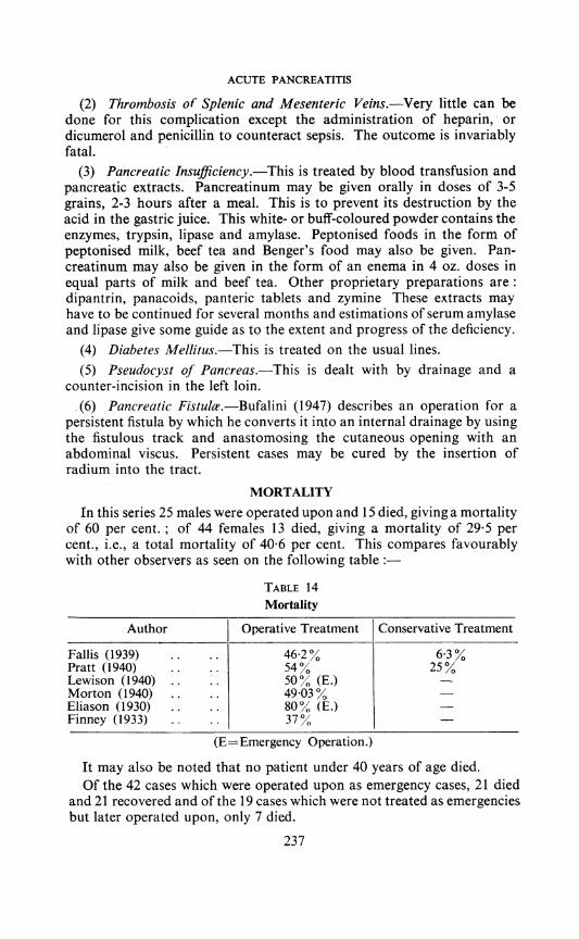
MORTALITYIn this series 25 males were operated upon and 15 died, giving a mortality
of 60 per cent. ; of 44 females 13 died, giving a mortality of 29-5 percent., i.e., a total mortality of 406 per cent. This compares favourablywith other observers as seen on the following table:
TABLE 14Mortality
Author Operative Treatment Conservative Treatment
Fallis (1939) .. .. 462% 6-3 %Pratt (1940) .. 54% 25%Lewison (1940) .. .. 50% (E.)Morton (1940) .. .. 49-03oEliason (1930) 80% (E.)Finney (1933) .. 37%
(E=Emergency Operation.)
It may also be noted that no patient under 40 years of age died.Of the 42 cases which were operated upon as emergency cases, 21 died
and 21 recovered and of the 19 cases which were not treated as emergenciesbut later operated upon, only 7 died.
237

R. A. RUSSELL TAYLOR
Of the 38 cases operated upon with symptoms of less than 24 hoursduration, 17 died and of these, 12 were subjected to a more extensiveoperation than a simple laparotomy. Twenty-one of the cases whichrecovered were submitted to the following operations
TABLE 15
Laparotomy and Laparotomy with Suprapubic Drainage .. 12 casesCholecystostomy .. .. .. 4 casesCholecystostomy and Drainage of Lesser Sac .. .. 2 casesDrainage of Lesser Sac .. .. .. .. .. 1 caseCholecystectomy .. .. .. .. .. .. .. 2 cases
Of the 27 patients whose symptoms had been present for 1-4 days,19 recovered and 8 died, but it will be noted that only one of these 8fatal cases had a simple laparotomy.
CONCLUSIONS,Etiology.-It must reluctantly be admitted that in spite of all the
present available evidence, not one of the current theories of the genesisof acute hemorrhagic pancreatitis adequately explains the mechanismof its production. As Smyth (1940) pointed out, the solution of thisproblem is more likely to be found in the experimental studies in whichthe earliest stages of the disease can be investigated rather than byexamining the materials obtained at autopsy.
Pathology.-It will be noted that the associated pathology is invariablyconfined to the biliary and gastro-intestinal tracts. Haemorrhage into thepancreas and fat necrosis is not pathognomonic of this condition.
Signs and Symptoms.-If a stoutish elderly patient of either sex with aprevious history of indigestion has an acute attack of severe upperabdominal pain, marked epigastric tenderness without muscular rigidity,upper abdominal distension, distressing flatulence, frequent vomiting, arapid pulse of extremely poor volume and tension, increased urinarydiastase, and a fall in serum calcium level; acute pancreatitis should besuspected. One should, however, be cautious in diagnosing acute pan-creatitis if the patient already has a laparotomy scar.
Treatment.-fn common with other observers, there is no doubt in myown mind that conservative treatment is definitely preferable, but it issuggested that the following are definite indications for surgical inter-vention after suitable pre-operative treatment
(1) Uncertainty in diagnosis.(2) Failure to respond to conservative treatment as shown by failing
circulation and evidence of renal damage.(3) Traumatic pancreatitis with involvement of other organs.(4) Evidence of spreading peritonitis-biliary or suppurative.(5) Deterioration of the patient's general condition which could be
attributed to necrosis or suppuration of the pancreas.(6) Jaundice.(7) Associated biliary tract disease.
238

ACUTE PANCREATITIS
(8) Retroperitoneal involvement as shown by discoloration in theloins.
(9) Distension of lesser peritoneal cavity or pseudocyst of the pancreas.(10) Pancreatic fistula-after prolonged conservative treatment has
failed.High serum amylase indicates severe pancreatic disease but this in itself
is not an indication for operation because the decision to operate must bebased on clinical grounds.
REFERENCESABELL, I. (1938) Surg. Gynec. Obstet. 66, 348.ACKERMAN, L. V. (1942) Arch. Path. 34, 6, 1065.BLAUVELT, H. (1946) Brit. J. Surg. 34, 134, 207.BUFALINI (1947) Arch. Ital. Chir. 69, 441.COLE, W. H. (1938) Int. Abstr. Surg. 67, 31.DINSMORE, R. S., and NOSIK, W. A. (1939) Surg. Clin. N. Amer. 19, 120.DITTLER, E. L., and MCGAVACK (1938) Amer. Heart J. 16, 354.EDMONSON, H. A., and BERNE, C. J. (1944) Surg. Gynec. Obstet. 72, 240.ELIASON, E. L. (1930) Surg. Gynec. Obstet. 51, 183.FALLIS, L. S. (1939) Amer. J. Surg. 46, 593.
and PLAIN, G. (1939) Amer. J. Surg. 5, 358.FINNEY, J. M. T. (1933) Ann. Surg. 98, 750.FORTY, F. (1939) Lancet 2, 370.HOWARD, J., and JONES, R. (1947) Amer. J. Med. Sci. 214, 6, 617.IRENEUS, C. (1941) Arch. Surg. 42, 126.JENSENN, K. (1946) Minnesota Med. 29, 10, 1047.KAUFMANN, N. (1927) Surg. Gynec. Obstet. 44, 15.LEWISON, E. F. (1940) Arch. Surg. 41, 4, 1008.LOEFFLER, W., and ESSELUER, A. (1946) Gastroenterologia, 71, 257.MCWHORTER, G. L. (1932) Arch. Surg. 25, 958.MORTON, J. J. (1940) New York St. J. Med. 40, 4, 255.
-_____- - and WIDGER, S. (1940) Ann. Surg. 111, 851.PAXTON, J. R., and PAYNE, J. H. (1948) Surg. Gynec. Obstet. 86, 1, 69.POPPER, et al (1948) Surg. Gynec. Obstet. 87, 79.PRATT, J. H. (1940) New Eng. J. Med. 222, 47.RICH, A. R., and DUFF, G. L. (1936) Bull. Johns Hopk. Hosp., 58, 212.SHUMACKER, H. B. (1940) Ann. Surg. 112, 117.SMEAD, L. F. (1940) J. Int. Coll. Surg. 3, 138.SMYTH, C. J. (1940) Arch. Path. 30, 651.WIGHTMAN, K. J. R. (1948) Med. Clin. N. Amer. 32, 518.
SAYINGS OF TIE GREAT
"Not gone, but dead before."-Confucius-on a recently retired anduninspired Professor. (Submitted by Douglas Robb, F.R.C.S.)
" Confidence and hope do more good than physic."-Galen.(Submitted by C. Allan Birch, M.D., F.R.C.P.)
" Everything can always be done better than it is being done."-Henry Ford. (Submitted by C. Allan Birch, M.D., F.R.C.P.)"No idea is wholly new: what is new is getting people to adopt it
and to act upon it."-Harvey Cushing. (Submitted by Professor LambertRogers.)
239