Embed Size (px)
Citation preview

Advanced Sequencing Technologies: methods and goals
Jay Shendure^, Rob Mitra*, Chris Varma^, George M. Church^+
^ Harvard Medical School, 77 Ave Louis Pasteur, Boston, MA 02115, USA.* Dept. of Genetics, Washington University School of Medicine, 4566 Scott Avenue, St. Louis, MO 63110, USA+ Corresponding author, email: http://arep.med.harvard.edu/gmc
Preface
Nearly three decades have passed since the invention of electrophoretic methods for DNA sequencing. The exponential growth in the cost-effectiveness of sequencing has been driven by automation and refinement, rather than through the invention of new methods. A variety of novel sequencing technologies are currently under development, each aspiring to drop costs to the point where the genomes of individual humans could be sequenced as part of routine health care. Here we review these technologies, and discuss the potential impact of such a “Personal Genome Project” on both the research community and society.
Introduction
In the Human Genome Project (HGP), early investments in the development of cost-effective sequencing methods contributed to its resounding success. Over the course of a decade, through the refinement, parallelization, and automation of established sequencing methods, the HGP motivated a 100-fold reduction of sequencing costs, from 10 dollars per finished base to 10 finished bases per dollar1. The relevance and utility of high-throughput sequencing and sequencing centers in the wake of the HGP has been a subject of recent debate. Nonetheless, a number of academic and commercial efforts are developing for new ultra-low-cost sequencing (ULCS) technologies that aim to reduce the cost of DNA sequencing by several orders of magnitude2. Here we discuss the motivations for ULCS and review a sampling of the technologies themselves.
Emerging ULCS technologies can be generally classified into one of four groups: (a) micro-electrophoretic methods, (b) sequencing-by-hybridization, (c) cyclic-array sequencing, and (d) single-molecule sequencing. Most of these technologies are still at relatively early stages of development, such that it is difficult to gauge the time-frame before any given method will truly be practical and living up to expectations. Yet there is an abundance of potential, and a number of recent technical breakthroughs have contributed to increased momentum and community-interest. Until recently, the motivations for pursuing ULCS technologies have generally been defined in terms of the needs and goals of the biomedical and bioagricultural research communities. This list is long, diverse, and potentially growing (Box 1). In more recent years, the primary justification for these efforts has shifted to the notion that the technology could become so affordable that sequencing the full genomes of individual patients would be justified from a health-care perspective 3-6. “Full individual genotyping” has great potential to impact health-care via contributions to clinical diagnostics and prognostics, risk assessment and disease prevention. Here we use the phrase “Personal Genome Project” (PGP) to describe this goal. As we contemplate the routine sequencing of individual human genomes we must consider the economic, social, legal and ethical issues raised by this technology. What are the potential health-care benefits? At what cost-threshold does the PGP become viable? What are the risks with respect to issues such as consent, confidentiality, discrimination, and patient psychology? In addition to reviewing technologies, we will try to address aspects of these questions.

Traditional Sequencing
In 1977, two groups familiar with peptide and RNA sequencing methods made a leap forward by harnessing the amazing single-base resolution separation power of gel electrophoresis7,8. Electrophoretic sequencing was widely adopted and rapidly improved. In 1985, a small group of scientists set the audacious goal of sequencing the entire human genome by 20051,9. The proposal met with considerable skepticism from the wider community10,11. At the time, many felt that the cost of DNA sequencing was far too high (about $10 per base) and the sequencing community too fragmented to complete such a vast undertaking. Such “large-scale biology” also represented a significant diversion of resources from the traditional question-driven approach that had been so successful in laying molecular biology’s foundations.
Five years ahead of schedule and slightly under the $3 billion budget, a useful draft sequence of the human genome was published in 2000. While the entire project costs include years of “production” using weaker technologies, the bulk of the sequencing cost was about $300 million. Amongst the factors underlying the HGP’s achievement was the rapid pace of technical and organizational innovation. Automation in the form of commercial sequencing machines, process miniaturization, optimization of biochemistry, and robust software were all crucial to the exponential “ramp-up” of sequencing throughput. Managerial and organizational challenges were successfully met at both the level of the coordination of the full HGP and within individual sequencing centers. Possibly more significant was the appearance of an “open” culture with respect to technology, data, and software1. In refreshing contrast to the competition and consequent secrecy that has traditionally characterized many scientific disciplines, the major sequencing centers freely shared technical advances and engaged in near-instantaneous data-release (i.e. the Bermuda Principles). The approach not only broadened support for the HGP, but also undoubtedly expedited its completion. With respect to both technology development and “large-scale biology” projects, the HGP perhaps provides excellent lessons for how the scientific community can proceed in future endeavors.
Why continue sequencing?
Sequencing the biosphere. Through comparative genomics, we are learning a great deal about our own molecular program, as well as those of other organisms in the biosphere12,13. There are currently 2x10¹⁰ bases in international databases14. The genomes of over 160 organisms have been fully sequenced, as well as parts of the genomes of over 100,000 taxonomic species. It is both humbling and amusing to compare that number to the full complexity of sequences on earth. By our estimate, a global biomass of over 2x10¹⁸ g contains a total biopolymer sequence on the order of 1038 residues. While sequencing the entire biosphere is obviously unnecessary and impractical, it seems clear that we have only sequenced a very small fraction of interesting and useful nucleotides.
Impact on biomedical research. A widely-available ULCS technology would improve existing biological and biomedical investigations and expedite the development of a variety of new genomic and technological studies (Box 1). Foremost amongst these goals might be efforts to determine the genetic basis of susceptibility to both common and rare human diseases. It is occasionally claimed that all we can afford (and hence all that we want) is information on "common" (i.e. > 1% in a population) single nucleotide polymorphisms, SNPs, or the arrangements of these (haplotypes) 15 in order to understand so-called multifactorial or complex diseases16. In a non-trivial sense all diseases are components of "complex diseases". As we get better at genotyping and phenotyping, we simply get better at finding the factors contributing to ever lower penetrance and expressivity. A focus on common alleles will probably be successful for alleles maintained in human populations by heterozygote advantage (such as the textbook relationship between sickle-cell anemia and malaria) but would miss most of the genetic diseases documented so far17. In any case, even for diseases that are amenable to the HAPLOTYPE MAPPING approach, ULCS would allow geneticists to move more quickly from a haplotype that is linked to a phenotype to the causative SNP(s). Diseases confounded by genetic heterogeneity
could be investigated by sequencing of specific candidate loci, or whole genomes, across populations of affected individuals18,19. It is possible that the cost of doing accurate genotyping (e.g. $5K for 500,000 SNPs95 and/or 30,000 genes) for tens of thousands of individuals will make more sense in the context of normal health care than stand-alone epidemiology. Whether SNPs or personal genomes, this will require high levels of informed consent and security20.
Another broad area that ULCS could significantly impact is cancer biology. Cancer is fundamentally a disease of the genome: cycles of somatic mutation followed by clonal expansion give rise to malignant cells. Epidemiology suggests that mutations in three to seven genes are necessary to cause malignancy, but it is also clear that different sets of genes are mutated in different cancers. Furthermore, phenomena of genomic instability such as aneuploidy or chromosomal rearrangements can play a role in cancer progression21. Although a remarkable variety of different genomic mutations and aberrations have been found in tumors, patterns are beginning to emerge. By looking for disrupted pathways rather than just individual genes, researchers are obtaining a better understanding of tumorigenesis22. The ability to sequence and compare complete genomes from a large number of normal, neoplastic, and malignant cells would allow us to exhaustively catalogue the molecular pathways and checkpoints that are mutated in cancer. Such a comprehensive approach would help us to more fully decipher the combinations of mutations that in concert give rise to cancer, and thus facilitate a deeper understanding of the cellular functions that are perturbed during tumorigenesis.
ULCS has the potential to facilitate new research paradigms. Mutagenesis in model and non-model organisms would be more powerful if one could inexpensively sequence large genomic regions or complete genomes across large panels of mutant pedigrees. In studying the diversity of the natural mutagenesis of a specific immune response, sequencing of rearranged B-cell and T-cell receptor loci in a large panel of B-cells and T-cells could be routine, rather than a major undertaking. ULCS would also benefit the emerging fields of SYNTHETIC BIOLOGY and genome engineering, both of which are becoming powerful tools for perturbing or designing complex biological systems. This would enable the rapid selection or construction of new enzymes, new genetic networks, or perhaps even new chromosomes. Even further afield than the above synthetics looms DNA computing23 and DNA as ultracompact memory. DNA computing uses only standard recombinant techniques for DNA editing, amplification, and detection but because these techniques operate on strands of DNA in parallel, the result is highly efficient and massively parallel molecular computing24. Furthermore, since a gram of dehydrated DNA contains approximately 1021 bits of information, DNA could potentially store data at a density of eleven orders of magnitude higher than today’s DVD’s24.
The Personal Genome Project. Perhaps the most compelling reason to pursue ULCS technology is the impact that it could have on human health via the sequencing of “personal genomes” as a component of individualized health-care. The current level of health-care spending for the general U.S. population is approximately $5,000 per capita per year25. Amortized over the 76-year average lifespan for which it is useful, a $1,000 genome would only have to produce $13 benefit per year to “break-even” in terms of cost-effectiveness. Straightforward ways in which “full individual genotypes” could benefit patient-care include clinical diagnostics and prognostics for both common and rare inherited conditions, risk assessment and prevention, informing pharmacogenetic contraindications, etc. Our growing understanding of how specific genotypes and their combinations impact and determine the phenome will only increase the value of personal genomes. If only even rare inherited mutations can be comprehensively surveyed for less than some threshold cost (e.g. $5000), it is likely that an autocatalytic paradigm shift could occur with each new genome/phenome fact found making the process more attractive, hence more genomes analyzed. The issue now is how might this catalysis get started?
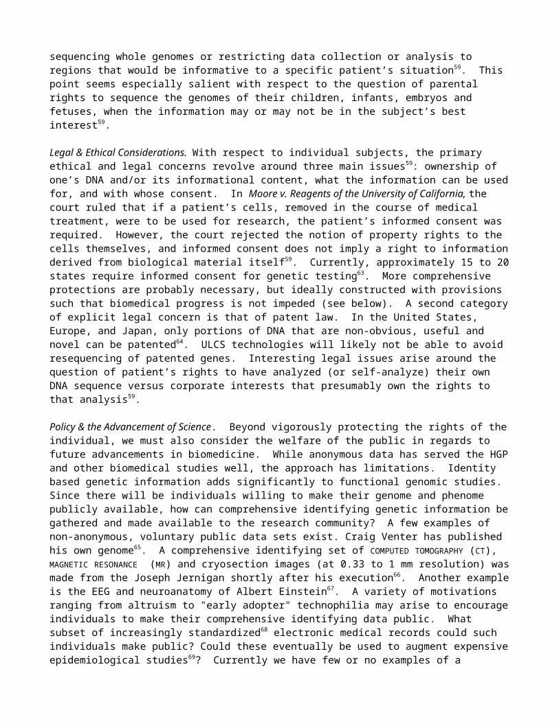
Is the PGP feasible? One reason for the overwhelming success of sequencing is that the number of nucleotides that can be sequenced at a given price has increased exponentially for the past 30 years (Figure 1). This exponential trend is by no means guaranteed and realizing a PGP in the next five years probably requires a higher commitment to technology development than was available in the

pragmatic and production-oriented HGP (Figure 1). How might this be achieved? Obviously we cannot review technologies that are secret, but a number of truly innovative approaches have now been made fully or partially public, marking this as an important time to compare and to conceptually integrate these innovative strategies. We review four major approaches below (also see Figures 2 and 3)
Emerging ULCS technologies
What are the specifications for a ULCS technology that is capable of delivering low-cost human genomes? Key considerations are (a) cost per raw base, (b) throughput per instrument, (c) accuracy per raw base, and (d) read-length per independent read. With respect to these parameters, let us consider what would be required to resequence a human genome with reasonably high accuracy at a cost of $1000. Accuracy goals will depend on the application, ranging from 21 base RNA tags26 to nearly error-free genomes (<1e-10). At a minimum, the error rate of any resequencing or genotyping method must be considerably lower than the level of variation that one is trying to detect27. As human chromosomes differ at ~1 in every 1,000 bases, an error rate of 1/100,000 bp is a reasonable goal. If individual errors are truly random with probability p, then the overall error rate for r reads is approximately:
.
If a given method can achieve ~99.7% accuracy (on par with the state-of-the-art), then 3x coverage of each base will yield the desired error rate. However, to ensure a minimum 3x coverage of >95% of bases of a diploid genome (6e9 bp) requires ~6.5x coverage, or ~40 billion raw bases. Achieving an accurate $1000 genome will thus require that costs approach ~40 million raw bases per dollar, a 4 to 5 log improvement over current methods. Although they could someday potentially approach the cost of a $2K computer, today’s integrated genomics devices typically cost $50K to $500K. Assuming that the capital and operating costs of any new instrument will be similar to that of conventional electrophoretic sequencers, the bulk of improvement will have to derive from an increase in the rate of sequence acquisition per device. In this scenario, the rate of data acquisition per device will have to increase from ~12 bases per instrument-second to ~450,000 bases per instrument-second. With respect to read-length, it is substantially advantageous to be resequencing rather than de novo sequencing a genome. No assembly is required; resequencing requires only that one can match sequencing reads to unique locations within an assembled canonical genome sequence, and then determine if and how a given sequencing read differs from its corresponding canonical sequence. In a random base model, one would expect that nearly all 20 bp reads would be unique in the genome (420 >> 3x10⁹). Probably due to repetitive elements, tandem repeats, low-complexity sequence, and the substantial fraction of recently duplicated sequence28, only ~73% of 20 bp genomic “reads” can in fact be assigned to a single unique location in the current draft of the human genome. To achieve >95% uniqueness, a modest goal, will require ~60 bp reads. There are diminishing returns with longer read-lengths; achieving >99% uniqueness will require >200 bp reads. It is also worth noting that if one is only concerned with n-mers derived from protein-coding sequences, ~88% of 20-mers and ~93% of 30-mers can be matched to a unique location in the genome.
Although this is only one scenario, alternative scenarios will require generally involve some trade-off (e.g. lower accuracy at higher throughput, or higher accuracy at lower throughput). With the above assumptions, a resequencing instrument capable of delivering a $1000 human genome of reasonable coverage and high accuracy will need to achieve >60 bp reads with 99.7% raw base accuracy, acquiring data at a rate of ~450,000 bases per second. Keeping these numbers in mind, let us review each of the technologies of interest.
Micro-electrophoretic Sequencing. The vast preponderance of DNA sequence has been obtained via the Sanger method, based on electrophoretic separation of dNTP fragments with single-base resolution. Using 384-capillary automated sequencing machines, costs for heavily optimized sequencing centers are currently approaching $1 per 1000 bp raw sequencing read and a throughput of
~12 bases per instrument-second. Typically, 99.99% accuracy can be achieved with as few as three RAW READS covering a given nucleotide. Regions of sequence that have proven difficult for Sanger sequencing can be rendered accessible via mutagenesis techniques29. A number of teams, including the Mathies group and the Whitehead BioMEMS laboratory, are currently investigating whether costs can be further reduced by additional multiplexing and miniaturization30,31. Borrowing microfabrication techniques developed by the semiconductor industry, they are working to create single devices that can perform DNA amplification, purification, and sequencing in an integrated fashion32.
The primary advantage of this approach is that the fundamental principles of the sequencing method are so well tested. Electrophoretic sequencing has already been used to successfully sequence on the order of 1011 nucleotides. Although the approaches being taken (e.g. miniaturization and process integration) will certainly yield significant cost-reductions, achieving 4 to 5 logs of improvement may require some more radical changes with respect to the underlying engineering of electrophoretic sequencers. Nevertheless, given that other ULCS methods are still far from proven, micro-electrophoretic sequencing may be a relatively safer bet, with a higher short-term probability of delivering reasonably low-cost genome resequencing (i.e. “the “$100,000 genome”).
Hybridization sequencing. There are several efforts to develop Sequencing By Hybridization (SBH) into a robust and genome-scale sequencing method. One approach is to immobilize the DNA to be sequenced on a membrane or glass chip, and then perform serial hybridizations with short probe oligonucleotides (e.g. 7-mers). The extent to which specific probes bind can be used to decode the sequence. The strategy has been applied to both genome resequencing and de novo sequencing33,34. Affymetrix and Perlegen have pioneered a different approach to SBH by hybridizing sample DNA to microfabricated arrays of immobilized oligonucleotide probes. The current maximum density of Affymetrix arrays is about one oligonucleotide "feature" per 5 micron square; each feature contains approximately 100,000 copies of a defined 25 base pair oligonucleotide. For each base pair of a reference genome to be resequenced, there are four features on the chip. The middle base pair of these four features is either an “A”,”C”,”G”, or “T”. The sequence that flanks the variable middle base is identical for all four features and matches the reference sequence. By hybridizing labeled sample DNA to the chip and determining which of the four features yields the strongest signal for each base pair in the reference sequence, a DNA sample can be rapidly resequenced. This approach to genome resequencing was first commercialized in the Affymetrix HIV chip in 199535. Miniaturization, bioinformatics, and the availability of a reference human genome sequence permitted Perlegen to greatly extend this approach and develop an oligonucleotide array for resequencing of human chromosome 2136. This technology possesses a unique set of advantages and unique challenges. The experiments impressively apply sequencing-by-hybridization (SBH) to obtain a non-trivial amount of sequence from multiple distinct chromosomes (> 109 bases). Although specific numbers on “bases per second” are not provided, the method of data-collection imaging, via scanning fluorescence of target DNA hybridized to a wafer-array of probe sequences, seems compatible with the necessary throughput. Read-length requirements are entirely avoided, as probes designed to query specific genomic bases are synthesized at defined positions. The primary challenges that SBH will face is designing probes or strategies that avoid cross-hybridization of probe to the incorrect targets due to repetitive elements or chance similarities. These factors render a substantial fraction of Chromosome 21 (30-60%) inaccessible36, and may also contribute to the 3% false-positive SNP detection rate. It is also worth noting that sequencing-by-hybridization does not escape sample preparation steps, as the relevant fraction of the genome must be PCR-amplified prior to hybridization. In the near-term, SBH may have the greatest potential as a technology to query the genotype of a focused set of genomic positions; for example, the ~10 million "common" SNPs in the human population37,38.
Cyclic array sequencing (Pyrosequencing; FISSEQ; MPSS). Key unifying features of these approaches, including multiplexing in space and time and the avoidance of bacterial clones, emerged as early as 198439. Although early methods in this class led to the first commercially sold genome40, a
dependence on electrophoresis ultimately proved limiting. Cyclic sequencing methods that have developed since have been non-electrophoretic. In both FISSEQ and Pyrosequencing, progression through the sequencing reaction is externally controlled by stepwise (i.e. cyclical), polymerase-driven addition of a single type of nucleotide triphosphate to an array of amplified, primed templates. Pyrosequencing, introduced in 1996, detects extension via the luciferase-based real-time monitoring of pyrophosphate release41,42. In FISSEQ (fluorescent in situ sequencing), extensions are detected off-line (i.e. not real-time) via fluorescent groups reversibly coupled to deoxynucleotides43. In both cases, repeated cycles of nucleotide extension are used to progressively infer the sequence of individual array features (based on patterns of extension / non-extension over the course of many cycles). We note that both FISSEQ and Pyrosequencing have previously been classified as “sequencing-by-synthesis” methods. However, as nearly all of the methods reviewed here have critical “synthesis” steps, we choose to emphasize “cycling” as the distinguishing feature of this class.
A third method in this class is based not on cycles of polymerase extension, but instead on cycles of restriction digestion and ligation. In Massively Parallel Signature Sequencing (MPSS), array features are sequenced at each cycle by employing a Type IIs restriction enzyme to cleave within a target sequence, leaving a four base-pair overhang. Sequence-specific ligation of a fluorescent linker is then used to query the identity of the overhang. The accuracy is quite high and the achievable 16 to 20 base-pair read-lengths (i.e. 4 to 5 cycles) are adequate for many purposes44.
An additional uniting feature of these methods, one that distinguishes them from several of the single-molecule projects discussed below, is that all rely on some method of isolated, i.e. clonal, amplification. After amplification, each feature to be sequenced contains thousands to millions of copies of an identical DNA molecule (thus clonal), but features must be spatially distinguishable. The amplification is necessary to achieve sufficient signal for detection. Although the method for clonal amplification is generally independent of the method for cyclic sequencing, all groups seem to have taken different (and creative) routes. In scaling up Pyrosequencing, 454 Corp. employed a PicoTiter plate to simultaneously perform hundreds of thousands of picoliter volume PCR reactions45. This was recently applied to the resequencing of the adenovirus genome, but cost and accuracy estimates for this project are not available46. For FISSEQ, clonal amplification was achieved via the polony technology, in which PCR is performed in situ within an acrylamide gel47. Because the acrylamide restricts the diffusion of the DNA, each single molecule included in the reaction produces a spatially distinct micron-scale colony of DNA (a polony) which can be independently sequenced48. For MPSS, each single molecule of DNA in a library is labeled with a unique oligonucleotide tag. After PCR amplification of the library mixture, a proprietary set of paramagnetic “capture beads” (with each bead bearing an oligonucleotide complimentary to one of the unique oligonucleotide tags) is used to separate out identical PCR products. The Vogelstein group recently developed BEAM, a fourth method for achieving clonal amplification that has great potential49.
It is worth emphasizing that in the above implementations of cyclic array sequencing, the methods developed for amplification and sequencing are potentially independent. It is therefore interesting to contemplate possibilities for mixing and matching. For example, one could imagine signature-sequencing polonies, or Pyrosequencing DNA-loaded paramagnetic beads.
The success or failure of these methods to achieve ULCS will depend on a variety of factors. Pyrosequencing is close to the required read-lengths, while FISSEQ has only been demonstrated to 5 to 8 base-pairs. Methods that rely on real-time monitoring or manufactured arrays of wells may be difficult to multiplex and miniaturize to the required scale. Crucially, both Pyrosequencing and FISSEQ-based methods must contend with discerning the lengths of homopolymeric sequences (i.e. consecutive runs of the same base). Although Pyrosequencing has made significant progress in tackling this challenge via signal quantification, the best answer may lie in development of reversible terminators (defined as a nucleotide that terminates polymerase extension, e.g. through modification of the 3’ hydroxyl group, but is designed in such a way that the termination-properties can be chemically or enzymatically reversed). Reversible terminators would also be required for any system in which all
four dNTPs (labeled with different fluorophores) could be used simultaneously. As development of reversible terminators with the necessary properties has proven to be a non-trivial problem50,51, recent progress by several groups (see below) is quite exciting.
Single molecule sequencing (cyclic-array related; nanopore). Each of the methods discussed above requires either an in vitro or in vivo amplification step, such that the DNA to be sequenced is present at sufficient copy number to achieve the required signal. A method for directly sequencing single molecules of DNA would eliminate the need for costly and often problematic procedures such as cloning and PCR amplification.
A number of groups, including Solexa, Genovoxx, the Webb group at Cornell, and the Quake group at Caltech, are developing cyclic-array methods that are related to those discussed above, but attempt to dispense with the amplification step. Each method relies on extension of a primed DNA template by a polymerase with fluorescently-labeled nucleotides, but they differ in the specifics of biochemistry and signal detection. Additionally, both Solexa and Genovoxx have invested heavily in developing reversibly-terminating nucleotides, which would solve the problem (for single-molecule methods as well as amplified cyclic-array methods) of deciphering homopolymeric sequences, by limiting each extension step to a single incorporation. In so far as their research has been revealed at public conferences, Solexa has data on reversible terminators and has shown single molecule detection with an impressive signal-to-noise ratio. The Genovoxx team has shown the possibility of using standard optics for single-molecule detection and has given details on one class of reversible terminators52. In the academic sector, the Quake group has recently demonstrated that sequence information can be obtained from single DNA molecules using serial single base extensions and the clever use of fluorescence resonance energy transfer (FRET) to improve their signal-to-noise ratio53. The Webb group has recently shown the real-time detection of nucleotide-incorporation events via a nanofabricated zero-mode wave-guide. By performing the reaction in a zero-mode waveguide, only a zeptoliter volume of the reaction is excited by the laser so that in principle, one is only detecting fluorescent triphosphates that reside in the DNA polymerases active site54.
With respect to ease and reliability of detecting extension events, cyclic-array methods that sequence amplified molecules have an obvious advantage over single-molecule methods. However, there are several advantages of the single-molecule approach. Although all polymerase-based methods still require the introduction of some flanking “common” sequence (such that a single sequencing primer can be hybridized), single-molecule methods avoid a PCR amplification step, thereby reducing costs and avoiding potential biases. All polymerase-synthesis-driven methods will likely experience both a low frequency of nucleotide misincorporation events and non-incorporation events. For amplified-molecule methods, these manifest as eventual signal decay via “dephasing” of the identical individual templates within a single feature. For single molecule methods, in contrast, there is no risk of dephasing. A misincorporation event will manifest as a “dead” template that will not extend further, while non-incorporation events will simply appear as a “pause” in the sequence.
Another advantage of single-molecule methods is that they might require less starting material than other ULCS contenders and conventional sequencing52. Relevant to all technologies, we should take note that methods for amplification of large DNAs by MULTIPLE DISPLACEMENT AMPLIFICATION (MDA) or WHOLE GENOME AMPLIFICATION (WGA) is improving rapidly96,97. This will enhance our ability to get complete sequence from single cells even when they are dead or hard to grow in culture55,56.
Cyclic array platforms operate via spatial separation of single molecules or amplified single molecules. As a consequence of this focus on single molecules, they also allow us to determine combinations of structures which are hard to disentangle in pools of molecules. For example, alternative RNA splicing contributes extensively to protein diversity and regulation but is poorly assayed by pooled RNAs on microarrays, while amplified single molecules allow accurate measures of over 1000 alternative spliceforms in RNAs like CD4457. Similarly haplotype (or diploid genotype) combinations of SNPs can be determined accurately from DNA molecules (or single cells)48.
A creative single-molecule approach that is quite unlike all of the above methods is nanopore sequencing, currently being developed by Agilent, and the Branton and Deamer groups. As DNA passes through a 1.5 nm nanopore, different base-pairs obstruct the pore to varying degrees, resulting in fluctuations in the pore’s electrical conductance. The pore conductance can be measured and used to infer the DNA sequence. The accuracies of base-calling range from 60% for single events to 99.9% for 15 events58. However, the method has thus far been limited to the terminal base-pairs of a specific type of hairpin.
Implications of sequencing human genomes
Although a thorough consideration of the ELSI implications of the PGP is available elsewhere59, we address a few additional issues here.
Clinical Pros & Cons. As discussed above, the PGP has the potential to impact patient care in a variety of ways, perhaps the most important of which is by informing diagnostics, prognostics, and risk assessment for rare and common diseases with genetic components. The extent of its usefulness will be a function of the number of genotypes that we can link to phenotypes. Causative mutations have already been discovered for hundreds of rare conditions60, and genetic risk factors have been defined for at least 10 common diseases15. ULCS technology can be expected to accelerate the rate of discovery. There are also potentially adverse consequences of having one’s genome sequenced. Most simply, it may provide more medical information about a patient than he wants to know or wants recorded in his medical record. Many patients will not want to know about late-onset diseases or genetically-influenced behavioral traits, both of which might require lifestyle changes to ameliorate59. Even if laws are passed preventing genomic information from negatively affecting insurability and employment62, such laws do not guarantee that one’s genomic information will never be misused. A debate may thus rise around the question of whether we should be sequencing whole genomes or restricting data collection or analysis to regions that would be informative to a specific patient’s situation59. This point seems especially salient with respect to the question of parental rights to sequence the genomes of their children, infants, embryos and fetuses, when the information may or may not be in the subject’s best interest59.
Legal & Ethical Considerations. With respect to individual subjects, the primary ethical and legal concerns revolve around three main issues59: ownership of one’s DNA and/or its informational content, what the information can be used for, and with whose consent. In Moore v. Reagents of the University of California, the court ruled that if a patient’s cells, removed in the course of medical treatment, were to be used for research, the patient’s informed consent was required. However, the court rejected the notion of property rights to the cells themselves, and informed consent does not imply a right to information derived from biological material itself59. Currently, approximately 15 to 20 states require informed consent for genetic testing63. More comprehensive protections are probably necessary, but ideally constructed with provisions such that biomedical progress is not impeded (see below). A second category of explicit legal concern is that of patent law. In the United States, Europe, and Japan, only portions of DNA that are non-obvious, useful and novel can be patented64. ULCS technologies will likely not be able to avoid resequencing of patented genes. Interesting legal issues arise around the question of patient’s rights to have analyzed (or self-analyze) their own DNA sequence versus corporate interests that presumably own the rights to that analysis59.
Policy & the Advancement of Science. Beyond vigorously protecting the rights of the individual, we must also consider the welfare of the public in regards to future advancements in biomedicine. While anonymous data has served the HGP and other biomedical studies well, the approach has limitations. Identity based genetic information adds significantly to functional genomic studies. Since there will be individuals willing to make their genome and phenome publicly available, how can comprehensive identifying genetic information be gathered and made available to the research community? A few examples of non-anonymous, voluntary public data sets exist. Craig Venter has published his own
genome65. A comprehensive identifying set of COMPUTED TOMOGRAPHY (CT), MAGNETIC RESONANCE (MR) and cryosection images (at 0.33 to 1 mm resolution) was made from the Joseph Jernigan shortly after his execution66. Another example is the EEG and neuroanatomy of Albert Einstein67. A variety of motivations ranging from altruism to "early adopter" technophilia may arise to encourage individuals to make their comprehensive identifying data public. What subset of increasingly standardized68 electronic medical records could such individuals make public? Could these eventually be used to augment expensive epidemiological studies69? Currently we have few or no examples of a publicly available human genome plus phenome70. A framework survey and forum for potential volunteers to discuss risks and benefits might be a crucial reality check at this point71. Will the response be tiny or will it be as resounding as the Public Library of Science, open source, and Free Software Foundation (FSF) 72?
Conclusions. Affordable, personal human genomes as a motivation for developing ULCS technology is a relatively new concept, one that is becoming viewed as possible only in the wake of the HGP. Given where the technologies stand today, and given where they need to be, we should endeavor to be conservative in making projections about when one or more of the ULCS contenders will actually deliver. At the same time, we need to recognize that there have been both a number of recent breakthroughs and broadening interest in this field. If the PGP is truly something that we want, then this seems like a good time-point to begin investing more resources in these technologies. ULCS has the potential to catalyze a revolution with respect to bringing genomics to every bedside. Simultaneously, there are clearly risks with respect to privacy and misuse of genetic information. In case the PGP does turn out to be right around the corner, we should begin thinking clearly about policy guidelines that balance patients’ interests in terms of confidentiality with patients’ interest in terms of better medicine.
FIGURES (1 – 3) and BOXES (1)
Figure 1. Exponential growth in computing & sequencing. The dark blue plot indicates the Kurzweil/Moore's Law73 for computer instructions per sec per dollar (IPS/$) doubling about every 18 months. The magenta plot indicates an exponential growth in number of base pairs of DNA sequence per unit cost (bp/$) as a function of time1. To some extent the doubling time for DNA mimics the IPS/$ curve because it is dependent on it. The yellow curve is the even steeper curve of the WWW (doubling time of 4 months) 74 which illustrates how fast a technology can explode when a sharable protocol spreads via an existing infrastructure. The light cyan plot is an "open source" case study of polony FISSEQ43 in bp/min on simple test templates (doubling time of 1 month).

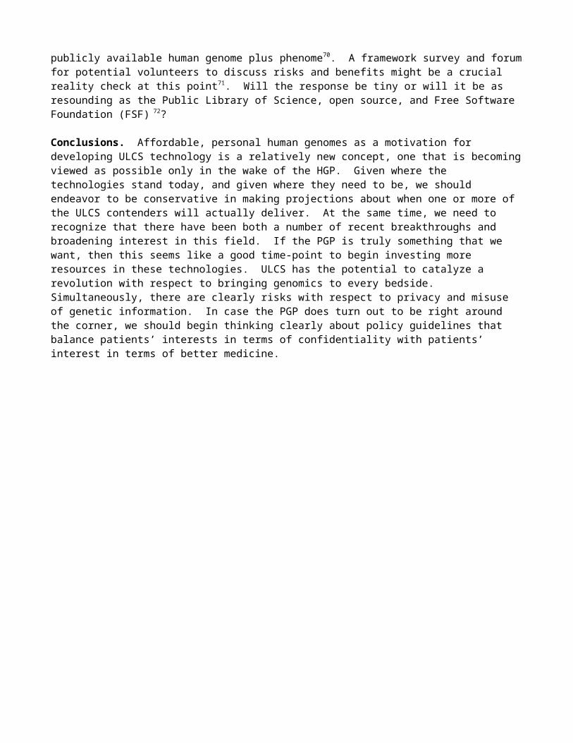
Figure 2. Examples of various potential ULCS technologies. (a) A microfabricated wafer for 384-well capillary electrophoresis sequencing. Reproduced from Emrich et al 30. (b) A single polynucleotide passing through a hemolysin nanopore can be detected as a transient blockade of the base-line ionic current. Reproduced from Winters-Hilt et al. 58 (c) Successive cycles of polymerase extension with reversibly fluorescent nucleotides can be used parallelize the sequencing of amplified single molecules. The principle is identical for single-molecule methods that employ a cyclic array strategy. Reproduced from Mitra et al 43. (d) Sequencing by hybridization. Genotyping data is obtained via differential hybridization of genomic DNA to a set of features that differ only at their “query base”. Reproduced from Cutler et al.27.
(a) (b)
(c) (d)(d)
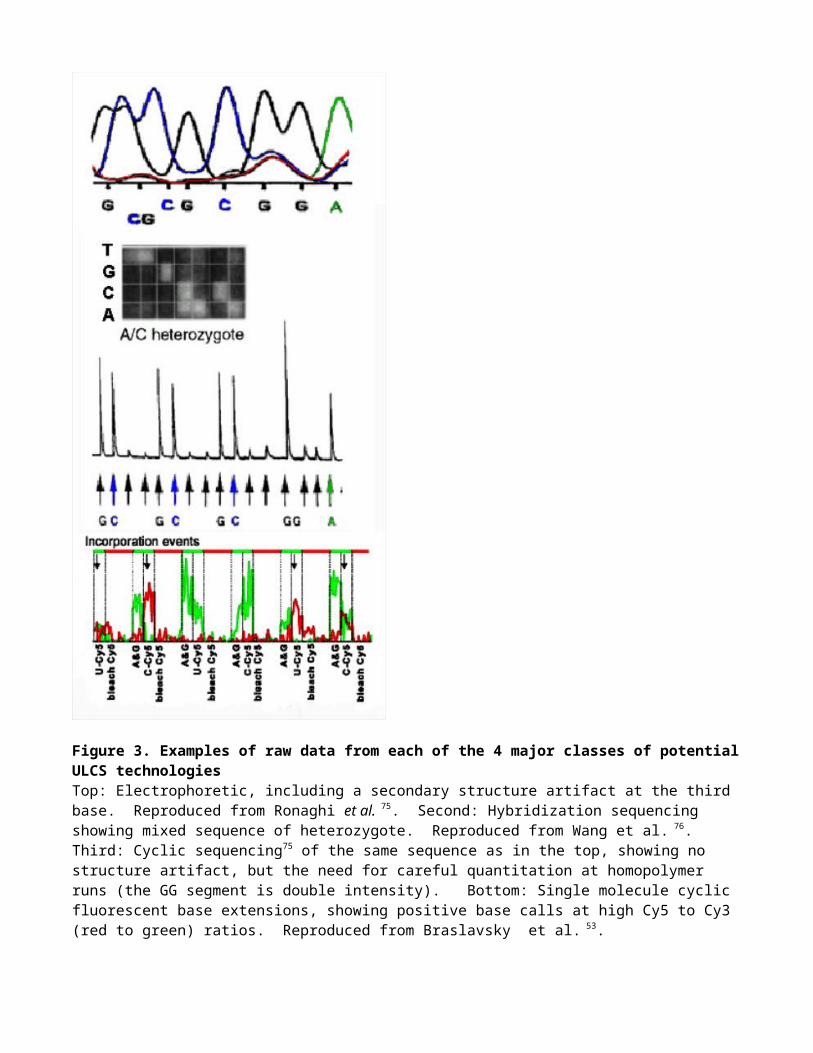
Figure 3. Examples of raw data from each of the 4 major classes of potential ULCS technologiesTop: Electrophoretic, including a secondary structure artifact at the third base. Reproduced from Ronaghi et al. 75. Second: Hybridization sequencing showing mixed sequence of heterozygote. Reproduced from Wang et al. 76. Third: Cyclic sequencing75 of the same sequence as in the top, showing no structure artifact, but the need for careful quantitation at homopolymer runs (the GG segment is double intensity). Bottom: Single molecule cyclic fluorescent base extensions, showing positive base calls at high Cy5 to Cy3 (red to green) ratios. Reproduced from Braslavsky et al. 53.
Box 1Partial List of Applications of Nucleic Acid Technology- Sequencing of individual human genomes as a component of preventative medicine4,5.- In vitro and in situ gene expression profiling across the full range of spatiotemporal variables in the
development of a multicellular organism77.- Cancer: determination of comprehensive mutation sets for individual clones78; loss of heterozygosity
analysis79; profiling sub-types for diagnosis and prognosis80,81.- Temporal profiling of B- & T-cell receptor diversity, both clinically and in lab antibody selection.- Identification of known and novel pathogens82; biowarfare sensors83.- Detailed annotation of the human genome via PHYLOGENETIC SHADOWING84.- Quantitation of alternative splice variants in transcriptomes of higher eukaryotes85,57. - Definition of epigenetic structures (e.g. chromatin modifications and methylation patterns)86.- Rapid hypothesis testing for genotype-phenotype associations.- In situ or ex vivo discovery of patterns of cell lineage87,88.- Characterization of microbial strains subjected to extensive “directed evolution” 89,90.- Exploration of microbial diversity towards agricultural, environmental, and therapeutic goals91.- Annotation of microbial genomes via selectional analysis of tagged insertional mutants92,94.- Aptamer technology for diagnostics and therapeutics93.- DNA Computing23,24.
GLOSSARY
COMPUTED TOMOGRAPHY (CT) is an imaging technology that uses computer processing of X-ray images to visualize cross-sectional (transverse) slices of internal structures, with the advantage relative to conventional radiography being the ability to eliminate superimposition.
HAPLOTYPE MAPPING utilizes combinations of "common" (e.g. >1% in a genetic population) DNA polymorphisms to find blocks of association with phenotypic traits.
BEAM is an acronym for beads emulsion, amplification, magnetic method useful for in vitro cloning of DNA molecules49.
MAGNETIC RESONANCE IMAGING is a non-invasive technique for generating multi-dimensional proton density images of internal organs, structures, and lesions.
RAW READS is the actual nucleotide sequence that is generated by a sequencing instrument, as opposed to finished sequence, which is the product of reducing sequencing errors by obtaining the consensus sequence of multiple, overlapping raw reads that provide information on a given base-pair.
SYNTHETIC BIOLOGY embraces the emerging capabilities to design, synthesize, and evolve novel genomes or biomimetic systems.
MULTIPLE DISPLACEMENT AMPLIFICATION is a technique for achieving whole genome amplification that utilizes a strand-displacing polymerase to catalyze isothermal amplification of DNA.
WHOLE GENOME AMPLIFICATION is defined by the in vitro amplification of a full genome sequence, ideally with even representation of the genome in the amplified product. Techniques for achieving WGA include PCR primed with random or degenerate oligonucleotides, or multiple displacement amplification.
PHYLOGENETIC SHADOWING is a technique related to phylogenetic footprinting, in which a genome is annotated via multiple comparisons to the genomes of several closely related species.
References
1. Collins, F. S., Morgan, M. & Patrinos, A. The human genome project: lessons from large-scale biology. Science 300, 286-290 (2003).
2. http://grants1.nih.gov/grants/guide/rfa-files/RFA-HG-04-003.html http://grants.nih.gov/grants/guide/rfa-files/RFA-HG-04-002.html
3. Joneitz, E. Personal Genomes. Technology Review (2001).4. Pray, L. A cheap personal genome? The Scientist Oct 2002.5. Pennisi, E. Gene Researchers Hunt Bargains, Fixer-Uppers. Science 29, 735-736 (2002).6. Salisbury, M. W. Fourteen Sequencing Innovations that could change the way you work. Genome
Technology (2003).7. Gilbert, W. DNA sequencing and gene structure. Science. 214, 1305-1312 (1981).8. Sanger, F. Sequences, sequences, and sequences. Annu Rev Biochem 5, 1-28 (1988).9. Cook-Deegan, R. M. The Alta summit, December 1984. Genomics 5, 661-663 (1989).10. Leder P. Can the human genome project be saved from its critics ... and itself? Cell 63,1-3 (1990)11. Davis B. D. The human genome and other initiatives. Science 249, 342-343 (1990).12. Carroll, S. B. Genetics and the making of Homo sapiens. Nature 422, 840-857 (2003).13. Ureta-Vidal, A., Ettwiller, L., Birney, E. Comparative genomics: genome-wide analysis in metazoan
eukaryotes. Nat Rev Genet 4, 251-62 (2003).14.http://www.ncbi.nlm.nih.gov/Taxonomy/txstat.cgi?uncultured=hide&unspecified=hide&m=0
http://www.ncbi.nlm.nih.gov/entrez/query.fcgi?db=Nucleotide http://wit.integratedgenomics.com/GOLD/,
15. Gibbs, R.A., et al. The International HapMap Project. Nature. 426, 789-796 (2003).16. Holtzman N.A. & Marteau T.M. Will genetics revolutionize medicine? N Engl J Med 343, 141-144
(2000).17. Vitkup, D., Sander, C. & Church, G.M. The amino-acid mutational spectrum of human genetic
disease. Genome Biol 4, R72 (2003).18. Farooqi I.S. et al. Clinical spectrum of obesity and mutations in the melanocortin 4 receptor gene. N
Engl J Med 348, 1085-1095 (2003)19. Smirnova, I. et al. Assay of locus-specific genetic load implicates rare Toll-like receptor 4 mutations
in meningococcal susceptibility. Proc Natl Acad Sci U S A 100, 6075-6080 (2003).20. Merz J. F., McGee G. E. & Sankar P. "Iceland Inc."?: On the ethics of commercial population
genomics. Soc Sci Med 58, 1201-1209 (2004).21. Rajagopalan, H., Nowak, M. A., Vogelstein, B., Lengauer, C. The significance of unstable
chromosomes in colorectal cancer. Nat Rev Cancer 3, 695-701 (2003).22. Hanahan, D. & Weinberg, R. A. The Hallmarks of Cancer. Cell 10, 51-70 (2000).23. Braich, R. S., Chelyapov, N., Johnson, C., Rothemund, P. W. & Adleman, L. Solution of a 20-
variable 3-SAT problem on a DNA computer. Science 296, 499-450 (2002).24. Reif, J.H. Computing: Successes and challenges. Science 296, 478-479 (2002).25. http://www.oecd.org/dataoecd/1/33/2957315.xls26. Saha S., et al. Using the transcriptome to annotate the genome. Nat Biotechnol 20, 508-512 (2002).27. Cutler, D. J., et al. High-throughput variation detection and genotyping using microarrays. Genome
Res 11, 1913-1925 (2001).28. Bailey, J. A., et al. Recent Segmental Duplications in the Human Genome. Science 297, 1003-
1007 (2002).29. Keith, J. M. et al. Unlocking hidden genomic sequence. Nucleic Acids Res 32, e35.30. Emrich, C. A., Tian, H., Medintz, I. L. & Mathies, R. A. Microfabricated 384-lane capillary array
electrophoresis bioanalyzer for ultrahigh-throughput genetic analysis. Anal Chem. 74, 5076-5083 (2002).
31. Koutny, L. et al. Eight hundred-base sequencing in a microfabricated electrophoretic device. Anal Chem 72, 3388-3391 (2000)
32. Paegel, B. M., Blazej, R. G., Mathies, R. A. Microfluidic devices for DNA sequencing: sample preparation and electrophoretic analysis. Curr Opin Biotechnol 14, 42-50 (2003).
33. Drmanac, S. Accurate sequencing by hybridization for DNA diagnostics and individual genomics. Nat Biotechnol 16, 54-58 (1998).
34. Drmanac, R., et al. DNA sequencing by hybridization with arrays of samples or probes. Methods Mol Biol 170, 173-179 (2001).
35. Lipshutz, R. J., et al. Using oligonucleotide probe arrays to access genetic diversity. Biotechniques 19, 442-7 (1995).
36. Patil, N., et al. Blocks of limited haplotype diversity revealed by high-resolution scanning of human chromosome 21. Science 294, 1719-1723 (2001).
37. Kruglyak, L. & Nickerson, D.A. Variation is the spice of life. Nature Genet 27, 234-236 (2001)38. Reich, D. E., Gabriel, S. B., Althshuler, D. Quality and completeness of SNP databases. Nature
Genet 33, 457-458 (2003).39. Church, G. M. & Gilbert, W. Genomic sequencing. Proc Natl Acad Sci U S A 81, 1991-1995 (1984).40. Nowak, R. Getting the bugs worked out. Science 267, 172-4 (1995).41. Ronaghi M., Karamohamed S., Pettersson B., Uhlen M., & Nyren P. Real-time DNA sequencing
using detection of pyrophosphate release. Anal Biochem 242, 84-89 (1996).42. Ronaghi, M., Uhlen M. & Nyren, P. A sequencing method based on real-time pyrophosphate.
Science 281, 363-365 (1998)43. Mitra, R.D., Shendure, J., Olejnik, J., Olejnik, E. K., and Church, G.M. Fluorescent in situ
Sequencing on Polymerase Colonies. Analyt Biochem 320, 55-65 (2003).44. Brenner, S., et al. Gene expression analysis by massively parallel signature sequencing (MPSS) on
microbead arrays. Nat Biotechnol. 18, 630-634 (2000).45. Leamon J. H., et al. A massively parallel PicoTiterPlate based platform for discrete picoliter-scale
polymerase chain reactions. Electrophoresis 24, 3769-2777 (2003). 46. Sarkis, G. et al. Sequence Analysis of the pAdEasy-1 Recombinant Adenoviral Construct. Using
the 454 Life Sciences Sequencing-by-Synthesis Method. NCBI AY370911 gi:34014919 (2003).
47. Mitra, R. D. & Church, G. M. In situ localized amplification and contact replication of many individual DNA molecules. Nucleic Acids Res 27:e34, 1-6 (1999).
48. Mitra, R. D., et al. Digital Genotyping and Haplotyping with Polymerase Colonies. Proc Natl Acad Sci 100, 5926-5931 (2003).
49. Dressman, D., Yan, H., Traverso, G., Kinzler, K. W. & Vogelstein, B. Transforming single DNA molecules into fluorescent magnetic particles for detection and enumeration of genetic variations. Proc Natl Acad Sci U S A. ??, 8817-8822 (2003).
50. Metzker M. L., Raghavachari R., Richards S., Jacutin S. E., Civitello A., Burgess K. & Gibbs R.A. Termination of DNA synthesis by novel 3'-modified-deoxyribonucleoside 5'-triphosphates. Nucleic Acids Res 22, 4259-67 (1994)
51. Welch, M. & Burgess, K. Synthesis of fluorescent, photolabile 3'-O-protected nucleoside triphosphates for the base addition sequencing scheme. Nucleosides & Nucleotides 18: 197-199 (1999).
52. Fagin, U., Hennig, C. & Cherkasov, D. Massive parallel single molecule sequencing with reversibly terminating nucleotides. [submitted] (2004).
53. Braslavsky, I., Hebert, B., Kartalov, E. & Quake, S. R. Sequence information can be obtained from single DNA molecules. Proc Natl Acad Sci U S A. 100, 3960-3964 (2003).
54. Levene, M. J., et al. Zero-mode waveguides for single-molecule analysis at high concentrations. Science 299, 682-686 (2003).
55. Sorensen, K. J., Turteltaub, K., Vrankovich, G., Williams, J., Christian, A. T. Whole-genome amplification of DNA from residual cells left by incidental contact. Anal Biochem 324, 312-314 (2004).
56. Rook M. S., Delach S. M., Deyneko G., Worlock A. & Wolfe J. L. Whole Genome Amplification of DNA from Laser Capture-Microdissected Tissue for High-Throughput Single Nucleotide Polymorphism and Short Tandem Repeat Genotyping. Am J Pathol 164, 23-33 (2003).
57. Zhu, J., Shendure, J., Mitra, R. D., & Church, G.M. Single Molecule Profiling of Alternative Pre-mRNA Splicing. Science 301, 836-838 (2003).
58. Winters-Hilt S., et al. Accurate classification of basepairs on termini of single DNA molecules. Biophys J 84, 967-976 (2003).
59. Robertson, J.A. The $1000 genome: ethical and legal issues in whole genome sequencing of individuals. The American Journal of Bioethics 3(3):InFocus (2003).
60. http://archive.uwcm.ac.uk/uwcm/mg/docs/hahaha.html62. Oak Ridge National Laboratory. Human Genome Project Information: Genetics Privacy and
Legislation. http://www.ornl.gov/TechResources/Human_Genome/elsi/legislat.html63. Gostin, L.O., Hodge, J.G., Calvo, C. Genetics Policy & Law: A report for policymakers. Washington,
D.C.: National Council of State Legislators (2001).64. Biotechnological Process Patent Act. Pub. L. No. 104-41, 104th Cong., lst Sess. (Nov. 1, 1995)65. Venter, J.C. A part of the human genome sequence. Science 299, 1183-1184 (2003).66. National Library of Medicine. The Visible Human Project.
http://www.nlm.nih.gov/research/visible/visible_human.html67. Witelson, S. F., Kigar, D. L. & Harvey, T. Lancet 19, 2149-2153 (1999).68. http://www.whitehouse.gov/omb/egov/press/chi_march.htm69. http://www.nurseshealthstudy.org/70. Freimer, N. & Sabatti,C. The Human Phenome Project. Nat. Gen 3, 15-21 (2003).71. http://arep.med.harvard.edu/PGP/pub.html72. http://www.plos.org/cgi-bin/plosSigned.pl, http://sourceforge.net/, http://www.gnu.org/73. http://www.kurzweilai.net/meme/frame.html?main=/articles/art0184.html74. http://www.mit.edu/people/mkgray/net/web-growth-summary.html75. Ronaghi, M., Nygren, M., Lundeberg, J. & Nyren, P. Analyses of secondary structures in DNA by
pyrosequencing. Anal Biochem 267, 65-71 (1999).76. Wang D.G. et al. Large-scale identification, mapping, and genotyping of single-nucleotide
polymorphisms in the human genome. Science 280, 1077-1082 (1998)77. Reymond, A., et al. Human chromosome 21 gene expression atlas in the mouse. Nature 420, 582-
586 (2002).78. Hahn, W. C. & Weinberg, R. A. Mechanisms of Disease: Rules for Making Human Tumor Cells. N
Engl J Med 34, 1593-1603 (2002).79. Paulson, T. G., Galipeau, P. C., Reid, B. J. Loss of heterozygosity analysis using whole genome
amplification, cell sorting, and fluorescence-based PCR. Genome Res 9, 482-491 (1999).
80. Golub, T.R., et al. Molecular classification of cancer: class discovery and class prediction by gene expression monitoring. Science 286, 531-537 (1999).
81. Ramaswamy, S., et al. A molecular signature of metastasis in primary solid tumors. Nat Genet 33, 49-54 (2003).
82. Weber, G., Shendure, J., Tanenbaum, D. M., Church, G. M., & Meyerson M. Microbial sequence identification by computational subtraction of the human transcriptome. Nature Genet 30, 141-142 (2002).
83. Stenger, D. A., Andreadis, J. D., Vora, G. J. & Pancrazio, J. J. Potential applications of DNA microarrays in biodefense-related diagnostics. Curr Opin Biotechnol 13, 208-212 (2002).
84. Boffelli, D., et al. Phylogenetic shadowing of primate sequences to find functional regions of the human genome. Science. 299, 1391-1394 (2003).
85. Roberts, G. C. & Smith, C. W. Alternative splicing: combinatorial output from the genome. Curr Opin Chem Biol 6, 375-383 (2002).
86. Robyr, D., et al. Microarray deacetylation maps determine genome-wide functions for yeast histone deacetylases. Cell 109, 437-446 (2002).
87. Yatabe, Y., Tavare, S. & Shibata, D. Investigating stem cells in human colon by using methylation patterns. Proc Natl Acad Sci USA 9, 10839-10844 (2001).
88. Dymecki, S. M., Rodriguez, C. I. & Awatramani, R. B. Switching on lineage tracers using site-specific recombination. Methods Mol Biol 18, 309-334 (2002).

89. Lenski, R. E., Winkworth, C. L. & Riley, M. A. Rates of DNA Sequence Evolution in Experimental Populations of Escherichia coli During 20,000 Generations. J Mol Evol 56, 498-508 (2003).
90. Cooper, T. F., Rozen, D. E. & Lenski, R. E. Parallel changes in gene expression after 20,000 generations of evolution in Escherichia coli. Proc Natl Acad Sci U S A 100, 1072-1077 (2003).
91. Gillespie, D. E., et al. Isolation of antibiotics turbomycin a and B from a metagenomic library of soil microbial DNA. Appl Environ Microbiol 68, 4301-4306 (2002).
92. Badarinarayana, V., et al. Selection analyses of insertional mutants using subgenic-resolution arrays. Nature Biotechnology 1, 1060-1065 (2001).
93. Cerchia, L., Hamm, J., Libri, D., Tavitian, B., & de Franciscis, V. Nucleic acid aptamers in cancer medicine. FEBS Lett 528, 12-16 (2002).
94. Sassetti CM, Boyd DH, Rubin EJ. Genes required for mycobacterial growth defined by high density mutagenesis. Mol Microbiol. 48, 77-84 (2003).
95. Kruglyak L. Prospects for whole-genome linkage disequilibrium mapping of common disease genes. Nat Genet 22, 139-144 (1999).
96. Dean, F. B., et al. Comprehensive human genome amplification using multiple displacement amplification. Proc Natl Acad Sci U S A. 99, 5261-5266 (2002).
97. Nelson, J. R., et al. TempliPhi, phi29 DNA polymerase based rolling circle amplification of templates for DNA sequencing. Biotechniques suppl, 44-47 (2002)
AcknowledgementsThe authors thank members of the polony community and Christian Hennig for sharing unpublished work and Ting Wu for helpful discussions.
Competing interests statement. The authors declare that they have potential competing financial interests based on advisory roles and/or patent royalties from Affymetrix, Agencourt, Agilent, Caliper, Genovoxx, Helicos, Lynx and Pyrosequencing.