Embed Size (px)
Citation preview

7/28/2019 TCRM-2008-4(5)-1960_Neubert
http://slidepdf.com/reader/full/tcrm-2008-45-1960neubert 1/12
© 2008 Dove Medical Press Limited. All rights reserved
Therapeutics and Clinical Risk Management 2008:4(5) 1085–1095 1085
R E V I E W
Bowen’s disease – a review of newer treatmentoptions
Thorsten Neubert
Percy Lehmann
Zentrum für Dermatologie,Allergologie und Umweltmedizin,Helios Klini kum Wuppertal, Klinikumder Universität Witten-Herdecke,Wuppertal, Germany
Correspondence: Thorsten NeubertZentrum für Dermatologie,Allergologie und Umweltmedizin,Helios Klinikum Wuppertal, Klinikumder Universität Witten-Herdecke,Heusnerstr. 40, D-42283Wuppertal,GermanyTel +49 202 896 3507Email [email protected]
Abstract: Bowen’s disease (squamous cell carcinoma in situ) has a 3%–5% risk to develop
into invasive squamous cell carcinoma. Non-melanoma skin cancer is the most common cancer
among Caucasians and its incidence has increased during the last decades dramatically. Multiple
treatment options for Bowen’s disease have been described and are established with advantages
and disadvantages. Bowen’s disease occurs more often in elderly patients (with a higher risk
of comorbidities) and is frequently located on body sites with poor wound healing. Therefore
there is need for non-invasive/non-destructive but effective treatment options.
We would like to give an overview of established therapies and more detailed information
about the newer treatment options for Bowen’s disease with topical diclofenac, topical imiquimod
and photodynamic therapy.
Keywords: Bowen’s disease, photodynamic therapy, imiquimod, diclofenac
IntroductionBowen’s disease (BD) is an in situ squamous cell carcinoma (SCC) which was first
described in 1912 by JT Bowen.
Clinically a typical BD is a slowly enlarging erythematous patch or plaque which is
well demarcated and has a scaling or crusted surface. In some cases it can be pigmented
or verrucous. It is commonly located on the lower limbs and on the head and neck. But
BD is also seen subungual or periungual, palmar, genital and perianal. Usually BD
is a solitary lesion, but in 10% to 20% it occurs at multiple sites (Thestrup-Pedersen
et al 1988; Cox et al 2007). The risk of progression into an invasive carcinoma is 3%
to 5% in extragenital lesions and about 10% in genital lesions (Kao 1986; Cox et al
1999). BD is very common in the Caucasian population with an incidence of 1.42 per
1000 in some populations (Reizner et al 1994).
Several etiological factors of BD have been reported, such as irradiation (ultraviolet
irradiation, radiotherapy, photochemotherapy), carcinogens (eg, arsenic), immunosuppression
(eg, after organ transplantation, AIDS), viral (strong association of perianal and genital
lesions with HPV; 47% of acral and 24% of nonacral extragenital BD contain HPV genome)
and some others like chronic injury or dermatoses (Clavel et al 1999; Cox et al 2007).
Treatment options for Bowen’s diseaseEfficacy comparison and evaluation of different treatment options and treatment studies
of Bowen’s disease are difficult because there is a variety of different protocols and the
success of a treatment modality is dependent on several factors (eg, body site, lesion
size and thickness, different equipment).
The choice of treatment should be guided by efficacy, location and size of BD,
number of lesions, availability of the therapy, the clinician’s expertise, patient factors
(age, immune status, concomitant medication, comorbidities and compliance), cosmetic
outcome and the patient’s preference.
7/28/2019 TCRM-2008-4(5)-1960_Neubert
http://slidepdf.com/reader/full/tcrm-2008-45-1960neubert 2/12
Therapeutics and Clinical Risk Management 2008:4(5)1086
Neubert and Lehmann
The different treatment options for BD are cryotherapy,
curettage with cautery, excision, 5-fluorouracil (5-FU),
radiotherapy, laser, photodynamic therapy (PDT), imiqui-
mod and some other therapies that were described in some
case reports or small numbers of patients. Up to now none
of the treatment options has been unequivocally proven to
be superior to any other.
This paper is focused on the newer treatment options for
BD: topical diclofenac and imiquimod and photodynamic
therapy.
CryotherapyThe clearance and recurrence rates of cryotherapy vary
between different studies. The reasons for this variety of
results are different techniques and regimens (especially
freezing time and number of freeze-thaw cycles (FTC)),
lesion size and location of BD. In a retrospective study a
clinical clearance of 61% was achieved (Bell and Rhodes
1999). Excellent results in combination with low recurrence
rates were described for 30s freeze-thaw cycle liquid
nitrogen cryotherapy (Plaza de Lanza et al 1980; Holt
1988). Lower recurrence rates were achieved by longer and
repeated freezing cycles whereby problems like prolonged
wound healing and poor cosmetic outcome (scaring,
hypopigmentation) increased (Plaza de Lanza et al 1980; Holt
1988; Cox and Dyson 1995; Ball and Dawber 1998).
In comparative studies cryotherapy showed clearance
rates from 50% to 100% by one to three 20s freeze-thaw
cycles and recurrence rates from 10% (one 20s FTC, fol-
low up 12 months) up to 36% (two 5 to 10s FTC, follow
up 24 months) (Morton et al 1996; Ahmed et al 2000).
Wound healing was faster with cryotherapy compared to
radiotherapy, but slower than curettage and cautery regimen
(Cox and Dyson 1995; Ahmed et al 2000). In comparison
with PDT after cryotherapy ulceration was seen in 25% of
the treated lesions (no ulceration in the PDT group) and the
clearance rate of a single PDT was significantly higher than
one 20s FTC (Morton et al 1996).
In conclusion cryotherapy with liquid nitrogen is an
effective, commonly used option in the treatment of BD
entailing only low costs. Especially for single and small
BD located in well healing sites cryotherapy is a favorable
treatment option.
Curettage with cauteryThe reported cure rates and recurrence rates of this method
differ due to different regimes, equipment used and skill of the
clinician. Cure rates ranged from 81% for curettage up to 93%
to 98% for curettage and cautery with a follow up of 2.5 to
4 years (Honeycutt and Jansen 1973; Thestrup-Pedersen
et al 1988; Morton al 1996). In a comparative study with a
follow up of 2 years there was a recurrence in the curettage
and cautery group in 4 of 44 lesions and in the cryotherapy
group (two 5 to 10 s FTC) in 13 of 36 lesions (Ahmed et al
2000). In this study curettage and cautery was associated
with a better and faster wound healing, less discomfort and
pain and a lower rate of complications. Additional anecdotal
reports described a therapy regime that combined curettage
and cryotherapy (Nordin 1999).
In summary curettage and cautery is a safe and effective
therapy of BD with a very good cost-benefit analysis. It is
suitable especially for small single BD.
ExcisionUp to now no randomized, comparative studies have been
published for surgery of BD. In 2 retrospective studies
recurrence rates ranged from 4.6% up to 19% (Graham and
Helwig 1961; Thestrup-Pedersen et al 1988). Higher rates
were reported for perianal BD. Mohs micrographic surgery
is especially performed in sites of the body where tissue-
sparing surgery is necessary (eg, fingers and nail unit, penis).
The recurrence rates differed from 6.3% up to 21%–28%
(follow up 1 to 5 years) due to different body sites, viral
etiology, large wound defects and lesion size. Perianal BD
showed in a retrospective study of 47 cases a recurrence
rate for wide excision of 23%, for local excision 53% and
for laser therapy 80% (treatment with radiotherapy was not
included)(Marchesa et al 1997). The most frequently used
therapy for perianal BD is wide excision and the surgical
treatment has been advocated as the treatment of choice
(Cleary et al 2000; Cox et al 2007).
In conclusion surgical excision of BD is one of the
standard treatments especially for small and single, digital
and perianal BD. The main advantage is the securing of
histological free excision margins. Possible limitations are
cosmetic and functional outcome and prolonged or compli-
cated wound healing in some body areas.
FluorouracilFor the treatment with topical 5-FU the reported clearance
and cure rates showed a wide range due to the use of different
therapy regimens and concentrations. The commercially
available and in most studies used concentration is a 5%
5-FU preparation. Once or twice daily application for vari-
able periods of time (1 week to 2 months or even longer)
is described, with preference for the twice daily usage and
7/28/2019 TCRM-2008-4(5)-1960_Neubert
http://slidepdf.com/reader/full/tcrm-2008-45-1960neubert 3/12
Therapeutics and Clinical Risk Management 2008:4(5) 1087
Bowen’s disease treatment options
longer treatment periods (8 weeks) with clearance rates up
to 92% (Bargmann and Hochmann 2003).
Pulse therapies with 5-FU have been reported with a
limited evidence base. A randomized trial for the treatment of
BD with either PDT or 5-FU (1 or if required 2 cycles of once
daily application for 1 week and twice daily for week 2–4)
reported an initial clinical clearance for PDT in 88% and for
5-FU in 67%, with a clearance rate at follow up (12 months)
of 82% (PDT) and 48% (5-FU) (Salim et al 2003).
Increasing effectiveness has been reported when 5-FU was
combined with other therapeutic modalities like occlusive
application, iontophoresis, pretreatment with erbium:YAG
laser, cryotherapy, imiquimod and acitretin with response
rates up to 96,2% (Heising 1979; Welch et al 1997; Khandpur
and Sharma 2003; Wang et al 2004). The advantage of 5-FU
cream is that it can be easily applied by the patients them-
selves accompanied by frequent clinical controls. The therapy
with 5-FU is limited by local inflammation with erosion and
ulceration which may last for several weeks.
In summary the commercially available 5% 5-FU prepa-
ration has shown its efficacy in short- and long-term studies
and can be used for the treatment of BD in good or even bad
healing sites and for special sites like fingers or penis.
RadiotherapyDifferent radiotherapy techniques (external beam
radiotherapy, Grenz rays, radioactive skin patches) have
been used in the treatment of BD with reported cure rates
between 94% and 100% (Stevens et al 1977; Lee et al
1997; Turpin 1999; Chung et al 2000; Lukas VanderSpek
et al 2005). For BD located at poor healing sites (especially
lower leg) radiotherapy should be avoided due to poor
healing/failure-to-heal rates in 20% to 25 % of treated lesions
(Cox and Dyson 1995; Dupree et al 2001). Severe toxicity
of radiotherapy (eg, cartilage/bone necrosis) was reported
for hypofractionated regimens after treatment of BD of the
extremities (Lukas VanderSpek et al 2005).
To avoid some of the disadvantages and adverse effects
of conventional radiotherapy skin patches equipped with
the β-emitter holmium-166 were designed for the treatment
of BD and other skin cancers (Lee et al 1997; Turpin 1999;
Chung et al 2000). In 29 biopsy-confirmed BD histological
clearance was reported at 5 month without any recurrence after
10–24 month (Chung et al 2000). In this study the functional
and cosmetic outcome was good with no serious adverse effects
(only desquamation, erythema and erosion were seen).
For anal/perianal BD radiotherapy has been reported as
a reasonable treatment of choice particularly when other
therapies (eg, surgery) are difficult to carry out (Cox et al
1999; Papillon and Chassard 1992; Lukas VanderSpek et al
2005. But there is no strong evidence supporting this point
of view.
In conclusion radiotherapy should not be used for poor
healing sites (especially not for the lower leg) but it can be an
alternative with high efficacy when other treatment options
(eg, surgery) are difficult or not possible (eg, very old patients
with comorbidities and medication with anticoagulants,
special sites like perianal or penile BD).
Laser So far no randomized controlled trials to evaluate laser
therapy in BD have been reported. The published data result
from case reports and small series. Argon, CO2and Nd:YAG
laser have been used for the treatment of BD.
Complete response of BD of the digits by CO2
laser
with no recurrence in the 0.5 to 7.7 years follow up was
published in some studies with good functional and cosmetic
outcome (Gordon et al 1994; Gordon et al 1996; Tantikun
2000), although others reported some failures (1 of 5 digi-
tal lesions) (Gordon et al 1994) or progression to invasive
SCC of lower leg lesions after 100% healing at 2 months
and complete response at 6 months (3/16 patients) (Dave
et al 2003). Genital lesions showed complete response after
treatment with argon, CO2
and Nd:YAG laser (Landthaler
et al 1986) with more recent data that reported a recurrence
rate of 26% (van Bezooijen et al 2001). For perianal BD
the results of laser therapy were poor (Cleary et al 2000),
although a case report showed a clearance and no recurrence
within 14 month after treatment with argon laser (Boynton
and Bjorkman 1991).
In summary especially the CO2laser can be used for penile
or digital BD with only limited data of recurrence rates.
Other therapiesAs some of the most standard therapies for BD mentioned
above are not evaluated by controlled randomized trials,
data of other less commonly used therapies result from case
reports or only small numbers of patients.
Local hyperthermia was tried by the use of chemical
pocket warmers applied on the lesions under pressure with
initial complete clinical clearance in six of eight patients
but with histological clearance in only three of eight cases
(Hiruma and Kawada 2000).
In 20 BD lesions (large and lower leg lesions included)
an ultrasonic surgical aspirator was used with no recurrence
during the follow up (12 to 26 months, mean 20 months)
7/28/2019 TCRM-2008-4(5)-1960_Neubert
http://slidepdf.com/reader/full/tcrm-2008-45-1960neubert 4/12
Therapeutics and Clinical Risk Management 2008:4(5)1088
Neubert and Lehmann
(Otani et al 2001). After 10 months acitretin therapy in one of
the two treated patients 90% of the multiple lesions cleared.
The other patient did not tolerate the acitretin and no improve-
ment was seen (Yerebakan et al 2002). In other reports for
example, the use of topical (in combination with liquid
nitrogen) and intralesional bleomycin (Dyall-Smith 1998;
Ota et al 2002) and oral isotretinoin in combination with
subcutaneous interferon-α (1 patient treated for 3 months for
multiple BD, no recurrence 15 month after therapy) (Gordon
et al 1997) was reported.
Newer treatment options
DiclofenacDiclofenac inhibits cyclooxygenase enzymes and thereby
downstream byproducts of arachidonic acid (AA)
metabolism are decreased. In the promotion of epithelial
tumor growth these metabolites of AA have shown to play
an essential role by several mechanisms and pathways
(eg, inhibition of apoptosis and immune surveillance,
stimulation of angiogenesis, etc) (Gately 2000; Masferrer
et al 2000).
Diclofenac 3% gel has been successfully used in the
treatment of actinic keratosis (Wolf et al 2001; Smith et al
2006). Two cases of BD were treated with 3% diclofenac
gel twice daily for 80 to 90 days with no residual disease
clinically and histologically (Dawe et al 2005).
In another series of 5 patients the biopsy proven BD
were treated once daily with diclofenac 3% gel for 8 weeks.
The treatment was well tolerated with mild inflammation
after 6 weeks and mild side effects like itching and dryness.
Complete clinical and histological clearance was proven by
biopsies taken 4 weeks after end of treatment (Pantel and
Stockfleth 2007).
However these promising data have to be proven in
randomized controlled trials and optimum regimens (eg,
once vs twice daily application, duration of application)
and recurrence rates in long-term follow-up have to be
investigated.
ImiquimodImiquimod 5% cream is a topical immune response modifier
with approval in most pharmaceutical markets for the treat-
ment of anogenital warts, actinic keratoses and superficial
basal cell carcinoma.
Imiquimod is heterocyclic imidazoquinoline amid with
antiviral and antitumor effects. Imiquimod itself does not
have antiviral or antiproliferative abilities, its efficacy is
ascribed to the stimulation of innate and acquired immunity.
Binding of imiquimod to toll-like receptor(TLR)-7 and -8
induces production of cytokines like interferon (IFN) alfa,
tumor necrosis factor alfa, interleukin (IL)-1α, IL-1 recep-
tor antagonist, IL-12 and IFN-γ resulting in a T-lymphocyte
helper type 1 coordinated cell mediated immunity (Hengge
et al 2001; Temmi et al 2002). During partial regression of
BD a T-lymphocyte-rich infiltrate was seen in the dermis
with epidermal apoptosis (Habetts et al 1989; Murata et al
1996). Furthermore resolution of BD was observed after
treatments promoting cell-mediated immunity and apoptosis
(Raaf et al 1967; Gordon et al 1997). Topical application on
the skin results in a keratinocyte secretion of IL-6, Il-8, and
IFN-α. The antiviral effects of imiquimod are mediated by
IFN-α. IFN-α induces proteins that activate RNAses and are
pivotal for antiviral activity. Furthermore Imiquimod induces
maturation of Langerhans cells and migration to the regional
lymph nodes and an increase of antigen presentation. Imiqui-
mod has indirect effects on T-cells and T-cell cytokines (eg,
IL-2, IL-4, IL-5) by the induction of IFN-γ that results in a
production of these cytokines in peripheral mononuclear
cells. These cytokines again increase cell-mediated immune
responses. Imiquimod is able to induce antigen specific
B-lymphocytes. Proliferation of B-cells is increased and the
expression of MHC class II and B7.2 that are important for
antigen presentation (Meykadeh and Hengge 2003).
Recently it has been reported that imiquimod can directly
induce apoptosis in SCC cell lines, independently of inflam-
matory cells (Schön et al 2003). In this apoptotic reaction a
cytosolic translocation of mitochondrial membrane protein
cytochrome c by a Bcl-2-dependent pathway was observed.
In the treatment of basal cell carcinoma with imiquimod this
Bcl-2-depletion and a higher rate of apoptotic keratinocytes
were seen (Urosevic et al 2003; Vidal et al 2004).
In a phase II open label study (16 patients with BD treated
with imiquimod) the CD4/CD8 lymphocyte ratio in pre- and
post-treatment biopsy specimens reversed from 2:1 to 1:2.2,
indicating a recruitment of CD8+ T lymphocytes (cytotoxic/
suppressor T lymphocytes) in the lesion (Mackenzie-Wood
et al 2001). In this study 16 BD (15 located an the legs, with
6 on the shin and one on the shoulder; maximum diameter
1 to 5.4 cm) were treated once daily up to 16 weeks
(10 patients 16 weeks, 6 patients 4 to 8 weeks because of
local side effects) with 93% (14 of 15 patients who completed
the study) clinical and histological clearance. Local skin
reactions appeared in 15 of the 16 patients. 6 patients
stopped the treatment after 4–8 weeks due to marked skin
reactions like superficial erosions with hemorrhagic crusts.
All of these areas healed within 6 weeks after treatment
7/28/2019 TCRM-2008-4(5)-1960_Neubert
http://slidepdf.com/reader/full/tcrm-2008-45-1960neubert 5/12
Therapeutics and Clinical Risk Management 2008:4(5) 1089
Bowen’s disease treatment options
was stopped. In 4 other patients the treatment site healed
without any residual crusting, although the treatment was
not discontinued during the 16 weeks. In another 3 patients
the treatment was discontinued for 6 to 13 days because of
local reactions, but after the rest periods they completed the
treatment without any flare up. In most of the patients the
surrounding normal skin was not affected, but in patients
with chronic sun damage satellite reactions were seen. During
the study no remarkable changes in laboratory tests and no
systemic side effects were seen.
In a randomized, double-blind, placebo-controlled trial
(daily placebo/imiquimod for 16 weeks) 9 of 12 (75%) (11 of
15 resolved as per intention-to-treat analysis) patients in
the imiquimod group and none in the placebo group had
complete clearance of BD with no recurrence during
the 9 months follow up. Three patients dropped out (1 was
lost to follow-up, 1 patient with chronic photodamage had
extended marked inflammatory reaction and a third patient
had a localized Staphylococcus aureus infection, resolving
after oral antibiotic therapy and stopping the imiquimod
treatment). The local side effects (mild itching-edema with
erosion) were similar to other studies (Patel et al 2006).
In another randomized, double-blind, placebo-controlled
trial in high risk renal transplant recipients in the verum
group imiquimod 5% cream was applied 3 times per week
for 16 weeks with reduction of skin atypia in 7 of 14 cases
(placebo group 1 of 6 cases) and a lower frequency of
squamous skin tumors (Brown et al 2005).
Alternative regimens (application of imiquimod 5%
cream 2 to 3 times per week for 6 weeks) with 87% clinical
clearance (Sierra-Valenti 2003) and cyclic application
(application of imiquimod 5% cream 3 times per week for
3 weeks, rest period of 4 weeks, second cycle if necessary)
with maintaining good results (Chen and Shumack 2003)
were assessed with a clear reduction of side effects and a
better acceptance.
In a small series with 3 patients imiquimod 5% cream was
applied occlusive with a 30% reduction of treatment duration
but with systemic side effects like fever, flu-like symptoms,
and psychic depression (suggesting significant imiquimod
absorption under occlusion) (Muzio et al 2004).
Imiquimod 5% cream was used in combination with
5-fluorouracil in kidney transplant recipients (Smith et al
2001) and in combination with sulindac (COX-2 inhibitor)
in immunosuppressed patients with chronic lymphocytic
leukemia (Smith et al 2001).
In addition to the randomized, double-blind, placebo-
controlled trial in high risk renal transplant recipients treated
with 5% imiquimod cream and the combined therapy regimen
(imiquimod and 5-FU) mentioned above, other publications
showed efficacy of imiquimod 5% cream in transplant
patients as well (Stockfleth et al 2003; Prinz et al 2004).
In other case reports successful treatment of genital
BD/erythroplasia of Queyrat (Cook-Bolden and Weinberg
2002; Orengo et 2002; Schroeder and Sengelmann 2002;
Thai and Sinclair 2002; Arlette 2003; Danielsen et al 2003;
Mandekou-Lefaki et al 2003; Micali et al 2003), anal/perianal
BD (Phoushek and Smith 2001; Gutzmer et al 2002; Kreuter
et al 2004), large facial BD (Kossard 2003), and BD of the
eyelid (Brannan et al 2005) was shown with good functional
and cosmetic outcome.
Despite these good results, 2 cases have been published
in which invasive SCC developed after the treatment of BD
with imiquimod (Goh 2006).
A summarized overview of studies and some case reports
is given in Table 1.
In conclusion topical imiquimod 5% cream is an effective
alternative treatment option for patients and body sites that
are unsuitable for other treatments like surgery. Nevertheless
further large randomized, double-blind controlled-
prospective studies with long-term follow-up are required
to verify the results mentioned above and to figure out an
optimal dosing scheme (duration of treatment, number of
weekly applications) with high efficacy and decreased local
side effects and an optimal cost-to-benefit analysis.
Photodynamic therapyPhotodynamic therapy (PDT) is a well established therapeutic
option for actinic keratoses, basal cell carcinoma (superficial
and nodular) and BD (Braathen et al 2007).
PDT is based on the combination of light and light
sensitive agents (eg, porphyrins) in the presence of oxygen.
The energy of photons is absorbed by porphyrins and then
transferred to surrounding oxygen molecules. The formation
of cytotoxic oxygen species (eg, singulet oxygen) and free
radicals results in cell death (Szeimies et al 1995). Exact
mechanisms of topical PDT in NMSC at cellular level are
not completely known but apoptosis (Noodt et al 1996;
Webber et al 1996; Gad et al 2001; Kuzelova et al 2004)
as well as necrosis (Noodt et al 1996) have been described
after PDT.
Currently used topical photosensitizer precursors are
5-aminolevulinic acid (ALA) and methyl aminolevulinate
(MAL). MAL-PDT is approved for the treatment of BD
in 22 European countries. After topical application and
accumulation in neoplastic lesions ALA and MAL are

7/28/2019 TCRM-2008-4(5)-1960_Neubert
http://slidepdf.com/reader/full/tcrm-2008-45-1960neubert 6/12
Therapeutics and Clinical Risk Management 2008:4(5)1090
Neubert and Lehmann
converted into photoactive porphyrins (protoporphyrin IX)
with a higher selectivity for neoplastic lesions with MAL
(Fritsch et al 1998; Sorensen et al 1998; Peng et al 2001).
For the production and accumulation of these photoactive
porphyins a sufficient period of time is necessary before the
light activation (for MAL 3 hours, for ALA 4 hours up to
14 to 18 hours were stipulated by some authors) (Braathen
et al 2007). With MAL-PDT no significant systemic uptake
was detected (Sorensen et al 1998).
The light sources used for PDT should match one of the
absorption peaks of the photoactive porphyrins. Visible blue
light (405 nm), green light (540 nm) and red light (630 nm)
from different light sources are used for PDT with a deeper
light penetration in the skin for longer wavelengths. In a
randomized comparison study red light was more effective
than green light for ALA-PDT of BD (initial clearance 94% vs
72%, 12 month clearance 88% vs 48%) (Morton et al 2000).
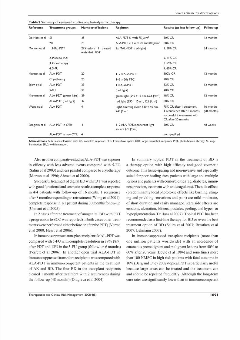
Several studies provided evidence for the use of topical
PDT in BD (summarized overview in Table 2) with initial
clearance rates between 88% and 100% (Morton et al 1996;
Morton et al 2000; Salim et al 2003).
The reported recurrence rates after 12 months were 15%
for MAL-PDT (cryotherapy 21%, 5-FU 17%) (Morton et al
2004) and 0%–12% for ALA (cryotherapy 10%, 5-FU 18%)
(Morton et al 1996; Morton et al 2000; Salim et al 2003).
Reports about long-term clearance rates are rare. In a
retrospective study (617 patients with BD) the recurrence rates
after more than 5 years were 5% for surgery, 6% for radiother-
apy, 14% for 5-FU, 19% for curettage and 34% for cryotherapy
(Thestrup-Pedersen et al 1988). For PDT a relapse rate of 17%
after 64 months was reported (Leman et al 2002) suggesting
a comparable long-term efficacy of PDT and other established
treatment options.
In the largest randomized, placebo-controlled, multicenter
study the initial cure was 93% for MAL-PDT (5-FU 83%,
cryotherapy 86%, 21% placebo-PDT) (Morton et al 2004).
At the 24 months follow-up the complete response rate was
68% (5-FU 59%, cryotherapy 60%, 11% placebo-PDT). In
this trial the percentage of an excellent or good cosmetic
outcome at 3 months was 89% in the PDT group (cryotherapy
47%, 5-FU 76%) with an improvement after 12 month to 97%
for PDT (cryotherpy 62%, 5-FU 94%). The tolerability of
MAL-PDT was excellent and superior to cryotherapy and
5-FU (pain was experienced by most PDT patients, but the
degree was less than in the cryotherapy group; 5-FU had a poor
local tolerability because of eczematous reactions, erosion
and ulceration) (Morton et al 2004). In the patients treated
with MAL-PDT no serious adverse events were seen (in the
cryotherapy group two therapy-related adverse events were
reported: lymphangitis and necrosis).
Table 1 Summary of reviewed studies and case reports on topical imiquimod
Reference Treatment groups Number
of lesions
Regimen Results (at
last follow-up)
Follow-up Notes
Patel et al Imiquimod 5%
Placebo (vehicle)
15
16
Imiquimod 5% once
daily for 16 weeks
75% (9/12)
0% CR
9 months 3 drop outs in the
imiquimod group
Mackenzie-Wood et al Imiquimod 16 Imiquimod 5% once
daily for 16 weeks
93% CR (13/14) 6 months 2 patients died from
unrelated illness
Smith et al Imiquimod 5% + 5-FU 5 Alternating Imiquimod
3 × and 5-FU 4 × per
week for 5–7 weeks
100% CR 3–15 months Renal transplant recipients
Smith et al Imiquimod 5% + sulin-
dac + valacyclovir
5 Imiquimod 5%
3 ×/week + sulindac
200 mg 2 ×/d +
valacyclovir 1000 mg/d
for 16 weeks
100% CR 5–14 months Patients with chronic
lymphatic leukemia
Muzio et al Imiquimod 5% 3 Imiquimod 5% under
occlusive dressing
(changed every 3 days)
for 60–75 days
100% CR 267–423 days Systemic side effects
in 2 patients
Prinz et al Imiquimod 5% 4 Imiquimod 5%
3 ×/week (daily
application in caseof no clinical response
after 2 weeks)
75% CR (3/4) 6 months Organ transplant
recipients; 1 recurrence
after 10 months
Abbreviations: CR, complete response; FU, 5-fluorouracil.
7/28/2019 TCRM-2008-4(5)-1960_Neubert
http://slidepdf.com/reader/full/tcrm-2008-45-1960neubert 7/12
Therapeutics and Clinical Risk Management 2008:4(5) 1091
Bowen’s disease treatment options
Also in other comparative studies ALA-PDT was superior
in efficacy with less adverse events compared with 5-FU
(Salim et al 2003) and less painful compared to cryotherapy
(Morton et al 1996; Ahmed et al 2000).
Successful treatment of digital BD with PDT was reported
with good functional and cosmetic results (complete response
in 4/4 patients with follow-up of 16 month, 1 recurrence
after 8 months responding to retreatment (Wong et al 2001);
complete response in 1/1 patient during 30 months follow-up
(Usmani et al 2005).
In 2 cases after the treatment of anogenital BD with PDT
a progression to SCC was reported (in both cases other treat-
ments were performed either before or after the PDT) (Varma
et al 2000; Heart et al 2006).
In immunosuppressed transplant recipients MAL-PDT was
compared with 5-FU with complete resolution in 89% (8/9)
after PDT and 13% in the 5-FU group (follow-up 6 months)
(Perrett et al 2006). In another open trial ALA-PDT in
immunosuppressed transplant recipients was compared with
ALA-PDT in immunocompetent patients in the treatment
of AK and BD. The four BD in the transplant recipients
cleared 1 month after treatment with 2 recurrences during
the follow-up (48 months) (Dragieva et al 2004).
In summary topical PDT in the treatment of BD is
a therapy option with high efficacy and good cosmetic
outcome. It is tissue-sparing and non-invasive and especially
suited for poor-healing sites, patients with large and multiple
lesions and patients with comorbidities (eg, diabetes, immu-
nosupression, treatment with anticoagulants). The side effects
(predominantly local phototoxic effects like burning, sting-
ing and prickling sensations and pain) are mild-moderate,
of short duration and easily managed. Rare side effects are
erosions, ulceration, blisters, pustules, peeling, and hyper- or
hypopigmentation (DeHaas al 2007). Topical PDT has been
recommended as a first-line therapy for BD or even the best
treatment option of BD (Salim et al 2003; Braathen et al
2007; Lehmann 2007).
In immunosuppressed transplant recipients (more than
one million patients worldwide) with an incidence of
cutaneous premalignant and malignant lesions from 40% to
60% after 20 years (Boyle et al 1984) and sometimes more
than 100 NMSC in high risk patients with fatal outcome in
10% (Berg and Otley 2002) topical PDT is particularly useful
because large areas can be treated and the treatment can
and should be repeated frequently. Although the long-term
cure rates are significantly lower than in immunocompetent
Table 2 Summary of reviewed studies on photodynamic therapy
Reference Treatment groups Number of lesions Regimen Results (at last follow-up) Follow-up
De Haas et al SI 25 ALA-PDT SI with 75 J/cm2 80% CR 12 months
2FI 25 ALA-PDT 2FI with 20 and 80 J/cm2 88% CR
Morton et al 1. MAL-PDT 275 lesions 111 treated
with MAL-PDT
2x MAL-PDT (red-light) 1. 68% CR 24 months
2. Placebo-PDT 2. 11% CR3. Cryotherapy 3. 59% CR
4. 5-FU 4. 60% CR
Morton et al ALA-PDT 20 1–2 ×ALA-PDT 100% CR 12 months
Cryotherapy 20 1–3 × 20s FTC 90% CR
Salim et al ALA-PDT 33 1 ×ALA-PDT 82% CR 12 months
5-FU 33 (red light) 48% CR
Morton et al ALA-PDT (green light) 29 green light (540 ± 15 nm, 62.6 J/cm2) 48% CR 12 months
ALA-PDT (red light) 32 red light (630 ± 15 nm, 125 J/cm2) 88% CR
Wong et al ALA-PDT 4 Light-emitting diode 630 ± 40 nm,
240 J/cm2
75% CR after 1 treatment,
1 recurrence after 8 months
successful 2 treatment with
CR after 20 months
16 months
(20 months)
Dragieva et al ALA-PDT in OTR 4 1–2 ALA-PDT, incoherent light
source (75 J/cm2)
50% CR 48 weeks
ALA-PDT in non-OTR 4 not specified
Abbreviations: ALA, 5-aminolevulinic acid; CR, complete response; FTC, freeze-thaw cycles; ORT, organ transplant recipients; PDT, photodynamic therapy; SI, single
illumination; 2FI, 2-fold illumination.

7/28/2019 TCRM-2008-4(5)-1960_Neubert
http://slidepdf.com/reader/full/tcrm-2008-45-1960neubert 8/12
Therapeutics and Clinical Risk Management 2008:4(5)1092
Neubert and Lehmann
patients (Dragieva et al 2004), PDT is an excellent treatment
option in transplant recipients because other standard
therapies are limited in these patients as well.
DiscussionThe conventional treatment options (cryotherapy, curettage
with cautery, excision, 5-Fluorouracil, radiotherapy, laser)
appear to have generally similar efficacy and recurrence
rates with no single therapy being superior for all clinical
situations. They all have their advantages and they all are con-
nected with some certain side effects and adverse events (eg,
ulceration, infection, scarring and hypopigmentation after
cryotherapy; toxicity of radiotherapy and prolonged healing
depending on the used technique; “fragile scalp syndrome”
with atrophic epidermis and erosions after extensive carbon
dioxide laser; inflammation with erosion and ulceration
during 5-FU).
As BD is often located on the lower limb of an older
person, there is need for non-invasive treatment options with
only mild-moderate local side effects or adverse events even
in poor-healing sites.
Diclofenac 3% gel is a non-destructive therapy that was
already successfully used in the treatment of actinic keratosis
(Wolf et al 2001; Smith et al 2006).
In the seven published cases of BD treated with diclof-
enac 3% gel mentioned above the therapy was very effective
(100% complete clinical and histological clearance) and it
was well tolerated with only mild side effects (inflammation,
itching and dryness) (Pantel and Stockfleth 2007). However,
these promising data have to be proven in randomized con-
trolled trials and optimum regimens (once vs twice daily
application, duration of application, etc) and recurrence rates
in long-term follow-up have to be investigated. Diclofenac
3% gel is not yet licensed for BD.
The use of topical imiquimod is an effective alternative
treatment option for patients and body sites that are unsuit-
able for other treatments like surgery.
As extragenital BD contains in about 47% of acral and
in about 24% of nonacral lesions the HPV genome, imiqui-
mod might have a double effect in these cases because of its
antiviral and antitumor activity through stimulation of innate
and acquired immunity.
Nevertheless further and larger prospect ive studies
with long-term follow-up are required to verify the results
mentioned above and to figure out an optimal dosing
scheme (duration of treatment, number of weekly applica-
tions) with high efficacy and decreased local side effects
(common local adverse effects: erythema, pruritus, erosion,
ulceration vesicle formation). The risk of systemic side
effects (eg, fever, flu-like symptoms, lymphopenia) due
to absorption of imiquimod has to be considered when
imiquimod is used under occlusion. Imiquimod is not yet
licensed for BD.
In comparative studies topical PDT had a similar efficacy
and less adverse events in comparison with cryotherapy and
PDT was more effective with fewer adverse events when
compared with 5-FU in the treatment of BD. The most
common side effect during PDT is pain (reported by most
of the patients as mild to moderate and by a minority as
severe or even intolerable). Other common local side effects
are erythema, edema and crust formation but in most of the
studies no ulceration, infection or other more serious adverse
events were seen.
Cosmetic outcome after PDT was typically superior com-
pared to the existing standard therapies. PDT can be repeated
easily if required and it is popular with patients.
PDT is especially suited for poor-healing sites, patients
with large and multiple lesions and patients with comor-
bidities (eg, diabetes, immunosupression, treatment with
anticoagulants).
Another special group of patients profiting from the
(repetitive) treatment with PDT are immunosuppressed
transplant recipients with their multiple premalignant and
malignant cutaneous lesions and their higher risk to develop
potentially fatal squamous cell carcinoma from AK and BD
(although the evidence base in these patients is limited and
the results showed a higher recurrence rate during follow-up
compared with immunocompetent patients).
Thus there is no single definite “right way” for all
patients with BD, the choice of treatment should be guided
by its efficacy, location and size of BD, number of lesions,
availability of the therapy, the clinicians expertise, patient
factors (age, immune status, concomitant medication,
comorbidities and compliance), cosmetic outcome and the
patients preference. In consideration of a 10% recurrence
rate for most treatment options a risk-adapted follow-up is
recommended.
DisclosuresThe authors declare no conflicts of interest.
ReferencesAhmed I, Berth-Jones J, Charles-Holmes S, et al. 2000. Comparison of
cryotherapy with curettage in the treatment of Bowen’s disease:a
prospective study. Br J Dermatol , 143:759–66.
Arlette JP. 2003. Treatment of Bowen’s disease and erythroplasia of Queyrat.
Br J Dermatol , 149:43–7.
7/28/2019 TCRM-2008-4(5)-1960_Neubert
http://slidepdf.com/reader/full/tcrm-2008-45-1960neubert 9/12
Therapeutics and Clinical Risk Management 2008:4(5) 1093
Bowen’s disease treatment options
Ball SB, Dawber RP. 1998. Treatment of cutaneous Bowen’s disease
with particular emphasis on the problem of lower leg lesions.
Australas J Dermatol , 39:63–8.
Bargmann H, Hochmann J. 2003. Topical treatment of Bowen’s disease
with 5-Fluorouracil. J Cutan Med Surg , 7:101–5.
Bell HK, Rhodes LE. 1999. Bowen’s disease – a retrospective review of
clinical management. Clin Exp Dermatol , 24:338–9.
Berg D, Otley CC. 2002. Skin cancer in organ transplant recipients:epidemi-
ology, pathogenesis, and management. J Am Acad Dermatol , 47:1.
Boyle J, MacKie RM, Briggs JD. 1984. Cancer, warts, and sunshine in renal
transplant patients. A case-control study. Lancet , 31:702.
Boynton KK, Bjorkman DJ. 1991. Argon laser therapy for perianal Bowen’s
disease:a case report. Laser Surg Med , 11:385–7.
Braathen LR, Szeimies RM, Basset-Seguin N, et al. 2007. Guidelines on
the use of photodynamic therapy for nonmelanoma skin cancer: An
international consensus. J Am Acad Dermatol , 56:125–43.
Brannan PA, Anderson HK, Kersten RC. 2005. Bowen disease of the
eyelid successfully treated with imiquimod. Ophthal Plast Reconstr
Surg , 21:321–2.
Brown VL, Atkins CL, Ghali L. 2005. Safety and efficacy of 5% imiquimod
cream for the treatment of skin dysplasia in high risk renal transplant
recepiens:randomized, double-blind, placebo-controlled trial. Arch
Dermatol , 141:985–93.
Chen K, Shumack S. 2003. Treatment of Bowen’s disease using a cycle
regimen of imiquimod 5% cream. Clin Exp Dermatol , 28:10–2.
Chung YL, Lee JD, Bang D, et al. 2000. Treatment of Bowen’s disease with aspecially designed radioactive skin patch. Eur J Nucl Med 27:842–6.
Clavel C, Pham-Huu V, Durlach A, et al. 1999. Mucosal oncogenic human
papillomaviruses and extragenital Bowen’s disease. Cancer , 86:282.
Cleary RK, Schaldenbrand JD, Fowler JJ, et al. 2000. Treatment options
for perianal Bowen’s disease:survery of American Society of Colon
and Rectal Surgeon Members. Am Surg , 66:686–8.
Cook-Bolden F, Weinberg JM. 2002. Topical imiquimod 5% cream in
the treatment of Bowen’s disease of the penis. J Am Acad Dermatol ,
46:146–7.
Cox NH, Dyson P. 1995. Wound healing on the lower leg after radiotherapy
or cryotherapy of Bowen’s disease and other malignant skin lesions.
Br J Dermatol , 133:60–5.
Cox NH, Eedy DJ, Morton CA. 1999. Guidelines for management of
Bowen’s disease. Br J Dermatol , 141:633–41.
Cox NH, Eedy DJ, Morton CA. 2007. Guidelines for management of Bowen’s disease: 2006 update. Br J Dermatol , 156:11–21.
Danielsen AG, Sand C, Weismann K. 2003. Treatment of Bowen’s
disease of the penis with imiquimod 5% cream. Clin Exp Dermatol ,
28:7–9.
Dave R, Monk B, Mahaffey P. 2003. Treatment of Bowen’s disease with
carbon dioxide laser. Laser Surg Med , 32:335.
Dawe SA, Salisbury JR, Higgins E. 2005. Two cases of Bowen’s disease
successfully treated topically with 3% diclofenac in 2.5% hyaluronan
gel. Clin Exp Dermatol , 30:712–3.
DeHaas ERM, Sterenborg HJCM, Neumann HAM, et al. 2007. Response of
Bowen Disease to ALA-PDT using a single and a 2-fold illumination
scheme. Arch Dermatol , 143:264–5.
Dragieva G, Hafner J, Dummer R, et al. 2004. Topical photodynamic therapy
in the treatment of actinic keratoses and Bowen’s disease in transplant
recipients. Transplantation, 77:115–21.Dupree MT, Kiteley RA, Weismantle K, et al. 2001. Radiation therapy for
Bowen’s disease:lessons for lesions of the lower extremity. J Am Acad
Dermatol , 45:401–4.
Dyall-Smith D. 1998. Intralesional bleomycin. Australas J Dermatol ,
39:123–4.
Fritsch C, Homey B, Stahl W, et al. 1998. Preferential relative porphyrin
enrichment in solar keratoses upon topical application of delta-
aminolevulinic acid methylester. Photochem Photobiol , 68:218–21.
Gad F, Viau G, Boushira M, et al. 2001. Photodynamic therapy with
5-aminolevulinic acid induces apoptosis and caspase activation in
malignant T cells. J Cutan Med Surg , 5:8–13.
Gately S. 2000. The contributions of cyclooxygenase-2 to tumor
angiogenesis. Cancer Metastasis Rev, 19:19–27.
Goh MSY. 2006. Invasive squamous cell carcinoma after treatment of
carcinoma in situ with 5% imiquimod cream. Australas J Dermatol ,
47:186–8.
Gordon KB, Garden JM, Robinson JK. 1996. Bowen’s disease of the distal
digit. Outcome of treatment with carbon dioxide laser vaporization.
Dermatol Surg , 22:723–8.
Gordon KB, Robinson J. 1994. Carbon dioxide laser vaporization for
Bowen’s disease of the finger. Arch Dermatol , 130:1250–2.
Gordon KB, Roenigk HH, Gendleman M. 1997. Treatment of multiple
lesions of Bowen disease with isotretinoin and interfon alfa. Efficacy
of combination chemotherapy. Arch Dermatol , 133:691–3.
Graham JH, Helwig EB. 1961. Bowen’s disease and its relationship to
systemic cancer. Arch Dermatol , 83:76–96.
Gutzmer R, Kaspari M, Vogelbruch M, et al. 2002. Successful treatment
of anogenital Bowen’s disease with the immunomodulator imiquimod,
and monitoring of therapy by DNA image cytometry. Br J Dermatol ,
147:160–5.
Habetts JMW, Tank B, Van Joost T. 1989. Characterization of the
mononuclear infiltrate in Bowen’s disease (squamous cell carcinoma
in situ):evidence of a T-cell-mediated anti-tumor response. Virchows
Arch, 415:125–30.
Heising RA. 1979. Treatment of Bowen’s disease of the ear by the combined
use of cryosurgery and topical 5-fluorouracil. Cutis, 24:271–5.
Hengge UR, Benninghoff B, Ruzicka T, et al. 2001. Topicalimmunomodulators- progess treating inflammation, infection and
cancer. Lancet Inf Dis, 1:189–98.
Herat A, Shirato K, Damian DL, et al. 2006. Invasive squamous cell carci-
noma arising in refractory perianal Bowen’s disease in a HIV-positive
individual. Australas J Dermatol , 47:120–3.
Hiruma M, Kawada A. 2000. Hyperthermic treatment of Bowen’s disease
with disposable chemical pocket warmers:a report of 8 cases. J Am
Acad Dermatol , 43:1070–5.
Holt PJ. 1988. Cryotherapy for skin cancer:results over a 5-year period using
liquid nitrogen spray cryosurgery. Br J Dermatol , 119:231–40.
Honeycutt WM, Jansen GT. 1973. Treatment of squamous cell carcinoma
of the skin. Arch Dermatol , 108:670–2.
Kao GF. 1986. Carcinoma arising in Bowen’s disease. Arch Dermatol ,
122:1124–6.
Khandpur S, Sharma VK 2003. Successful treatment of multiple pre-malignantand malignant lesions in arsenical keratosis with a combination of
acitretin and intralesional 5-fluorouracil. J Dermatol , 30:730–4.
Kossard S. 2003. Treatment of large facial Bowen’s disease:a case report.
Clin Exp Dermatol , 28(Suppl 1):13–5.
Kreuter A, Hochdofer B, Stücker M, et al. 2004. Treatment of anal intraepi-
thelial neoplasia in patients with acquired HIV with imiquimod 5%
cream. J Am Acad Dermatol , 50:980–1.
Kuzelova K, Grebenova D, Pluskalova M, et al. 2004. Early
apoptotic features of K562 cell death induced by 5-aminolaevu-
linic acid-based photodynamic therapy. J Photochem Photobiol B,
73:67–78.
Landthaler M, Haina D, Brunner R, et al. 1986. Laser therapy of
bowenoid papulosis and Bowen’s disease. J Dermatol Surg Oncol ,
12:1253–7.
Lee JD, Park KK, Lee MG, et al. 1997. Radionuclide therapy of skin cancersand Bowen’s disease using a specially designed radioactive skin patch.
J Nucl Med 38:697–702.
Lehmann P. 2007. Nebenwirkungen der topischen photodynamischen
Therapie. Hautarzt , 58:597–603.
Leman JA, Mackie RM, Morton CA. 2002. Recurrence rates following
aminilaevulinic acid-photodynamic therapy for intraepidermal
squamous cell carcinoma compare favourably with outcome following
conventional modalities. Br J Dermatol , 147 (Suppl 62):35.
Lukas VanderSpek LA, Pond GR, Wells W, et al. 2005. Radiation
therapy for Bowen’s disease of the skin. Int J Radiat Oncol Biol Phys,
63:505–10.

7/28/2019 TCRM-2008-4(5)-1960_Neubert
http://slidepdf.com/reader/full/tcrm-2008-45-1960neubert 10/12
Therapeutics and Clinical Risk Management 2008:4(5)1094
Neubert and Lehmann
Mackenzie-Wood A, Kossard S, de Launey J, et al. 2001. Imiquimod 5%
cream in the treatment of Bowen’s disease. J Am Acad Dermatol ,
44:462–70.
Mandekou-Lefaki I, Delli F, Koussidou-Eremondi TH, et al. 2003. Treat-
ment of Bowen’s disease with imiquimod cream 5%. J Eur Acad
Dermatol Venereol , 17:238–9.
Marchesa P, Fazio VW, Oliart S, et al. 1997. Perianal Bowen’s disease:
a clinicopathologica study of 47 patients. Dis Colon Rec tum ,
40:1286–93.
Masferrer JL, Leahy KM, Koki AT, et al. 2000. Antiangiogenetic and
antitumor activities of cyclooxygenase-2 inhibitors. Cancer Res,
60:1306–11.
Meykadeh N, Hengge UR, 2003. Topische Immunmodulation in der
Dermatologie. Hautarzt , 54:641–62.
Micali G, Nasca MR, Tedeschi A. 2003. Topical treatment of intraepithelial
penile carcinoma with imiquimod. Clin Exp Dermatol , 28:4–6.
Morton CA, Horn M, Leman J, et al. 2004. A randomized, placebo-controlled,
European study comparing MAL-PDT with cryotherapy and
5-fluorouracil in subjects with Bowen’s disease. J Eur Acad Dermatol
Venereol , 18 (Suppl 2):415.
Morton CA, Whitehurst C, Moore J, et al. 1996. Photodynamic therapy vs
cryotherapy in the treatment of Bowen’s disease. Clin Exp Dermatol ,
21:79.
Morton CA, Whitehurst C, Moore JV, et al. 2000. Comparison of red and
green light in the treatment of Bowen’s disease by photodynamic
therapy. Br J Dermatol , 143:767–72.Morton CA, Whitehurst C, Moseley H, et al. 1996. Comparison of photo-
dynamic therapy with cryotherapy in the treatment of Bowen’s disease.
Br J Dermatol , 135:766–71.
Murata Y, Kumano K, Sashikata T. 1996. Partial spontaneous regression
of Bowen’s disease. Arch Dermatol , 132:429–32.
Muzio G, Ciambellotti A, Rebora A. 2004. Occlusive medication with
imiquimod in Bowen’s disease. Acta Derm Venereol , 84:168–9.
Noodt BB, Berg K, Stokke T, et al. 1996. Apoptosis and necrosis induced
with light and 5-aminolaevulinic acid-derived protoporphyrin IX. Br
J Cancer , 74:22–9.
Nordin P. 1999. Curettage-cryosurgery for non-melanoma skin cancer o the
external ear:excellent 5-year results. Br J Dermatoo, 140:291–3.
Orengo I, Rosen T, Guill CK. 2002. Treatment of squamous cell carcinoma
in situ of the penis with 5% imiquimod cream: a case report. J Am Acad
Dermatol , 47:225–8.Ota M, Kawashima M, Mitsuishi T. 2002. Multiple Bowen’s disease of the
fingers. Eur J Dermatol , 12:275–7.
Otani K, Ito Y, Sumiya, et al. 2001. Treatment of Bowen’s disease using the
ultrasonic surgical aspirator. Plast Reconstr Surg , 108:68–72.
Pantel MJ, Stockfleth E. 2007. Does progession from actinic keratosis and
Bowen’s disease end with treatment:diclofenac gel, an old drug in a
new environment? Br J Dermatol , 156(Suppl 3):53–6.
Papillon J, Chassard JL. 1992. Respective roles of radiotherapy and surgery
in the management of epidermoid carcinoma of the anal margin. Dis
Colon Rectum, 35:422–9.
Patel GK, Goodwin R, Chawla M, et al. 2006. Imiquimod 5% cream
monotherapy for cutaneous squamous cell carcinoma in situ (Bowen’s
disease):A randomized, double-blind, placebo-controlled trial. J Am
Acad Dermatol , 54:1025–32.
Peng Q, Soler AM, Warloe T, et al. 2001. Selective distribution of porhpyrinsin skin thick basal cell carcinoma after topical application of methyl
5-aminolevulinate. J Photochem Photobiol B Biol , 62:140–5.
Perrett CM, McGregor J, Proby C, et al. 2006. A comparative study of
topical 5-fluorouracil and topical photodynamic therapy using methyl-
aminolevulinate for actinic keratosis and Bowen’s disease in organ
transplant recipients [abstract]. J Am Acad Dermatol , 54:A7.
Phoushek J, Smith KJ. 2001. Imiquimod and 5% fluorouracil therapy for
anal and perianal squamous cell carcinoma in situ in an HIV-1-positive
man. Arch Dermatol , 137:14–6.
Plaza de Lanza M, Ralfs I, Dawber R. 1980. Cryosurgery for Bowen’s
disease of the skin. Br J Dermatol , 103:14.
Prinz BM, Hafner J, Dummer R, et al. 2004. Treatment of Bowen’s disease
with imiquimod 5% cream in transplant recipients. Transplantation,
77:790–1.
Raaf JH, Krown SE, Pinsky CM, et al. 1967. Treatment of Bowen’s
disease with topical dinitrochlorobenzene and 5-fluorouracil. Cancer,
37:1633–42.
Reizner GT, Chuang TY, Elpern DJ, et al. 1994. Bowen’s disease (squamous
cell carcinoma in situ) in Kaui, Hawaii: a population-based incidence
report. J Am Acad Dermatol , 31:596–600.
Salim A, Leman JA, McColl JH, et al. 2003. Randomized comparison of
photodynamic therapy with topical 5-fluorouracil in Bowen’s disease.
Br J Dermatol , 148:539–43.
Schön M, Bong AB, Drewniok D, et al. 2003. Tumor-selective induction of
apoptosis and the small-molecule immune response modifier imiqui-
mod. J Natl Cancer , 95:1138–49.
Schroeder TL, Sengelmann RD. 2002. Squamous cell carcinoma in situ of
the penis successfully treated with imiquimod 5% cream. J Am Acad
Dermatol , 46:545–8.
Sierra-Valenti X. 2003. Efficacy of imiquimod 5% cream for the treatment
of Bowen’s disease and basal cell carcinoma. J Eur Acad Dermatol
Venereol , 17:351.
Smith KJ, Germain M, Skelton H. 2001. Bowen’s disease (squamous cell
carcinoma insitu) in immunosuppressed patients treated with imiquimod
5% cream and a COX inhibitor, sulindac:potential applications for this
combination of immunotherapy. Dermatol Surg , 27:143–6.
Smith KJ, Germain M, Skelton H. 2001. Squamous cell carcinoma in situ(Bowen’s disease) in renal transplant patients treated with 5% imiqui-
mod and 5% fluorouracil therapy. Dermatol Surg , 27:561–4.
Smith SR, Morhenn VB, Piacquadio DJ, et al. 2006. Bilateral comparison
of the efficacy and tolerability of 3% diclofenac sodium gel and 5%
5-fluorouracil cream in the treatment of actinic keratoses of the face
and scalp. J Drugs Dermatol , 5:156–9.
Sorensen R, Juzenas P, Iani V, et al. 1998. Formation of protoporphyrin IX
in mouse skin after topical application of 5-aminolevulinic acid and its
methyl ester. SPIE , 3563:77–81.
Stevens DM, Kopf AW, Gladstein A, et al. 1977. Treatment of Bowen’s
disease with Grenz rays. Int J Dermatol , 16:329–39.
Stockfleth E, Ulrich C, Schmook T, et al. 2003. The therapeutic impact of
immune-response-modifiers (IRM`s) in organ transplant patients. J Eur
Acad Dermatol Venereol , 17:75–6.
Szeimies RM, Abels C, Fritsch C, et al. 1995. Wavelength dependency of photodynamic effects after sensitization with 5-aminolevulinic acid in
vitro and in vivo. J Invest Dermatol , 105:672–7.
Tantikun N. 2000. Treatment of Bowen’s disease of the digit with carbon
dioxide laser. J Am Acad Dermatol , 43:1080–3.
Temmi H, Kaisho T, Takeuchi O, et al. 2002. Small antiviral compounds
activate immune cells via the TLR7 MyD88-dependent pathway. Nature
Immunol , 3:196–200.
Thai KE, Sinclair RD. 2002. Treatment of Bowen’s disease of the penis
with imiquimod. J Am Acad Dermatol , 46:470–1.
Thestrup-Pedersen K, Ravnborg L, Reymann F. 1988. Morbus Bowen.
A description of the disease in 617 patients. Acta Derm Venereol ,
68:236–9.
Turpin IM. 1999. Radionuclide therapy of skin cancers and Bowen’s
disease using a specially designed skin patch. Plast Reconstr Surg ,
103:1333.Urosevic M, Maier T, Benninghoff B, et a.l 2003. Mechanism underlying
imiquimod-induced regression of basal cell carcinoma in vivo. Arch
Dermatol , 139:1325–32.
Usmani N, Stables GI, Telfer NR, et al. 2005. Subungual Bowen’s disease
treated by topical aminolevulinic acid-photodynamic therapy. J Am
Acad Dermatol , 53:273–6.
van Bezooijen BP, Horenblas S, Meinhardt W, et al. 2001. Laser therapy
for carcinoma in situ of the penis. J Urol , 166:1670–1.
Varma S, Holt PJA, Anstey AV. 2000. Erythroplasia of Queyrat treated by
topical aminolaevulinic acid photodynamic therapy:a cautionary tale.
Br J Dermatol , 142:825–6.

7/28/2019 TCRM-2008-4(5)-1960_Neubert
http://slidepdf.com/reader/full/tcrm-2008-45-1960neubert 11/12
Therapeutics and Clinical Risk Management 2008:4(5) 1095
Bowen’s disease treatment options
Vidal D, Matias-Guiu X, Alomar A. 2004. Efficacy of imiquimod for the
expression of Bcl-2, Ki67, p53 and basal cell carcinoma apoptosis.
Br J Dermatol , 151:656–62.
Wang KH, Fang JY, Hu CH, et al. 2004. Erbium:YAG laser pretreat-
ment accelerates the response of Bowen’s disease treated by topical
5-fluorouracil. Dermatol Surg , 30:441–5.
Webber J, Luo Y, Crilly R, et al. 1996. An apoptotic response to
photodynamic therapy with endogenous protoporphyrin in vivo.
J Photochem Photobiol B, 35:209–11.
Welch ML, Grabski WJ, McCollough ML, et al. 1997. 5-Fluorouracil
iontophoretic therapy for Bowen’s disease. J Am Acad Dermatol ,
36:956–8.
Wolf JE, Jr, Taylor JR, Tschen E, et al. 2001. Topical 3.0% diclofenac
in 2.5% hyaluronan gel in the treatment of actinic keratoses. Int J
Dermatol , 40:709–13.
Wong TW, Sheu HM, Lee JY. 2001. Photodynamic therapy for Bowen’s
disease (squamous cell carcinoma in situ) of the digit. Dermatol Surg ,
27:452–6.
Yerebakan O, Ermis O, Yilmaz E, et al. 2002. Treatment of arsenical keratosis
and Bowen’s disease with acitretin. Int J Dermatol , 41:84–7.

7/28/2019 TCRM-2008-4(5)-1960_Neubert
http://slidepdf.com/reader/full/tcrm-2008-45-1960neubert 12/12