Embed Size (px)
Citation preview
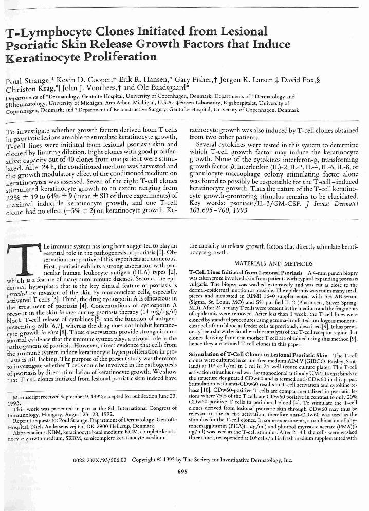
T -Lymphocyte Clones Initiated from Lesional Psoriatic Skin Release Growth Factors that Induce Keratinocyte Proliferation
paul Strange,* Kevin D. Cooper,t Erik R. Hansen,* Gary FisherJ Jorgen K. Larsen,t David Fox,§ Christen Krag, ~ John J. Voorhees, t and O le Baadsgaard * Departments of 'De~matology, Gentofte Hospital, Univ~rsi.ty of Copenhag~n, Denmark; Departments.of tDermatology and §Rheumatology, Ul11vemty of Mlclugan, Ann Arbor, Mlclugan, U.S.A.; :j:Fll1sen Laboratory ,. R,gshospltalet, Ul11vemty of Copenhagen, Denmark; and ~Department of Reconstructive Surgery, Gentofte Hospital, Ul1lVerslty of Copenhagen, Denmark
To investigate whether growth factors derived from T cells in psoriatic lesions .ar~ .able to stimula.te keratin<:>c~te g~owth, T-cell lines were InItiated from leslOnal psonasls skill and cloned by limiting dilution. Eight clones wi~h good pro~iferative capacity out of 40 clones from one patient were stimulated. After 24 h, the conditioned medium was harvested and the growth modulatory effect of the condi~ioned medium on keratinocytes was assessed. Seven of the eight T-cell clones stimulated keratinocyte growth to an extent ranging from 22% ± 19 to 64% ± 9 (mean ± SD of three experiments) of maximal inducible keratinocyte growth, and one T-cell clone had no effect (- 5% ± 2) on keratinocyte growth. Ke-
The immune system has long been suggested to play an essential role in the pathogenesis of p~oriasis [1]. Observations supportive of this hypotheSIS are numerous. First, psoriasis exhibits a strong association with particular human leukocyte antigen (HLA) types [2],
which is a feature of many autoimmune diseases. Second, t~e ~p!dermal hyperplasia that is the key clinical feature of psonaSlS IS preceded by invasion of the skin by mononucl.ear ~ells, esp.eClal!y activated T cells [3]. Third, the drug cyclosponn A IS efficaclO~s In
the treatment of psoriasis [4]. Concentrations of cyclosponn A present in the skin ill "iva during psoriasis therapy. (14 mg/~g/d) block T-cell release of cytokines [5] and the function of antlgenpresenting cells [6,7], whereas the dru~ does no~ inhibit ke~atinoeyte grow~h ill vitro [8] . T.hese observations prOVide strong Circumstantial eVidence that the Immune system plays a pIvotal role In the pathogenesis of psoriasis. However, direct evidence that cell.s from the immune system induce keratinocyte hyperprohferatlon In psoriasis is still lacking. The purpose of the present study was therefore to investigate whether T cells could be involved in the pathogenesis of psoriasis by direct stimulation ofkeratinocyte growth. We show that T-cell clones initiated from lesional psoriatic skin indeed have
Manuscript received September 9, 1992; accepted for publication June 23, 1993.
This work was presented in part at the 8th International Congress of Immunology, Hungary, August 23-28,1992.
Reprint requests to: Poul Strange, Department of Dermatology , Gentofte Hospital, Niels Andersens vej 65, DK-2900 Hellerup, Denmark. .
Abbreviations: KBM, keratinocyte basal medIUm; KGM, complete keratlnoeyte growth medium, SKBM, semicomplete keratinocyte medium.
ratinocyte growth was also induced by T-cell clones obtained from two other patients .
Several cytokines were tested in this system to determine which T-cell growth factor may induce the keratinocyte growth. None of the cytokines interferon-g, transforming growth factor-p, interleukin (IL}-2, IL-3, IL-4, IL-6, IL-8, or granulocyte-macrophage colony stimulating factor alone was found to possibly be responsible for the T -cell- induced keratinocyte growth. Thus the nature of the T -cell keratinocyte growth-promoting stimulus remains to be elucidated. Key words: psoriasis/IL-3/GM-CSF. ] Imlest Dermatol 101:695-700, 1993
the capacity to release growth factors that directly stimulate keratinocyte growth.
MATERIALS AND METHODS
T-Cell Lines Initiated from Lesional Psoriasis A 4-mm punch biopsy was taken from involved skin from patients with typical expanding psoriasis vulgaris. The biopsy was washed extensively and was cut as close to the dermal-epidermal junction as possible. The epidermis was cut in many small pieces and incubated in RPM I 1640 supplemented with 5% AB-serum (Sigma, St. Louis, MO) and 5% purified IL-2 (Pharmacia, Silver Spring, MD) .. After24 h many T cells were present in the medium and the fragments of epidermIS were removed. After less than 1 week, the T-cell lines were cloned by standard procedures using gamma-irradiated autologous mononuclear cells from blood as feeder cells as previously described [9]. It has previously been. shown by Southern blot analysis of the T-cell receptor region that clones denvl1lg from one mother T cell are obtained using this method [9], hence they are termed T-cell clones in this paper.
Stimulation ofT-Cell Clones in Lesional Psoriatic Skin The T-cell clones wer~ cultured in serum-free medium AIM V (GIBCO, Paisley, Scotland) at 10 .cells/mlm 1 mlm 24-well tissure culture plates. The T-cell activatIOn stm1Ulus used was the monoclonal antibody UM4D4 that binds to the struc.ture d.es 'gnated CDw60 and is termed anti-CDw60 in this paper. StimulatIOn With antl-C!?w60 results in T-cell activation and cytokille release (10]. CDw60-posltlve T cells are compartmentalized in psoriatic lesions where 75% of the T .cells are CDw60 positive in contrast to only 20% CDw60-posltlve T cells 111 peripheral blood [4]. To stimulate the T-cell clones denved from leslOnal psoriatic skin through CDw60 may thus be relevant to the itl vivo activation, therefore anti-CDw60 was used as the stimulus for the T-cell clones. In some experiments, a combination of phytohemagglutinin (PHA)(1 .ug/ ml) and phorbol myristate acetate (PMA)(3 ng/ml) was used as the T-cell stimulus. After 2-4 h the cells were washed three times, resuspended at 106 cells/ml in fresh medium supplemented with
0022-202X/93/S06.00 Copyright © 1993 by The Society for Investigative Dermatology, Inc.
695

696 STRANGE ET AL
2000 2
R >0.99
~ I.L '000 a: . "
" ,
.' " ,
" .' 0
,
o 2000 4000 6000
KERA TINOCYTES SEEDED
Figure 1. Correlation between number ofkeratinocytes seeded and relative fluorescence intensity (RFU). Kerannocytes were seeded In a flat-bottomed microtiter plate and allowed to attach for 6 h. After two washes the keratinocytes were incubated with 50 ,ug/ ml methyl-umbelliferone-heptanoate at 37 ' C for 30 min. The RFU at 450 nm aftcr ultraviolet radiation excitation at 365 nm was read. Bars, mean ± SO of triplicate determinations; dotted lil/e, linear regression line. T he relative fluorescent units is a very good estimate of relative cell number. Representative experiment of two.
2 U /ml rhIL-2, and incubated as described above in a new tissue cultnre plate . Concurrently, control medium also containing rhIL-2 was incubated. After 24 h the supernatant was harvested and spun at 1000 X g for 30 min to clear all cel ls and debris, and was storcd at -80 'C for later use.
Cultivation of Keratinocytes Primary cultures of keratinocytes were initiated from patients undergoing reduction mammoplasty and were cultured in serum-free medium keratinocyte growth medium (KGM; Clonetics, San Diego, CAl by standard methods that eliminate cell types other than keratinocytes [11]. The keratinocytes were used for assay after three to four passages.
Keratinocyte Growth Assay The serum-free keratinocyte medium consisted of a chemically defined basal medium keratinocyte basal medium (KBM) that supports minimal or no growth ofkeratinocytes. The KBM was supplemented with suboptimal concentrations of growth factors: hydrocortisone (10 nl/ml), insulin (100 nl/ml), and bovine pituitary extract (1.0-1.3 ,ug/ml), below termed semicomplete keratinocyte medium (SKBM). SKBM was used because it permits both up- and down-regulation of keratinocyte proliferation (12] .
Determination of Keratinocyte Growth Using a Fluorometric Assay To assess keratinocyte growth we used a slight modification of a fluorometric method developed by Detmar el al [13]. Two or three thousand keratinocytes were seeded in flat-bottom microtiter plates in SKBM with va.rying dilutions of the T-cell supernatant or recombinant cytokine to be tested for keratinocyte growth-modulatory property. After 3 d the microtiter plates were washed twice and incubated with methyl-umbelliferoneheptanoate (Sigma) at 50 ,ug/ml in phosphate-buffered saline (PBS) . After 30 min at 37 ' C the relative fluorescence intensity (RFU) at 450 nm after ultraviolet radiation excitation at 365 nm was read using a Microfluor reader (Dynatech Laboratories, Chanti lly, VA). The coefficient of correlation between RFU and the number of keratinocytes seeded and allowed to attach for 6 h was r2 > 0.99 (F ig 1).
Counting of Keratinocytes Using a Hemocytometer Keratinocytes were seeded in 6-well plates at the same density in each well and grown to semiconfluence in KGM, switched to SKBM for 24 h, and finally switched to T -cell - conditioned medium or added in 1 : 5 concentration to SKBM. After 3 d the cells were harvested and counted in a hemocytometer.
T-Cell Lines, Cytokines. and Cytokine Assays The T-cell lines MOLT -4 [14] and CEM [15] were obtained from the American Type Culture Collection (Rockville, MD) and C91/PL [16] was kindly provided by Claus Nielsen, Statens Seruminstitut, Copenhagen, Denmark. The cytokines rhIL-3, rhGM-CSF, rhIL-4, rhIL-6, and rhIFN-g were obtained from Genzyme (Cambridge, MA), rh lL-2 was from Cetus and rhlL-8 was from Endogen (Boston, MA). The B9 hybridoma cells for the IL-6 bioassay were kindly provided by Dr. Aarden, Central Laboratory of the Netherlands Red
THE JOURNAL OF INVESTIGATIVE DERMATOLOGY
100 -
X 9 0 -~ ~
8 0 -u. 0 7 0 -
*" 6 0 -
Z 5 0 -0 i= 4 0 -(.) ~ Q 3 0 -~ :I: 2 0 -I-~ 10 -0 II: o -CI
-1 0 -
+_. MEDIUM K 1 K 2 K4 K6 K 13 K15 K28 K 3 3
Figure 2. T -cell clones from involved psoriatic epidermis vary in their keratinocyte growth-modulatory capacity. Each T-cell clone was stimulated with anti-CDw60 in two or three separate experiments. The T -cell clone supernatants were tested for keratinocyte growth induction in several dilutions from 1 : 5 to 1 : 200,000. After 3 d incubation the relative ke ratinocyte ccl l number was estimated using the fluorometric assay. The stimulation of the keratinocytcs by the supernatants in the dilution 1 : 5 was calculated as percent of maximal inducible keratinocyte growth, i.e., keratinocytc stimulation in completely growth-factor-supplemented medium KGM (-'- '-" 100% growth). The arithmetic mean ± SO of these individually determined growth- induction percentages from each T-cell clone were calculated and are expressed on the y-axis .
Cross Blood Transfusion Service. The assay was performed as described by Aarden el al [17] and the rhlL-6 was found to be biologically active.
Cytokine contents were determined by radioimmunoassay (RIA) and enzyme-linked immunosorbent assay (ELISA) kits. The TGF-a RIA kit was obtained from Biomedical Technologies Inc. (Stoughton, MA), the IL-3 ELISA kit was from R&D Systems (Minneapolis, MN), and the granulocyte-macrophage colony stimulating factor (GM-CSF) ELISA kit was from Genzyme.
RESULTS
T -Cell Clones Derived from Lesional Psoriatic Skin Stimulate Keratinocyte Growth From one psoriasis patient 40 lesional T -cell clones were obtained. Eight of these T-cell clones grew sufficiently well to be used in experiments. All eight T-cell clones were CDw60+ and responded to anti-CDw60 by aggregating in clusters within 3 h. Each of these T-cell clones was stimulated with anti-CDw60 in two or three separate experiments. The supernatants from each experiment were tested for their keratinocyte growth-modulatory property. Seven T -cell clones were reproducibly found to stimulate keratinocyte growth and one T-cell clone had no effect on keratinocyte growth (Fig 2). All but one of the T-cell clones (PSIK1) were immunostained for CD4/CD8 expression and they were all found to be CD4+. The T-cell clones were CD8- except PS1K15, which showed contamination with < 1 % CD8+ cells, indicating that it is an oligoclone.
To better characterize the growth-promoting effect. one well growing T-cell clone derived from lesional psoriatic skin that induced keratinocyte proliferation was further studied. The supernatant from this T-cell clone was tested on three different keratinocyte sources and induced proliferation in all of them. This is shown in Fig 3, where the left, middle, and right curves show the doseresponse curve of added T-cell supernatant to the different keratinocyte sources. The potencies of the supernatant on the adult keratinocyte sources (at 20% di lution) (mean ± SD) left figure. 54% ± 19; middle figure, 65% ± 29 and the neonate keratinocytes (at 30% di lution). right figure, 41 % ± 3 were approximately the same. Thus the ability of the T cells to stimulate keratinocyte growth seems to be independent of the keratinocyte source.
T-cell lines were initiated and T-cell clones were obtained from involved skin of two other patients with typica l psoriasis vulgaris.

V OL. 101. N O.5 NOVEMBER 1993
4000 1600 3400
._'- '- '-'-'-3200
1400 3000
3000 "}
:J u.. 1200 ~ ...... a: 2800 "
"-"-"-"1 2000
1000 , ..... 2600 ~·-··l~'-·- ~ "1 "-,,-,,-,,,:
800 2400 1000 L...... __ --=-...... 0% 10% 20% 0% 10% 20% 0% 30%
DILUTION OF SUPERNATANT
Figure 3. T-ceIl stimulation of keratinocyte growth is independent of keraeinocyte source. The T-cell supernatant (--) and con~rol medlllm ( ___ ) w ere tested for keratinocyte growth induction on the different kerati
ocyte sources using the fluorometric assay. The keratinocytes used in the ~ and middle wrves were normal human adult and the keratinocytes used in the right wrve were normal human neonate. The dilution of the supernatant
d control medium is depicted on the x-axis and the relative fluo rescence (directly proportional to ce ll number) on the y-axis: -'-'- ' , maximal kerat. ocyee growth in completely supplemented keratll10cyte medIum (KGM); ~ .. _ . . _ .. - .. -, keratinocyte growth in semicompleted medium (SKBM). Error bars, mean ± SD of triplicate well determinations.
The T-ceJl cl o nes w ere also stimul ated with anti-CDw60 and th e supe rnatants w ere test.ed for keratinocyte growth-modulatory pro p e rties. In both patients T-cell clones that stimulated T-cell gro wth w ere present. Supernatant w as harvested from one of these T-cell clones after 24, 48, and 72 h . The dose-response curves of the kera tinocyte growth-stimulatin g effects of these supernatants harvest e d at different times after stimulation with antl-CDw60 are shown in Fi g 4 . The keratinocyte stimulatory effect had peaked by 24 h (Fig 4 , compare level of stimulation after 24 [33% ± 19], 48
[27 % ± 17], and 72 h [43% ± 16]).
The Keratinocyte Stimulation by T Cells Is Not Specific to psoriasis Supernatants from human immo rtal T-cell l!nes ~ere subsequently studied . The T-cell line MOLT-4, which IS den ved fro m peripheral blood from a patient With acute lymphoblastic leu-
:::l !la:
1800
1500
1200
900 L-_--"-__ ....
0.00 0.10 0.20 0.10 0 .20 0.100.20
DILUTION OF SUPERNATANT
Figure 4. The keratinocyte-stimulating effect released from T-cell clones are at maximum at 24 h T-cell stimulation. The T-cell clone was stimulated with anti-CDw60 and the conditioned media were harvested from different wel ls after 24 (--), 48 ( -..:..- ), or 72 (-- """,) h and tested for keratinoeyee growth-modulatory effect on the same keratinocyte source in the same experiment. ---, control medium incubated 24 h. -'-'-' , maximal keratinocyte growth in completely supplemented keratinocyte medium (KGM); and _ .. _ .. _ .. _ .. _ , keratinocyte growth in semicompletcd medium
(SKBM).
T CELLS STIMULAT E KERATINOCYTE GRO W T H 697
1700 1700 1500
1500 1475
1300
::J 1300 1250 II.. a::
1100 1100 1025
900~--""'" 900 L..-__ __
800 '-----
0% 25% 0% 25% 0% 25%
DILUTION OF SUPERNATANT
Figure 5. T~cell growth induction of keratinocytes is not specific to T ce lls cloned from II1 vo lved psoriatic epidermis, Twenty-four - hour supernatants (--) from the T ce ll lines MO LT-4 (lift m rlle) , C9 1/PL (middle m rve) , and CEM (nght CII nle) , and controlmcdium (-- -) were tested for ke ratinocytc growth induction in the fluorometric assay. The T-cell line MOLT-4 induced kerannocyte growth whereas the rwo other cell lines did not, T his was the representative cxperiment of two experiments of each ce ll line using the same source of keratinocytes ,
ken~ia [14] , stimul ated keratinocyte growth. The keratinocyte stimulatIOn IS sho~n as a dose-respo nse curve in Fig 5 (lift curve), Two other T-cell "~l es tested C91/ PL and CEM (Fig 5, middle and right CIIrves! respectively) did not stimulate keratinocyte growth . The keratmocyte growth-modulato ry effect o f these three T-cell lines were not affected by the addition of PHA and PMA (data no t shown) .
The ability o f T cells to release g rowth factors that stimulate kerat1110cyte gr~wth is thus not specific to either epidermal T cells or psori aSIS. ~hls does not , however, eliminate th e possibility that T-cell 111ductlon o f keratinocyte grow th plays a significant ro le in the pathogenesis of psorias is.
The Capacity of Various Cytokines to Modulate Keratinocyte Growth To investiga te which T-cell cyto kines could m ediate the keratinocyte growth-stimul ato ry effect , addi t io n o f blocking antibodies to cyto kmes w as attempted. These experiments were abandoned because the assay sys tem proved very sensitive to the addition of antibodies . Even isotype control antibodies inhibited keratinocyte growth despite extensive dialys is of the antibody prior to addition to the keratinocytes (data not show n).
Instead , several reco_mbinant humall cytokines w ere tested for growth-modulatory effect on the keratinocytes in this culture system . These data are shown .by the dose-respo nse curves of the various cy tokl1~es tested 1\1 Fig 6 . The keratinocytes used fo r the ~ytok111e expenments w ere the sam e as those used in Fig 2 and, Importantly , they responded with enhanced growth to the T -cell supernatant. The cyto~ines IL-2, IL-4, and IL-8 (Fi g 6, top l~(t, 1I1Iddie left, and.bottom hft, respectively) had no effect o n keratinocyte growth m tl11S assay sys tem . The cytokines IFN-g and TGF-p downregulated keratinocyte growth in th e concentratio n ranges 0 .01 -1000 Jll ml and 1-10 ng/ml , respectively (data no t shown) . Both IL-3 and GM-C SF (Fig 6 , top righI, bottom right) had a modest but reproducible growth-enhancin g effect o n keratinocyte growth , whereas IL-6 at low concentratio ns slig htly stimulated keratinocyte growth (Fig 6 , middle right) . The keratinocyte grow th-enh ancing effect of IL-3 and GM-C SF w as, however , o nly a small fractio n o f the substantial stimulatio n by the T-cell clone supernatants (Fig 2).
To test whether a combination o f IL-3 and GM-C SF p resent at the same time in the supernatants could induce keratinocyte growth , the concentratio ns o f IL-3 and GM-C SF in various T-cell supernatants w ere m easured using ELISA kits. As shown in Fig 7, it

698 STRANGE ET AL
1100
:::l ""\ .... a:
600
900
700
' ._ .. _ .. _1._ .. _ .. 04 500 '---------------------
0 . 1 10 100 1000 0.01 0 .1 10 100
INTERLEUKIN- 2/u/ml INTERLEUKIN- 3/ng/ml
2400
2100 :::l .... a:
1800
1500 '--_______________ --L
100 1000 10 10 0 1000
INTERLEUKIN-4/u/ml INTERLEUKIN- 6/u/ml
2S00 ____ ._. __ ._. ___ ._.____ 1100
:::l SOO ~ 2 100
1 400 '--=---.. ____ --' ________ -.-l 500 '----------------
0.2 10 0.0020.01 0 . 1 10
INTERLEUKIN- S/ng/ml GM-CSF/ng/ml
Figure 6. Keratinocyte growth-modulatory properties of various recombinant human cytokines. The recombinant cytokines were diluted in SKBM and added to the keratinocytes cultured in SKBM. The keratinocytes were from the same so~rces that were found to be stimulated by the T-cell supernatants. After 3 d mcubatton the relattve cell number was estimated using the fluorometnc assay. Note that IL-2, IL-4, and IL-8 alone do not induce ~eratinocyte growth in. this assay system. IL-6 at low concentrations may mduce growt~ of keratmocytes, whereas IL-3 and GM-CSF induce slight (but repro?uclble) growth of keratinocytes. Each curve is representative of two expenments.
is unlikely that either IL-3 or GM-CSF are responsible for the keratinocyte growth stimulation. T-cell clone supernatants (PS1K4 and PS1K6, Fig 2) that induced keratinocyte growth as depicted on the y-axis of Fig 7 contained neither measurable IL-3 (Fig 7, top curve, dot, and triangle) nor GM-CSF (Fig 7, bottom ClIme, dot, and triarlgle) and thus contained far less than the amounts of rhIL-3 and rhGMCSF required for keratinocyte stimulation (Fig 7) .
To test the possibility that the keratinocyte stimulatory effect was due to TGF-O', we measured the content ofTGF-O' in the T-cell supernatants. TGF-O' presence was below the detectable level (data not shown) in all of the T-cell supernatants.
DISCUSSION
The data presented in this article demonstrate both that T-cell clones are capable of releasing cytokines that stimulate growth of normal keratlllocytes a~d that T cells with this capability are present m.the skill III the keratlllocyte hyperproliferative skin disease psoriaS Is.
T cells have previously been postulated to re lease cytokines that stimulate keratinocyte growth [12]. However, T cells for those experiments were obtained from purified peripheral blood mono~1Uclear cells that contain contami~~nts of other cytokine-produclllg cell types. Furthermore, dendntic cells were added to stimulate the purified T cells. Therefore, effects of supernatants harvested from these cells cannot with certainty be attributed to T cells.
We demonstrate the keratinocyte growth-promoting effect to be released by T-cell clones consisting of pure T-cell populations. Although we used autologous gamma-irradiated mononuclear cells as feeder cells, it is very unlikely that the cell types herein are contaminatants, because the T-cell clones were used for supernatant har-
x < :E u. o <f.
z o i= u ::::I o ~
:z: I-~ o c: (!J
THE JOURNAL OF INVESTIGATIVE DERMATOLOGY
60
40 •
20
o
-20 o 1 2 3 4 5 6
INTERLEUKIN - 3 / ng/ml
6 0
40 • 2 0
o
-20 o 2 3 4 5 6
GM-CSF/ng/ ml
Figure 7. GM-CSF and IL-3 modulation of keratinocyte growth and ELISA-detem-uned concentratIOns of GM-CSF and IL-3 in selected T-cell supernatants. The x-axis depicts ELISA-determined content of IL-3 (top Cllrl/e) or GM-CSF (bottom Cllrl/e) of the T-ce ll supernatants from the T-cell clones: P~ 1 K4 (dot) , PS1K6 (triallgle), PS1K15 (square) (see Fig 1) and PS3K5 (diamond) . The cytokme concentrations are plotted against the keratmocyte modulatory effect of the respective T-cell clones expressed as percent of max1Inalmducibie growth. (--), keratinocyte growth-modulatory capacity of various concentrations of rhIL-3 (top CIIrve) and rhGMCSF (bottom CIIrve) . Error bars are mean ± SD of triplicate determinations.
vesting at least 10 d later. The substantial expansion of the T-cell clones following stimulation and the heavy irradiation with 3600 rads makes it even more unlikely that any feeder cells remain and are capable of producing cytokines. Furthermore, the T -cell line MOLT -4, whic.h does not exhibit reverse transcriptase activity, stimulated keratmocyte growth (Fig 5). This cell line is free from contaminating non-T cells. . The immune cellular infiltrate in psoriasis consists almost exclu
Sively of T cells and antigen-presenting macrophages. In a culture system .almost identical to ours it has been shown that antigenpresent~ng cells are not capab.le of enhancing keratinocyte DNA sYI~thesl~ [12] . We have preVIOusly shown that lesional psoriatic epidermiS ~l~S an increased ability to stimulate autologous T cells and that tillS IS due to non-Langerhans antigen-presenting cells [l S]. It therefo~e s~ems probable that the macrophages in the psoriatic mfiltrate ~11 villa a~tlvate T cell s, which then enhance keratinocyte growth Ilia cytokme release.
. Our observations that IL-6 and IL-S do not stimulate or barely stimulate. keratl11~cyte . growth (Fig 6) differ from .findings of other laboratones [19]. TI1lS may be explallled by a difference in assa systems. The IL-S investigation used an assay time of 2 weeks * which is l~Ot comparable to our 3-d system. In the IL-6 study [19] the expenments were performed with growth-arrested keratinocytes and the parameter measured was 3H-thymidine incorporation,
• Reuch MK, St~dt~ann M, Schroder J-M, Sticherling M, Christophen E: NAP /Interleukm-8 IS a potent mitogen for human keratinocytes in vitrQ (abstr) . J Invest Dermato l 95:485, 1990 (abstr.) .
VOL. 101, NO.5 NOVEMBER 1993
hich is not necessarily correlated with division of cells [20]' in w ntrast to the methods used in this paper, which are correlated to ~~1l number. These differences in design may explain the conflict-
ing data. Our data confirm the observations by Hancock et 01 [12] that IL-3 d GM-CSF could induce keratinocyte growth, but we found that
~cell clones that stimulated keratinocyte growth contained undetable levels ofIL-3 and GM-CSF, suggesting that these cytokines
~~ unlikely to be responsible for the T-cell induction ofkeratino-eyte growth.
Although IFN-g contents were not measured in the conditioned dia from the T -cell clones, it may be assumed that at least some of
~e clones are IFN-g producers. Because IFN-g is known to inhibit ~e~atinocyte growth it may seem paradoxical that seven of e.ight I nes stimulate kerat1l10cyte growth. We measured the comb1I1ed
c frect of all factors in the conditioned media from the T -cell clones. ~ ossible growth-inhibitory effect from IFN-g may have been ov~rriden by the concurrent presence of keratinocyte growthstimulatory factors.
The nature of the stimulus from the T cell s that induce keratino-te proliferation remains elusive. One presently unidentified and
cy sibly even unknown growth factor, or perhaps a combination of PO:eral growth factors, could be responsible. Such a combination of se eral growth factors could even include the cytokines that in this sev er were found not to stimulate keratinocyte growth alone. If the f?cell-derived keratinocyte stimulus is one growth factor, identifi-ation is possible by fractionation.
c The fact that the T-cell supernatants contained undetectable levels ofTGF-Cl', IL-3, and ~M-C~F does not exclude the possibil. that these cytokl11es partiCipate 111 the regulation of the keratllloItyte proliferation. TGF-Cl' and GM-CSF can be produced by kerati%cytes [21 ,22]. Thus, the T-cell supernatant might induce keratinocyte production of these cyto~ines, which then might stim-
I te keratinocyte growth III an autocnne manner. Alternatively, the ~ ~cell supernatant may induce the receptors for these cytok~nes, thus increasing the sensitivity of the keratinocytes to the cytokllles, resul ting in increased growth. At ~east With rega~d .to TGF-Cl', this h pothesis is supported by the find1l1g that III psonas Ls both the EGF r!ceptor [23] and its li.gand, TGF-Cl', are o~erexpressed [24]. In tim
nnection we found 111 preltm1l1ary expenments that T -cell superco tants stimulating keratinocyte growth increased keratinocyte exna ssion of EGF receptor by 13% as measured by flow cytometric ~:lys is. Thus the . keratinocyt~-stimulating activity from lesional
soriatic T cells 111L ght be mediated through the TGF-Cl'-EGF re~eptor system, in spite of the fact that the T-cell supernatants did not contain any TGF-Cl'. .
Only eight of 40 clones grew to suffiCient numbers. to execute experiments. Whether these eight clones are representative of the T cells in the psoriatic lesion in terms ofkeratlllocyte growth modu lation is unknown. However, T-cell clones obtallled With thiS method exhibit the same CD4/CD8 ratio as the T cells itl siw [9] , which suggests that the T-cell clones may be representative of T cells in the psoriatic lesion. The final proof awaits identification of the keratinocyte-stimulatory factor(s) and demonstration of the presence of this/these factor(s) in situ.
We have shown that lesional psoriatic T-cell clones in vitro have rhe capacity to secrete growth factors that stimulate keratinocyte growth. Whether the same process o.ccurs i" l: iIlO. re~a1l1s to be elucidated. The growth rate of keratlllocytes It! SltH IS less than maximal and .we have attempted to mimic this charact~ristic by using sub-optimal addltlon of growth factors to our medium. We have circumstantial evidence that lesional psoriatic T cells release cytokines in 1Ii110. Clusters oflesional psoriatic keratinocytes express rhe surface molecule HLA-DR [25]. The only cytokine known to be able to induce HLA-DR 011 keratinocytes is IFN-g [26], and the only cell type known to produce IFN-g is the T-Iymphocyte. Thus, rhe presence of HLA-DR+ keratinocytes suggests that IFN-g is locally produced by T cells. The point is underscored by rhe demonstrated presence in vivo of enhanced levels of IFN-g in involved psoriatic skin [27] and the finding that lesional psoriatic T cells with
T CELLS STIMULATE KERATINOCYTE GROWTH 699
the capacity to release IFN-g itl lIilro induce HLA-DR and other surface molecules characteristic of the psoriatic phenotype on normal keratinocytes [9] .
In this paper, we have provided evidence to support the hypothesis that T cells playa pivotal role in the pathogenesis of psoriasis and that the T -cell - derived keratinocyte growth-promoting effect is not specific to psoriasis. Many dermatoses are characterized by the presence of activated T cells, e.g., contact dermatitis, lichen planus, atopic dermatitis, and cutaneous T-cell lymphoma concurrent with epidermal hyperplasia. We propose that T-cell-derived cytokines play an important role in the generation of the epidermal hyperplasia in the~e diseases and that this common pathway is upregulated in psonasls 111 particular, leading to the extreme epidermal hyperplasia.
This work was supported ill part by grallts Jro", The Dallish Medica/ Research COI/llciI12-9676; The FOl/lldatioll established by grosserer L.F. Foglh; The NO/loNordisk FOI/lldotioll; Erik Horslell alld his wife Birgit Horslev's FOl/lldatioll; The DOllish Hospital FOl/lldotioll Jor Medical Research, Regioll oj Copell /,agell , Th e Faroe Islallds alld Greel1/atld; The Leo FOI/lldatioll; alld t.he DatlisiI Psoriasis Research Foulldatiotl.
We are gratifl//lo Ca lhy Yohe Jor editorial assistallcc.
REFERENCES
1. Valdimarsson H, Baker BS, Jonsdottir I, Fry L: Psoriasis: a disease of abnormal keratmocyte proliferation induced by T lymphocytes. 1111111111101 Today 7:256-259, 1986
2. White SH, Newcomer VD, Mickey MR, Terasaki PI: Disturbance of HL-A antigen frequency in psoriasis. N EllgI) Mod 287:740- 743, 1972
3. Ragaz A ,. Ackerman AB: Evolution, maruration, and regression of lesions of psoriaSIS. Alii) Derlllot0l'athol 1:199 - 214, 1979
4. Ellis CN, Gorsulowsky DC, Hamilton TA, Billings JK. Brown MD, Headington JT, Cooper KD, Baadsgaa rd 0, Duell EA, Annesley TM, Turcotte JG, Voorhees JJ: Cyclosponne improves psoriasis in a double-blind study. )AMA 256:3110-31 16, 1986
5. Kronke M, Leonard WJ, Depper JM, Arya SK, Wong-Staal F, Gall o RC, Waldmann TA, Greene WC: Cyclosporin A inhibits T-cell growth factor gene expressIOn at the level of mRNA transcription. Prot Nod Acad Sci USA 81 :5214-5218,1984
6. Fu.'ue M, Katz SI: The effect of cyclosporinc on epidermal cell s. Cyclosporine mlllbltS accessory cell functions of Langerhans cells in vitto.) lrmlllmol 140:4139-4 143, 1988
7. Cooper KD, Baadsgaard 0, Ellis CN, Duell E, Voorhees J): Mechanisms of cyclosporinc A inhibition of antigen prese nting activity in uninvolved :U1d lesional psoriatic epidermis. ) Illvesl Derlllolol94:649 - 656, 1990
8. Gupta AK, Baadsgaard 0, Ellis CN, Voorhees JJ, Cooper KD: Lymphocytes and m~crophagcs of the epidermis and dermis in Icsio nal psoriatic skin. but not epIdermal Langerhans cells, arc depleted by treatment with cyclosporin A. Arch Derlllalol Res 281:219-226,1989
9. Baadsgaard 0 , Tong 1', Elder JT, HallSen ER, 1-10 V, Hammerberg C, Vejlsgaard GL, Fox DA, Fisher G, VoorheesJJ, Cooper KD: UM4D4+ (CDw60) T-cells a.re compartmentahzed IIltO psoriatic sbn and release Iymphokines that induce keratlnocyte phenotype expressed in psoriatic lesions.) ltlllest DermatoI95:275 -282, 1990
10. Higgs JB, Zeldes W, Kozarsky K, Schteingarr M, Kan L, Bohlke P, Krieger K, DaVIS W' . Fox DA: A novel pathway of human T-Iymphocyte activation. IdentificatIOn by a monoclonal antibody generated against a rheumatoid synovial T cell line.) [,1111111/10/140:3758 -3765,1988
11. Boyce ST, Ham RG: Normal human epidermal keratinocytes. In: Weber MM, Sekely L (cds.). III Vilro Modelsfor Co II cer Research, Ill. CRC Press, Boca Raton, Florida , 1983, PI' 245 -274
12. Hancock GE, Kaplan G, Cohn ZA: Keratinocyte growth regulation by the producrs of Immune ce lls.) Exp MeclI68:1395-1402, 1988
13. Stadler R, Detmar M, Stephanek K, Bangcmann C , Orfanos CEo A rapid fluorometnc assay for the determination of keratinocytc proliferation in vitro.] hIVest Dermato/93:532-534,1989
14. Mi~lowada J , Ohnuma T, Moore GE: Rosette-forming human lymphoid cell hnes. I. Estabhshment and evidence for origin of thymus-derived lymphocytes. ) NaIl Callcer [IISI 49:891-895. 1972
15. Foley CE, Lazarus H, Farber S, U,man BG, Boone BA, McCarthy RE: Continuous culture ofhumanlymphoblasts from peripheral blood of a child with acute leukemia. CO ll cer 18:522-529,1965
16. Popovic M, Sarin PS, Robert-GurrofFM. Kalyanaraman VS, Mann 0, Minowada j. Gallo ftC: Iso lation and transmiss ion of human retrovirus (human T-cell leukemia vinlS). Scicllce 219:856-859, 1983
17. Aarden LA, Degroot ER, Schaap OL, Landsorp PM: Production of hybridoma growth factor by human monocytes. Ellr) ],1111111110/17: 1411 - 1416. 1987
18. Baadsgaard 0, Gupta AK, Taylor RS. Ellis C N . Voorhees JJ, Cooper KD: Psoriatic epidermal cells demonstrate increased numbers and function of non-Langerhans antigen-presenting cel ls. ) Illvest Derllloto/92:190 - 195, 1989

700 STRANGE ET AL
19. Grossman RM. Krueger J. Youris!. D. Granelli -PipernoA. Murphy DP. May LT. Kupper TS. Sehgal PB. Gottlieb AB: Interleukin 6 is expressed in high levels in psoriatic skin and stimulates proliferation of cultured human keratinocytes. Proc Natl Acad Sci USA 86:6367 - 6371. 1989
20. Davison p. Liu SC. Karasek M: Limitations in the USe of 3H-thymidine incorporation of normal keratinocytes in a skin equivalent model in vitro. Cell Tiss lle Ki"ecI2:605 - 615. 1979
21. Coffey RJ. Derynck R. WilsonJN. Bringman TS. Goustin AS. Moses HL. Pittelkow MR: Production and auto-induction of transforming growth factor alpha in human keratinocytes. Natu re 328:817 - 820.1987
22. Kupper TS. Lee F. Birchall N. Clark S. Dower S: Interleukin 1 binds to specific receptors on human keratinocytes and induces granulocyte macrophage colony-stimulating factor mRNA and protein. A potential role for interleukin 1 in epidermis. ] Clill [,west 82:1787 - 1792. 1988
23. Nanney LB. Stoschheck CM. Magid M. King LE: Altered (1251) epidermal
THE JOURNAL OF INVESTIGATIVE DERMATOLOGY
growth factor binding and receptor distribution in psoriasis. J Ill vest Dennarol 86:260-265.1986
24. Elder JT. Fisher GJ. Lindquist PB. Bennett GL. Pittelkow MR. Coffey RJ. Ellingsworth L. Derynck R. Voorhees JJ: Overexpression of transforming growth factor alpha in psoriatic epidermis. Scieuce 243:811-814. 1989
25. Gottlieb AB. Lifschitz B. Fu SM. Staiano-Coico L. Wang CY. Carter OM: Expression of HLA-DR molecules by keratinocytes. and presence of Langerhans cells in the dermal infiltrate of active psoriatic plaques. ] Exp M.d 164:1013 -1028. 1986
26. Czernielewski JM. Bagot M: C lass II MHC antigen expression by human kerarinocy tes results from lympho-epidermal interactions and gamma interferon production. C/ill Exp [m"'"lIo/ 66:295 - 302. 1986
27. Barker JNWN. Karabin GD. Stoor TJ. Sarma VJ. Dixit VM. Nickoloff BJ: Detection of interferon-gamma mRNA in psoriatic epidermis by polymerase chain reaction. J Dermato/ Sci 2:106-111. 1991