Embed Size (px)
Citation preview
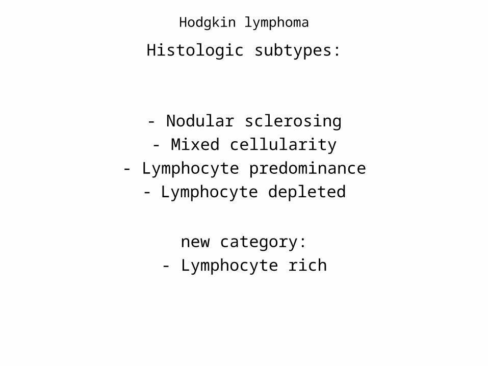
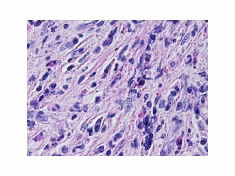
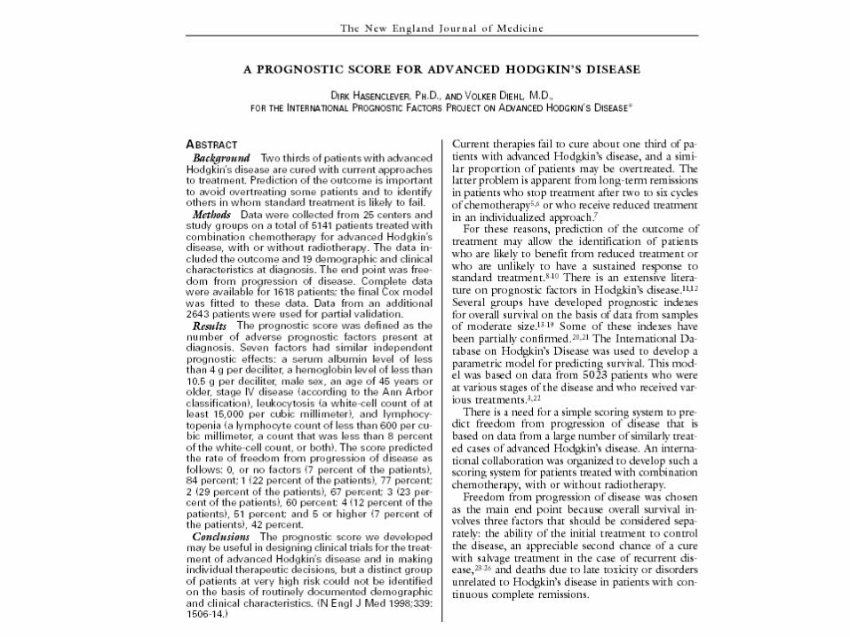
Hodgkin lymphoma
Histologic subtypes:
- Nodular sclerosing
- Mixed cellularity
- Lymphocyte predominance- Lymphocyte depleted
new category:
- Lymphocyte rich
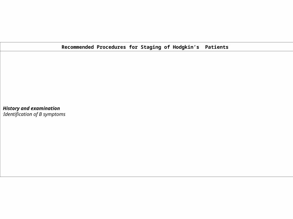
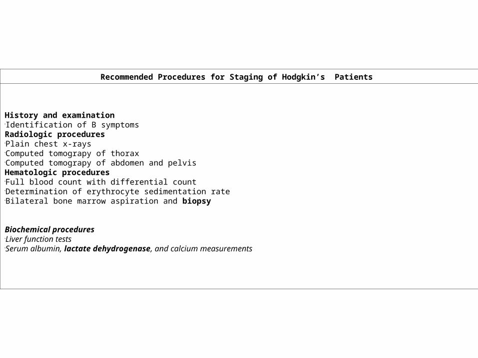
Recommended Procedures for Staging of Hodgkin’s Patients
History and examination Identification of B symptoms
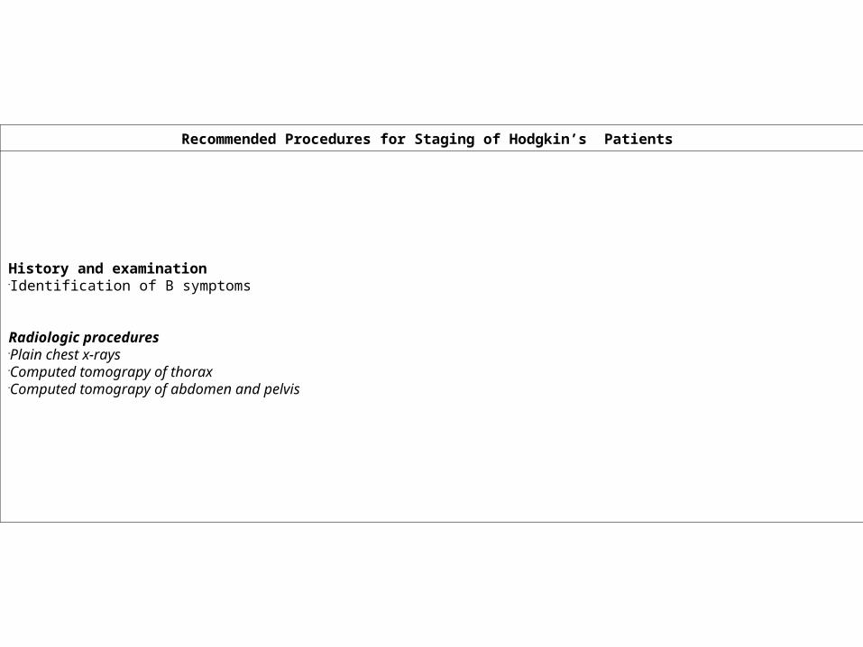
Recommended Procedures for Staging of Hodgkin’s Patients
History and examination Identification of B symptoms
Radiologic procedures Plain chest x-rays Computed tomograpy of thorax Computed tomograpy of abdomen and pelvis
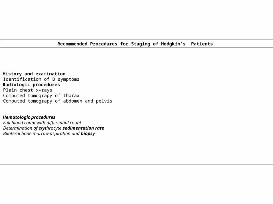
Recommended Procedures for Staging of Hodgkin’s Patients
History and examination Identification of B symptoms Radiologic procedures Plain chest x-rays Computed tomograpy of thorax Computed tomograpy of abdomen and pelvis
Hematologic procedures Full blood count with differential count Determination of erythrocyte sedimentation rate Bilateral bone marrow aspiration and biopsy
Recommended Procedures for Staging of Hodgkin’s Patients
History and examination Identification of B symptoms Radiologic procedures Plain chest x-rays Computed tomograpy of thorax Computed tomograpy of abdomen and pelvis Hematologic procedures Full blood count with differential count Determination of erythrocyte sedimentation rate Bilateral bone marrow aspiration and biopsy
Biochemical procedures Liver function tests Serum albumin, lactate dehydrogenase, and calcium measurements
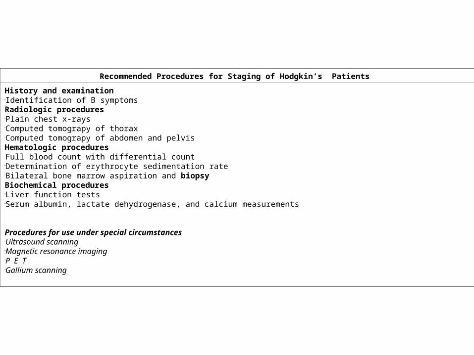
Recommended Procedures for Staging of Hodgkin’s Patients
History and examination Identification of B symptoms Radiologic procedures Plain chest x-rays Computed tomograpy of thorax Computed tomograpy of abdomen and pelvis Hematologic procedures Full blood count with differential count Determination of erythrocyte sedimentation rate Bilateral bone marrow aspiration and biopsy Biochemical procedures Liver function tests Serum albumin, lactate dehydrogenase, and calcium measurements
Procedures for use under special circumstances Ultrasound scanning Magnetic resonance imaging P E TGallium scanning
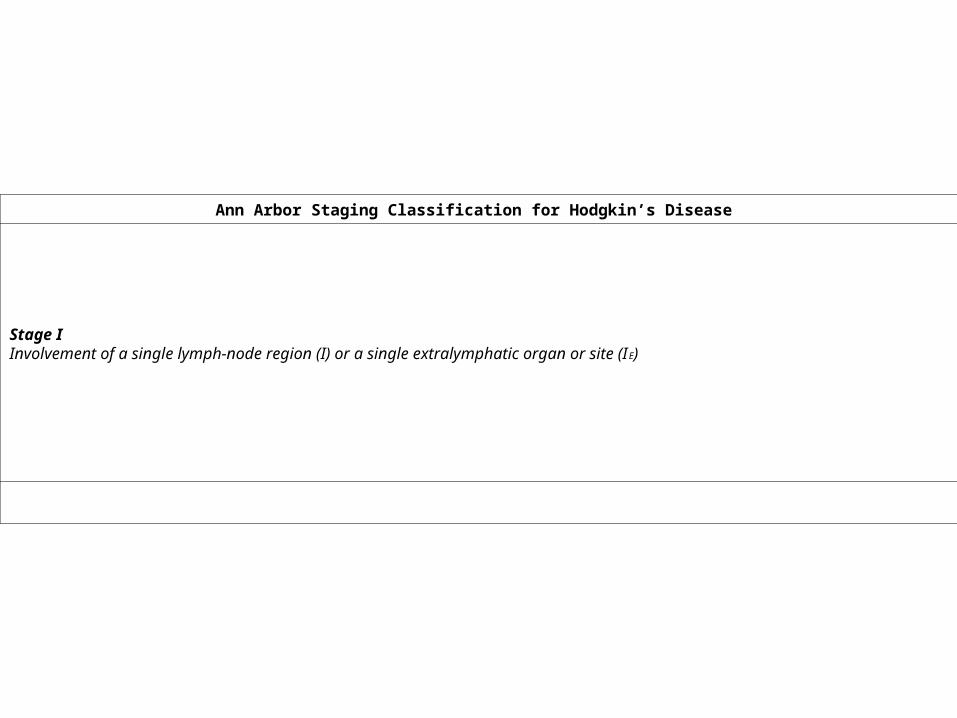
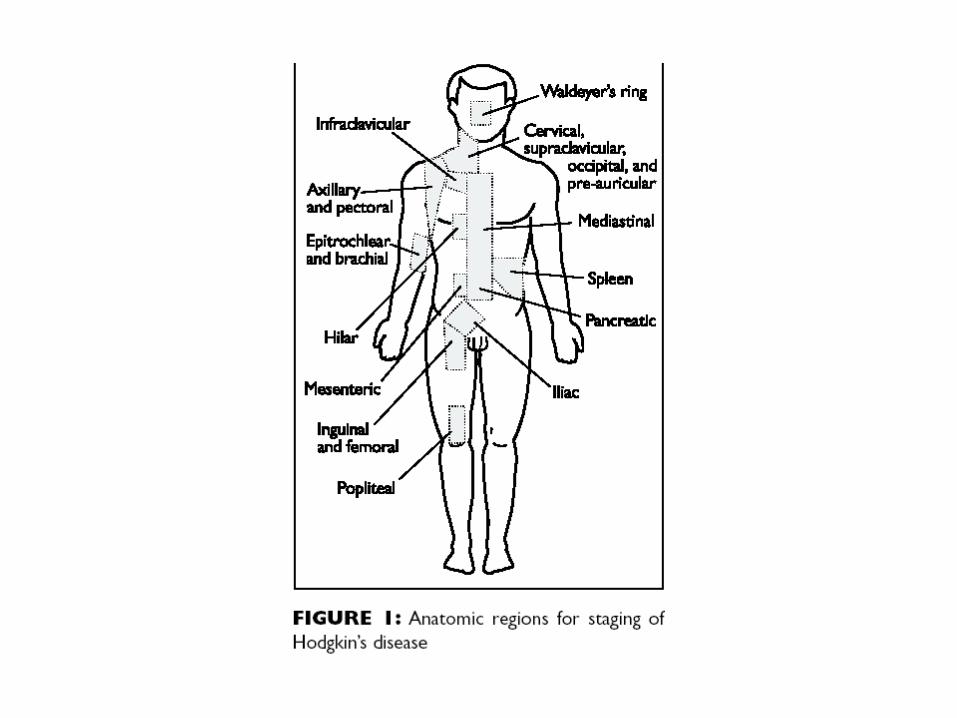
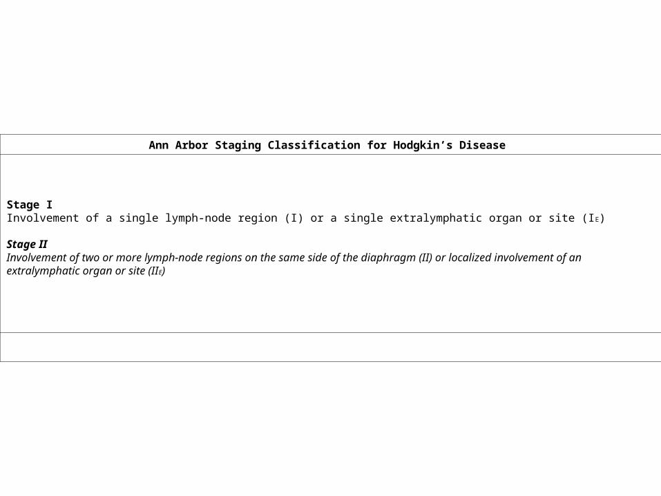
Ann Arbor Staging Classification for Hodgkin’s Disease
Stage I Involvement of a single lymph-node region (I) or a single extralymphatic organ or site (IE)
Ann Arbor Staging Classification for Hodgkin’s Disease
Stage I Involvement of a single lymph-node region (I) or a single extralymphatic organ or site (IE)
Stage II Involvement of two or more lymph-node regions on the same side of the diaphragm (II) or localized involvement of an extralymphatic organ or site (IIE)
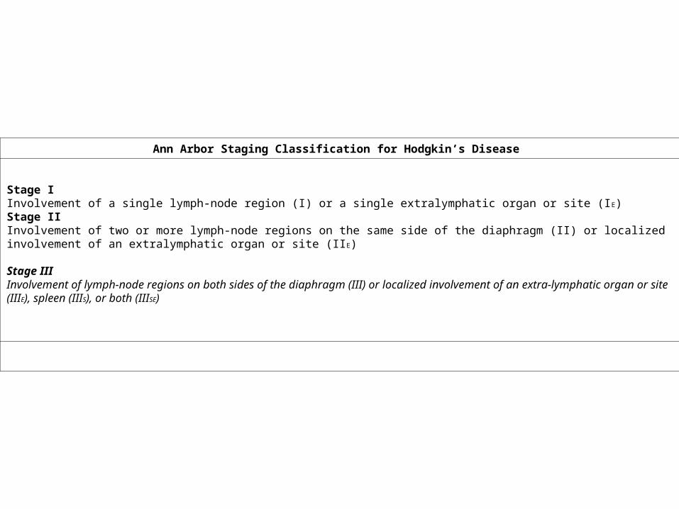
Ann Arbor Staging Classification for Hodgkin’s Disease
Stage I Involvement of a single lymph-node region (I) or a single extralymphatic organ or site (IE) Stage II Involvement of two or more lymph-node regions on the same side of the diaphragm (II) or localized involvement of an extralymphatic organ or site (IIE)
Stage III Involvement of lymph-node regions on both sides of the diaphragm (III) or localized involvement of an extra-lymphatic organ or site (III E), spleen (IIIS), or both (IIISE)
Ann Arbor Staging Classification for Hodgkin’s Disease
Stage I Involvement of a single lymph-node region (I) or a single extralymphatic organ or site (IE) Stage II Involvement of two or more lymph-node regions on the same side of the diaphragm (II) or localized involvement of an extralymphatic organ or site (IIE) Stage III Involvement of lymph-node regions on both sides of the diaphragm (III) or localized involvement of an extra-lymphatic organ or site (III E), spleen (IIIS), or both (IIISE)
Stage IV Diffuse or disseminated involvement of one or more extralymphatic organs, with or without associated lymph-node involvement; the organ(s) involved should be identified by a symbol: (P) pulmonary, (O) osseous, or (H) hepatic.
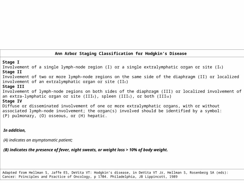
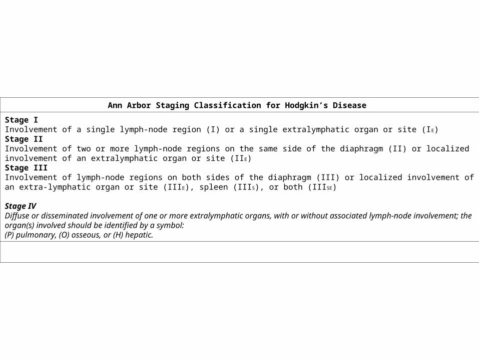
Ann Arbor Staging Classification for Hodgkin’s Disease
Stage I Involvement of a single lymph-node region (I) or a single extralymphatic organ or site (IE) Stage II Involvement of two or more lymph-node regions on the same side of the diaphragm (II) or localized involvement of an extralymphatic organ or site (IIE) Stage III Involvement of lymph-node regions on both sides of the diaphragm (III) or localized involvement of an extra-lymphatic organ or site (III E), spleen (IIIS), or both (IIISE) Stage IV Diffuse or disseminated involvement of one or more extralymphatic organs, with or without associated lymph-node involvement; the organ(s) involved should be identified by a symbol: (P) pulmonary, (O) osseous, or (H) hepatic.
In addition,
(A) indicates an asymptomatic patient;
(B) indicates the presence of fever, night sweats, or weight loss > 10% of body weight.
Adapted from Hellman S, Jaffe ES, DeVita VT: Hodgkin’s disease, in DeVita VT Jr, Hellman S, Rosenberg SA (eds): Cancer: Principles and Practice of Oncology, p 1704. Philadelphia, JB Lippincott, 1989
Radiotherapy
Chemiotherapy
A B M T
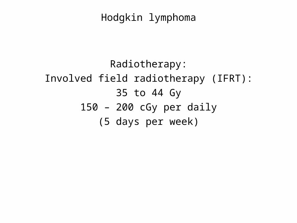
Hodgkin lymphoma
Radiotherapy:
Involved field radiotherapy (IFRT):
35 to 44 Gy
150 – 200 cGy per daily
(5 days per week)
Radiotherapy
Chemiotherapy
A B M T
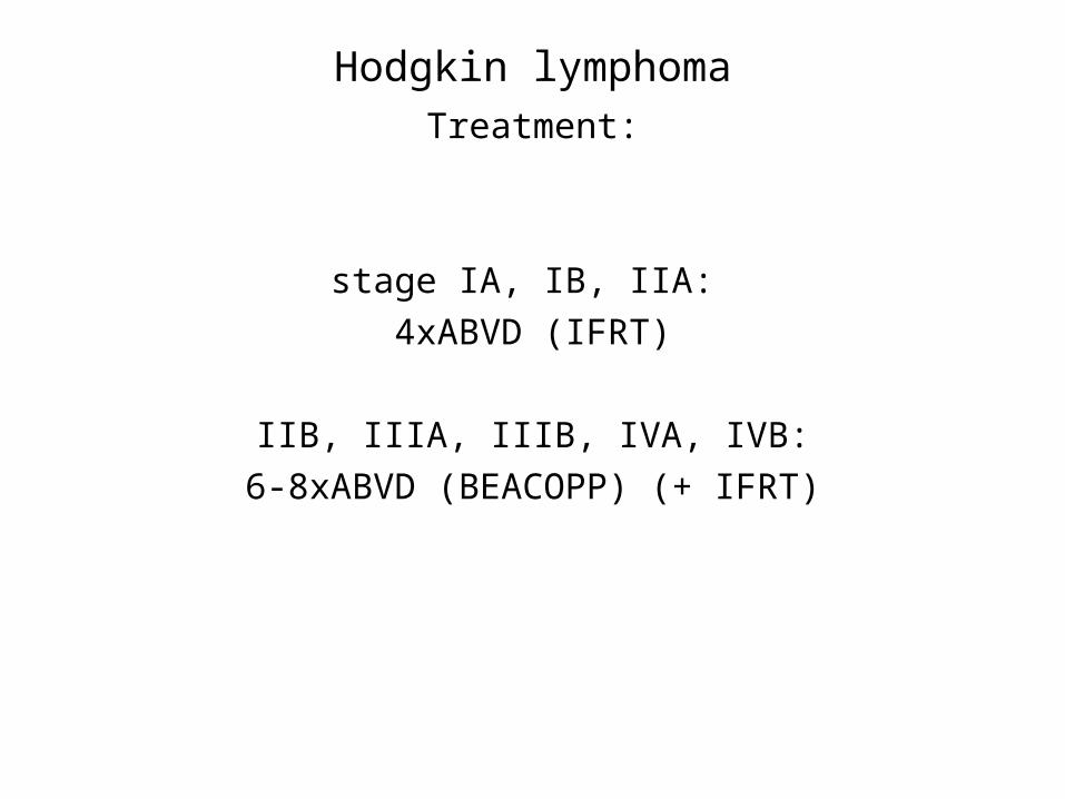
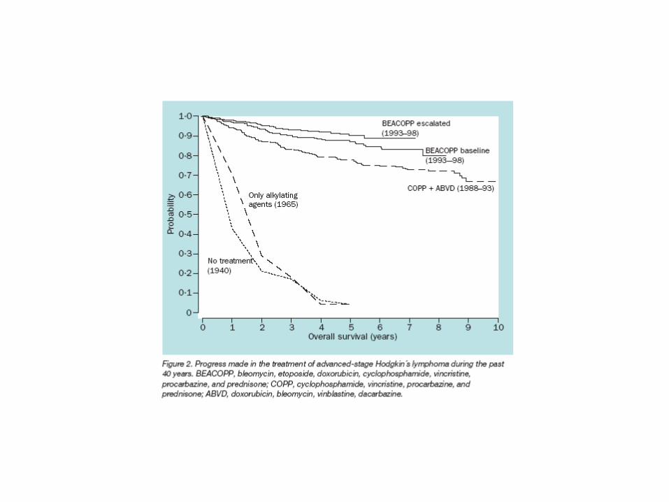
Hodgkin lymphomaTreatment:
stage IA, IB, IIA:
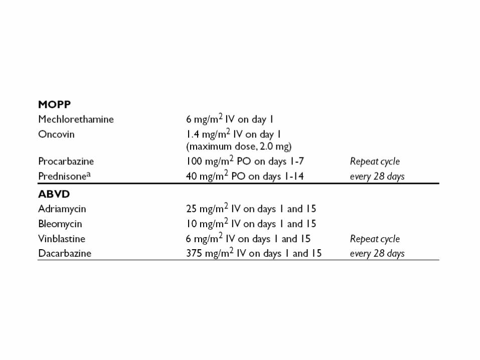
4xABVD (IFRT)
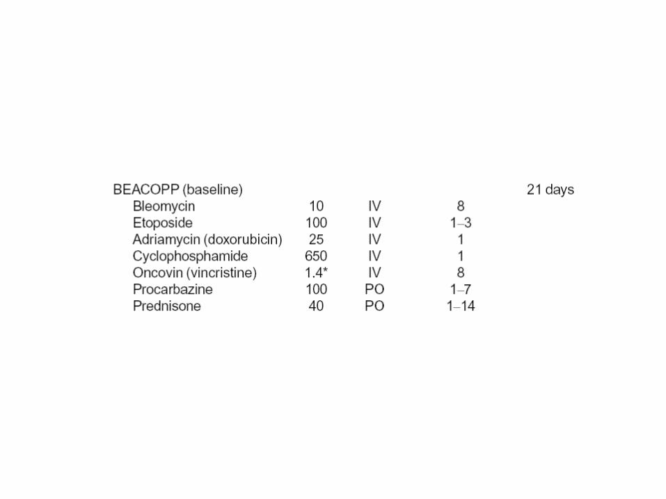
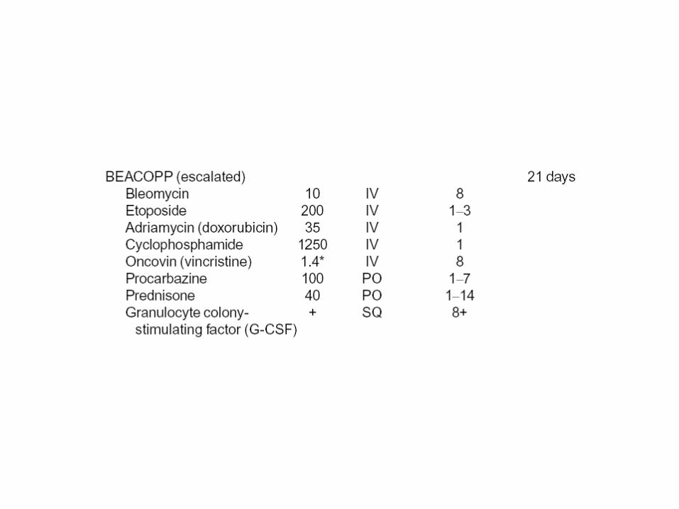
IIB, IIIA, IIIB, IVA, IVB:
6-8xABVD (BEACOPP) (+ IFRT)
Radiotherapy
Chemiotherapy
A B M T


![Predominance of Islam [Fath-i Islam]](https://img.dokumen.tips/doc/110x75/577d29a71a28ab4e1ea76c95/predominance-of-islam-fath-i-islam.jpg)


