Embed Size (px)
Citation preview
Surgical Technique for Osteocutaneous Pedicle Flap Transfer for Salvage of Transtibial Amputation After
Severe Lower-Extremity Injury
by Heather A. Vallier, Brendan M. Patterson, and John K. Sontich
JBJS Essent Surg TechVolume 2(4):e21October 10, 2012
©2012 by The Journal of Bone and Joint Surgery, Inc.
Heather A. Vallier et al. JBJS Essent Surg Tech 2012;2:e21
©2012 by The Journal of Bone and Joint Surgery, Inc.
Heather A. Vallier et al. JBJS Essent Surg Tech 2012;2:e21
©2012 by The Journal of Bone and Joint Surgery, Inc.
Heather A. Vallier et al. JBJS Essent Surg Tech 2012;2:e21
©2012 by The Journal of Bone and Joint Surgery, Inc.

Heather A. Vallier et al. JBJS Essent Surg Tech 2012;2:e21
©2012 by The Journal of Bone and Joint Surgery, Inc.

Heather A. Vallier et al. JBJS Essent Surg Tech 2012;2:e21
©2012 by The Journal of Bone and Joint Surgery, Inc.
Heather A. Vallier et al. JBJS Essent Surg Tech 2012;2:e21
©2012 by The Journal of Bone and Joint Surgery, Inc.
Heather A. Vallier et al. JBJS Essent Surg Tech 2012;2:e21
©2012 by The Journal of Bone and Joint Surgery, Inc.
Heather A. Vallier et al. JBJS Essent Surg Tech 2012;2:e21
©2012 by The Journal of Bone and Joint Surgery, Inc.
Heather A. Vallier et al. JBJS Essent Surg Tech 2012;2:e21
©2012 by The Journal of Bone and Joint Surgery, Inc.
Heather A. Vallier et al. JBJS Essent Surg Tech 2012;2:e21
©2012 by The Journal of Bone and Joint Surgery, Inc.
Heather A. Vallier et al. JBJS Essent Surg Tech 2012;2:e21
©2012 by The Journal of Bone and Joint Surgery, Inc.
Heather A. Vallier et al. JBJS Essent Surg Tech 2012;2:e21
©2012 by The Journal of Bone and Joint Surgery, Inc.
Heather A. Vallier et al. JBJS Essent Surg Tech 2012;2:e21
©2012 by The Journal of Bone and Joint Surgery, Inc.
Heather A. Vallier et al. JBJS Essent Surg Tech 2012;2:e21
©2012 by The Journal of Bone and Joint Surgery, Inc.
Heather A. Vallier et al. JBJS Essent Surg Tech 2012;2:e21
©2012 by The Journal of Bone and Joint Surgery, Inc.
Heather A. Vallier et al. JBJS Essent Surg Tech 2012;2:e21
©2012 by The Journal of Bone and Joint Surgery, Inc.
Heather A. Vallier et al. JBJS Essent Surg Tech 2012;2:e21
©2012 by The Journal of Bone and Joint Surgery, Inc.

Heather A. Vallier et al. JBJS Essent Surg Tech 2012;2:e21
©2012 by The Journal of Bone and Joint Surgery, Inc.
Heather A. Vallier et al. JBJS Essent Surg Tech 2012;2:e21
©2012 by The Journal of Bone and Joint Surgery, Inc.

Heather A. Vallier et al. JBJS Essent Surg Tech 2012;2:e21
©2012 by The Journal of Bone and Joint Surgery, Inc.
Heather A. Vallier et al. JBJS Essent Surg Tech 2012;2:e21
©2012 by The Journal of Bone and Joint Surgery, Inc.
Heather A. Vallier et al. JBJS Essent Surg Tech 2012;2:e21
©2012 by The Journal of Bone and Joint Surgery, Inc.
Heather A. Vallier et al. JBJS Essent Surg Tech 2012;2:e21
©2012 by The Journal of Bone and Joint Surgery, Inc.
Heather A. Vallier et al. JBJS Essent Surg Tech 2012;2:e21
©2012 by The Journal of Bone and Joint Surgery, Inc.
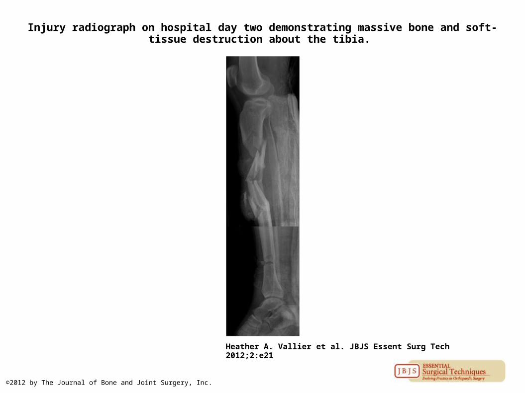
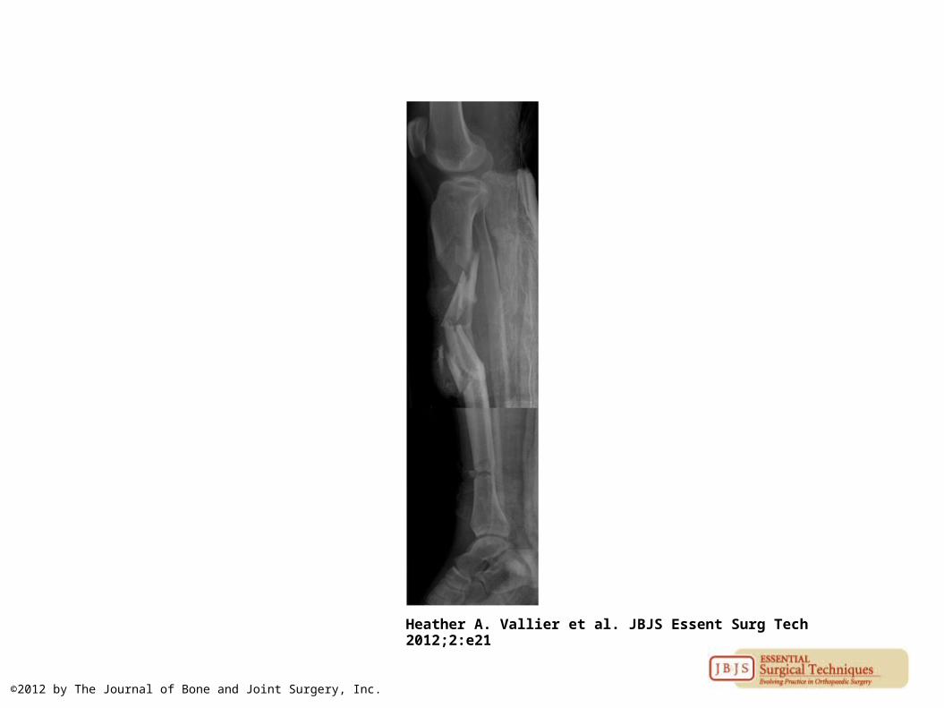
Injury radiograph on hospital day two demonstrating massive bone and soft-tissue destruction about the tibia.
Heather A. Vallier et al. JBJS Essent Surg Tech 2012;2:e21
©2012 by The Journal of Bone and Joint Surgery, Inc.
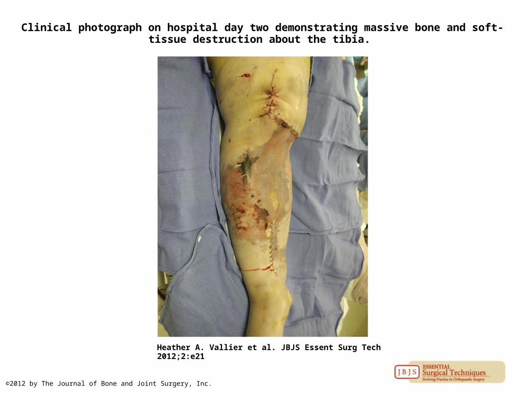
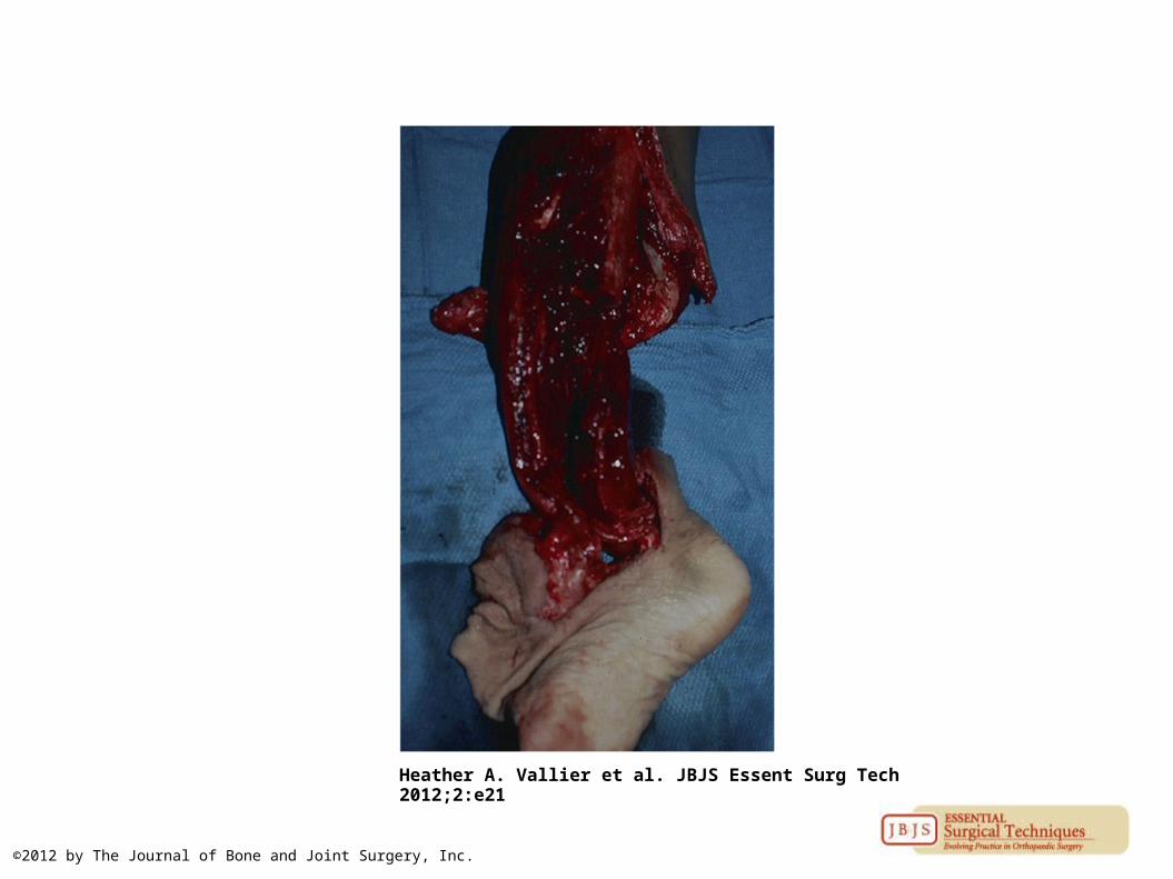
Clinical photograph on hospital day two demonstrating massive bone and soft-tissue destruction about the tibia.
Heather A. Vallier et al. JBJS Essent Surg Tech 2012;2:e21
©2012 by The Journal of Bone and Joint Surgery, Inc.
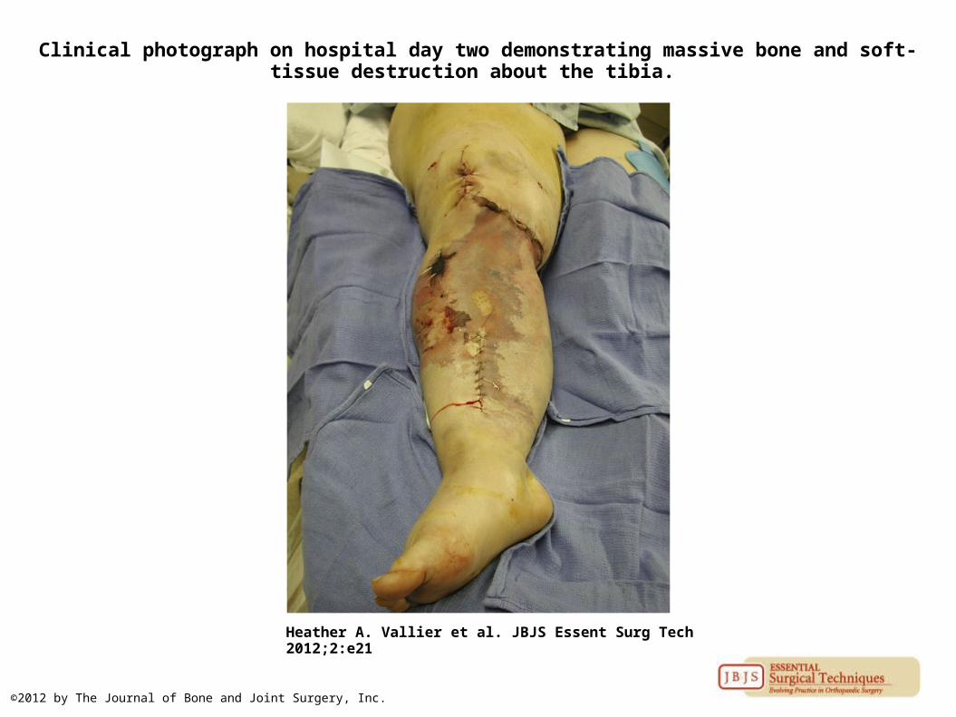
Clinical photograph on hospital day two demonstrating massive bone and soft-tissue destruction about the tibia.
Heather A. Vallier et al. JBJS Essent Surg Tech 2012;2:e21
©2012 by The Journal of Bone and Joint Surgery, Inc.
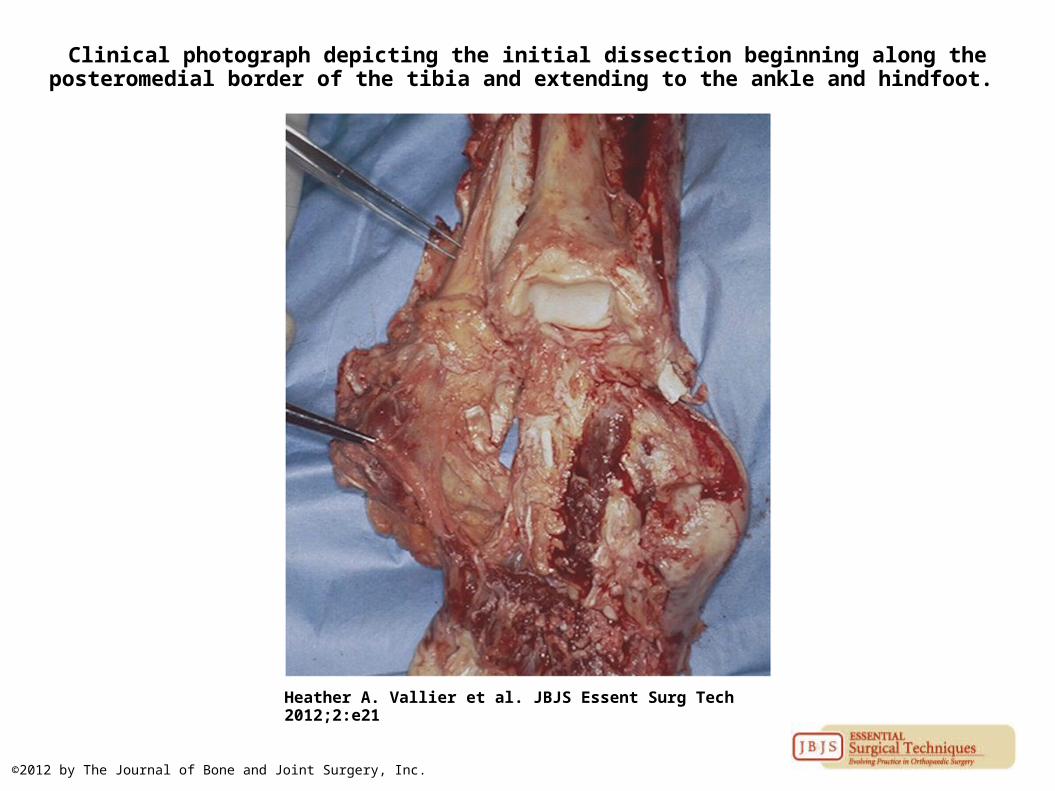
Clinical photograph depicting the initial dissection beginning along the posteromedial border of the tibia and extending to the ankle and hindfoot.
Heather A. Vallier et al. JBJS Essent Surg Tech 2012;2:e21
©2012 by The Journal of Bone and Joint Surgery, Inc.
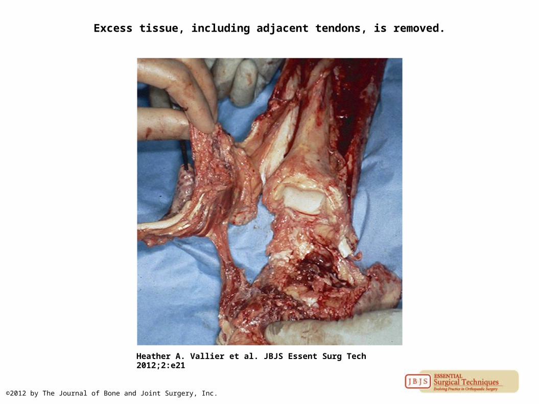
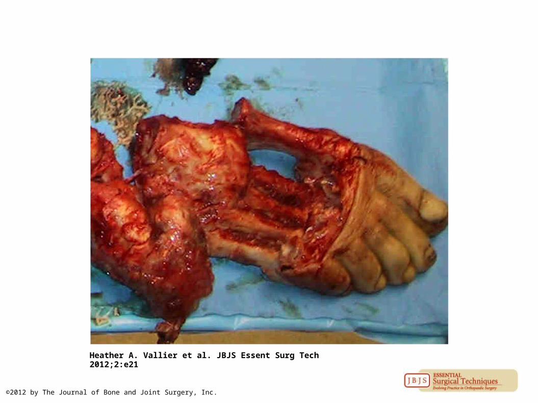
Excess tissue, including adjacent tendons, is removed.
Heather A. Vallier et al. JBJS Essent Surg Tech 2012;2:e21
©2012 by The Journal of Bone and Joint Surgery, Inc.
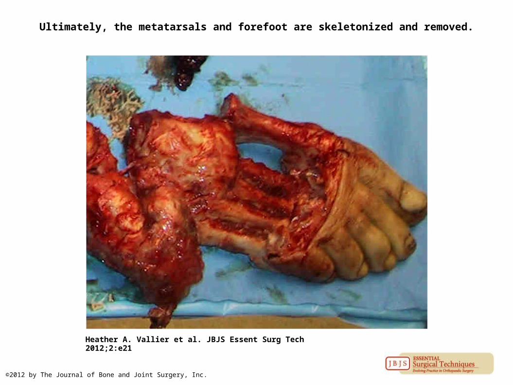
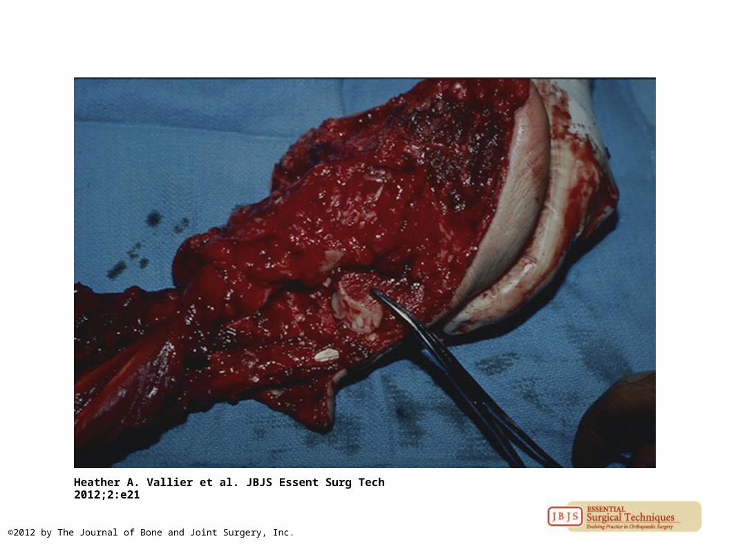
Ultimately, the metatarsals and forefoot are skeletonized and removed.
Heather A. Vallier et al. JBJS Essent Surg Tech 2012;2:e21
©2012 by The Journal of Bone and Joint Surgery, Inc.
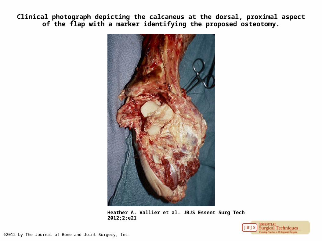
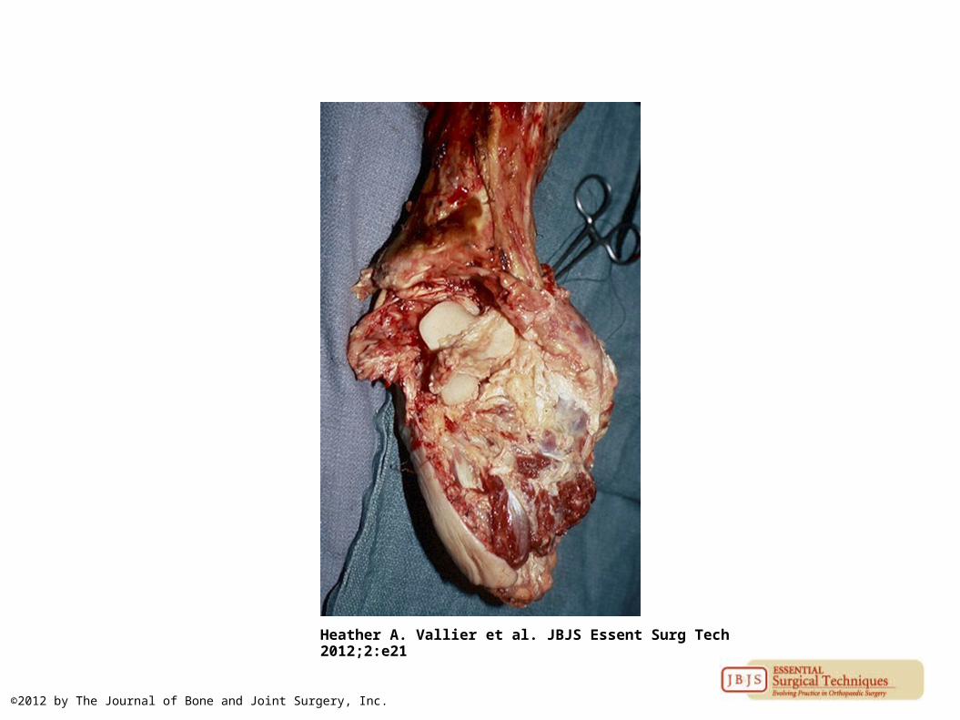
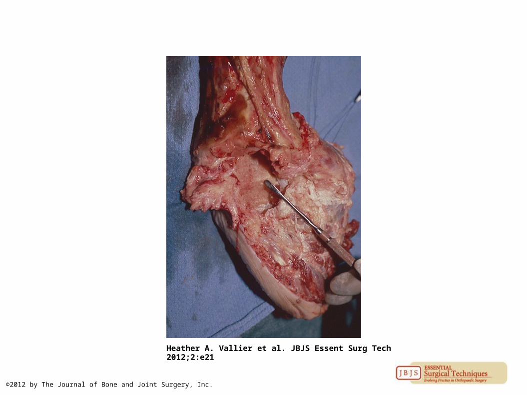
Clinical photograph depicting the calcaneus at the dorsal, proximal aspect of the flap with a marker identifying the proposed osteotomy.
Heather A. Vallier et al. JBJS Essent Surg Tech 2012;2:e21
©2012 by The Journal of Bone and Joint Surgery, Inc.
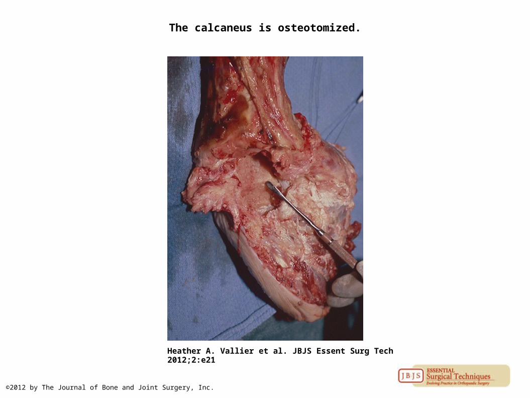
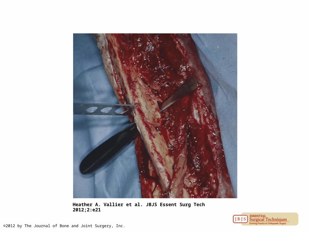
The calcaneus is osteotomized.
Heather A. Vallier et al. JBJS Essent Surg Tech 2012;2:e21
©2012 by The Journal of Bone and Joint Surgery, Inc.
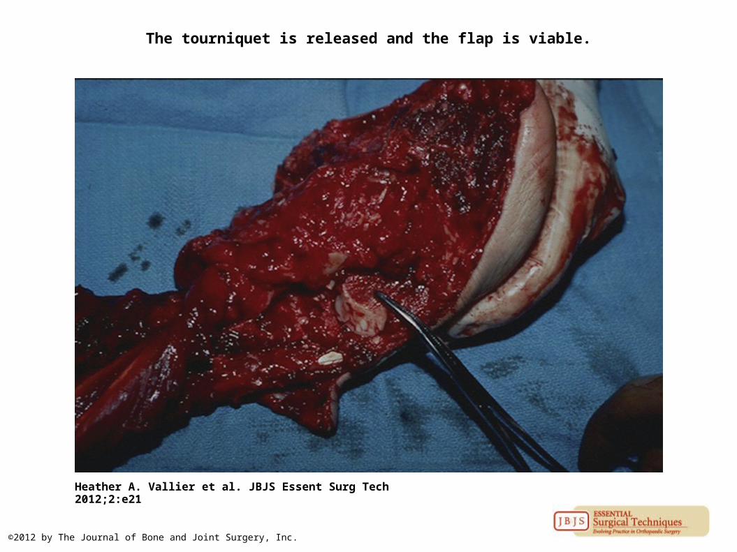
The tourniquet is released and the flap is viable.
Heather A. Vallier et al. JBJS Essent Surg Tech 2012;2:e21
©2012 by The Journal of Bone and Joint Surgery, Inc.
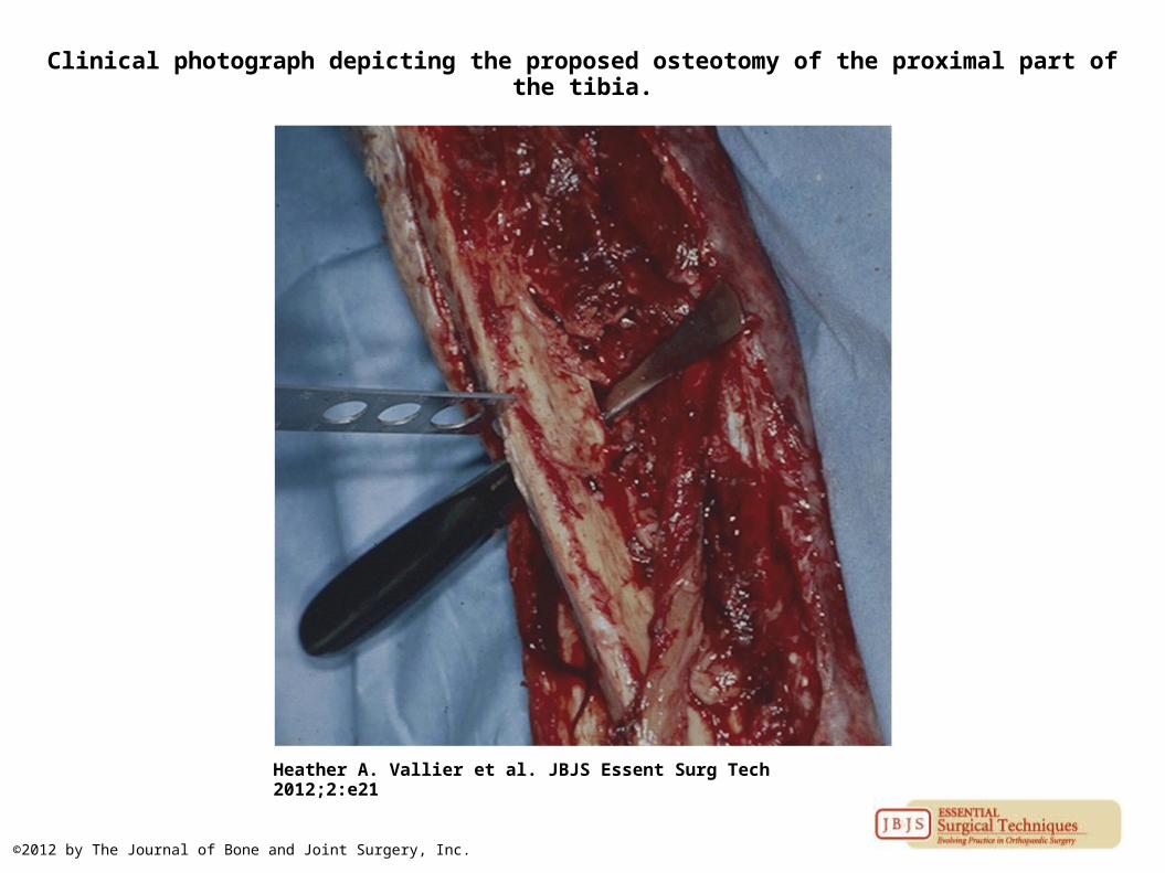
Clinical photograph depicting the proposed osteotomy of the proximal part of the tibia.
Heather A. Vallier et al. JBJS Essent Surg Tech 2012;2:e21
©2012 by The Journal of Bone and Joint Surgery, Inc.
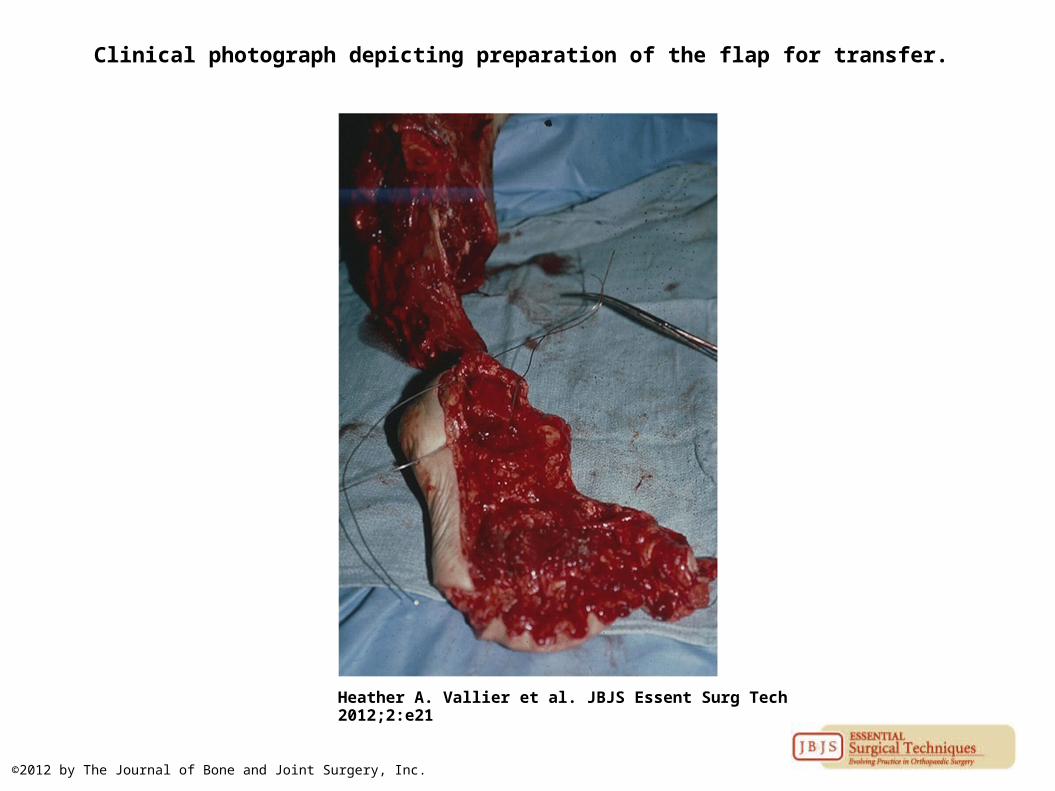
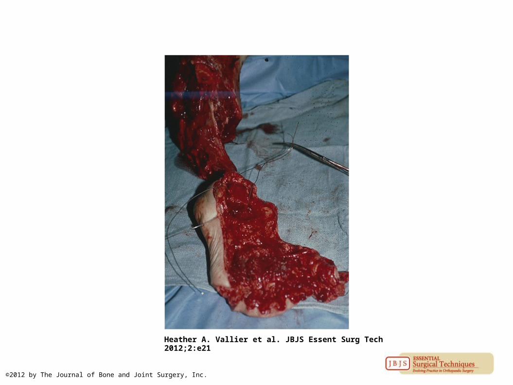
Clinical photograph depicting preparation of the flap for transfer.
Heather A. Vallier et al. JBJS Essent Surg Tech 2012;2:e21
©2012 by The Journal of Bone and Joint Surgery, Inc.
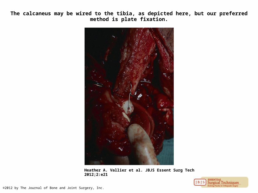
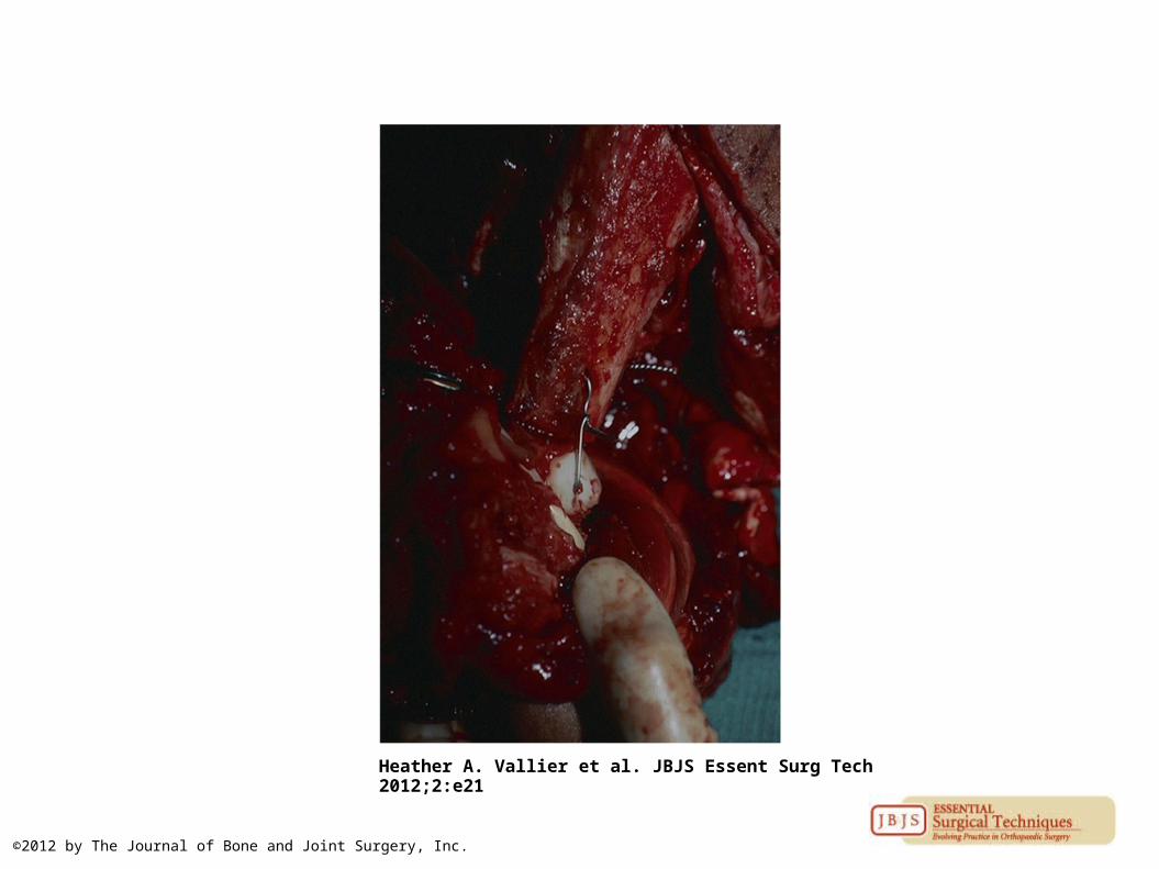
The calcaneus may be wired to the tibia, as depicted here, but our preferred method is plate fixation.
Heather A. Vallier et al. JBJS Essent Surg Tech 2012;2:e21
©2012 by The Journal of Bone and Joint Surgery, Inc.

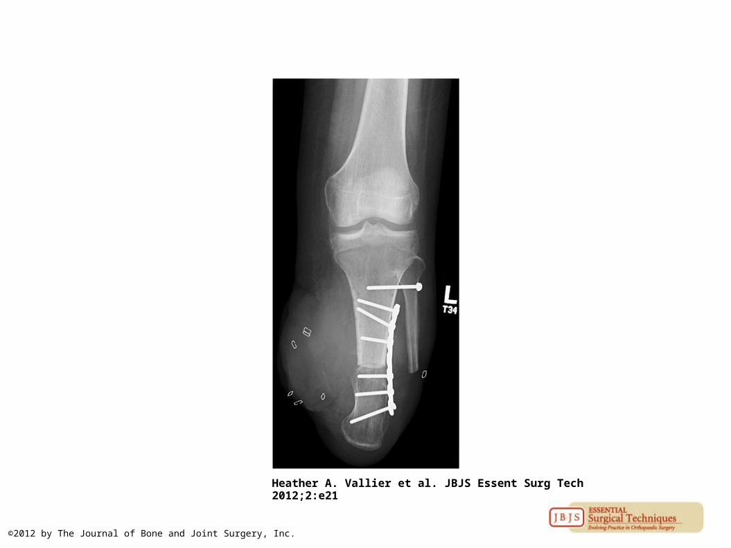
Anteroposterior radiograph depicting plate fixation, our preferred method.
Heather A. Vallier et al. JBJS Essent Surg Tech 2012;2:e21
©2012 by The Journal of Bone and Joint Surgery, Inc.
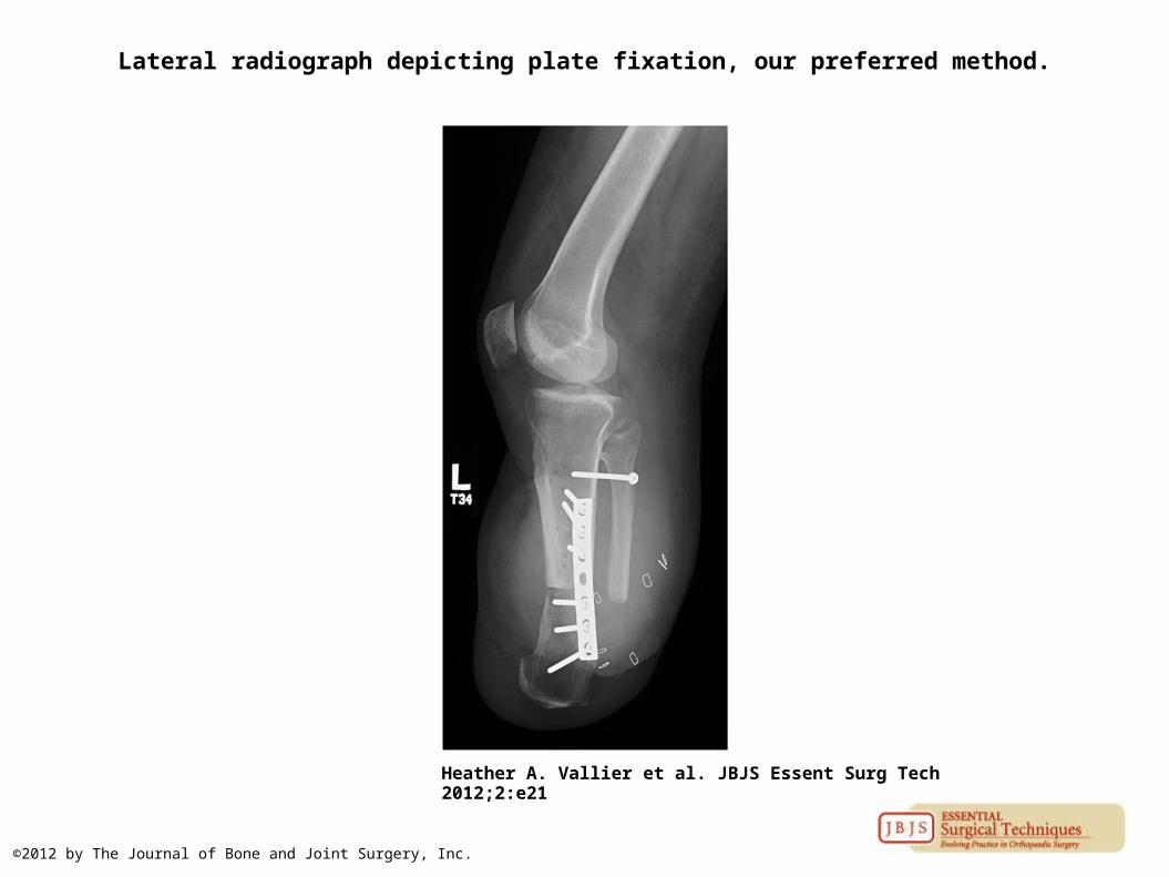
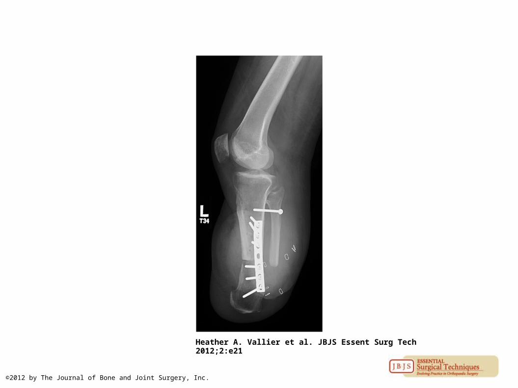
Lateral radiograph depicting plate fixation, our preferred method.
Heather A. Vallier et al. JBJS Essent Surg Tech 2012;2:e21
©2012 by The Journal of Bone and Joint Surgery, Inc.
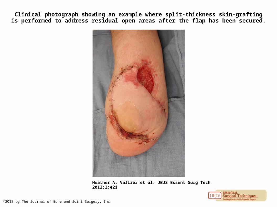
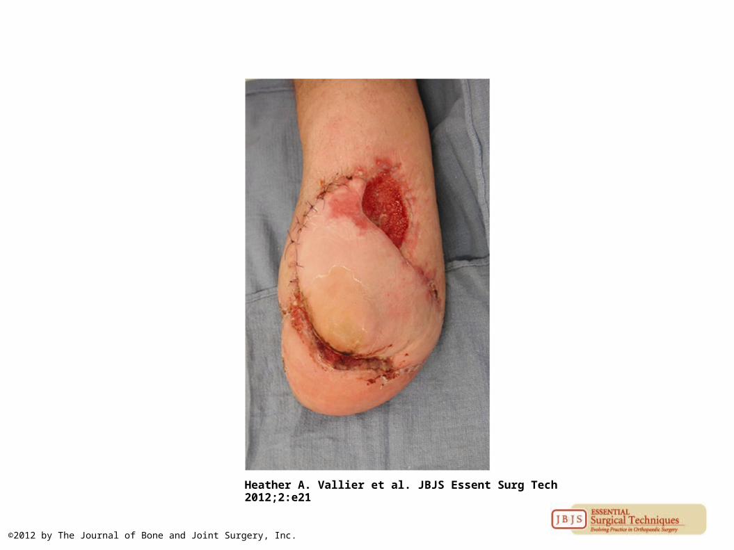
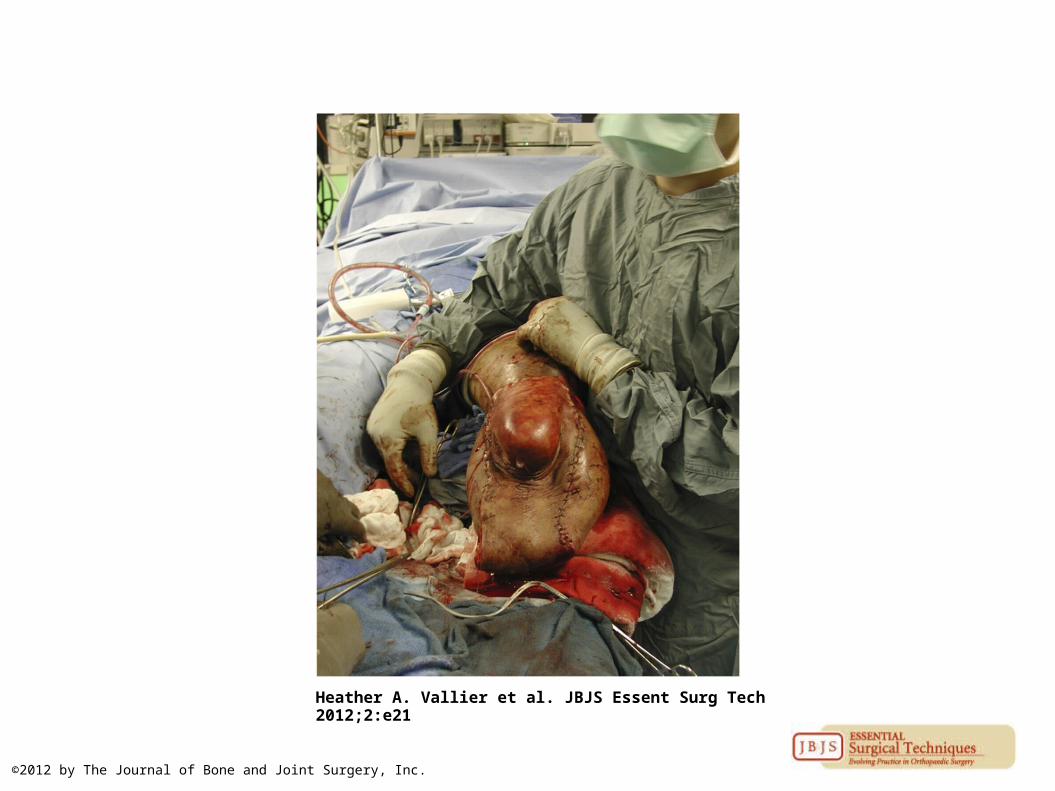
Clinical photograph showing an example where split-thickness skin-grafting is performed to address residual open areas after the flap has been secured.
Heather A. Vallier et al. JBJS Essent Surg Tech 2012;2:e21
©2012 by The Journal of Bone and Joint Surgery, Inc.
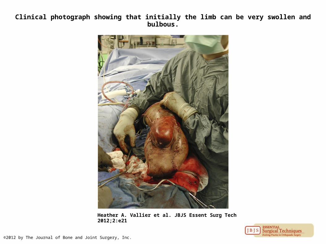
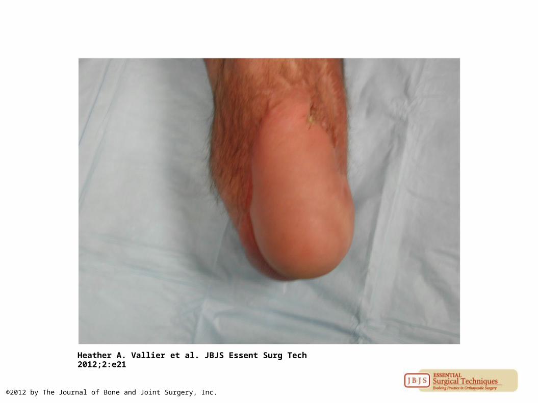
Clinical photograph showing that initially the limb can be very swollen and bulbous.
Heather A. Vallier et al. JBJS Essent Surg Tech 2012;2:e21
©2012 by The Journal of Bone and Joint Surgery, Inc.
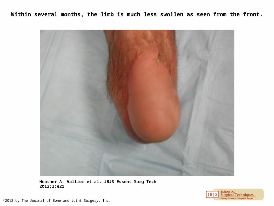
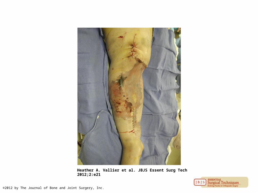
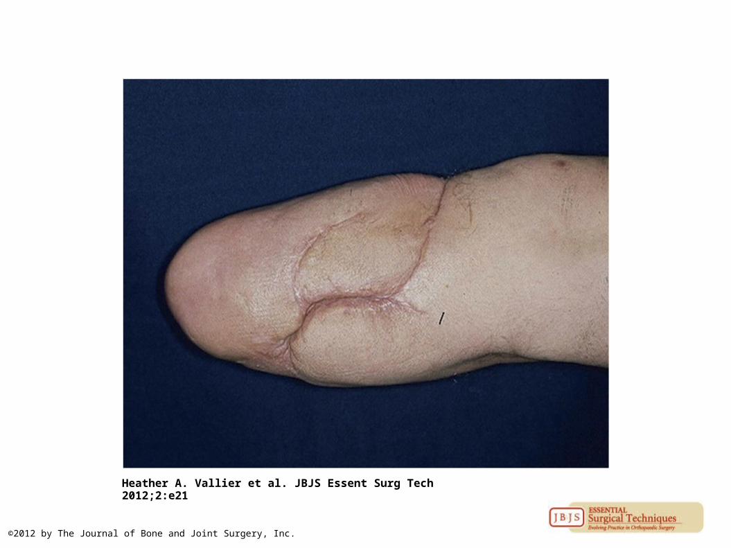
Within several months, the limb is much less swollen as seen from the front.
Heather A. Vallier et al. JBJS Essent Surg Tech 2012;2:e21
©2012 by The Journal of Bone and Joint Surgery, Inc.
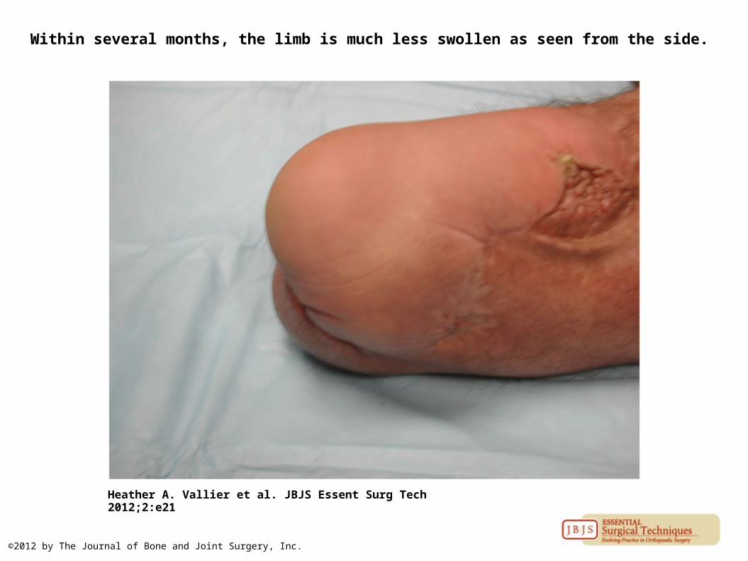
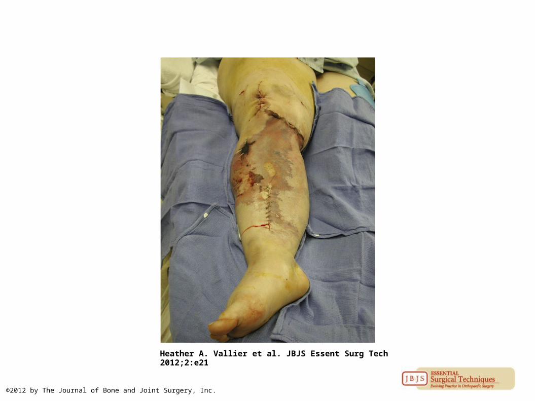
Within several months, the limb is much less swollen as seen from the side.
Heather A. Vallier et al. JBJS Essent Surg Tech 2012;2:e21
©2012 by The Journal of Bone and Joint Surgery, Inc.

Lateral view of knee extension after healing.
Heather A. Vallier et al. JBJS Essent Surg Tech 2012;2:e21
©2012 by The Journal of Bone and Joint Surgery, Inc.
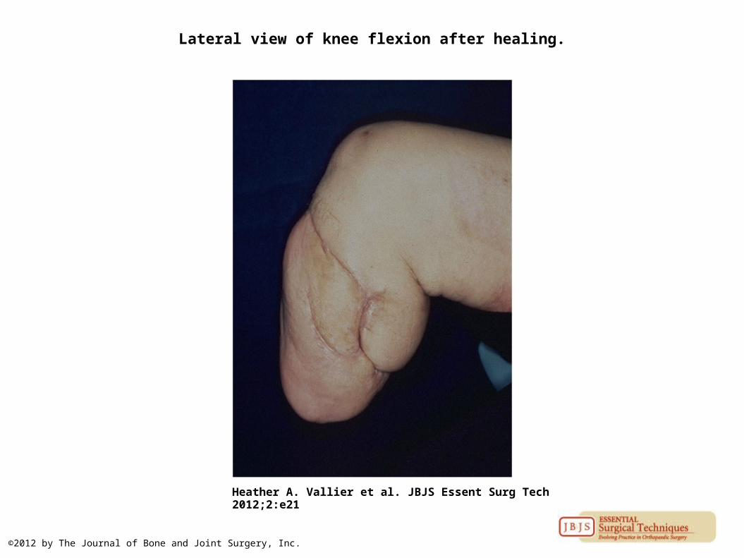
Lateral view of knee flexion after healing.
Heather A. Vallier et al. JBJS Essent Surg Tech 2012;2:e21
©2012 by The Journal of Bone and Joint Surgery, Inc.

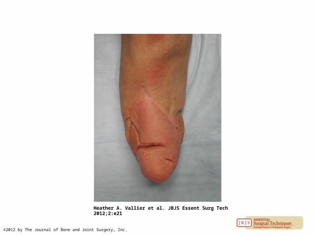
The stump is very durable.
Heather A. Vallier et al. JBJS Essent Surg Tech 2012;2:e21
©2012 by The Journal of Bone and Joint Surgery, Inc.

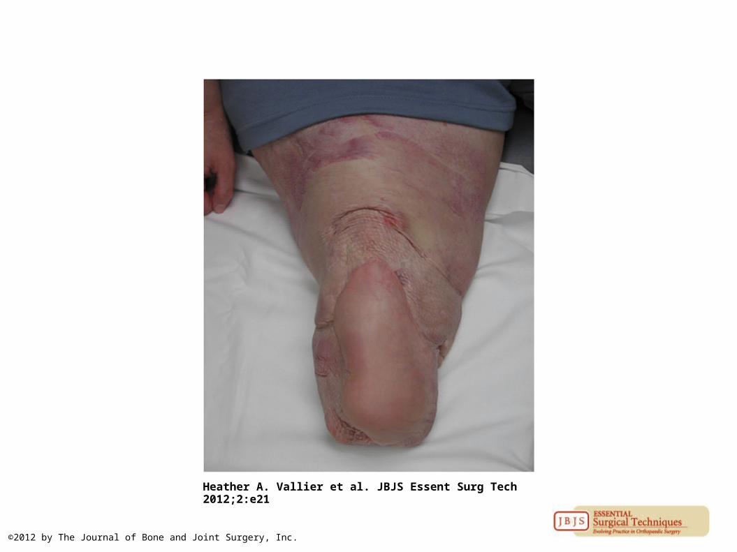
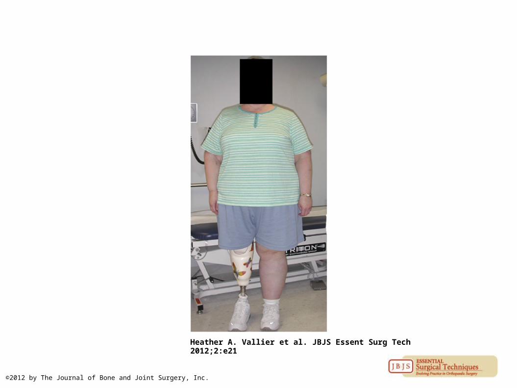
The stump is very durable even in patients who are large and/or have heavy activity demands.
Heather A. Vallier et al. JBJS Essent Surg Tech 2012;2:e21
©2012 by The Journal of Bone and Joint Surgery, Inc.

Clinical photograph depicting redundant lateral hindfoot skin, which often has a tenuous blood supply.
Heather A. Vallier et al. JBJS Essent Surg Tech 2012;2:e21
©2012 by The Journal of Bone and Joint Surgery, Inc.
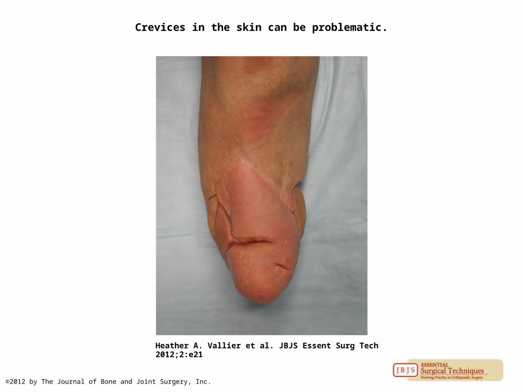
Crevices in the skin can be problematic.
Heather A. Vallier et al. JBJS Essent Surg Tech 2012;2:e21
©2012 by The Journal of Bone and Joint Surgery, Inc.