Embed Size (px)
Citation preview
Cellular/Molecular
STIM1 Regulates Somatic Ca2� Signals and Intrinsic FiringProperties of Cerebellar Purkinje Neurons
X Changhyeon Ryu,1,2,3,4* X Dong Cheol Jang,1,3,5* X Dayoon Jung,1,2,3* X Yong Gyu Kim,1,2,3 X Hyun Geun Shim,1,2,3
Hyun-Hee Ryu,1,8 Yong-Seok Lee,1,2,4 X David J. Linden,6 Paul F. Worley,6,7 and X Sang Jeong Kim1,2,3,4
Departments of 1Physiology and 2Biomedical Sciences, 3Neuro-Immune Information Storage Network Research Center, and 4Neuroscience ResearchInstitute, Medical Research Center, Seoul National University College of Medicine, Jongno-gu, Seoul 03080, Korea, 5Department of Brain and CognitiveSciences, College of Natural Sciences, Seoul National University, Kwanak-gu, Seoul 08826, Korea, Departments of 6Neuroscience and 7Neurology, JohnsHopkins University School of Medicine, Baltimore, Maryland 21205, and 8Department of Life Science, College of Natural Sciences, Chung-Ang University,Dongjak-gu, Seoul 06974, Korea
Control of Ca 2� flux between the cytosol and intracellular Ca 2� stores is essential for maintaining normal cellular function. It has beenwell established in both neuronal and non-neuronal cells that stromal interaction molecule 1 (STIM1) initiates and regulates refillingCa 2� into the ER. Here, we describe a novel, additional role for STIM1, the regulation of free cytosolic Ca 2�, and the consequent controlof spike firing in neurons. Among central neurons, cerebellar Purkinje neurons express the highest level of STIM1, and they fire contin-uously in the absence of stimulation, making somatic Ca 2� homeostasis of particular importance. By using Purkinje neuron-specificSTIM1 knock-out (STIM1 PKO) male mice, we found that the deletion of STIM1 delayed clearance of cytosolic Ca 2� in the soma duringongoing neuronal firing. Deletion of STIM1 also reduced the Purkinje neuronal excitability and impaired intrinsic plasticity withoutaffecting long-term synaptic plasticity. In vestibulo-ocular reflex learning, STIM1 PKO male mice showed severe deficits in memoryconsolidation, whereas they were normal in memory acquisition. Our results suggest that STIM1 is critically involved in the regulation ofthe neuronal excitability and the intrinsic plasticity of the Purkinje neurons as well as cerebellar memory consolidation.
Key words: cerebellum; intrinsic excitability; intrinsic plasticity; memory consolidation; Purkinje neuron; STIM1
IntroductionCa 2� flux originating from both the extracellular space and fromintracellular stores modifies cytosolic Ca 2� (Verkhratsky, 2005).
The endoplasmic reticulum (ER) is the largest intracellular Ca 2�
store and contributes to the dynamics of cytosolic Ca 2� by releas-ing stored Ca 2� through activation of ryanodine receptors(RyRs) and inositol-triphosphate receptors (IP3Rs). These formsof Ca 2� release are allowed only when ER Ca 2� stores are filled.Thus, refilling Ca 2� into the ER is an essential process for appro-priate cytosolic Ca 2� dynamics. Stromal interaction molecule(STIM), an ER membrane-bounded protein, plays a central rolein the refilling process by sensing Ca 2� concentration in the ERlumen ([Ca 2�]ER). In response to low [Ca 2�]ER, STIM oligomer-
Received Dec. 28, 2016; revised July 19, 2017; accepted July 31, 2017.Author contributions: C.R., D.C.J., D.J., and S.J.K. designed research; C.R., D.C.J., D.J., H.G.S., and H.-H.R. per-
formed research; C.R., D.C.J., D.J., and Y.G.K. analyzed data; C.R., D.C.J., D.J., Y.-S.L., D.J.L., P.F.W., and S.J.K. wrotethe paper.
*C.R., D.C.J., and D.J. contributed equally to this work.The authors declare no competing financial interests.This work was supported by grants (the Medical Research Center, Grant 2012R1A5A2A44671346; Small Grant for
Exploratory Research, Grant 2016R1D1A1A02937282; Global Ph.D. Fellowship Program, Grant 2013H1A2A1034318;and the Medical Researchers Support Program) from the National Research Foundation of Korea. We thank ProfessorSascha du Lac (Johns Hopkins University) and her research group for helping with the installation of the VOR testsystem. We also thank Geehoon Chung, Jewel Park, Junho Hwang, Sang Bin Hong, and Ji Han Kim for comments ona prior version of the manuscript and for correcting errors in this manuscript.
Correspondence should be addressed to Sang Jeong Kim, Departments of Physiology, Seoul National UniversityCollege of Medicine, Jongno-gu, Seoul 03080, Korea. E-mail: [email protected].
DOI:10.1523/JNEUROSCI.3973-16.2017Copyright © 2017 the authors 0270-6474/17/378876-19$15.00/0
Significance Statement
Stromal interaction molecule 1 (STIM1), which regulates the refilling of ER Ca 2�, has been investigated in several systemsincluding the CNS. In addition to a previous study showing that STIM1 regulates dendritic ER Ca 2� refilling and mGluR1-mediated synaptic transmission, we provide compelling evidence describing a novel role of STIM1 in spike firing Purkinje neu-rons. We found that STIM1 regulates cytosolic Ca 2� clearance of the soma during spike firing, and the interruption of thiscytosolic Ca 2� clearing disrupts neuronal excitability and cerebellar memory consolidation. Our results provide new insights intoneuronal functions of STIM1 from single neuronal Ca 2� dynamics to behavior level.
8876 • The Journal of Neuroscience, September 13, 2017 • 37(37):8876 – 8894

izes and initiates the ER store refilling process in which ER andSTIM localize in close apposition to the plasma membrane inassociation with proteins including store-operated Ca 2� chan-nels (SOCs) and sarco/endoplasmic reticulum Ca 2�-ATPase(SERCA; Soboloff et al., 2012). While well studied in other cellu-lar systems, the neuronal functions of STIM have just begun to beidentified (Hartmann et al., 2014; Sun et al., 2014; Zhang et al.,2015).
The Purkinje neuron (PN) is the central neuron of the cere-bellar cortex. It broadly integrates sensory-motor informationand provides the sole output of the cerebellar cortex (Dean et al.,2010). The ER in PNs is widely distributed, extending from den-dritic spines, where synaptic plasticity occurs, to the soma, wherefinal characteristics of neuronal output are determined (Terasakiet al., 1994). Cellular mechanisms responsible for learning andmemory have been explained by the regulation of single neuronalinput and output, most well represented by synaptic plasticityand intrinsic plasticity, respectively (Zhang and Linden, 2003;Kandel et al., 2014). Therefore, it would seem to be obvious thatSTIM in PNs could contribute to cerebellar learning and memoryvia Ca 2� dynamics involving the ER.
Although PNs have the highest expression of type 1 STIM(STIM1) among all brain regions (Skibinska-Kijek et al., 2009),only one study has reported the role of STIM1 in PNs. Hartmannet al. (2014) proposed STIM1 as a linker of metabotropic glutamatereceptor type 1 (mGluR1) to downstream factors. Their model wasbased on the canonical function of STIM1, which regulates SOCs(TRPC/Orai channels) to refill ER Ca2� stores, and provided us withthe new insight that STIM1 as an mGluR1 linker performs roles inregulating normal synaptic transmission by maintaining ER Ca2�
stores. However, the experiments that suggested this model wereperformed in dendritic regions and in voltage-clamp (VC; �70 mV)cells where spiking was completely suppressed.
Several types of neurons including PNs are featured to firespikes spontaneously. Spikes inevitably accompany Ca 2� influxfrom extracellular space into the soma where action potentials(APs) occur (Raman and Bean, 1999). To maintain spike firingproperly, such neurons are equipped to handle this Ca 2� influx.STIM1 is strongly expressed in the soma of PNs (Hartmann et al.,2014; Fig. 1B). In this study, we asked how STIM1 participates inthe handling of somatic Ca 2� influx and regulates intrinsic firingof PNs. By using PN-specific STIM1 knock-out (KO; STIM1 PKO)mice, we found that STIM1 contributes to SERCA-dependentcytosolic Ca 2� clearance of the soma in firing PNs, which affectsintrinsic firing properties. Furthermore, the deletion of STIM1caused a severe impairment in the intrinsic plasticity of PNs.STIM1 PKO mice showed defects in memory consolidation of sev-eral types of vestibulo-ocular reflex (VOR) learning, includinggain-up, gain-down, and phase reversal. Our study not only com-plements the roles of STIM1 in cerebellar learning, but also elu-cidates the function of STIM1 that is unique to neurons.
Materials and MethodsAnimalsSTIM1 PKO mice were generated by crossing the homozygous PCP2-Creline (B6.129-Tg(Pcp2-cre)2Mpin/J line from The Jackson Laboratory)with the STIM1-floxed line (C57BL/6 background). The first filial gen-eration (F1) was crossed again with the STIM1-floxed line. Among thesecond filial generation (F2), male mice that were homozygous forfloxed-STIM1 alleles were used for this study. We used male mice in all ofthe experiments. All procedures were approved by the Institutional An-imal Care and Use Committee of Seoul National University College ofMedicine.
ImmunohistochemistryAnesthetized 9- to 13-week-old mice were perfused with PBS (Life Technol-ogies) and again with 4% paraformaldehyde (PFA; T&I). Brains were takenout and fixed in 4% PFA overnight. After embedding the tissues in par-affin, we obtained 5-�m-thick sagittal slices on slides by using a mi-crotome (RM2145, Leica). Paraffin was removed with 100% xylene(Junsei Chemical), and xylene was washed out with 99.9%, 95%, 90%,80%, and 70% ethanol (diluted from absolute ethanol; Sigma-Aldrich),sequentially. Afterward, slides were submerged in distilled water for hy-dration. Epitope retrieval was performed with heated citrate buffer, pH6.0 (T&I). Next, slices were blocked with a serum solution containingPBS-T (0.3% Triton X-100) and 5% goat serum (Vector Laboratories)for 1 h at room temperature. The slices were then incubated overnight at4°C with diluted primary antibodies, anti-STIM1 (rabbit, 1:500; CellSignaling Technology) and calbindin (mouse, 1:500; Abcam). Afterwashing in PBS, fluorescence-conjugated secondary antibodies, AlexaFluor-488 and 568 (anti-rabbit, 1:500; anti-mouse, 1:500; Abcam), wereused to treated the slices for 1 h at room temperature. Primary antibodiesand secondary antibodies were diluted in serum solution and PBS, re-spectively. Images were acquired and processed using a confocal micro-scope (A1, Nikon) and NIS-Element software (Nikon).
ElectrophysiologySlice preparation. Sagittal slices of the cerebellar vermis (250 �m thick)were obtained from 5- to 9-week-old mice using a vibratome (VT1200,Leica) after isoflurane anesthesia and decapitation. The slices were cutwith ice-cold cutting solution containing the following (in mM): 75 su-crose, 75 NaCl, 2.5 KCl, 7 MgCl2, 0.5 CaCl2, 1.25 NaH2PO4, 26 NaHCO3,and 25 glucose, bubbled with 95% O2 and 5% CO2. The slices wereimmediately moved to artificial CSF (ACSF) containing the following (inmM): 125 NaCl, 2.5 KCl, 1 MgCl2, 2 CaCl2, 1.25 NaH2PO4, 26 NaHCO3,and 10 glucose bubbled with 95% O2 and 5% CO2. For recovery, sliceswere incubated at 32°C for 30 min and for a further 1 h at room temper-ature. All recordings were obtained within 8 h from recovery.
Cell-attached and whole-cell recordings. Brain slices were placed in asubmerged chamber and perfused with ACSF for at least 10 min beforerecording. Somatic whole-cell VC and current-clamp (CC) recordingswere made at 29.5�30°C. We used recording pipettes (3– 4 M�) filledwith the following (in mM): 9 KCl, 10 KOH, 120 K-gluconate, 3.48MgCl2, 10 HEPES, 4 NaCl, 4 Na2ATP, 0.4 Na3GTP, and 17.5 sucrose, pH7.25, for testing synaptic plasticity and current-clamp recordings; Cs-methanesulfonate 4 NaCl, 0.5 CaCl2, 10 HEPES, 2 MgATP, and 5 EGTA,pH 7.3, for miniature EPSC (mEPSC) recording, and nothing but 140Cs-methanesulfonate was replaced by 140 CsCl for miniature IPSC(mIPSC) recording. For cell-attached recordings, experiments were per-formed at 32–32.5°C by using recording pipettes (4 –5 M�) filled withthe following (in mM): 125 NaCl, 10 HEPES, 3 KCl, and 2 CaCl2. LowCa 2�-ACSF contained a lower Ca 2� concentration (100 �M), and areduced amount of CaCl2 was substituted with an equivalent amount ofMgCl2. Data were acquired using an EPC9 patch-clamp amplifier(HEKA Elektronik) and PatchMaster software (HEKA Elektronik) with asampling frequency of 20 kHz, and the signals were filtered at 2 kHz. Allelectrophysiological recordings were acquired in Lobule III–V of cerebel-lar central vermis, except for the data presented in Figure 5A.
Stimulations. To induce long-term synaptic plasticity in the Purkinjeneuron, parallel fibers (PFs) and climbing fibers (CFs) were stimulatedby ACSF-containing glass pipettes placed onto the molecular layer (ML)and the granule cell layer (GCL), respectively. PF-stimulated EPSC as atest pulse was evoked every 15 s to avoid any plasticity in whole-cell VCmode. The baseline recording of the EPSC was made for 5–10 min, andthe mode was switched to CC mode for plasticity induction. After induc-tion, EPSC amplitude was monitored in VC mode. Recordings wereexcluded if the series resistance or paired-pulse ratio varied by �20%.
Drugs. In the experiments for measuring intrinsic excitability (IE), allof the recordings were performed within the ACSF containing 10 �M
2,3-dihydroxy-6-nitro-7-sulfonyl-benzo[f]quinoxaline and 100 �M pi-crotoxin to block excitatory and inhibitory synaptic inputs, respectively.Other experiments that required synaptic event were performed with
Ryu et al. • STIM1-Mediated Excitability of Purkinje Neurons J. Neurosci., September 13, 2017 • 37(37):8876 – 8894 • 8877

appropriate drug composition. All drugs used in the experiments, exceptfor picrotoxin (Sigma-Aldrich), were purchased from Tocris Bioscience.
Data analysis. All patch-clamp data, except for the data from mEPSCand mIPSC recordings, were imported and analyzed using Igor Pro(WaveMetrics). The mEPSC and mIPSC data were analyzed using MiniAnalysis (Synaptosoft). Pooled data underwent further analysis usingsoftware custom built with LabView (National Instruments) and Matlab(MathWorks) routines. Membrane capacitance (Cm) was measured from acurrent trace of a 5 mV depolarizing voltage step (50 ms duration), and inputresistance (Rin) was measured from the end of the voltage trace of 100 pAhyperpolarizing current step (1 s duration). Active membrane propertieswere analyzed from the action potential train induced by a 600 pA depolar-izing current injection (1 s duration). The threshold was determined bymeasuring the membrane potential where its velocity entered the range of30–60 mV/ms (Kim et al., 2012). AP amplitude was calculated as a differ-ence between the threshold and positive peak. Upstroke and downstrokewere measured as the maximal derivative of the voltage with respect to time
(dV/dt) ratio of the rising and falling phases, respectively. The slope of post-spike depolarization was measured as the slope between the negative peak ofthe target spike and the threshold of the next spike that covers the interactionpotential window, while the interspike interval (ISI) was assessed as the timedifference between positive peaks of the target spike and the next spike thatcovers both action potentials and interaction potential windows. Fast after-hyperpolarization (fAHP) and medium AHP (mAHP) were measured bysubtracting the negative peak of the end of the action potential and the actionpotential train from both the threshold and �70 mV baseline. The coeffi-cient of variation (CV) and the coefficient of variation of adjacent intervals(CV2) of ISIs were calculated by the method from a previous study (Wulff etal., 2009).
Behavioral testsSurgical procedure. Mice at the age of 7–12 weeks were used. A headfixation pedestal was formed with two nuts (M2) and four screws(M1.2 � 5.5). Nuts were placed on bregma and lambda of skull, and
Figure 1. STIM1 deletion altered spike-evoked somatic Ca 2� dynamics of PNs. A, Schematic strategy for generating PN-specific STIM1 PKO mice. B, STIM1 was completely abolished in STIM1 PKO
PNs. Calbindin (green), STIM1 (red), merged from left to right. The inset shows a magnified single-cell image. Scale bars, 200 �m. C, Purkinje neuron morphology was not different in both groups.Representative image of PNs in wild-type and STIM1 PKO mice. Area of soma and dendrite was the same (wild-type, n � 34; STIM1 PKO, n � 25; soma, p � 0.264; dendrite, p � 0.754). Scale bars,50 �m. D, Ca 2� propagation patterns by different stimulations. Ca 2� measured sites in a single PN (inset). All sites showed Ca 2� transient by 500 ms of depolarization (left). While Ca 2�
continuously accumulated in the soma (black), there was almost no response in dendrites (red, blue, and purple) in 24 Hz of AP clamping (right). E, Somatic Ca 2� accumulation under samespontaneous firing rate. STIM1 PKO mice showed more rapid accumulation of Ca 2� during 24 Hz of AP clamping (left). At the 30 s point, the changed Ca 2� signal ratio of STIM1 PKO was significantlyhigher than that of wild-type (wild-type, n � 7; STIM1 PKO, n � 7; p � 0.001, right). Lines and shaded areas show mean and SEM. F, Somatic Ca 2� transient after 500 ms of depolarization.Representative trace with single exponential curve fitting (left). Decaying � of STIM1 PKO mice was significantly longer than that of wild-type littermates (wild-type, n � 15; STIM1 PKO, n � 13; p �0.008, center) with unchanged peak amplitude ( p � 0.554, right). G, Dendritic Ca 2� transient after 500 ms of depolarization. We analyzed confined regions of dendrites (distance from the centerof soma,65 �m) to exclude the deviation by spatial location of dendrites. No difference was found in both decaying � and peak amplitude between STIM1 PKO and wild-type littermates (wild-type,n � 10; STIM1 PKO, n � 8; decaying �, p � 0.13; center: peak, p � 0.31, right). Independent-samples t test used for all bar graphs. Error bars denote SEM. **p � 0.01; ***p � 0.001.
8878 • J. Neurosci., September 13, 2017 • 37(37):8876 – 8894 Ryu et al. • STIM1-Mediated Excitability of Purkinje Neurons

screws were implanted between the nuts. Mice were under isofluraneanesthesia during surgery. After surgery, at least 24 h of recovery time wasgiven to mice.
Instrumentation. The image of the eye of the mouse was taken by CCDcamera (IPX-VGA210, IMPERX) with a lens (VS-LD 35, VST) and aninfrared (IR) filter (LP830), and was processed into a PC through acamera link grabber board (PCI-1426, National Instruments). IR light-ing was generated by IR-LED (DR4 –56R-IR85, LVS), and an additionalsingle IR-LED was placed around the camera to generate reference cor-nea reflex for calibration, which is described below. Optokinetic stimu-lation was applied by a drum, 50 cm in diameter, mounted on a motor(AKM22E-VBBNR-00, Kollomorgen). A custom-made turntable wasalso mounted on another motor (D061M-12-1310, Kollomorgen) forvestibular stimulation. Since both stimuli were generated by indepen-dent motors, visuo-vestibular mismatch stimulation could be applied.Data acquisition (DAQ) PCI board (PCI-6230, National Instruments)was responsible for the input and output between PC and motion. Theacquired image data were processed by several virtual instruments writ-ten in LabView (National Instruments).
Recording preparation. Before every recording, physostigmine salicy-late solution (Eserine, Sigma-Aldrich) was treated for pupil dilatationcontrol with brief isoflurane anesthetization. The concentration of Eser-ine solution was constantly increased from 0.1% to 0.15% and 0.2%because of drug resistance. To effectively washout the side effect of theanesthetic, mice were given a recovery phase for at least 20 min afterEserine treatment. After recovery, mice were restrained in a custom-builtanimal holder. The holder was placed in the center of the turntable.
Acclimation and calibration. Acclimation began at least 24 h after sur-gery. Two sessions of acclimation were performed. During acclimation,the mouse was fixed onto a custom-made restrainer for 15 min withoutany stimulation. Calibration was performed during the day after 2 d ofacclimation. Briefly, the purpose of calibration was to convert linear eyeposition to angular eye positions. As the results of calibration, we couldcalculate the radius of the pupil, which is an important value for calcu-lating the gain and phase values of eye movement. The equation was usedand the procedure for calibration was performed according to the studyby Stahl et al., (2000). At the recording after calibration, the mouse andholder were placed in the position where calibration was performed.
Eye movement recordings. Three basal ocular–motor responses, whichare optokinetic response (OKR), VOR in dark (dVOR), and VOR in light(lVOR), were measured. For OKR, drum stimulation was provided insinusoidal rotation with 5° of rotation amplitude. For dVOR andlVOR, turntable stimulation was applied in sinusoidal rotation with 5°of rotation amplitude. The only difference between dVOR and lVOR wasunder conditions of light off and on, respectively. Each response wasrecorded at four different rotating frequencies (0.1, 0.25, 0.5, and 1.0 Hz).
Learning protocols. Associative visuo-vestibular stimulation was ap-plied to induce dVOR learning at 0.5 Hz frequency. Drum and tablesimultaneously rotated with 5° of amplitude in and out of phase. Forgain-down and gain-up learning, the protocols contained three 10 mintraining sessions and four check points (see Figs. 12A, 13A). After dailylearning, mice were placed in a completely dark condition for 24 h untilthe next learning session. Afterward, dVOR was measured again as pre-learning check point and another daily learning session began. For phasereversal learning, the dVOR was recorded six times, before learningstarted and after finishing five learning sessions (see Fig. 13D). Whilegain-up and gain-down learning keep the protocol until the end of learn-ing, the protocol of phase reversal contained different daily drum stim-ulation. On the first day, the turntable and drum rotated in phase with 5°of amplitude. The next day, the drum rotated 2.5° more than the turnta-ble. On the third day, the final learning day, the drum rotated 5° than theturntable totally. Caging in the completely dark condition between learn-ing sessions was the same as gain-down and gain-up. Each mouse wastrained by only one protocol and did not learn multiple visuo-vestibularstimulations.
Data analysis. The given stimulus and the response were fitted to sinecurves. In the fitted curves, the gain value was obtained by calculating theratio of the response amplitude to the stimulus amplitude. The time lagand the lead of response (Phase) were determined by calculating the
phase difference between the two sine curves. For all these procedures, weused a data analysis tool custom-built in LabView. To measure the levelof memory consolidation, ratio of the percentage of remaining memoryto learned memory was calculated.
Two-photon Ca 2� imagingImage acquisition. For two-photon Ca 2� imaging, we used a multipho-ton microscope (LSM 7 MP, Zeiss) equipped with a Ti:Sapphire laser(Chameleon Vision II, Coherent). Wide-field images were taken using aCCD camera (UM-300, Uniq Vision). Microscopes were equipped withwater-immersion objectives [W Plan-Apochromat 20�/1.0 differentialinterference contrast (DIC) M27 75 mm, W Plan-Apochromat 63�/1.0M27; Zeiss]. In this recording, depolarization stimuli were evoked by anEPC-8 amplifier (HEKA Elektronik). Slice preparation was the same aspreviously described. Cs-containing pipette solution was used for abrief depolarization experiment using the following (in mM): 150 Cs-methanesulfonate, 2 MgCl2*6H2O, 10 HEPES, 2 Na2ATP, and 0.4Na3GTP with 200 �M Oregon BAPTA 488 Green-1 (OGB-1, Invitrogen)and 25 �M Alexa Fluor-594 hydrazide (Sigma-Aldrich). A K-containingpipette solution, which was used in synaptic plasticity and current-clamprecordings, with added OGB-1 and Alexa Fluor-594 was used for the APclamping experiment. AP clamping was performed by using PatchMastersoftware (HEKA Elektronik), and the template of whole-cell spontane-ous firing was chosen from the recordings that we had previously ob-tained from PNs. For sufficient dye diffusion, PNs were dialyzed for atleast 30 min after membrane rupture. We obtained simultaneously bothCa 2�-sensitive images (by 200 �M OGB-1) and Ca 2�-insensitive images(by 25 �M Alexa Fluor-594) via two different optical filters (filter 1,500 –550 nm bandpass filter; filter 2, 575– 610 nm bandpass filter).
Image analysis. We obtained the ratio (r) of OGB-1 signals (G) to AlexaFluor-594 signals (R; r � G/R). rmin (0.136) and rmax (0.926) were mea-sured using a pipette solution with 200 �M OGB-1 and 25 �M AlexaFluor-594 containing 10 mM BAPTA or 10 mM CaCl2, respectively. All ofour Ca 2� imaging data did not exceed r � 0.8. We also measured theratios at an intermediate Ca 2� concentration with several combinationsof EGTA and CaCl2 (at 32°C: 1 mM EGTA and 0.3 mM CaCl2 for 91.6 nM
free Ca 2�; 1 mM EGTA and 0.5 mM CaCl2 for 213.2 nM free Ca 2�). Ca 2�
concentration was calculated using MAXCHELATOR (http://web.stanford.edu/~cpatton/). We ensured that our data are within the linearrange from the rmin (0.136) to r � 0.801 (213.2 nM free Ca 2�; linearregression, R 2 � 0.964). Data acquisition and analysis were performedusing Zen 2010 software (Zeiss) and customized Matlab code.
Real-time quantitative PCR and Western blotSampling. Sagittal slices of the cerebellar vermis (250 �m thick) wereobtained from 7-week-old mice. Whole regions of the vermis were mi-crodissected into Purkinje neuronal layer (PNL) plus ML and GCL pluswhite matter according to the visually detectable borderline betweenPNL and GCL under an illuminated stereomicroscope (Fig. 2A). To an-alyze the gene expression in Purkinje neurons while minimizing anycontamination from other cell types, we used PNL plus ML only forquantitative PCR (qPCR).
cDNA extraction and qPCR. Total RNA was isolated from PNL plus MLtissues using the RNeasy Mini Kit (QIAGEN) according to the manufac-turer instructions. To eliminate genomic DNA, DNase I (QIAGEN) wastreated. A total of 350 ng of total RNA per sample was reverse transcribedinto cDNA using SuperScript III First-Strand (Invitrogen). qPCR wasperformed using the CFX Connect system (Bio-Rad) with SYBR Premix(TaKaRa). Primers were designed by using Primer3 (http://primer3.ut.ee/) or were selected from the Harvard Primer Bank (http://pga.mgh.harvard.edu/primerbank/index.html). Primer sequences are availableupon request. The mRNA expression levels of STIM1 PKO relative to wildtype were normalized according to the ��Ct method. All �Ct valueswere normalized by Ct value of GAPDH.
Western blot. Tissues were lysed with 1% Triton X-100, 300 mM NaCl,50 mM Tris-HCl, 500 mM NaF, 200 Mm Na3VO4, protease inhibitorcocktail (P8340, Sigma-Aldrich) and phosphatase inhibitors (P5726 andP0044, Sigma-Aldrich). After quantification using BCA assay (PierceBCA kit, Life Technologies), an equal amount of protein was loaded onto
Ryu et al. • STIM1-Mediated Excitability of Purkinje Neurons J. Neurosci., September 13, 2017 • 37(37):8876 – 8894 • 8879

SDS-PAGE 8 –12% acrylamide gels. After blocking using 5% skim milkin TBS-T (TBS with 0.1% Tween 20), blots were incubated overnightwith primary antibodies at 4°C. The primary antibodies used were asfollows: monoclonal mouse anti-calbindin (1:1000; CB955, Abcam); poly-clonal goat anti-parvalbumin (1:1000; Swant); and monoclonal mouse anti-�-tubulin (1:10,000; TU-02, Santa Cruz Biotechnology). After washing withPBS-T, blots were then incubated for 1 h with secondary antibodies. Thesecondary antibodies used were as follows: HRP-conjugated goat anti-mouse IgG (1:1000-10000; Bio-Rad); HRP-conjugated goat anti-rabbit (1:1000; Bio-Rad); and HRP-conjugated rabbit anti-goat IgG (1:2000; Bio-Rad). Images were acquired using an Imager 600 (GE Healthcare) and werestored in JPG and TIFF file format. Protein quantification was performedusing ImageJ software (National Institutes of Health), and each band densitywas normalized to �-tubulin in the same lane.
StatisticsExtracted data were managed with Origin 8.5 (OriginLab), with whichsimple statistics and curve fitting were carried out. Further statisticalanalysis was performed using SPSS Statistics 21 (IBM) and SigmaPlotversion 12 (Systat Software). In parametric cases, an independent-samples t test was used and obtained two-tailed p values for comparing
two groups, and ANOVA was used for comparison of more than threegroups. In nonparametric cases, a Mann–Whitney U test was used.
ResultsTo generate PN-specific STIM1 KO (PCP2Cre;STIM1flox/flox,STIM1 PKO) mice, we selectively removed loxP-floxed exon 6(which is translated into the transmembrane domain of STIM1)of the Stim1 gene using the PCP2(L7)-Cre transgenic mouse line(Fig. 1A). Immunostaining showed that STIM1 protein was com-pletely abolished only in PNs (Fig. 1B). Immunoreactivity ofgranule cell layers for STIM1 remained in both groups, showingthat STIM1 knockout was confined to PNs (Fig. 1B, middle). Weasked whether STIM1 deletion might result in morphologicalchanges that could affect electrophysiological properties of neu-rons by comparing the shape and size of PNs in STIM1 PKO micewith those in wild-type littermates (STIM1flox/flox). There was nosignificant difference in the size of the soma and in dendritegrowth between PNs of two groups (wild-type, n � 34;STIM1 PKO, n � 25; soma: wild-type, 416.710 12.410 �m 2;
Figure 2. STIM1 deletion did not significantly change the mRNA expression of Ca 2� clearance/influx sources. A, Slices of the cerebellar vermis (left) were microdissected into PNL � ML andGCL � white matter (middle) according to the visually detectable borderline between PNL and GCL under an illuminated surgery microscope. A DIC microscopic image showed that a piece ofseparated PNL � ML tissue contained PNs (right). B, RT-PCR (30 cycles) with primers of Purkinje neuronal marker [L7(PCP2)] and granule cell marker (GABRA6) showed that separated PNL � MLshowed a lack of genomic contents from granule cells. C, qPCR analysis of representative Ca 2� clearance and influx sources of PNs (number of samples from different animals: wild-type, n � 3;STIM1 PKO, n � 3). From left to right: SERCA2 ( p � 0.700), SERCA3 ( p � 0.700), PMCA2 ( p � 0.700), NCX2 ( p � 0.999), P/Q-type VGCCs (Cav2.1; p � 0.100), T-type VGCCs (Cav3.1; p � 0.999),R-type VGCCs (Cav2.3; p � 0.999). Mann–Whitney U test was used for C. Error bars denote the SEM.
8880 • J. Neurosci., September 13, 2017 • 37(37):8876 – 8894 Ryu et al. • STIM1-Mediated Excitability of Purkinje Neurons
STIM1 PKO, 396.365 12.533 �m 2, p � 0.254; dendrite: wild-type, 14345.2 470.737 �m 2; STIM1 PKO, 14,140.2 406.102�m 2; p � 0.743, independent-samples t test; Fig. 1C).
Altered cytosolic Ca 2� dynamics of STIM1-deleted PN somaPacemaking activity, which generates simple spikes spontane-ously without synaptic input, is a distinguishing feature of PNs.Every single AP is accompanied by the opening of voltage-gatedCa 2� channels (VGCCs; Raman and Bean, 1999), and ER Ca 2�
stores could be consistently restored by these abundant Ca 2�
sources in excitable neurons. Therefore, the functional signifi-cance of STIM1 in spontaneously firing neurons may be differentfrom that in nonexcitable cells, of which Ca 2� stores are mainlyrestored via SOCs. A previous study showed that dendritic Ca 2�
level was not affected by simple spike firing because Na� and K�
channels distributed in the soma and dendrites of PNs limit theback-propagation of simple spikes to their dendrites (Stuart andHausser, 1994; Brenowitz et al., 2006; Ohtsuki et al., 2012; Masoliet al., 2015). Thus, we focused on somatic Ca 2� dynamics offiring PNs. The observation of Ca 2� transients by every single APis an ideal way to identify the effect of STIM1 on somatic Ca 2�
dynamics during neuronal firing; however, the duration of a sin-gle AP of PNs is too short (�1 ms) to detect the Ca 2� kinetics.Therefore, we observed the Ca 2� kinetics of clustered APs. Weadopted the action potential-shaped voltage-clamp (AP clamp)technique, which applies voltage commands from real recordedAPs. By using this technique, we were able to compare cytosolicCa 2� kinetics between two groups during the firing of APs that
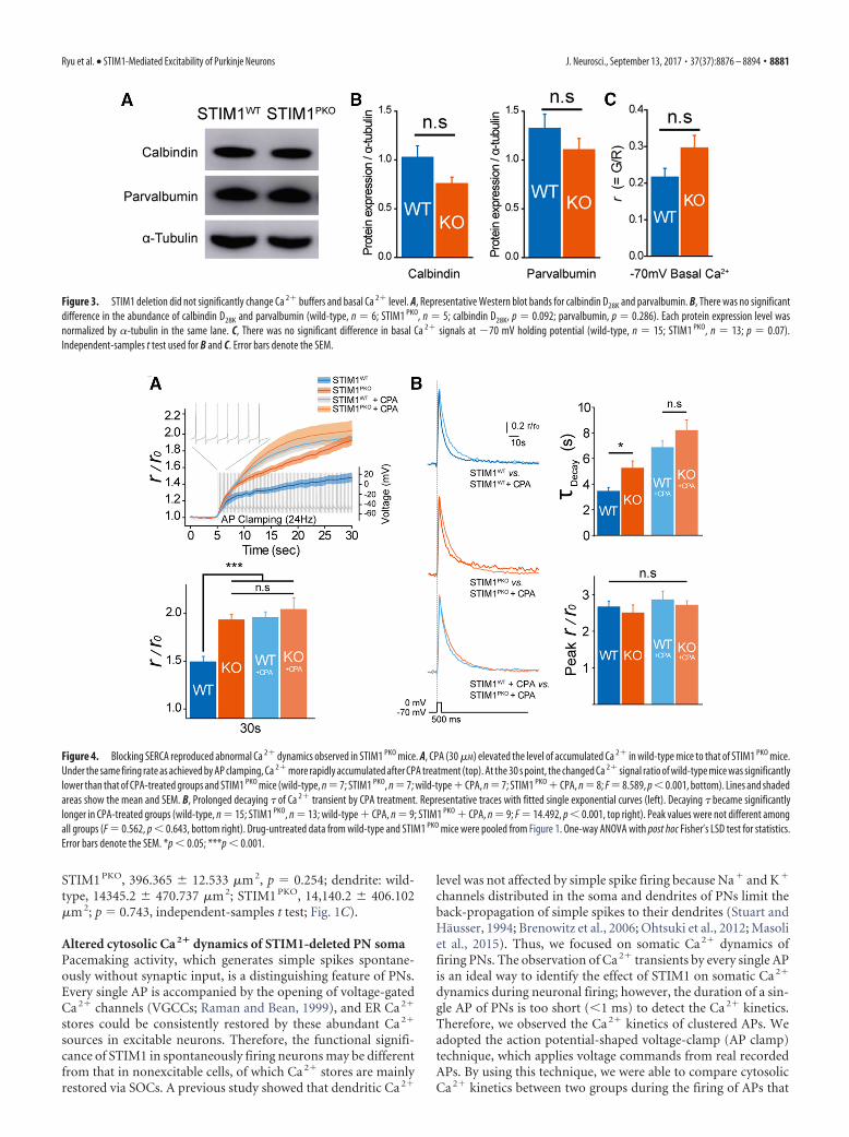
Figure 3. STIM1 deletion did not significantly change Ca 2� buffers and basal Ca 2� level. A, Representative Western blot bands for calbindin D28K and parvalbumin. B, There was no significantdifference in the abundance of calbindin D28K and parvalbumin (wild-type, n � 6; STIM1 PKO, n � 5; calbindin D28K, p � 0.092; parvalbumin, p � 0.286). Each protein expression level wasnormalized by �-tubulin in the same lane. C, There was no significant difference in basal Ca 2� signals at �70 mV holding potential (wild-type, n � 15; STIM1 PKO, n � 13; p � 0.07).Independent-samples t test used for B and C. Error bars denote the SEM.
Figure 4. Blocking SERCA reproduced abnormal Ca 2� dynamics observed in STIM1 PKO mice. A, CPA (30 �M) elevated the level of accumulated Ca 2� in wild-type mice to that of STIM1 PKO mice.Under the same firing rate as achieved by AP clamping, Ca 2� more rapidly accumulated after CPA treatment (top). At the 30 s point, the changed Ca 2� signal ratio of wild-type mice was significantlylower than that of CPA-treated groups and STIM1 PKO mice (wild-type, n � 7; STIM1 PKO, n � 7; wild-type � CPA, n � 7; STIM1 PKO � CPA, n � 8; F � 8.589, p � 0.001, bottom). Lines and shadedareas show the mean and SEM. B, Prolonged decaying � of Ca 2� transient by CPA treatment. Representative traces with fitted single exponential curves (left). Decaying � became significantlylonger in CPA-treated groups (wild-type, n � 15; STIM1 PKO, n � 13; wild-type � CPA, n � 9; STIM1 PKO � CPA, n � 9; F � 14.492, p � 0.001, top right). Peak values were not different amongall groups (F � 0.562, p � 0.643, bottom right). Drug-untreated data from wild-type and STIM1 PKO mice were pooled from Figure 1. One-way ANOVA with post hoc Fisher’s LSD test for statistics.Error bars denote the SEM. *p � 0.05; ***p � 0.001.
Ryu et al. • STIM1-Mediated Excitability of Purkinje Neurons J. Neurosci., September 13, 2017 • 37(37):8876 – 8894 • 8881

were exactly the same. Since more frequent firing of APs causesmore chances of Ca 2� influx into the cytosol, we had to fix thesame firing rate of APs in both groups to compare the differencein Ca 2� dynamics during repetitive firing. We chose 24 Hz spon-taneous firing of APs from our current-clamp recording as atemplate for the AP clamp. To confirm that the AP clamp voltagestimulus does not evoke a dendritic Ca 2� response, we comparedCa 2� signals between prolonged depolarization and spike-likedepolarization by AP clamp (Fig. 1D). While prolonged depolar-ization (�70 to 0 mV, 500 ms) could evoke both somatic anddendritic Ca 2� transients (Fig. 1D, left), AP clamp voltage stim-ulus could evoke only somatic Ca 2� transients (Fig. 1D, right). Inaccordance with a previous study (Brenowitz et al., 2006), theseresults confirmed that simple spikes induce negligible effects ondendritic Ca 2� signals. In the AP clamp recordings, cytosolicCa 2� of the soma is accumulated much more rapidly inSTIM1 PKO PNs than in wild type (wild type, 1.492 0.058, n �7; STIM1 PKO, 1.932 0.056, n � 7; p � 0.001, independent-samples t test; Fig. 1E). Either increased Ca 2� influx or delayed
Ca 2� decay can account for this overaccumulation of cytosolicCa 2� in STIM1 PKO PNs. We measured the cytosolic Ca 2� tran-sient of the soma after depolarizing voltage step (500 ms; �70 to0 mV) in both wild-type and STIM1 PKO PNs to validate whichCa 2� kinetics is affected by STIM1. The decaying kinetics ofCa 2� following depolarization was significantly delayed inSTIM1 PKO PNs without a difference in peak value of the Ca 2�
transient (wild type, n � 15; STIM1 PKO, n � 13; �decay: wild-type,3.434 0.305 ms; STIM1 PKO, 5.232 0.583 ms, p � 0.001; peak:wild-type, 2.665 0.167; STIM1 PKO, 2.503 0.217, p � 0.554,independent-samples t test; Fig. 1F).
To examine whether STIM1 deletion also affects dendriticcalcium signaling, we recorded dendritic Ca 2� transients follow-ing depolarization (Fig. 1G). There was no difference in the de-caying kinetics or amplitudes of dendritic Ca 2� transientsbetween wild-type and STIM1 PKO PNs, suggesting that STIM1deletion does not affect dendritic calcium dynamics by depolar-ization (wild type, n � 10; STIM1 PKO, n � 8; �decay: wild type,0.722 0.038 ms; STIM1 PKO, 0.644 0.026 ms, p � 0.131; peak:
Figure 5. Attenuated spontaneous and current injection-evoked spike firing in STIM1-deleted PNs. A, Representative traces of spontaneous firing from attached recording in the lobule III–V(left). Spontaneous simple spike firing frequency was considerably reduced in STIM1 PKO. Reduced firing frequency of STIM1 PKO was not only observed in anterior lobule (III–V; wild-type, n � 55;STIM1 PKO, n � 43; p � 0.001), but also lobule X and the flocculus. (wild-type, n � 19; STIM1 PKO, n � 30; p � 0.001). B, Distribution plot of corresponding interspike interval (left). CV wassignificantly increased in STIM1 PKO mice (wild-type, n � 31; STIM1 PKO, n � 26; p � 0.007), but CV2 was unaffected ( p � 0.878). C, Representative trace at 600 pA of current injection. D, Evokedfiring rate was decreased in STIM1 PKO mice (wild-type, n � 60; STIM1 PKO, n � 43; F � 7.221, p � 0.008, left). Bar graph at 600 pA of current injection ( p � 0.002, right). E, The amplitude of mAHPsignificantly increased in STIM1 PKO (F � 28.220, p � 0.001, left). Bar graph at 600 pA injection ( p � 0.001, right). F, STIM1 PKO showed strengthened SFA. Wild-type mice have the shorter durationof 10 spikes than STIM1 PKO (left). Instantaneous firing frequency more steeply decreased in STIM1 PKO (F � 11.367, p � 0.001, right). One-way ANOVA was used for A. Independent-samples t testwas used for B. Two-way repeated-measures ANOVA was used for D–F, and asterisks in bar graphs in A, D, and E were marked by post hoc Tukey’s test pairwise comparison. Error bars denote theSEM. **p � 0.01; ***p � 0.001.
8882 • J. Neurosci., September 13, 2017 • 37(37):8876 – 8894 Ryu et al. • STIM1-Mediated Excitability of Purkinje Neurons

wild-type, 1.858 0.079; STIM1 PKO, 1.982 0.089, p � 0.312,independent-samples t test; Fig. 1G). Previous studies showedthat STIM1 inhibits L-type VGCC (Cav1.2; Park et al., 2010; Wang etal., 2010). Unlike other cortical neurons, mature PNs express P/Q-type VGCCs (Cav2.1) as the predominant high voltage-activatedVGCC, instead of the L-type VGCC (Usowicz et al., 1992). So far,there has been no report on whether STIM1 could also inhibitP/Q-type VGCCs. Our data suggest that STIM1 deletion did notaffect the amount of Ca 2� influx through P/Q-type VGCCs.
We performed qPCR analyses to quantitatively compare thetranscript levels of several Ca 2� clearance/influx sources mainlyexpressed in PNs, such as SERCA2/3, plasma-membrane Ca 2�-ATPase 2 (PMCA2), Na�–Ca 2� exchanger 2 (NCX2), andVGCCs [P/Q-type (Cav2.1), T-type (Cav3.1) and R-type (Cav2.3)].Tissues containing PNL and ML were microdissected to specifi-cally measure the mRNA levels in PNs (Fig. 2A). We confirmedthe specificity of tissue dissection by using reverse transcriptionPCR (RT-PCR) with primers for a Purkinje neuronal marker[L7(PCP2)] and a granule cell marker [GABAA receptor �6 sub-unit (GABRA6); Boyden et al., 2006; Fig. 2B]. Wild-type andSTIM1 PKO mice showed comparable mRNA levels for thosegenes (wild-type, n � 3; STIM1 PKO, n � 3; SERCA2: wild-type,1.007 0.012; STIM1 PKO, 0.090 0.075, p � 0.700; SERCA3:wild-type, 1.010 0.098; STIM1 PKO, 0.087 0.082, p � 0.700;PMCA2: wild-type, 1.004 0.033; STIM1 PKO, 1.094 0.085,p � 0.700; NCX2: wild-type, 1.100 0.266; STIM1 PKO, 1.087 0.417, p � 0.999; P/Q-type VGCC: wild-type, 1.091 0.303;STIM1 PKO, 0.588 0.084, p � 0.100; T-type VGCC: wild-type,1.012 0.043; STIM1 PKO, 1.037 0.076, p � 0.999; R-typeVGCC: wild-type, 1.017 0.089; STIM1 PKO, 0.900 0.200, p �0.999; Mann–Whitney U test; Fig. 2C). However, it may be wor-thy to note that we observed a fairly clear tendency of decreasedmRNA expression level of P/Q-type VGCCs in the STIM1 PKO
group even though it did not reach a statistically significant level.In discordance with the qPCR result, our Ca 2� imaging datashowed no significant difference of Ca 2� influx by depolarizationbetween the two groups (Fig. 1F,G). These results indicated thatgene expression levels could not explain the alterations in Ca 2�
signals in STIM1 PKO PNs.Since PNs were well known for their high endogenous Ca 2�
buffering (Fierro and Llano, 1996), delayed decaying kinetics ofSTIM1 PKO PNs could be caused by alterations in the capacity ofendogenous Ca 2� buffering. We measured the protein expres-sion levels of calbindin D28K and parvalbumin, two representa-tive Ca 2� buffers in PNs (Schmidt et al., 2003). There was atendency toward decreased calbindin D28K and parvalbumin ex-pression in STIM1 PKO PNs, but it was not statistically significant(calbindin D28K: wild-type, 1.026 0.117, n � 6; STIM1 PKO,0.757 0.066, n � 5, p � 0.092; parvalbumin: wild-type, 1.324 0.147, n � 6; STIM1 PKO, 1.105 0.116, n � 5, p � 0.286,independent-samples t test; Fig. 3A,B). We also quantified thebasal levels of Ca 2� signals at �70 mV holding potential. Thebasal Ca 2� level of STIM1 PKO PNs was slightly increased but wasnot statistically significant either (wild-type: 0.216 0.024, n �15; STIM1 PKO: 0.295 0.034, n � 13, p � 0.07, independent-samples t test; Fig. 3C).
STIM1-mediated cytosolic Ca 2� dynamics of soma dependson SERCADelayed decaying kinetics indicates delayed clearance of Ca 2�
from the cytosol after Ca 2� influx. Among various moleculesassociated with STIM1-driven ER Ca 2� store refilling (Soboloffet al., 2012), SERCA is the most appropriate candidate responsi-
ble for cytosolic Ca 2� clearance of PNs (Fierro et al., 1998). Pre-vious studies demonstrated that SERCA, a final executor of therefilling process, is assisted by STIM1 (Krapivinsky et al., 2011).We tested the effect of blocking SERCA by cyclopiazonic acid(CPA) on somatic Ca 2� dynamics. We applied CPA during 24 HzAP clamping in both groups. The accumulation of Ca 2� of wild-type PNs with CPA was similar to that of STIM1 PKO with CPA atall time points. There were no significant differences among thethree groups (wild-type with CPA, STIM1 PKO with CPA, andSTIM1 PKO without CPA) at late time points [wild-type withCPA, n � 7; STIM1 PKO, n � 7; STIM1 PKO with CPA, n � 8; 30 s,wild-type with CPA, 1.958 0.060; STIM1 PKO with CPA,2.041 0.123; wild-type with CPA vs STIM1 PKO, p � 0.831;STIM1 PKO vs STIM1 PKO with CPA, p � 0.359; wild-type withCPA vs STIM1 PKO with CPA, p � 0.484, post hoc Fisher’s leastsignificant difference (LSD) test after one-way ANOVA; Fig. 4A].We also compared the effect of SERCA blocking on somatic Ca 2�
dynamics in 500 ms depolarization. CPA application delayed thedecay kinetics of cytosolic Ca 2� more in both groups (wild-typewith CPA, n � 9; STIM1 PKO with CPA, n � 9; �decay: wild-typewith CPA, 6.858 0.507 ms; STIM1 PKO with CPA, 8.186 0.837ms; wild-type vs wild-type with CPA, p � 0.001; STIM1 PKO vsSTIM1 PKO with CPA, p � 0.001; peak: wild-type with CPA,1.922 0.239; STIM1 PKO with CPA, 2.073 0.117, post hocFisher’s LSD test after one-way ANOVA; Fig. 4B). However, de-caying kinetics delayed by CPA of wild-type was not significantlydifferent from that of STIM1 PKO PNs (p � 0.132, post hoc FisherLSD test after one-way ANOVA; Fig. 4B). These results indicatethat STIM1 deletion caused hypofunction of SERCA-dependentcytosolic Ca 2� clearance.
Altered neuronal excitability in STIM1 PKO
To examine the functional consequence of STIM1 deletion inPNs, we measured the simple spike firing rate of STIM1 PKO mice
Table 1. Passive and active membrane properties of wild-type and STIM1 PKO mice
Spike Wild-type mice STIM1 PKO mice p Value
Cm (pF) 644.9 12.7 634.5 14.0 0.586Rin (M�) 43.9 1.1 38.1 1.0 �0.001***First spike latency (ms) 16.5 0.5 18.0 0.9 0.137Rheobase (pA) 261.1 9.1 281.6 11.2 0.159Threshold (mV) 1st spike �47.0 0.3 �46.0 0.5 0.080
25th spike �43.1 0.4 �41.9 0.5 0.070AP amplitude (mV) 1st spike 82.0 1.0 82.4 1.1 0.775
25th spike 70.6 1.0 71.4 1.0 0.545FWHM (ms) 1st spike 0.205 0.004 0.195 0.003 0.031*
25th spike 0.226 0.005 0.211 0.003 0.012*Upstroke (V/s) 1st spike 542.6 10.1 570.1 10.3 0.059
25th spike 421.0 9.8 448.5 8.8 0.039*Downstroke (V/s) 1st spike �452.9 10.9 �466.0 9.6 0.368
25th spike �368.8 10.3 �385.9 9.1 0.214Slope of postspike
depolarization (V/s)1st spike 1.69 0.03 1.62 0.05 0.23425th spike 1.19 0.03 1.01 0.04 �0.001***
ISI (ms) 1st spike 10.4 0.2 10.7 0.3 0.40225th spike 14.4 0.3 17.3 0.8 �0.001***
fAHP (mV) 1st spike 14.6 0.3 14.3 0.4 0.58125th spike 15.8 0.3 15.9 0.4 0.830
mAHP (mV) 8.5 0.3 10.9 0.3 �0.001***
Several parameters, including Rin, slope of postspike depolarization, ISI, and mAHP were significantly different inSTIM1 PKO mice compared to wild-type littermates ( p � 0.001). Active properties of the 25th spike showed moredifferences than those of the 1st spike. Slope of postspike depolarization, especially, were remarkably lowered inSTIM1 PKO mice, which could explain the increase of ISI despite the shortened action potential waveform ( p �0.05).Up-stroke and down-stroke, rising and falling phase of action potential, respectively. Data were from the experi-ment performed in Figure 5. Data are expressed as the mean SEM.
*p � 0.05, independent-samples t test.
***p � 0.001, independent-samples t test.
Ryu et al. • STIM1-Mediated Excitability of Purkinje Neurons J. Neurosci., September 13, 2017 • 37(37):8876 – 8894 • 8883

against that of wild-type mice whileblocking both excitatory and inhibitorysynaptic input by using cell-attached re-cording. PNs of STIM1 PKO mice showedsignificantly lower firing rates in anteriorlobules of cerebellar central vermis (Lob-ule III–V: wild-type, 37.308 1.454 Hz,n � 55; STIM1 PKO, 22.039 1.344 Hz,n � 43; p � 0.001, post hoc Tukey’s testafter one-way ANOVA; Fig. 5A). It is wellknown that the firing rates of PNs corre-late with localized zebrin (aldolase C) ex-pression patterns (Zhou et al., 2014). Toconfirm whether reduced firing rates ofSTIM1 PKO PNs depend on the location ofPNs, we recorded the firing rates of PNs inlobule X and the flocculus, which werewell known as purely zebrin-positive re-gions (Zhou et al., 2014). The firing rate isalso significantly reduced in STIM1 PKO
PNs compared with wild-type PNs inthese zebrin-positive regions (Lobule Xand the flocculus, wild-type, 25.407 1.629 Hz, n � 19; STIM1 PKO, 15.222 0.958 Hz, n � 30; p � 0.001, post hocTukey after one-way ANOVA; Fig. 5A),demonstrating that the reduction of firingrates in STIM1 PKO PNs does not dependon the regional specificity by zebrin ex-pression patterns. We also compared theregularity of PN firing. STIM1 PKO PNsshowed a larger CV for ISIs than inwild-type littermates (wild-type, 0.054 0.002, n � 31; STIM1 PKO 0.066 0.004,n � 26, p � 0.007, independent-samplest test; Fig. 5B, left). However, CV2 valuesfrom both groups showed no significantdifference (wild-type, 0.065 0.003;STIM1 PKO 0.066 0.004, p � 0.878,independent-samples t test; Fig. 5B,right).
To investigate the details of spikeproperties, we injected step currentsinto PNs in current-clamp recording(wild-type, n � 60; STIM1 PKO, n � 43;Fig. 5C–F). The firing frequency versusinjected current ( f–I) curve showed thereduced excitability of PNs in STIM1 PKO
mice [curve: p � 0.008, two-way repea-ted-measures (RM) ANOVA; �600 pA:wild-type, 69.133 1.304; STIM1 PKO,
Figure 6. Altered excitability of STIM1 PKO PNs required activation of Ca 2�-activated K � channels. A, Continuous cell-attachedrecording during the application of ACSF containing low Ca 2� concentration. Under low extracellular Ca 2� concentration (100�M) firing rates of both groups increased and reached a similar peak point within 5 min. When extracellular Ca 2� concentrationreturned to normal (2 mM), the firing rate slowly recovered to the initial level (wild-type, n � 15; STIM1 PKO, n � 14). B, Bar graphof low-Ca 2� contained ACSF application (right). Though PNs of STIM1 PKO mice had significantly lower baseline firing rates thanwild-type littermates at baseline ( p � 0.001), the firing rates finally met at the peak ( p � 0.949). When the firing rate recoveredunder normal ACSF, the gap between wild-type and STIM1 PKO mice was also recovered ( p � 0.003). C, Blocking SK channel byapamin (100 nM) increases the firing rates of both groups of PNs (wild-type, n � 3; STIM1 PKO, n � 13). D, E, Blocking BK channelalso raised spontaneous firing rate in both groups. D, Application of iberiotoxin (100 nM; wild-type, n � 3; STIM1 PKO, n � 4).E, Paxilline, a broader spectrum blocker that is able to block iberiotoxin-insensitive BK currents (100 nM; wild-type, n � 2;STIM1 PKO, n � 6) were not able to reduce the firing rate difference between two groups. F, Blocking both SK and BK channels withthe cocktail of apamin (100 nM) and paxilline (100 nM). The mixture of drugs narrows the gap in firing rates between wild-type andSTIM1 PKO mice (wild-type, n � 7; STIM1 PKO, n � 9). Although blocking both SK and BK channels suddenly changes the firingpattern (from tonic firing to burst firing), the firing frequencies of both groups became similar before pattern change (left). Bargraph in two time points (right). Baseline is before drug treatment, and maximum is the maximum firing rate before pattern change. In thecomparison of maximum firing rates, there was no statistical significance between wild-type and STIM1 PKO (baseline, p � 0.003;
4
maximum, p � 0.351). G, Potentiating KCa channels with1-EBIO (100 �M). 1-EBIO treatment decreased the differencebetween wild-type and STIM1 PKO mice without changing thefiring pattern (left; wild-type, n � 7; STIM1 PKO, n � 15). Thebar graph show the baseline and 15 min after drug application(right). At 15 min after 1-EBIO treatment, statistical signifi-cance between wild-type and STIM1 PKO mice disappeared(baseline, p � 0.004; 25 min, p � 0.078). Asterisks in all bargraphs were marked by the Mann–Whitney U test. Error barsdenote the SEM. **p � 0.01; ***p � 0.001.
8884 • J. Neurosci., September 13, 2017 • 37(37):8876 – 8894 Ryu et al. • STIM1-Mediated Excitability of Purkinje Neurons

61.860 2.361; p � 0.002, post hoc Tukey’s test after two-way RMANOVA; Fig. 5D] with considerably increased mAHP followingspike trains (curve: p � 0.001, two-way RM ANOVA; �600 pA:wild-type, 8.520 0.255; STIM1 PKO, 10.882 0.344, p � 0.001,post hoc Tukey’s test after two-way repeated-measures ANOVA;Fig. 5E). The properties of a single spike such as voltage thresholdor amplitude were similar between the two groups (Table 1);however, spike frequency adaptation (SFA) was more prominentin STIM1 PKO mice (p�0.001, two-way repeated-measures ANOVA;Fig. 5F). Altered SFA indicates that STIM1 deletion changed theexcitability of PNs qualitatively as well as quantitatively (Bendaand Herz, 2003; Pozzorini et al., 2013). Among several mecha-nisms explaining SFA, the Ca2�-activated K� current (KCa current)has been known as a determinant of mAHP in PNs (Belmeguenai etal., 2010). Based on both experimental data and theoretical model-ing, previous studies have shown that the KCa-dependent SFA mech-anism accompanies intracellular Ca2� dynamics, including bothCa2� flux to the cytosol and Ca2� removal from the cytosol (Bendaand Herz, 2003).
In AP-clamp recordings, we assumed that normal PNs andSTIM1-deleted PNs fire equally, more Ca2� in the cytosol ofSTIM1PKO PNs accumulated during repetitive firing of APs in thatcondition (Fig. 1E). However, in actual PN firing, STIM1PKO PNsfire more slowly than wild-type PNs (Fig. 5A). If STIM1PKO PNs arenot able to efficiently remove Ca2� influx by a single AP from thecytosol, PNs should engage more negative feedback mechanisms tomaintain the homeostasis of cytosolic Ca2� concentration([Ca2�]i). KCa current, which reduces the chances of Ca2� influx bydecelerating firing rates of neurons, is one of these feedback mecha-nisms. Reduced excitability in STIM1PKO PNs could be explained bythe enhanced negative feedback mechanisms such as KCa current,which is recruited more by excessive cytosolic Ca2� in STIM1-deleted PNs to prevent overaccumulation of cytosolic Ca2�.
Global effects of STIM1 deletion on Ca 2�-dependentionic currentsTo determine whether the altered Ca 2� dynamics causes thechanges in excitability of STIM1-deleted PNs, we recorded spon-taneous firing while replacing most of the extracellular Ca 2� withMg 2� to exclude Ca 2� effects on firing (wild-type, n � 15;STIM1 PKO, n � 14; Fig. 6A). Lowering extracellular Ca 2� levelsmade the firing rates of the two groups equal and narrowed thegap between the two groups recovered under reperfusion of nor-mal ACSF (baseline: wild-type, 28.165 1.640 Hz; STIM1 PKO,16.212 1.430 Hz, p � 0.001; peak: wild-type, 127.097 9.701
Hz; STIM1 PKO, 127.969 8.140 Hz, p �0.949; recovery: wild-type, 26.919 2.490Hz; STIM1 PKO, 16.202 1.397 Hz; p �0.001, Mann–Whitney U test; Fig. 6B).These results confirmed that the differ-ence in simple spike firing was attributedto Ca 2� dynamics. The changed Ca 2� dy-namics could affect diverse kinds of Ca 2�-dependent ionic currents. The broadeffects of the KCa current on the excitabil-ity of PNs have been well established inprevious studies (Edgerton and Reinhart,2003; Benton et al., 2013), and, as men-tioned above, altered parameters ofSTIM1PKO PNs, including increasedmAHP and SFA, have been known to berelated to KCa current (Benda and Herz,2003; Belmeguenai et al., 2010). We tested
the effects of KCa current on the firing of PNs in both groups byblocking or potentiating small and big conductance KCa channels[small-potassium (SK) and big-potassium (BK) channels, re-spectively]. During cell-attached recording, the application ofapamin, a selective inhibitor of SK channels, was not able tonarrow the gap in firing rates between the two groups (Fig. 6C).Additionally, two kinds of BK channel-selective blockers, iberio-toxin and paxilline, also could not narrow the gap in firing rates(Fig. 6D,E). Both groups fired equally while blocking both SKand BK channels (wild-type, n � 7; STIM1 PKO, n � 9; baseline:wild-type, 36.973 3.833 Hz; STIM1 PKO, 24.756 1.799 Hz;p � 0.003; maximum: wild-type, 151.429 20.300 Hz; STIM1PKO,123.333 14.337 Hz; p � 0.351, Mann–Whitney U test; Fig. 6F).However, in both groups, the application of an apamine–paxil-line cocktail induced burst firing of PNs (Fig. 6F), which is in-volved in dendritic Ca 2� signals (Brenowitz et al., 2006), so wecould not precisely estimate the contribution of KCa channels tosimple spike firing and somatic Ca 2� signals. 1-EBIO, a nonse-lective activator of KCa channels (Benton et al., 2013), decreasedthe firing rates of both groups to the same level without chang-ing the firing pattern (wild-type, n � 7; STIM1 PKO, n � 15;baseline: wild-type, 28.824 2.554 Hz; STIM1 PKO, 20.111 1.010 Hz, p � 0.004; 25 min: wild-type, 11.673 0.931 Hz;STIM1 PKO, 9.772 0.637 Hz; p � 0.078, Mann–Whitney U test;Fig. 6G). These results indicate that the alteration of PN activity inSTIM1 PKO cannot be explained by the modulation of a singlechannel, but several channels may be globally affected by changesin intracellular Ca 2� environments.
Alterations in excitability depend onSTIM1–SERCA interactionSTIM1 interacts with various molecules to maintain ER Ca 2�
stores (Soboloff et al., 2012). STIM1 recruits SOCs to bring Ca 2�
from extracellular space to cytosol, and then SERCA uptakes cy-tosolic Ca 2� into ER stores. Stored Ca 2� can be released to thecytosol by the influx of Ca 2�, which is called Ca 2�-induced Ca 2�
release (CICR), which is mediated by RyRs. During cell-attachedrecording, pharmacological blocking of RyRs had no effect onspontaneous firing rates in both groups (Fig. 7). These resultsshowed that CICR may not be involved in the reduced firing ratesof STIM1-deleted PNs.
We already showed that STIM1 deletion in PNs decreased theSERCA-dependent cytosolic Ca 2� clearance in the soma (Fig. 4).Few studies have revealed that sequestration of cytosolic Ca 2� bySERCA can influence the neuronal excitability (Cueni et al.,
Figure 7. Blocking Ca 2� release from ER store did not affect the spontaneous firing of PNs. A, Ca 2� transient after treatingcaffeine. Large Ca 2� transient was recorded by caffeine treatment (80 mM, puff application, 5 s, 5 psi) before the application ofdantrolene (50 �M; black). After 20 min of bath application of the drug, Ca 2� response after caffeine treatment was reduced (red),and the response was abolished after 40 min of dantrolene application (blue). B, Blocking ryanodine receptor by dantrolene(50 �M) was not able to change the firing rate of PNs in both groups (wild-type, n � 6; STIM1 PKO, n � 2).
Ryu et al. • STIM1-Mediated Excitability of Purkinje Neurons J. Neurosci., September 13, 2017 • 37(37):8876 – 8894 • 8885

2008), but the pacemaking activity ofPNs has never been studied in this re-gard. We examined whether SERCA-dependent Ca 2� signals could affectpacemaking activity. Blocking SERCA byCPA decreased the spontaneous firingrates of wild-type PNs to the level ofSTIM1 PKO PNs. However, importantly,CPA treatment did not change the firingrates of STIM1 PKO PNs (wild-type, n�11;STIM1PKO, n � 9; inset �frequency: wild-type, �8.862 1.616 Hz; STIM1 PKO,�1.141 0.979 Hz; p � 0.001, Mann–Whitney U test; Fig. 8A, bar graph; Table2). To examine whether CPA affectsSERCA in the soma or in dendrites, wealso recorded the spontaneous firing ratesduring local CPA puffing onto the somaor dendrites of PNs. Only CPA puffingonto the soma (n � 4) significantly de-creased the firing rates, whereas CPApuffing onto the dendrites (n � 4) did notdecrease firing rates in wild-type slices.Consistent with the bath application ex-periment, CPA puffing onto the soma(n � 4) did not also alter firing rates inSTIM1 PKO (reduced firing rate: wild-typesoma, 74.84 10.69; wild-type dendrite,103.7 5.792; STIM1 PKO soma, 105.5 3.500; p � 0.027, one-way ANOVA; Fig.8B). These data indicate that SERCAs inthe soma have critical roles in regulatingfiring rates. We injected step currents un-der bath application of CPA. The drug re-
Figure 8. Blocking SERCA reproduced the attenuated intrinsic excitability of STIM1-deleted PNs. A, CPA treatment during theattached recording. CPA (30 �M) decreases firing rates of wild-type PNs to the level of STIM1 PKO PNs, while PNs of STIM1 PKO seemunaffected by CPA (wild-type, n � 11; STIM1 PKO, n � 9). Reduced firing frequency of wild-type PNs was significantly larger than
4
that of STIM1 PKO PNs (p � 0.001, inset). Comparing initialfiring rates to 30 min after CPA treatment, the initial firingrates of wild-type PNs are remarkably higher than others.B, Somatic SERCAs are important for the regulation of sponta-neous firing rates. Local puffing of CPA (600 �M, 4 psi) onto thesoma (n � 4, circle, blue) caused a reduction of firing rates,but there was no change in local CPA puffing onto dendrites ofwild-type PNs (n � 4, square, blue) and the soma of STIM1 PKO
PNs (n � 4, triangle, red). After 10 min of treatment, the wild-type soma treatment group only showed significant reductionof firing rates (F � 5.556, p � 0.027). C, 30 min of CPA (30�M) preincubation reduces the evoked firing rate of wild-typelittermates. Wild-type littermates showed significantly higherevoked firing frequency than other groups (wild-type, n � 60;STIM1 PKO, n � 43; wild-type � CPA, n � 17; STIM1 PKO �CPA, n � 13). At the 600 pA point, the differences betweenwild-type mice and others are obvious. D, mAHP after prein-cubation of CPA. Only wild-type littermates were affected bythe drug, and mAHP of wild-type littermates became same asother groups. The representative bar graph at 600 pA of cur-rent injection shows that wild-type mice had considerablysmaller mAHP than other groups. E, CPA strengthened the SFAof wild-type PNs. The instantaneous firing frequency at the30th spike was significantly lower in the other three groupsthan wild-type littermates. Data from drug-untreated wild-type and STIM1 PKO mice, except A and B, were pooled fromFigure 5. One-way ANOVA was used for B, and other details ofdata and statistics see also Table 2. Error bars denote the SEM.*p � 0.05; **p � 0.01; ***p � 0.001.
8886 • J. Neurosci., September 13, 2017 • 37(37):8876 – 8894 Ryu et al. • STIM1-Mediated Excitability of Purkinje Neurons

duced the gain of the f–I curve and increased both mAHP andSFA of wild-type PNs to the level of STIM1 PKO PNs. Further-more, CPA administration did not lead to significant changes inthe gain of the f–I curve, mAHP, and SFA of STIM1 PKO PNs (Fig.8C–E; Table 2). These data strongly indicate that the reducedexcitability of STIM1 PKO PNs involved a SERCA-dependentmechanism. The results from blocking RyRs (Fig. 7) suggest thatthe altered excitability was not caused by the CPA-induced de-pletion of ER Ca 2� store and the consequent lack of Ca 2� releasefrom the ER. Instead, the Ca 2� uptake process of SERCA knownto remove and buffer cytosolic Ca 2� (Fierro et al., 1998; Higginset al., 2006) could be a crucial step in STIM1-dependent regula-tion of the excitability. Our data suggest that STIM1 deletioncauses less efficient SERCA-dependent cytosolic Ca 2� clearing,and delayed decaying kinetics of somatic Ca2� enhances Ca2�-dependent ionic currents, which decrease the excitability of PNs tolimit Ca2� influx.
Preserved basal synaptic transmission in STIM1 PKO
There was no difference between two groups in basal synaptictransmissions, including mEPSC (wild-type, n � 8; STIM1 PKO,n � 11; amplitude: wild-type, 17.405 0.761 pA; STIM1 PKO,16.211 0.696 pA, p � 0.206; frequency: wild-type, 3.412 0.681 Hz; STIM1 PKO, 3.204 0.506 Hz; p � 1.000, Mann–Whit-ney U test; Fig. 9A) and mIPSC (wild-type, n � 5; STIM1 PKO, n �7; amplitude: wild-type, 40.225 1.381 pA; STIM1 PKO, 42.800 4.111 pA; p � 0.639; frequency: wild-type, 2.287 0.503 Hz;STIM1 PKO, 2.913 0.745 Hz; p � 0.876, Mann–Whitney U test;Fig. 9B). We also recorded strong excitatory synaptic transmis-sion by CF, which is essential for long-term depression (LTD)induction (Coesmans et al., 2004), as well as for the induction ofother forms of PN output besides simple spikes, complex spikes(CSs). STIM1 PKO mice had no differences in properties of CSscompared with wild-type littermates (wild-type, n � 43;STIM1PKO, n � 34; spikelets: wild-type, 4.468 0.142; STIM1PKO,4.500 0.165; p � 0.884; CS duration: wild-type, 9.517 0.535;
STIM1PKO, 9.486 0.511; p � 0.968, independent-samples t test;Fig. 9C,D).
Intrinsic plasticity, but not synaptic plasticity, was impairedin STIM1 PKO miceLTD and long-term potentiation (LTP) at glutamatergic synapsesbetween PF inputs and PNs have been considered as critical cel-lular mechanisms underlying cerebellar learning and memory(Ito, 1982; De Zeeuw et al., 1998; Hansel et al., 2006; Jorntell andHansel, 2006). It has been well known that LTD induction re-quires Ca 2� release from the ER store through IP3Rs (Inoue et al.,1998). Considering the ER Ca 2� refilling by STIM1, we examinedwhether STIM1 deletion affects long-term synaptic plasticity. In-terestingly, STIM1 PKO mice showed levels of LTP (wild-type, n �11; STIM1 PKO, n � 8; p � 0.952, two-way repeated-measuresANOVA; Fig. 10A) and LTD (wild-type, n � 10; STIM1 PKO, n �11; p � 0.653, two-way repeated-measures ANOVA; Fig. 10B)comparable to those of their wild-type littermates. We hypothesizethat sufficient Ca2� stores to induce synaptic plasticity may be filledby other ER store refilling processes, for instance, Ca2� influxthrough VGCCs (further referred to in the Discussion section).
In addition to synaptic plasticity, intrinsic plasticity has beensuggested to be critical for learning and memory (Schonewille etal., 2010; Peter et al., 2016). We investigated whether STIM1deletion could also affect intrinsic plasticity as well as basal intrin-sic excitability. We recorded LTP of IE (LTP-IE) after LTP induc-tion (1 Hz PF stimulation for 5 min; Belmeguenai et al., 2010;Peter et al., 2016). Interestingly, LTP-IE was significantly impairedin STIM1PKO PNs compared with that in wild-type PNs (wild-type,n � 9; STIM1PKO, n � 9; p � 0.009, two-way repeated-measuresANOVA; Fig. 10C). These results indicate that STIM1 is required forintrinsic plasticity but not for synaptic plasticity.
Motor memory consolidation deficiency in STIM1 PKO miceThe previous study (Hartmann et al., 2014) showed thatSTIM1 PKO mice have defects in two types of motor learning tests,
Table 2. Statistical table of CPA treatment in Figure 8
Figure 8A Fig. 8C Fig. 8D Fig. 8EFiring frequency (Hz) Firing frequency (Hz) mAHP (mV) Firing frequency (Hz)
n Mean SEM n Mean SEM Mean SEM Mean SEM
WT 11 32.876 1.957 60 69.133 1.304 8.520 0.255 68.967 1.510KO 9 19.300 1.121 43 61.860 2.361 10.882 0.344 59.146 2.411WT � CPA 11 24.538 2.099 17 57.588 4.168 10.762 0.669 62.909 3.312KO � CPA 9 18.472 1.318 13 60.846 4.578 11.452 0.544 57.143 4.479
One-way ANOVA Two-way RM ANOVA
F p F p F p F p
StatisticsWT vs KO 14.338 �0.001 7.221 0.008 28.11 �0.001 11.367 0.001WT vs WT � CPA 12.021 �0.001 12.619 �0.001 5.491 0.022WT vs KO � CPA 8.939 0.004 21.503 �0.001 6.519 0.013KO vs WT � CPA 0.81 0.372 0.00163 0.968 0.109 0.743KO vs KO � CPA 0.51 0.478 1.25 0.269 0.0199 0.888WT � CPA vs KO � CPA 0.00749 0.932 0.734 0.399 0.154 0.698
p value ( post hoc Tukey’s test)WT vs KO �0.001 0.002 �0.001 �0.001WT vs WT � CPA 0.007 �0.001 �0.001 0.007WT vs KO � CPA �0.001 0.005 �0.001 0.003KO vs WT � CPA 0.177 0.276 0.848KO vs KO � CPA 0.989 0.631 0.373WT � CPA vs KO � CPA 0.092 0.683 0.400
First row, Mean and SEM values of bar graphs in each panel; second row, F and p values of each pair after ANOVA, but the values after one-way ANOVA were comparisons of the whole group; third row, p values of bar graphs in each panelafter post hoc Tukey’s test.
Ryu et al. • STIM1-Mediated Excitability of Purkinje Neurons J. Neurosci., September 13, 2017 • 37(37):8876 – 8894 • 8887

Figure 9. Basal synaptic transmission and climbing fiber activity remained intact in PNs of STIM1 PKO mice. A, Miniature EPSCs. Representative trace of mEPSCs recorded from wild-type andSTIM1 PKO (top). Cumulative distribution plots of frequency (bottom left) and amplitude (bottom right). In both frequency and amplitude, there was no statistical significance between both groups(wild-type, n � 8; STIM1 PKO, n � 11; amplitude, p � 0.206; frequency, p � 1.000). B, Miniature IPSCs. Data are presented same as in A, and there were no differences between wild-type andSTIM1 PKO mice (wild-type, n � 5; STIM1 PKO, n � 7; amplitude, p � 0.639; frequency, p � 0.876). C, The number of CS spikelets in both groups was the same. Representative trace of CSs inducedby CF stimulation from whole-cell recording (left). Average number ( p � 0.884, center) and probability (right) of the spikelets were almost the same (wild-type, n � 30; STIM1 PKO, n � 34).D, Properties of each spikelet showed no statistically significant differences between both groups. The peak amplitude of each spikelet was not significantly different between wild-type andSTIM1 PKO mice (first spike, p � 0.935; second spike, p � 0.792; third spike, p � 0.145; left). The times when each spikelet appeared (first spike, p � 0.412; second spike, p � 0.385; third spike,p � 0.494; fourth spike, p � 0.724; top, right) and the total spike duration ( p � 0.968, bottom, right) were also unchanged. A Mann–Whitney U test was used for bar graphs in A and B. Anindependent-samples t test was used for C and D. Error bars denote the SEM.
Figure 10. Intrinsic plasticity, but not synaptic plasticity, was impaired in STIM1 PKO mice. A, LTP was successfully induced in both groups (wild-type, n � 11; STIM1 PKO, n � 8). Representativetrace of EPSCs at preinduction and postinduction (left). Normalized EPSCs of both groups were the same in the entire time period (F � 0.004, p � 0.952, center) with unchanged paired-pulse ratio(right). B, LTD was also successfully induced in both groups with no differences (wild-type, n � 10; STIM1 PKO, n � 11; F � 0.208, p � 0.653). Data were presented as in A. C, Intrinsic plasticity wasabolished in STIM1 PKO mice. While LTP-IE was induced by the LTP induction protocol (1 Hz, 300 times, 5 min) in the wild-type group, this plasticity was unable to be induced in STIM1 PKO mice.Two-way repeated-measures ANOVA was used for plasticity graphs in all panels. An independent-samples t test was used for the bar graph in C. Error bars denote the SEM. *p � 0.05; **p � 0.01.
8888 • J. Neurosci., September 13, 2017 • 37(37):8876 – 8894 Ryu et al. • STIM1-Mediated Excitability of Purkinje Neurons

an elevated beam-balancing test and a rotarod test. However, anelevated beam-balancing test (Piot-Grosjean et al., 2001;Urakawa et al., 2007) or rotarod test (Rothwell et al., 2014; Scholzet al., 2015) reflects the function of various brain regions otherthan the cerebellum. Thus, these behavioral tests could not elu-cidate which specific steps of cerebellar learning are interruptedin STIM1 PKO mice, and more appropriate behavioral tests arerequired to specify the rules of STIM1 in cerebellar circuits re-lated to learning.
The VOR is a reflex movement of eyes that makes for theproper location of images on the retina during head movement.This reflex is characterized by its well known neural circuits in-volving the cerebellar cortex (Boyden et al., 2004; Ito, 2013). Theadaptive learning of VOR is achieved by plastic changes in neuralcircuits including the cerebellar cortex and vestibular nuclei(VNs) in the brainstem. As the sole output of the cerebellar cor-tex, PNs compute and integrate two different kinds of sensorystimuli [vestibular stimuli via mossy fibers (MFs) and retinal slip
via CF] and provide instructive signals forlearning to vestibular nuclei. Thus, theVOR has been regarded as a suitable be-havioral model to identify the cerebellarlearning mechanisms at the neural circuitlevel, along with the eye-blink condition-ing (Kloth et al., 2015).
Mice were prepared with a head-fixedpedestal for the test, and three differentbasal oculomotor responses were testedbefore the learning: OKR, dVOR, andlVOR (Fig. 11A). STIM1 PKO mice showedthe same levels as wild-type mice in theseparameters, and these results indicate thatSTIM1 PKO mice have normal visual andvestibular functions compared with wild-type mice (wild-type, n � 47; STIM1 PKO,n � 39; OKR: gain, p � 0.253; phase, p �0.203; dVOR: gain, p � 0.391; phase,p � 0.737; lVOR: gain, p � 0.052, phase:p � 0.611, two-way repeated-measuresANOVA; Fig. 11B–D).
Visual stimuli in the same direction ofvestibular stimuli induce a decrease ofVOR gain (the ratio of eye velocities tohead velocities; gain-down learning),whereas visual stimuli in the opposite di-rection of vestibular stimuli induce an in-crease of VOR gain (gain-up learning;Boyden et al., 2004). Adaptation of theVOR also occurs in the phase differencebetween the eye and head velocities as wellas VOR gain (Wulff et al., 2009). We ap-plied the gain-down protocol (Fig. 12A),and STIM1 PKO mice successfully de-creased gain to the same extent as wild-type littermates during learning sessions(wild-type, n � 11; STIM1 PKO, n � 10;p � 0.6, two-way repeated-measuresANOVA; Fig. 12B). The memory of re-duced gain was fully retained until 30 minor 1 h after the learning sessions in bothgroups (�30 min: wild-type, 100.053 4.318%; STIM1 PKO, 109.698 2.478%;p � 0.063; �1 h: wild-type, 114.380
3.575%; STIM1 PKO, 115.485 6.868%; p � 0.796, Mann–Whit-ney U test; Fig. 12C). To determine the effect of STIM1 PKO onlong-term memory retention, we extended learning procedure to4 d with three 24 h intervals. STIM1 PKO mice had much lowerlevels of retained memory than wild-type mice 24 h after the firstday of learning (day 1–2; Fig. 12D). In a further 2 d of learning,STIM1 PKO mice were able to catch up to the level of daily learningof wild-type mice but forgot most of the newly acquired memoryon the next day (day 2– 4; Fig. 12D). Therefore, the memoryconsolidation level (percentage change brought forward fromlearned memory of the previous day; Wulff et al., 2009) ofSTIM1 PKO mice was considerably lower than that of the wild-type mice on both day 1–2 and day 2–3 intervals (wild-type, n �14; STIM1 PKO, n � 14; day 1–2: wild-type, 65.013 5.817%;STIM1 PKO, 25.014 5.943%, p � 0.001, day 2–3: wild-type,66.891 5.225%; STIM1 PKO, 44.015 6.963%; p � 0.019, Man-n–Whitney U test; Fig. 12E). Because there are mouse modelsshowing defects only in specific types of eye movement learning
Figure 11. The basal ocular–motor performance of STIM1 PKO mice was comparable to that of wild-type mice. A, Recordingprocedures. OKR, dVOR, and lVOR. B, OKR responses in different drum rotating frequencies. Gain and phase values were notsignificantly different from each other group (wild-type, n � 47; STIM1 PKO, n � 39; gain: F � 1.324, p � 0.253; phase: F �1.649, p � 0.203). C, dVOR responses in different drum rotation frequencies. Gain and phase values were not significantly differentbetween groups (gain: F � 0.743, p � 0.391; phase: F � 0.114, p � 0.737). D, lVOR responses in different drum rotationfrequencies. Gain and phase values were not significantly different between groups (gain: F � 3.873, p � 0.052; phase: F �0.261, p � 0.611).
Ryu et al. • STIM1-Mediated Excitability of Purkinje Neurons J. Neurosci., September 13, 2017 • 37(37):8876 – 8894 • 8889

paradigms (Boyden et al., 2006; Hansel et al., 2006), the gain-uplearning protocol (Fig. 13A) was applied to check whether thesame phenomenon occurs in that learning. As seen in gain-downlearning, STIM1 PKO mice had daily learning ability comparableto that of wild-type mice but lost most of what they had learnedbefore 24 h, again showing reduced memory consolidation oflearning (wild-type, n � 11; STIM1 PKO, n � 10; day 1–2: wild-type, 84.291 9.483%; STIM1 PKO, 22.284 9.842%; p � 0.001;day 2–3: wild-type, 67.069 13.510%; STIM1 PKO, 32.161 7.585%; p � 0.040, Mann–Whitney U test; Fig. 13B,C). We askedwhether the defect of STIM1 PKO mice in memory consolidationexists not only in the amplitude (gain) of VOR, but also in thetiming (phase). Both groups were tested in phase reversal learn-ing, which reduces the gain on day 1 and afterward shifts thephase of VOR on days 2 and 3 (Fig. 13D; Wulff et al. (2009)). Theprotocol of the first-day learning was similar to that of gain-downlearning but with two more learning sessions. During the learn-ing on day 1, both groups performed the gain-down learningequally well without differences in both gain and phase values(day 1: wild-type, n � 11; STIM1 PKO, n � 11; Fig. 13E,G), butafter 24 h, the level of memory consolidation in gain was signifi-cantly different between two groups (day 1–2: wild-type,63.899 5.485%; STIM1 PKO, 44.884 5.567%; p � 0.028, Man-n–Whitney U test; Fig. 13F). These results confirmed the defectof STIM1 PKO mice by showing that the same phenomenon oc-curs even in more intensive learning. The phase was shifted fromthe second day of learning, and the memory consolidation defi-ciency of STIM1 PKO mice was also reproduced in the phase learn-ing (day 2– 4; Fig. 13G; day 1–2: wild-type, 57.544 8.372%;STIM1 PKO, 15.367 5.760%; p � 0.001; day 2–3: wild-type,83.535 6.823%; STIM1 PKO, 32.796 9.242%; p � 0.001, Man-n–Whitney U test; Fig. 13H). On the third day of learning, thedirection of learning in wild-type mice was changed from gain-
down-bound to gain-up-bound (day 3; Fig. 13E), as previously re-ported (Wulff et al., 2009), since the phase of VOR was completelyreversed as a consequence of accrued memory by repetitive phaseshifting (day 3; Fig. 13G). However, STIM1PKO mice were not able tochange the direction of alterations in gain due to the loss of thememory (day 3; Fig. 13E,G). Together, these results show that PN-specific STIM1 deletion significantly impairs the consolidation ofcerebellar memory without affecting the acquisition.
DiscussionMost previous studies on STIM function including studies in thenervous system have focused on its canonical function in ERCa 2� store refilling (Soboloff et al., 2012; Hartmann et al., 2014;Sun et al., 2014; Kraft, 2015; Zhang et al., 2015). Unlike other celltypes where there are limited routes for Ca 2� entry, excitableneurons can frequently bring extracellular Ca 2� into intracellu-lar space via VGCCs (Park et al., 2010; Harraz and Altier, 2014).In contrast to previous studies, our experiments have highlightedthe novel function of STIM1 in firing PNs (Fig. 14). We observedSTIM1-SERCA-dependent cytosolic Ca 2� kinetics of PN somawhere the neuronal output of PNs is determined and demon-strated how this Ca 2� signal has influence on the PN excitability,including pacemaking activity. As a previous study about burst-ing neurons in nucleus reticularis thalami (nRT; Cueni et al.,2008) showed, the sequestration of cytosolic Ca 2� by SERCA isthe significant factor for neuronal excitability, and we newlyfound that STIM1 mediates the Ca 2� sequestration and subse-quently regulates neuronal excitability. It cannot yet be answeredwhether this sequestration is important for neuronal excitabilityonly in high-frequency firing neurons, such as PNs and nRTneurons, or likewise in relatively slow-firing neurons such as cor-tical pyramidal neurons. However, it would be obvious that therelationship between this sequestration and neuronal excitability
Figure 12. STIM1 PKO mice showed memory consolidation deficiency in VOR learning. A, Gain-down learning protocol: 5° of in-phase paired turntable and drum stimulation was applied threetimes and 10 min each; 0.5 Hz dVOR was recorded in prelearning and postlearning and every interval between learning (arrows in top bar diagram). B, Gain-down learning in short term. In bothgroups, memory acquired by gain-down learning remained perfect until 30 min and 1 h (wild-type, n � 10; STIM1 PKO, n � 10; F � 0.284, p � 0.6). C, Both groups showed the same memoryconsolidation level for the gain value (right; 30 min, p � 0.063; 1 h, p � 0.796). D, Gain-down learning. STIM1 PKO mice could keep up with wild-type mice at the end of the daily learning, but therewere considerable differences between two groups in gain values on the next day (wild-type, n � 14; STIM1 PKO, n � 14). E, Consolidation levels were significantly reduced in STIM1 PKO mice (Day1–2, p � 0.001; Day 2–3, p � 0.019). Two-way repeated-measures ANOVA was used for B, and D and asterisks were marked by a post hoc Fisher’s LSD test pairwise comparison. Asterisks in C andE were marked by Mann–Whitney U test. Error bars denote the SEM. *p � 0.05; **p � 0.01; ***p � 0.001.
8890 • J. Neurosci., September 13, 2017 • 37(37):8876 – 8894 Ryu et al. • STIM1-Mediated Excitability of Purkinje Neurons

is critical for intracellular Ca 2� homeostasis in high-frequencyfiring neurons where Ca 2� influx via VGCC is more frequent.
In addition to its role in the soma, STIM1 in dendrites couldalso participate in the regulation of PN output through mGluR1-mediated slow current (Hartmann et al., 2014). Although thephysiological role of mGluR1-mediated slow current in PN out-put is not clear, this current evokes prolonged depolarization ofPNs after fast AMPA-mediated depolarization (Kim et al., 2003;Hartmann et al., 2008). Since, in the voltage-clamp condition, thiscurrent is missing in STIM1PKO PNs, STIM1-null PNs may be im-paired in the regulation of spikes induced by mGluR1-mediatedprolonged depolarization in response to intense glutamatergic syn-aptic transmission, which remains to be investigated further.
We observed severe deficiency of memory consolidation inSTIM1 PKO mice, and these phenomena were not limited to aspecific protocol, but occurred globally. It implies that the STIM1deletion in PNs may affect the common pathway for cerebellarmemory consolidation. STIM1-deleted PNs exhibited alterationsin the neuronal excitability and its plasticity. Our results accordwell with those of previous studies demonstrating that normaloutput of PNs is crucial for neuronal information transfer fromthe cerebellar cortex to the brainstem for successful memory con-solidation (Wulff et al., 2009; Yamazaki et al., 2015).
Cytosolic Ca 2� clearance and buffering by STIM1-assistedSERCA
There are several molecular mechanisms that extrude cytosolicCa 2� increased by depolarization, including SERCA, PMCA,NCX, and mitochondrial calcium uptake (Fierro et al., 1998).Among these mechanisms, SERCA and PMCA are known to beregulated by STIM1 combined with an adaptor protein calledPOST (partner of STIM1; Krapivinsky et al., 2011). Store deple-tion promotes the STIM1–POST complex to bind SERCA, keep-ing SERCA close to Ca 2� entry sites on the plasma membrane topromote ER Ca 2� refilling (Manjarres et al., 2010, 2011; Krapiv-insky et al., 2011). The STIM1–POST complex also inhibitsPMCA activity, which removes cytosolic Ca 2� to extracellularspace (Ritchie et al., 2012). With respect to cytosolic Ca 2� clear-ance, STIM1 seems to play opposite roles at the same time. How-ever, a previous study showed that SERCA contributes more tocytosolic Ca 2� clearance than PMCA, especially in high [Ca 2�]i
conditions in PNs (Fierro et al., 1998). Thus, we deduce that thenet effect of STIM1 on [Ca 2�]i after depolarization works towardreducing the cytoplasmic concentration, and our data supportthis inference. There is no evidence that the STIM1–POST com-plex draws SERCA to the vicinity of VGCCs (distinct fromSOCs). However, our data support this possibility because
Figure 13. STIM1 PKO mice showed memory consolidation deficiency in various VOR learning protocols. A, Gain-up learning protocol. Same as gain-down protocol but with out-of-phase pairedstimulation. B, Gain-up learning. STIM1 PKO mice learned the same as wild-type mice, but they lost almost all of learned memory by 24 h after learning (wild-type, n � 11; STIM1 PKO, n � 11).C, Comparison with wild-type littermates, memory consolidation levels are substantially lower in STIM1 PKO mice (Day 1–2, p � 0.001; Day 2–3, p � 0.040). D, Phase reversal learning protocol. Fivelearning sessions for each day. From day 1 to day 3, the rotating amplitude of the drum increases 2.5° each day from 5°. Turntable rotating amplitude was fixed to 5°. E, The normalized gain in phase reversallearning. At the beginning of days 2 and 3, the gain of STIM1 PKO mice was significantly higher than that of wild-type mice (wild-type, n�11; STIM1 PKO, n�11). F, Consolidation levels in gain values. STIM1 PKO
mice had considerably lower consolidation levels than wild-type mice in daily memory consolidation levels (Day 1–2, p�0.028; Day 2–3, p�0.034). G, Phase values in phase reversal learning. There were nodifferences in the end of entire learning (last point of the third day). Same as the gain graph; there were significant differences in the beginning point. H, Consolidation level in phase values. Wild-type mice haveamuchhigher leveloftrainedmemory(Day2–3, p�0.001;Day3– 4. p�0.001).Two-wayrepeated-measuresANOVAwasusedfor B, E,and G,andasterisksweremarkedby post hoc Fisher’sLSDtestpairwisecomparison. Asterisks in all bar graphs were marked by Mann–Whitney U test. Error bars denote the SEM. *p � 0.05; **p � 0.01; ***p � 0.001.
Ryu et al. • STIM1-Mediated Excitability of Purkinje Neurons J. Neurosci., September 13, 2017 • 37(37):8876 – 8894 • 8891

STIM1 does not affect the pumping activity of SERCA itself(Manjarres et al., 2011). Delayed cytosolic Ca 2� decay and over-accumulated cytosolic Ca 2� during repetitive firing (Figs. 1, 4)suggest that STIM1 could contribute to the coupling betweenSERCA and Ca 2� entry through VGCCs. Moreover, SERCA itselfhas Ca 2� buffering activity (Higgins et al., 2006), which affectsAHP and SFA. If STIM1 locates SERCA near VGCCs, SERCAcould buffer depolarization-evoked Ca 2� influx more quickly.Proper localization of SERCA by STIM1 could optimize Ca 2�
clearing and buffering function of SERCA, preventing excessivecytosolic Ca 2� during repetitive firing.
Intact LTP/LTD induction and related complementaryCa 2� refillingSince the induction of postsynaptic LTP is known to be largelyreliant on presynaptically released nitric oxide independently ofpostsynaptic Ca 2� (Lev-Ram et al., 2002), it seems plausible thatLTP induction of STIM1 PKO PNs is intact (Fig. 10A). To reachthe required [Ca 2�]i for inducing LTD, Ca 2� is driven by threemajor sources: influx from extracellular milieu through CF-induced VGCCs (Konnerth et al., 1992) or NMDA receptors lo-cated postsynaptically at CF–PN synapses (Piochon et al., 2010),and Ca 2� efflux via IP3R (Inoue et al., 1998). Given the result ofHartmann et al. (2014), which revealed the necessity of STIM1for the spontaneous refilling of ER Ca 2� stores in PNs at restingpotential, STIM1 might affect the induction of LTD. Contrary tothis expectation, LTD was normally induced in STIM1 PKO PNs(Fig. 10B). Depolarization-induced Ca 2� influx through VGCCscould compensate the mGluR1–STIM1-dependent inward cur-rent by transiently supercharging ER Ca 2� stores, as shown in the
study by Hartmann et al. (2014). Because this compensatoryrefilling is too transitory (diminished within 3 min; data notshown), temporal coincidence between Ca 2� release from ERand prior depolarization is necessary for the successful inductionof LTD. Hartmann et al. (2014) showed that STIM1 PKO PNs hada defect in IP3R-dependent Ca 2� release at resting potential (�70mV); however, PNs in vivo have pacemaking activity and areconstantly depolarized. As mentioned above, spontaneous sim-ple spike firing cannot evoke Ca 2� transience in dendrites (Bre-nowitz et al., 2006), but ER networks in PNs are continuouslyconnected from the soma to dendrites (Terasaki et al., 1994).Therefore, Ca 2� introduced by spontaneous firing in the somacould replenish ER Ca 2� stores in dendrites. Moreover, strongdepolarization evoked by CF during LTD induction might alsoperform this function. A recent study directly measured ER Ca 2�
stores that were supplemented by 1 Hz CF stimulation as thesame frequency we used in LTD induction (Okubo et al., 2015).As a result, Ca 2� release through IP3R needed for LTD inductioncould be sustained in vivo.
Memory consolidation of cerebellar learning andpossible mechanismsMany previous VOR studies have suggested that synaptic plastic-ity between PF and PN is crucial for memory acquisition (DeZeeuw et al., 1998; Hansel et al., 2006; Schonewille et al., 2010).Moreover, this acquired memory is stored instantly in the cere-bellar cortex, where PF–PN synapses exist (Kassardjian et al.,2005; Shutoh et al., 2006; Okamoto et al., 2011). A recent studysuggested that PF–PN plasticity in the cerebellar flocculus, whichis known as the main region involved in cerebellum-dependent
Figure 14. Schematic representation of STIM1 functions in the soma and dendrites of PNs. Left, A previous study (Hartmann et al., 2014) suggested that STIM1 is important for maintainingdendritic ER Ca 2� stores at rest. The regulation of dendritic Ca 2� homeostasis by STIM1 is essential for mGluR1-dependent synaptic transmission. Right, Our study described a novel, additionalSTIM1 function: the regulation of the somatic Ca 2� dynamics of firing neurons. The regulation of somatic Ca 2� dynamics, and thereby Ca 2�-sensitive ion channels, by STIM1 contributes to normalneuronal firing. Solid lines indicate well established interactions, and dashed lines indicate interactions not fully described in detail. Arrows indicate activation pathways, and a blunt end indicatesthe inhibition pathway.
8892 • J. Neurosci., September 13, 2017 • 37(37):8876 – 8894 Ryu et al. • STIM1-Mediated Excitability of Purkinje Neurons

eye movement learning, has specific timing window for the in-duction different from that in the cerebellar vermis (Suvrathan etal., 2016). In light of the finding by Suvrathan et al. (2016), wespeculate that STIM1 PKO mice may also show intact synapticplasticity in the flocculus since memory acquisition in VOR wasnormal in STIM1 PKO mice (Figs. 10, 12, 13). Intact short-term(1 h) memory was also observed in STIM1 PKO mice (Fig.12B,C), which is consistent with the time range of synaptic plas-ticity that we recorded. However, examining whether synapticplasticity is maintained for 24 h remains to be further investi-gated. It is interesting that STIM1 PKO mice show severe impair-ment in intrinsic plasticity. Since intrinsic plasticity is induced bythe same protocol for inducing synaptic plasticity, both forms ofplasticity may occur together during memory acquisition. Manymouse models had deficits in both forms of plasticity as well asbehavioral deficits in both acquisition and consolidation(Schonewille et al., 2010; Peter et al., 2016). To our knowledge,STIM1 PKO is the first mouse model to show a selective deficit inintrinsic plasticity with intact synaptic plasticity in the cerebel-lum. Together with behavior data showing a deficit in memoryconsolidation, but not in acquisition, our results strongly suggestthat intrinsic plasticity is a critical mechanism for memory con-solidation in VOR learning. We speculate that new informationmay be acquired through PF–PN plasticity, which is subsequentlyconsolidated in VNs through intrinsic plasticity.
In addition to intrinsic plasticity, the consolidation step mayalso require proper firing properties and patterns, which weresignificantly altered in STIM1 PKO mice. PN output relays twotypes of neuronal information coding, rate coding and temporalcoding (De Zeeuw et al., 2011). Previous studies showed thatintrinsic plasticity is important for cerebellar learning because italters both types of coding (Johansson et al., 2015; Grasselli et al.,2016). STIM1-deleted PNs showed the innate alterations both inrate (firing frequency) and temporal (CV and SFA) parameters.Diverse kinds of synaptic plasticity require highly precise timingfor the coincidence of presynaptic and postsynaptic neuronalactivity (Bi and Rubin, 2005). Several studies suggested that plas-ticity occurring beyond the cerebellar cortex is important for theconsolidation of motor memory, such as MF–VN plasticity (Porrilland Dean, 2007; Ito, 2013; Yamazaki et al., 2015). PN output pro-vides instructive signals to MF–VN plasticity (Pugh and Raman,2006; Yamazaki et al., 2015). If PN output is transferred improperlyand consequently induces temporal discord with postsynaptic activ-ity, it could be possible that MF–VN plasticity is impaired and mem-ory consolidation of the cerebellum is interrupted.
ReferencesBelmeguenai A, Hosy E, Bengtsson F, Pedroarena CM, Piochon C, Teuling E,
He Q, Ohtsuki G, De Jeu MT, Elgersma Y, De Zeeuw CI, Jorntell H,Hansel C (2010) Intrinsic plasticity complements long-term potentia-tion in parallel fiber input gain control in cerebellar Purkinje cells. J Neu-rosci 30:13630 –13643. CrossRef Medline
Benda J, Herz AV (2003) A universal model for spike-frequency adaptation.Neural Comput 15:2523–2564. CrossRef Medline
Benton MD, Lewis AH, Bant JS, Raman IM (2013) Iberiotoxin-sensitive and-insensitive BK currents in Purkinje neuron somata. J Neurophysiol 109:2528 –2541. CrossRef Medline
Bi GQ, Rubin J (2005) Timing in synaptic plasticity: from detection to inte-gration. Trends Neurosci 28:222–228. CrossRef Medline
Boyden ES, Katoh A, Raymond JL (2004) Cerebellum-dependent learning:the role of multiple plasticity mechanisms. Annu Rev Neurosci 27:581–609. CrossRef Medline
Boyden ES, Katoh A, Pyle JL, Chatila TA, Tsien RW, Raymond JL (2006)Selective engagement of plasticity mechanisms for motor memory stor-age. Neuron 51:823– 834. CrossRef Medline
Brenowitz SD, Best AR, Regehr WG (2006) Sustained elevation of dendriticcalcium evokes widespread endocannabinoid release and suppression ofsynapses onto cerebellar Purkinje cells. J Neurosci 26:6841–6850. CrossRefMedline
Coesmans M, Weber JT, De Zeeuw CI, Hansel C (2004) Bidirectional par-allel fiber plasticity in the cerebellum under climbing fiber control. Neu-ron 44:691–700. CrossRef Medline
Cueni L, Canepari M, Lujan R, Emmenegger Y, Watanabe M, Bond CT,Franken P, Adelman JP, Luthi A (2008) T-type Ca2� channels, SK2channels and SERCAs gate sleep-related oscillations in thalamic den-drites. Nat Neurosci 11:683– 692. CrossRef Medline
Dean P, Porrill J, Ekerot CF, Jorntell H (2010) The cerebellar microcircuit asan adaptive filter: experimental and computational evidence. Nat RevNeurosci 11:30 – 43. CrossRef Medline
De Zeeuw CI, Hansel C, Bian F, Koekkoek SK, van Alphen AM, Linden DJ,Oberdick J (1998) Expression of a Protein Kinase C Inhibitor in Pur-kinje Cells Blocks Cerebellar LTD and adaptation of the vestibulo-ocularreflex. Neuron 20:495–508. CrossRef Medline
De Zeeuw CI, Hoebeek FE, Bosman LW, Schonewille M, Witter L, KoekkoekSK (2011) Spatiotemporal firing patterns in the cerebellum. Nat RevNeurosci 12:327–344. CrossRef Medline
Edgerton JR, Reinhart PH (2003) Distinct contributions of small and largeconductance Ca2�-activated K� channels to rat Purkinje neuron func-tion. J Physiol 548:53– 69. CrossRef Medline
Fierro L, Llano I (1996) High endogenous calcium buffering in Purkinjecells from rat cerebellar slices. J Physiol 496:617– 625. CrossRef Medline
Fierro L, DiPolo R, Llano I (1998) Intracellular calcium clearance in Purkinjecell somata from rat cerebellar slices. J Physiol 510:499–512. CrossRefMedline
Grasselli G, He Q, Wan V, Adelman JP, Ohtsuki G, Hansel C (2016)Activity-dependent plasticity of spike pauses in cerebellar Purkinje cells.Cell Rep 14:2546 –2553. CrossRef Medline
Hansel C, de Jeu M, Belmeguenai A, Houtman SH, Buitendijk GH, AndreevD, De Zeeuw CI, Elgersma Y (2006) �CaMKII is essential for cerebellarLTD and motor learning. Neuron 51:835– 843. CrossRef Medline
Harraz OF, Altier C (2014) STIM1-mediated bidirectional regulation ofCa(2�) entry through voltage-gated calcium channels (VGCC) andcalcium-release activated channels (CRAC). Front Cell Neurosci 8:43.CrossRef Medline
Hartmann J, Dragicevic E, Adelsberger H, Henning HA, Sumser M,Abramowitz J, Blum R, Dietrich A, Freichel M, Flockerzi V, BirnbaumerL, Konnerth A (2008) TRPC3 channels are required for synaptic trans-mission and motor coordination. Neuron 59:392–398. CrossRef Medline
Hartmann J, Karl RM, Alexander RP, Adelsberger H, Brill MS, Ruhlmann C,Ansel A, Sakimura K, Baba Y, Kurosaki T, Misgeld T, Konnerth A (2014)STIM1 controls neuronal Ca2� signaling, mGluR1-dependent synaptictransmission, and cerebellar motor behavior. Neuron 82:635– 644.CrossRef Medline
Higgins ER, Cannell MB, Sneyd J (2006) A buffering SERCA pump in mod-els of calcium dynamics. Biophys J 91:151–163. CrossRef Medline
Inoue T, Kato K, Kohda K, Mikoshiba K (1998) Type 1 inositol 1,4,5-trisphosphate receptor is required for induction of long-term depressionin cerebellar Purkinje neurons. J Neurosci 18:5366 –5373. Medline
Ito M (1982) Cerebellar control of the vestibulo-ocular reflex—around theflocculus hypothesis. Annu Rev Neurosci 5:275–296. CrossRef Medline
Ito M (2013) Error detection and representation in the olivo-cerebellar sys-tem. Front Neural Circuits 7:1. CrossRef Medline
Johansson F, Carlsson HA, Rasmussen A, Yeo CH, Hesslow G (2015) Acti-vation of a temporal memory in Purkinje cells by the mGluR7 receptor.Cell Rep 13:1741–1746. CrossRef Medline
Jorntell H, Hansel C (2006) Synaptic memories upside down: bidirectionalplasticity at cerebellar parallel fiber-Purkinje cell synapses. Neuron 52:227–238. CrossRef Medline
Kandel ER, Dudai Y, Mayford MR (2014) The molecular and systems biol-ogy of memory. Cell 157:163–186. CrossRef Medline
Kassardjian CD, Tan YF, Chung JY, Heskin R, Peterson MJ, Broussard DM(2005) The site of a motor memory shifts with consolidation. J Neurosci25:7979 –7985. CrossRef Medline
Kim CH, Oh SH, Lee JH, Chang SO, Kim J, Kim SJ (2012) Lobule-specificmembrane excitability of cerebellar Purkinje cells. J Physiol 590:273–288.CrossRef Medline
Kim SJ, Kim YS, Yuan JP, Petralia RS, Worley PF, Linden DJ (2003) Activa-
Ryu et al. • STIM1-Mediated Excitability of Purkinje Neurons J. Neurosci., September 13, 2017 • 37(37):8876 – 8894 • 8893
tion of the TRPC1 cation channel by metabotropic glutamate receptormGluR1. Nature 426:285–291. CrossRef Medline
Kloth AD, Badura A, Li A, Cherskov A, Connolly SG, Giovannucci A, BangashMA, Grasselli G, Penagarikano O, Piochon C, Tsai PT, Geschwind DH,Hansel C, Sahin M, Takumi T, Worley PF, Wang SS (2015) Cerebellarassociative sensory learning defects in five mouse autism models. Elife4:e06085. CrossRef Medline
Konnerth A, Dreessen J, Augustine GJ (1992) Brief dendritic calcium signalsinitiate long-lasting synaptic depression in cerebellar Purkinje cells. ProcNatl Acad Sci U S A 89:7051–7055. CrossRef Medline
Kraft R (2015) STIM and ORAI proteins in the nervous system. Channels(Austin) 9:245–252. CrossRef Medline
Krapivinsky G, Krapivinsky L, Stotz SC, Manasian Y, Clapham DE (2011)POST, partner of stromal interaction molecule 1 (STIM1), targets STIM1to multiple transporters. Proc Natl Acad Sci U S A 108:19234 –19239.CrossRef Medline
Lev-Ram V, Wong ST, Storm DR, Tsien RY (2002) A new form of cerebellarlong-term potentiation is postsynaptic and depends on nitric oxide butnot cAMP. Proc Natl Acad Sci U S A 99:8389 – 8393. CrossRef Medline
Manjarres IM, Rodríguez-García A, Alonso MT, García-Sancho J (2010)The sarco/endoplasmic reticulum Ca2� ATPase (SERCA) is the thirdelement in capacitative calcium entry. Cell Calcium 47:412–418. CrossRefMedline
Manjarres IM, Alonso MT, García-Sancho J (2011) Calcium entry-calciumrefilling (CECR) coupling between store-operated Ca2� entry and sarco/endoplasmic reticulum Ca2�-ATPase. Cell Calcium 49:153–161. CrossRefMedline
Masoli S, Solinas S, D’Angelo E (2015) Action potential processing in adetailed Purkinje cell model reveals a critical role for axonal compartmen-talization. Front Cell Neurosci 9:47. CrossRef Medline
Ohtsuki G, Piochon C, Adelman JP, Hansel C (2012) SK2 channel modula-tion contributes to compartment-specific dendritic plasticity in cerebellarPurkinje cells. Neuron 75:108 –120. CrossRef Medline
Okamoto T, Endo S, Shirao T, Nagao S (2011) Role of cerebellar corticalprotein synthesis in transfer of memory trace of cerebellum-dependentmotor learning. J Neurosci 31:8958 – 8966. CrossRef Medline
Okubo Y, Suzuki J, Kanemaru K, Nakamura N, Shibata T, Iino M (2015)Visualization of Ca 2� filling mechanisms upon synaptic inputs in theendoplasmic reticulum of cerebellar Purkinje cells. J Neurosci 35:15837–15846. CrossRef Medline
Park CY, Shcheglovitov A, Dolmetsch R (2010) The CRAC channel activa-tor STIM1 binds and inhibits L-type voltage-gated calcium channels. Sci-ence (New York) 330:101–105. CrossRef Medline
Peter S, Ten Brinke MM, Stedehouder J, Reinelt CM, Wu B, Zhou H, Zhou K,Boele HJ, Kushner SA, Lee MG, Schmeisser MJ, Boeckers TM, Schone-wille M, Hoebeek FE, De Zeeuw CI (2016) Dysfunctional cerebellar Pur-kinje cells contribute to autism-like behaviour in Shank2-deficient mice.Nat Commun 7:12627. CrossRef Medline
Piot-Grosjean O, Wahl F, Gobbo O, Stutzmann JM (2001) Assessment ofsensorimotor and cognitive deficits induced by a moderate traumaticinjury in the right parietal cortex of the rat. Neurobiol Dis 8:1082–1093.CrossRef Medline
Piochon C, Levenes C, Ohtsuki G, Hansel C (2010) Purkinje cell NMDAreceptors assume a key role in synaptic gain control in the mature cere-bellum. J Neurosci 30:15330 –15335. CrossRef Medline
Porrill J, Dean P (2007) Cerebellar motor learning: when is cortical plastic-ity not enough? PLoS Comput Biol 3:e197. CrossRef Medline
Pozzorini C, Naud R, Mensi S, Gerstner W (2013) Temporal whitening bypower-law adaptation in neocortical neurons. Nat Neurosci 16:942–948.CrossRef Medline
Pugh JR, Raman IM (2006) Potentiation of mossy fiber EPSCs in the cere-bellar nuclei by NMDA receptor activation followed by postinhibitoryrebound current. Neuron 51:113–123. CrossRef Medline
Raman IM, Bean BP (1999) Ionic currents underlying spontaneous actionpotentials in isolated cerebellar Purkinje neurons. J Neurosci 19:1663–1674. Medline
Ritchie MF, Samakai E, Soboloff J (2012) STIM1 is required for attenuationof PMCA-mediated Ca2� clearance during T-cell activation. EMBO J31:1123–1133. CrossRef Medline
Rothwell PE, Fuccillo MV, Maxeiner S, Hayton SJ, Gokce O, Lim BK, Fowler
SC, Malenka RC, Sudhof TC (2014) Autism-associated neuroligin-3mutations commonly impair striatal circuits to boost repetitive behav-iors. Cell 158:198 –212. CrossRef Medline
Schmidt H, Stiefel KM, Racay P, Schwaller B, Eilers J (2003) Mutationalanalysis of dendritic Ca2� kinetics in rodent Purkinje cells: role of parv-albumin and calbindin D28k. J Physiol 551:13–32. CrossRef Medline
Scholz J, Niibori Y, Frankland PW, Lerch JP (2015) Rotarod training in miceis associated with changes in brain structure observable with multimodalMRI. Neuroimage 107:182–189. CrossRef Medline
Schonewille M, Belmeguenai A, Koekkoek SK, Houtman SH, Boele HJ, vanBeugen BJ, Gao Z, Badura A, Ohtsuki G, Amerika WE, Hosy E, HoebeekFE, Elgersma Y, Hansel C, De Zeeuw CI (2010) Purkinje cell-specificknockout of the protein phosphatase PP2B impairs potentiation and cer-ebellar motor learning. Neuron 67:618 – 628. CrossRef Medline
Shutoh F, Ohki M, Kitazawa H, Itohara S, Nagao S (2006) Memory trace ofmotor learning shifts transsynaptically from cerebellar cortex to nuclei forconsolidation. Neuroscience 139:767–777. CrossRef Medline
Skibinska-Kijek A, Wisniewska MB, Gruszczynska-Biegala J, Methner A,Kuznicki J (2009) Immunolocalization of STIM1 in the mouse brain.Acta Neurobiol Exp (Wars) 69:413– 428. Medline
Soboloff J, Rothberg BS, Madesh M, Gill DL (2012) STIM proteins: dynamiccalcium signal transducers. Nat Rev Mol Cell Biol 13:549 –565. CrossRefMedline
Stahl JS, van Alphen AM, De Zeeuw CI (2000) A comparison of video andmagnetic search coil recordings of mouse eye movements. J NeurosciMethods 99:101–110. CrossRef Medline
Stuart G, Hausser M (1994) Initiation and spread of sodium action poten-tials in cerebellar Purkinje cells. Neuron 13:703–712. CrossRef Medline
Sun S, Zhang H, Liu J, Popugaeva E, Xu NJ, Feske S, White CL 3rd, Bezproz-vanny I (2014) Reduced synaptic STIM2 expression and impaired store-operated calcium entry cause destabilization of mature spines in mutantpresenilin mice. Neuron 82:79 –93. CrossRef Medline
Suvrathan A, Payne HL, Raymond JL (2016) Timing rules for synaptic plas-ticity matched to behavioral function. Neuron 92:959 –967. CrossRefMedline
Terasaki M, Slater NT, Fein A, Schmidek A, Reese TS (1994) Continuousnetwork of endoplasmic reticulum in cerebellar Purkinje neurons. ProcNatl Acad Sci U S A 91:7510 –7514. CrossRef Medline
Urakawa S, Hida H, Masuda T, Misumi S, Kim TS, Nishino H (2007) Envi-ronmental enrichment brings a beneficial effect on beam walking andenhances the migration of doublecortin-positive cells following striatallesions in rats. Neuroscience 144:920 –933. CrossRef Medline
Usowicz MM, Sugimori M, Cherksey B, Llinas R (1992) P-type calciumchannels in the somata and dendrites of adult cerebellar Purkinje cells.Neuron 9:1185–1199. CrossRef Medline
Verkhratsky A (2005) Physiology and pathophysiology of the calciumstore in the endoplasmic reticulum of neurons. Physiol Rev 85:201–279.CrossRef Medline
Wang Y, Deng X, Mancarella S, Hendron E, Eguchi S, Soboloff J, Tang XD,Gill DL (2010) The calcium store sensor, STIM1, reciprocally controlsOrai and CaV1.2 channels. Science 330:105–109. CrossRef Medline
Wulff P, Schonewille M, Renzi M, Viltono L, Sassoe-Pognetto M, Badura A,Gao Z, Hoebeek FE, van Dorp S, Wisden W, Farrant M, De Zeeuw CI(2009) Synaptic inhibition of Purkinje cells mediates consolidation ofvestibulo-cerebellar motor learning. Nat Neurosci 12:1042–1049. CrossRefMedline
Yamazaki T, Nagao S, Lennon W, Tanaka S (2015) Modeling memory con-solidation during posttraining periods in cerebellovestibular learning.Proc Natl Acad Sci U S A 112:3541–3546. CrossRef Medline
Zhang H, Wu L, Pchitskaya E, Zakharova O, Saito T, Saido T, BezprozvannyI (2015) Neuronal store-operated calcium entry and mushroom spineloss in amyloid precursor protein knock-in mouse model of Alzheimer’sdisease. J Neurosci 35:13275–13286. CrossRef Medline
Zhang W, Linden DJ (2003) The other side of the engram: experience-driven changes in neuronal intrinsic excitability. Nat Rev Neurosci 4:885–900. CrossRef Medline
Zhou H, Lin Z, Voges K, Ju C, Gao Z, Bosman LW, Ruigrok TJ, Hoebeek FE,De Zeeuw CI, Schonewille M (2014) Cerebellar modules operate at dif-ferent frequencies. Elife 3:e02536. CrossRef Medline
8894 • J. Neurosci., September 13, 2017 • 37(37):8876 – 8894 Ryu et al. • STIM1-Mediated Excitability of Purkinje Neurons


![Successive Refinement of Abstract Sourcessuccessive refinement of abstract sources. Our characterization extends Csiszar’s result [´ 2] to successive refinement, and general-izes](https://img.dokumen.tips/doc/110x75/5f0328477e708231d407d2a1/successive-reinement-of-abstract-sources-successive-reinement-of-abstract-sources.jpg)