Embed Size (px)
Citation preview

~ 127 ~
Journal of Medicinal Plants Studies 2019; 7(4): 127-138
ISSN (E): 2320-3862
ISSN (P): 2394-0530
NAAS Rating: 3.53
JMPS 2019; 7(4): 127-138
© 2019 JMPS
Received: 21-05-2019
Accepted: 25-06-2019
Mequanente Dagnaw
Institute of Biotechnology,
University of Gondar, and
Gondar, Ethiopia
Berhanu Andualem
Institute of Biotechnology,
University of Gondar, Gondar,
Ethiopia
Correspondence
Mequanente Dagnaw
Institute of Biotechnology,
University of Gondar, and
Gondar, Ethiopia
Solid state fermentation of keratinolytic proteases
production using Bacillus spp. isolated from hair
and mud sample of traditional leather processing
ponds in North Gondar, Ethiopia
Mequanente Dagnaw and Berhanu Andualem
Abstract The objective of the present study was, production of keratinolytic protease (s) through solid state
fermentation using bacteria isolated from traditional leather processing stagnant ponds (Ponds are in use
for last several years). Samples were collected from four different locations of Sebaha, North Gondar and
isolated casein proteolytic bacteria. Partial purification enzymes was carried out using 80% saturated
ammonium sulfate. Morphological and biochemical techniques were used to characterize the bacteria.
Four isolated Bacillus species from two different regions showed highest proteolytic activity ranging
from 26.7 U/ml (Bacillus strain Hs-3), 23 U/ml (Bacillus strain ms-1), 21.1 U/ml (Bacillus strain ms-2),
17.1 U/ml (Bacillus strain Hs-1). The optimum pH for protease production and stability of Bacillus
species were 7 and 8 respectively. The optimum temperature for isolates ms-1 and ms-2 was found to be
37 °C, whereas for isolate Hs-1 and Hs-3, was at 30 °C. Maximum enzyme activity was observed at
0.2M NaCl. The optimum production time was 48 hours, Bacillus spp. grown best in wheat bran and rice
bran carbon sources and at 1:3 ratio of media to moisture content showed highest enzymatic activity.
Proteolytic activity of crude enzyme tested with and without traditional fruit extract (Lagenaria
abyssinica) were compared and tested on various substrates. Complete dehairing of cattle hide after 24h
of incubation and complete removal of blood stains was only observed with Bacillus spp. Hs-3 crude
enzyme along with fruit juice. The present study suggests that, the two isolates should be further
characterized and optimized for pure enzyme production.
Keywords: Bacillus spp., crude enzyme, de-hairing, leather industry, keratinolytic protease
Introduction Enzymes are also important for thousands of metabolic processes that sustain life (Robinson,
2015). While enzymes are definite for their substrates and speed up only a few reactions from
among many possibilities, the set of enzymes made in a cell are responsible for which
metabolic pathways occur in that body parts of living things. Organisms are also differentially
enriched in sets of enzymes to compartmentalize function within the cell. Enzymes are applied
in various fields, including technical use, food manufacturing, animal nutrition, cosmetics,
medication, and as tools for research and development. At present, almost thousands enzymes
are known (Robinson, 2015). Among enzymes that play great role for biochemical reactions,
proteases play significant catalytic roles for metabolism of proteins (Jisha et al., 2013) [10-16].
Proteases are hydrolytic enzymes found in every organism to carry out important physiological
functions. These include: cell division, regulating protein turnover, activation of zymogenic
performance, blood clotting, lysis of blood clot, processing and transport of secretary proteins
across membrane, nutrition, regulation of gene expression and virulence factors. Proteases
differ in their specific activities, substrate specificities, pH and temperature optima and
stability, active site, and catalytic mechanisms. All these features contributed in diversifying
their classification and practical applications in industries involving protein hydrolysis (Jisha
et al., 2013) [10-16].
Proteases represent one of the most important groups of industrial enzymes, because of their
widespread use in detergents and dairy industry and industrial sales of protease are estimated
at more than $350 million annually (Kumar et al., 2012) [27]. Proteases account for 65% of the
global industrial enzyme market (Cherry and Fidantsef, 2003). The proteases of industrial
importance are obtained from animals, plants and microorganisms. The proteolytic enzymes

~ 128 ~
Journal of Medicinal Plants Studies
hydrolyse the peptide links of proteins and peptides to form
smaller subunits of amino acids and are produced both
extracellularly as well as intracellularly (Gajju et al., 1996;
Kumar et al., 2003). The proteases play an important role in a
wide range of industrial processes viz., baking, brewing,
detergents, leather processing, pharmaceuticals, meat
tenderization, cosmetics and medical diagnosis (Bhalla et al.,
1999; Gupta et al., 2002; Kumar et al., 2003; Kumar and
Bhalla, 2005; Najafi et al., 2005).
Microbial proteases are among the most important,
extensively studied groups since the Development of
enzymology and currently they are further divided as
acidophilic, Neutralophilic and alkaliphilic. Neutralophilic
and alkaliphilic microbial alkaline proteases possess a
considerable industrial potential due to their biochemical
diversity and stability at extreme pH environments,
respectively (Moon et al., 1994) [32]. However, the demanding
industrial conditions for technological applications and cost of
protease production required continuous exercise for search of
new microbial resources. Enzyme cost is also the most critical
factor limiting wide use of protease for different applications.
A large part of this cost is accounted for the production cost
of the enzyme. Therefore, reduction in the production cost of
enzymes could greatly reduce the cost of the enzyme. In
submerged fermentation up to 40% of the total production
cost of enzymes is due to the cost of the growth substrate
(Enshasy et al., 2008) [14]. In this regard, SSF which uses
cheap agricultural residues have enormous potential in
reducing enzyme production cost. So, studies on protease that
are produced in SSF by microorganisms are scarce in
literature. As a result, it is of great importance to pursue such
studies. This type of fermentation process also does not
require highly caliber equipment and energy for agitation to
provide oxygen (Iqbal et al., 2011).
This study was focused on production of proteases through
SSF using cheap substrate. To produce protease in such a
way, isolation of protease producing bacteria using standard
methods is significant. Once protease producing bacteria
obtained, their enzyme activities has to be characterize in
order to evaluate the capacity of the enzyme for industrial
application. In this study, the sources of potential bacteria for
production of protease were stagnant pond used de-haring of
leather. Ethiopians have basic traditional leather processing
knowledge using stagnant water as microbial source for de-
hairing purpose. Here bacteria associated with stagnant water
may serve as protease source to de-hair leather.
Materials and Methods
Isolation of keratinolytic protease producing microbes
Samples were collected from hair of processing leather kept
for couple of days and from bottom mud of the sixteen
different traditional leather processing stagnant ponds of
Sabaha, North Gondar. Collected samples were transferred to
sterile labelled tubes and stored at 4°C until used.
Keratinolytic protease producing microorganisms were
isolated using a medium containing wheat bran 10g, K2HPO4
0.1g, MgSO4.7H2O 0.02g, CaCl2 0.01g and casein 1.0g in a
250 ml Erlenmeyer flask and 30 ml of water added to make
final bran to moisture ratio was 1:3 and thoroughly mixed,
and autoclaved at 121 ºC for 15 min. Then media was
inoculated with 3 ml of collected sample suspension and
incubated at 37°C for 5 days (Sharma et al., 2017).
1g of the above fermented sample was suspended in 5 ml of
sterilized distilled water in a glass tube and kept in shaker at
121 rpm for 30 minutes at room temperature. This suspension
was serially diluted (10-1 to 10-12) and spread on a sterile
nutrient agar plates and incubated for 24h. Individual colonies
were isolated and screened for keratinolytic protease
production.
Morphological characterization of the bacterial isolates
Macroscopic and microscopic characterization of isolates
After 24h of incubation on nutrient agar, colonies were
observed for configuration, margin, elevation, opacity,
pigment and cell shape (Duncan, 2005) [12], Gram staining
(Harley and Prescott, 2002) [23], endospore staining and
motility of microorganisms were studied microscopically.
Motility test Bacterial motility was observed on casein containing
semisolid nutrient agar. Highly motile organisms were spread
throughout the tube and growth of non-motile organisms
observe along the stab line only (Ali et al., 2017) [5].
Endospore staining
Endospore staining was carried out by preparing heat fixed
smears from a 24h old bacterial culture on clean microscopic
slides. The slides were then covered with Malachite green and
placed in a beaker that had been kept in a boiling water bath
for 3 to 5 minutes to allow the dye to penetrate the endospore.
After counterstaining the vegetative cells with Safranin
solution, the bacteria were observed using a standard
microscope (Harley and Prescott, 2002) [23].
Biochemical characterization of isolates
A loop-full of sample from an overnight culture was streaked
on to nutrient agar plate and incubated for 24h at 37°C and
the culture were used for different biochemical tests. Presence
or absence of changes in the media was recorded as positive
and negative, respectively, and the results were interpreted
using Berge’s Manual of Determinative Bacteriology (Holt et
al., 1994) [25].
Catalase test: Thick emulsions of each bacteria isolates were
prepared on a clean slide. Three drops of 3% hydrogen
peroxide were added on each of the slides. Formation of
bubbles was observed as positive result (Adetunji et al.,
2012).
Starch hydrolysis test
This test was carried out by dividing starch agar plate into
four equal sectors using a marker. After labeling the
organism’s name, the test organisms were spot inoculated and
incubated for 24 h (Harley and Prescott, 2002) [23]. Zone of
hydrolysis of starch was detected as a brownish clear zone in
a blue black background after flooding the starch agar plate
with Iodine solution. The presence of zone of hydrolysis on
the plate indicated the ability of the test organism to
metabolize starch.
Urea hydrolysis test
Urease test was carried out by preparing urea broth containing
phenol red as pH indicator. After inoculating the broth with
the test isolate and incubating the culture for 24h, color
change of the broth from red to pink was observed and
recorded as a positive result for urease test (Harley and
Prescott, 2002) [23].
Carbohydrate fermentation test Carbohydrate fermentation patterns of microorganisms were

~ 129 ~
Journal of Medicinal Plants Studies
studied using media containing different carbohydrates such
as glucose, lactose, galactose, D-xylose, mannitol and
cellulose as carbon source and phenol red as the pH indicator.
The experiment involved both control and experimental
groups. The fermentation of carbohydrates were observed for
a color change from red to yellow after 24h of incubation
(Harley and Prescott, 2002) [23].
Gas production using triple sugar iron test (TSI)
Gas production was detected using TSI agar slants which are
prepared from a mixture of agar, a pH-sensitive dye (phenol
red), 1% lactose, 1% sucrose, 0.1% glucose, sodium
thiosulfate and ferrous sulfate (Harley and Prescott, 2002;
Sharma, 2007) [45]. The bacterial isolate to be studied were
inoculated both by streaking on slant and stabbing the butt.
After incubating the inoculated TSI agar slant tubes for 24
hours, presence of H2 S, color change on the slant and in the
butt were observed and interpreted according to Sharma
(2007) [45]. Production of H2 S was indicated by the
blackening of the TSI medium.
Seed culture medium
For enzyme production, bacterial cells from a 24h culture
were inoculated into 100 ml Erlenmeyer flasks containing 50
ml of sterile inoculation medium containing glucose, CaCl2,
K2HPO4 and MgSO4 and casein. The composition of the
inoculum medium was the same as that of the medium
described for culture maintenance. The cultures were grown
at 37oC for 24h, 2% (v/v) of the culture was used to inoculate
the production medium (Ghaemi et al., 2007).
Screening for keratinolytic protease production
Bacterial colonies were screened for keratinolytic protease
production using casein-yeast extract peptone (CYP) agar
medium and the plates were incubated at 37°C for 48h.
Colonies with halo zone were considered as positive for
proteolytic activity and these colonies were isolated and
repeated till single uniform colonies were obtained Gessesse
A. e t a l., 2011) [20].
To screen bacterial colonies for high keratinolytic protease
activity, bacteria inoculated into 30 ml glass with 5 ml of
keratinolytic protease production medium and incubated 16h
at 121 rpm at room temperature and 5 % (v/v) of the 16h
inoculum was inoculated in to 50 ml of keratinolytic protease
production medium in 250 ml Erlenmeyer flask and incubated
at room temperature for 5 days under constant shaking at 121
rpm. Five ml of the fermented broth was taken and
centrifuged at 6000 rpm for 5 min and the cell free
supernatant was used as enzyme source.
Optimization of the growth conditions for production of
keratinolytic protease
Effect of fermentation time on the production of
keratinolytic protease: To determine the time for maximum
production of keratinolytic protease, the culture in the
medium containing wheat bran, peptone, yeast extracts,
casein, CaCl2, K2HPO4 and MgSO4 was incubated at 37°C for
24-72h and the keratinolytic protease activity was determined
at 12h intervals. Thus 2 ml of culture broth was collected after
each interval and keratinolytic protease activity was
determined (Serinet al., 2012).
Effect of temperature on the production of keratinolytic
protease The optimum temperature for keratinolytic protease
production was determined by incubating the culture at
different temperatures (i.e. 25, 30, 37, 40, 45 and 50°C), for
48h. At the end of incubation period, the cell free culture
filtrate was tested for keratinolytic protease activity (Muthu
and Christudhas, 2012) [34].
Effect of pH on the production of keratinolytic protease The effect of pH on the production of keratinolytic protease
was investigated by adjusting the pH of the growth medium to
pH 5.0, 6.0, 7.0, 8.0, and 9.0 and incubating at 37°C for 48h.
Effect of carbon source on the production of keratinolytic
protease: Glucose, rice bran, wheat bran, and sucrose were
used as carbon sources. The cultures were incubated at 37°C
for 48h (Akcan, 2012) [2-44].
Effect of different hair source on the production of
keratinolytic protease: Cow skin, Goat skin, Human hair and
Feathers were used as carbon sources. Human hair was
previously washed with distilled water. Feathers were washed
with 0.1% (v/v) Triton X-100 and distilled water and then cut
into small pieces to enhance the contact surface and the other
substrates were not pretreated (Syed et al. 2009)
Effect of nitrogen source on the production of
keratinolytic protease: Two different sources of nitrogen,
viz. organic nitrogen and inorganic nitrogen were tested for
their potentials to enhance keratinolytic protease production.
The production medium was initially supplemented with
different organic nitrogen sources such as yeast extract,
peptone, casein, each at 1% (w/v) and inorganic nitrogen
sources such as, (NH4)2SO4, and NH4Cl at 1% (w/v) were
tested after incubating the culture for 48h (Akcan, 2012) [2-44].
Effect of NaCl concentration on the production of
keratinolytic protease: NaCl was added at various
concentrations, i.e. 0.0, 0.2, 0.4, 0.6 and 0.8M, into the
keratinolytic protease production medium and crude enzyme
activity was checked after 48h of incubation (Agrawa et al.,
2012) [4].
Effect of moisture level on keratinolytic protease
production: The effect of moisture level on keratinolytic
protease production from the bacterial isolates (1% inoculum)
were determined by adding moistening medium to wheat bran
at level of 1:2, 1:3, 1:4 and 1:5 (w/v). SSF medium were
incubated at 37°C and the crude enzymes were harvested after
48h of fermentation time using centrifugation at 10,000 rpm
for 6 min. The activity of the crude enzymes was determined.
Effect of inoculums size on keratinolytic protease
production: The effect of inoculum size on protease
production from the selected bacterial isolates were checked
by inoculating the SSF medium (wheat bran to moistening
medium at 1:3 ratio) with inoculum size of 5%, 10%, 15%
and 20%. After incubating the medium at 37°C for 48h, crude
enzymes were harvested and enzyme activity was checked.
Solid State Fermentation (SSF)
SSF medium with (g/g): wheat bran 10; K2HPO4 0.1;
MgSO4.7H2O 0.02; CaCl2 0.01; and casein 1.0 ( What is the
pH of the medium or it is a standard medium used) was
prepared in a 250 ml Erlenmeyer flask and the solid substrate
moistened in 1:3 ratio and incubated at 37°C for 5 days. Then,
keratinolytic protease harvested by soaking the fermented

~ 130 ~
Journal of Medicinal Plants Studies
solid with ten volumes of distilled water per gram solid
substrate (wheat bran), under shaking at 121rpm for 30
minutes at room temperature. At the end of the extraction, the
suspension was hand squeezed through a double layered
muslin cloth and the particulate materials clarified by
centrifugation at 10,000 rpm for 5 minutes. Recovery
efficiency was calculated from the crude supernatant
keratinolytic protease activity by dividing total activity of at
each squeezing stage to the overall keratinolytic protease
activity at three stages (Roussos et al., 1992).
Characterization of keratinolytic protease
Casein hydrolysis test
Keratinolytic protease bacterial colonies were inoculated on
nutrient agar containing 1% casein (w/v) and incubated at
37ºC for 24 h. Casein hydrolysis was visualized by applying
30% trichloroacetic acid on the agar surface. A transparent
halo around the bacterial growth was considered as being a
positive reaction (Vishwannatha et al., 2010).
Determination of the keratinolytic protease activity of
selected isolates
Casein -TCA method
Hydrolysis on casein agar test were cultured at 37°C for 48h
on media consisting of (g/l): wheat bran (1.0), peptone (10.0),
yeast extract (0.2), CaCl2 (0.1), K2HPO4 (0.5) and MgSO4
(0.1). The culture broth was centrifuged at 10000 rpm for 15
min at 4°C and used as enzyme source for quantitative
studies. Keratinolytic protease activity was determined using
casein as a substrate as described by Hema and Shiny (2012) [24]. The reaction mixture contained a total volume of 2 ml
which in turn was composed of 1 ml of 1% (w/v) casein in 50
mM sodium phosphate buffer (pH 7) and 1 ml enzyme
solution. After 20 min of incubation at 37°C, the reaction was
terminated by adding 2 ml of 10% (w/v)) trichloroacetic acid
(TCA) and again incubated at 37°C for 20 min. After
separation of the un-reacted casein precipitate by
centrifugation at 10000 rpm for 15 min, 0.5 ml of clear
supernatant was mixed with 0.5 ml of 1N Folin-Ciocalteau’s
phenol reagent. After incubation for 20 min at 37°C,
absorbance was measured at 660 nm against a reagent blank.
One unit of protease activity is defined as the amount of
enzyme that releases 1 µg amino acid equivalent to tyrosine
per min under the standard assay conditions (Hema and
Shiny, 2012, Sevinc and Demirkan, 2011). As a reference to
keratinolytic protease enzyme activity, tyrosine standard
curve was generated using an appropriate amount of tyrosine
diluted in water. The suitably diluted samples (0.1 – 1.5
mg/ml) were treated similar to the experimental enzyme
catalyzed reaction mixture and then were measured using a
spectrophotometer at a wavelength of 660 nm (Hema and
Shiny, 2012).
Units/ml = μ mole of tyrosine x reaction volume
Sample volume X reaction time X volume assay (Source:
Folin and Ciocalteu, 1929)
Effect of pH on the activity and stability of keratinolytic
protease The crude keratinolytic protease was incubated at different pH
values of 5, 6, 7, 8, 9, 10 and 11 with phosphate buffer (pH
7.0). The effect on the activity was studied by incubating for
20 min and determining the remaining activity following the
standard keratinolytic protease assay procedures described
above. The effect on the stability was studied by pre-
incubating for 12h and determined the enzyme activity
(Ovievera et al., 2010).
Effect of temperature on the activity and stability of
keratinolytic protease This experiment was performed by incubating keratinolytic
protease at different temperatures viz.: 30, 40, 50 60, 70, 80
and 90ºC. The effect on the activity was studied by incubating
for 20 min and determined the enzyme activity. The effect on
the stability was studied by pre-incubating for 12h and
determined the enzyme activity.
Partial purification of crude enzyme
Partially purified enzymes were obtained by ammonium
sulfate precipitation and dialysis using membrane tube
(Saxena and Singh, 2011) [43]. Ammonium sulfate powder was
added slowly to the crude enzymes until reached 80%
saturation and crude enzymes were allowed to precipitate for
60 min with gentle mixing at room temperature. The
precipitates were recovered by centrifugation at 12,000 rpm
for 20 min at room temperature. The precipitates recovered
from ammonium sulfate precipitation were dissolved in 0.1 M
phosphate buffer (pH 7) for 4h. Using membrane dialysis
tube, the precipitates obtained from ammonium sulfate
precipitation were dialyzed overnight against the same buffer
and re-centrifuged. Finally, the supernatants were used as
partially purified enzymes for further study. Enzyme activity
was determined at each step.
Molecular weight determination of partially purified
keratinolytic protease using sodium doedcyl sulfate
polyacrylamide gel electrophoresis
Molecular weights of the partially purified keratinolytic
protease were determined by performing Sodium Doedcyl
Sulfate Polyacrylamide Gel Electrophoresis (SDS-PAGE)
with 10% polyacrylamide gel by following the method
described by (Annamalai et al., 2011). First, 80 ml 10%
resolving gel solution and 20 ml 4% stacking gel solution
were prepared. In the study, 1.5 mm gel thickness was
prepared and allowed to cast in a vertical plate system (8x7
cm). After casting the gel, samples (purified enzyme solution)
prepared with equal volume of 2x loading buffer along with
standard protein marker (6X Tris protein 100kDA) were
loaded into electrophoretic wells. Upper and lower tanks were
filled by tank buffer (pH 8.3) and electrophoresis was done at
room temperature using a constant current of 120 mA for 4h.
After completing running of the samples, the gel was
disassembled. Then, the gel was stained in a solution
containing 0.25% Coomassie Blue R-250 in 50% Methanol,
10% Glacial acetic acid and 40% H2O and distained in
solution containing 5% Methanol, 7.5% Glacial acetic acid
and 87.5% H2O at room temperature for 4 and 12h,
respectively. Finally, the distained gel was visualized through
gel documentation system and the molecular weights of the
enzymes were determined by comparing with size of standard
protein marker (10-100kDA).
Test for enzymatic dehairing of cow hide and feather
degradation Three sets of cow hides were washed with distilled water and
cut into 15x20 cm pieces. Control was treated with distilled
water, the second piece was treated with enzyme solution
alone and the third piece was treated with enzyme along with
traditional fruit for dehairing (Entelya) and fruit for softening

~ 131 ~
Journal of Medicinal Plants Studies
(gull fruit) at pH 7.0 and incubated under constant shaking at
121 rpm at room temperature and skin pieces were examined
at 12 hours and 24 hours and noted down the percentage
dehairing and amount of scud formation. Similar treatments
were carried out for percentage feather degradation.
Test for enzymatic degradation of blood stains
Blood drop was taken on cloth and allowed to dry and blood
stain was treated with either distilled water/ enzyme/
detergent/ enzyme along with gulo/ enzyme along with gulo
and Entelya at room temperature for five minutes. Then
washed with tap water and noted the percentage blood stain
removal.
Data Analysis
All the experiements were performed in triplicates and data
was tabulated and ANOVA test was performed using SPSS
(version 20.0) statistical software at 95 significance level (P ≤
0.05).
Results and Discussion
Isolation, screening and selection of keratinolytic protease
producing bacterial isolates:
Initially 215 colonies were isolated from 16 different samples
and out of that, 145 colonies (67.4%) were keratinolytic
protease positives (Table1). The isolates showed great
variation in clear zone of hydrolysis on casein agar plates
ranging from minimum 1mm to the maximum 20mm. Our
results was in good agreement with earlier studies reported
clear zone of casein hydrolysis ranging from 1-20mm
(Akpomie et al., 2012) [3] (Ogbonnaya and Odiase, 2012) [41].
Selection of the best keratinolytic protease producing
bacteria
Among the total of 145 positive isolates, 11 isolates with
relatively large clear zone of hydrolysis were selected for
further investigation. The selection of potent bacteria was
done by corresponding the isolates with each other in terms of
both their diameter of clear zone of hydrolysis and their
keratinolytic protease activities (Table1). The results showed
that the isolates with higher clear zone of hydrolysis also give
higher keratinolytic protease production (Table1). This step
resulted in selection of four potentially potent isolates, named
as ms-1 and ms-2 from mud, Hs-1 and Hs-3 from hair.
Table 1: Screening of the 11 keratinolytic protease producing isolates
Sample source Positive isolates Zone of clearance (mm) Protease activity after 48 hours (U/ml)
ms-1 11 4.23
ms-2 15.5 13.68
Mud sample ms-3 13 8.1
ms-4 09 2.4
ms-5 10 1.65
Hs-1 17 13.44
Hs-2 12 10.0
Hair sample Hs-3 14 15.5
Hs-4 13 11.7
Hs-5 11 12.1
Hs-6 11 10.5
Phenotypic characterization of the bacterial isolates
Several bacterial strains producing high keratinolytic protease
were identified. Bacterial cells were observed under light
microscopy after Gram’s and endospore staining.
Physiological and biochemical characteristics were studied
using Berge’s Manuals of Systematic Bacteriology. The
isolates ms-1, ms-2, Hs-1 and Hs-3 were identified as spore-
forming bacterial species, were catalase positive and could
grow under aerobic conditions confirmed as the genus
Bacillus.
Effect of culture conditions on keratinolytic protease
production under solid state fermentation
The effect of temperature, pH, moisture level, carbon sources,
nitrogen sources, NaCl concentration and inoculums size on
keratinolytic protease production from selected isolates were
determined.
Effect of carbon source, nitrogen source and moisture
ratio on the production of keratinolytic protease
Among the various carbon sources used for keratinolytic
protease production, complex carbon sources like wheat bran
and rice bran were found to be the best and easily available
substrates. Wheat bran showed maximum enzyme production
even better than glucose for isolates ms-1(10.8U/ml) ms-2 (13
U/ml), Hs-1 (13.75 U/ml) and Hs-3(17.18 U/ml) (Fig 1). The
effect of moisture level on enzyme production was
determined by growing the bacterial isolates on wheat bran
supplemented with moistening medium at different ratios
(w/v). In all isolates, maximum keratinolytic protease activity
was shown at moisture content 1:3 (8.9 U/ml, 11.5 U/ml,
11U/ml and 13.75 U/ml for isolates ms-1, ms-2, Hs-1 and Hs-
3 respectively). The outcome of various nitrogen sources
(organic and inorganic nitrogen sources) on keratinolytic
protease production of the four selected isolates (i.e. ms-1,
ms-2, Hs-1 and Hs-3) was examined. It was observed that the
growth medium containing casein yielded highest activity in
all isolates, 23 U/ml, 21.1 U/ml, 17.1 U/ml and 26.7 U/ml, for
ms-1, ms-2, Hs-1 and Hs-3 respectively. This was followed
by peptone, yeast extract, ammonium sulphate and
ammonium chloride (Fig.1). Within the enzymes of the
isolates, significant difference in keratinolytic protease
activity were shown except Hs-3 (P< 0.05). Based on this
study, organic nitrogen sources (casein, yeast extract and
peptone) were support better for keratinolytic protease
production compared to inorganic nitrogen sources
(ammonium sulphate and ammonium chloride). This
maximum keratinolytic protease production by casein,
peptone and yeast extract might be due to the presence of high
nutritional amino acids and compatible nitrogen source in
these organic nitrogen sources. In the other, least production
of keratinolytic protease was observed in SSF medium
supplemented with, ammonium sulphate and ammonium
chloride respectively. Therefore, casein was selected as
substrate for further optimization.

~ 132 ~
Journal of Medicinal Plants Studies
Fig (A)
Fig (B)
Fig 1(A, B, C): Effect of nitrogen, carbon sources and moisture ratio
on keratinolytic protease production. (n=? P=>0.05)
This might be due to the inability of the bacteria isolates to
utilize these nitrogen sources in degrading these nitrogen
sources into utilizable forms, which was in agreement with
results reported by Shyam et al., 2013. Similarly, Niadu and
Devi (2005) [35] also reported repressing ability of inorganic
nitrogen sources in the Bacillus isolate K-30. These authors
suggested inability of the Bacillus isolates to utilize inorganic
nitrogen sources.
Microbial growth medium for enzyme production at industrial
scale takes about 30-40% production cost Enshasy et al.,
2008. By using wheat bran alone, appreciable amount of
keratinolytic protease production can be achieved, implying
presence of enough nutrients in wheat bran that support the
growth of the isolate and keratinolytic protease production.
This observation was in agreement with previous studies
which suggested that larger amount of enzyme was
synthesized when carbon sources were poorly utilized for
growth purposes Tambekar and Tambekar, 2013. The
keratinolytic protease activity within the enzymes of the
isolates were not shown significant difference (P>0.05)
except ms-1. Among the several factors that are essential for
microbial growth and enzyme production under solid-state
fermentation, moisture level is one of the most critical factor
Pandey et al., 2000; Mrudula et al.( 2011) [33].
In the present study, in all isolates, high enzyme activity was
obtained when the substrate to moisture ratio maintained at
1:3. In all isolates, any further increase or decrease of
moisture ratio from the optimum (1:3) resulted in a slight
decline of enzyme production. This slight reduction of
enzyme yields at low moisture level might be due to clumping
of solid particles, reduction in solubility of the nutrients of the
substrate, low degree of swelling and higher water tension
Mrudula et al., 2011. The low enzyme activity at high
moisture level (at 1:5) might be due decreased oxygen
availability and steric hindrance of the growth of the isolates
by reduction in porosity of the wheat bran Mrudula et al.,
2011. Different studies showed difference in optimization of
moisture content for the production of keratinolytic protease.
(Paul et al. 2014) reported that 1:3 moisture content as an
optimum moisture ratio for enzyme production from Bacillus
species, which was in agreement with the present study. On
the other hand, Salwa et al., 2012 were reported that the
optimum moisture ratio for enzyme production from Bacillus
cereus and Bacillus species were 1:2 and 1:2.5, respectively.
These reports demonstrated slightly lower moisture ratio for
maximum enzyme production compared to the result of
present study. This might be due to the difference in the
nature of the solid substrates used for fermentation. The
keratinolytic protease activity within the enzymes of the
isolates were not shown significant difference (P>0.05).
Effect of initial pH, time and temperature on the
production of keratinolytic protease
The optimum pH for keratinolytic protease production for the
four isolates was 7.0 although the enzyme was active in the
pH range of 7- 11 (Fig. 2). At pH 7, the keratinolytic protease
activities for ms-1, ms-2, Hs-1 and Hs-3 were 22 u/ml, 25
u/ml, 22 u/ml and 26.44 u/ml respectively. However, previous
studies shown that the optimum pH range for keratinolytic
protease production was usually between 7 and 9 (Al-Shehri
and Mostafa, 2004; Qadar et al., 2009; Sevinc and Demirkan,
2011; Josephine et al., 2012) [26].
In the present study the optimum keratinolytic protease
production time for the four isolates was found to be 48 hours
corresponding to keratinolytic protease activity of 6.4 U/ml
for ms-1, 22.6 U/ml for ms-2, 20 U/ml for Hs-1 and 24 U/ml
for Hs-3 (Fig. 2). After 72 hours of incubation time, no further
increase in keratinolytic protease production; and no
pronounced drop in keratinolytic protease production was
observed. This might be due to the decrease in microbial
growth associated with the depletion of available nutrient,
loss of moisture content, production of toxic metabolites and
autolysis of produced keratinolytic protease (Sumantha et al.,
2006) [48].
The optimum temperature for isolates ms-1 and ms-2 was
found to be 37°C, corresponding to keratinolytic protease
activities of 16.2 U/ml and 13 U/ml, respectively. Whereas for
isolate Hs-1 and Hs-3 the maximum activities 20 and 23 were
obtained at 30°C.
However, considerable decreases in activity were observed
with further increase in temperature beyond the maximum for
the respective isolates (Fig.2). It might be due the fact that at
high temperature, the growth of the bacteria was hindered.

~ 133 ~
Journal of Medicinal Plants Studies
Fig (A)
Fig (B)
Fig 2 (A, B, C): The effect of initial pH, time and temperature of the
media on keratinolytic protease production (n=16; P=>0.05)
Supplementary increase in initial pH values resulted in the
decrement of keratinolytic protease production. This might be
the isolates prefer neutral pH for optimum growth
Gangadharan et al. (2006) [18]. Normally, Bacillus spp. prefer
neutral or slightly alkaline between 6.8 and 7.2 pH for
keratinolytic protease production at the initial stage of
fermentation Benjamin et al. (2013) [10]. For bacteria isolated
from meso philic environments, reports from earlier studies
revealed that an optimum pH for keratinolytic protease
production was pH 7 Meenakshi et al. (2009) [31] Ashwini et
al. (2011) [8] Siva Kumar et al. (2012) [27]. The keratinolytic
protease activity within the enzymes of the isolates were
shown significant difference (P< 0.05). These results are in
accordance with observations made by Dirhams, (1987),
Gessesse, (1997) [21] and Qadar et al. (2009) [38], where
maximum enzyme production was observed during
continuous growth of the culture at the late exponential phase
and early stationary phase of the growth and thereafter
number of viable cells decreased due to depletion of readily
available carbon sources and other nutrients. The keratinolytic
protease activity within the enzymes of the isolates were
shown significant difference (P< 0.05). According to the
report of Aiba et al. (1983) [1] high temperature may inactivate
the expression of the gene responsible for the synthesis of
protease enzyme. At relatively low temperature (< 25°C),
protease production was very low due to the reason at low
temperature bacterial growth was relatively slow
(Christiansson et al, 1989). Several reports indicate that
maximum keratinolytic protease production was achieved at
35-40°C for certain Bacillus spp. Qadar et al. (2009) [38]
Kumara et al. (2012) [27] Josephine et al., 2012. These results
suggest that isolates ms-1, ms-2, Hs-1 and Hs-3 belong to the
meso philic keratinolytic protease group. The keratinolytic
protease activity within the enzymes of the isolates were not
shown significant difference (P>0.05) except ms-2.
Effect of inoculum size and NaCl concentrations on
keratinolytic protease production
The size of inoculum plays an important role in the
production of high keratinolytic protease (Saxena and Singh,
2011; Shyam et al., 2013) [46]. In the present study, 10% was
found to be an optimum inoculum size for keratinolytic
protease production in all isolates (i.e 9.1 U/ml, 18.5 U/ml,
13.1 U/ml and 27 U/ml for isolates ms-1, ms-2, Hs-1 and Hs-
3 respectively).
Various NaCl concentrations (i.e. 0, 0.2, 0.4, 0.6, 0.8M) were
used to determine optimum level required for the production
of keratinolytic protease by the four selected isolates (i.e. ms-
1, ms-2, Hs-2 and Hs-3).
Fig (A)
Fig 3 (A, B): The effect of inoculums size and NaCl concentrations
on keratinolytic protease production (n=16; P=>0.05)
According to this study, inoculum size higher or lower than
10% has been shown to decrease keratinolytic protease
production. The decreased keratinolytic protease yield at
lower inoculum size might be due to the need of longer time
by the bacteria isolates to grow to an optimum number to
utilize the substrate and form the desired product. On the
other hand, the low keratinolytic protease production at higher
inoculums size (>20%) might be due to the stressful
~ 134 ~
Journal of Medicinal Plants Studies
conditions created by the microbial cells such as depletion of
nutrients, pH fluctuation, change in availability of oxygen and
competition to limited resources, which were in line with the
results reported by Kumar et a. (2010) and Shyam et al.,
2013) [46]. The keratinolytic protease activity within the
enzymes of the isolates were not shown significant difference
(P>0.05) except Ms-2. It was observed that the growth
medium containing 0.2M NaCl yielded the maximum activity
in all isolates (23 U/ml, 18 U/ml, 20 U/ml and 25 U/ml for.
ms-1, ms-2, Hs-1 and Hs-3 respectively) (Fig. 3). This was
followed by 0.00M of NaCl for isolates of ms-1 and ms-2
which resulted in activities of 5.3 U/ml and 6.2 U/ml,
respectively. Whereas for isolate Hs-1 and Hs-3 is 5.7, 6 U/ml
respectively. Similar report has been reported in alkali-
tolerant Bacillus patagoniensis Oliver et al., 2006, and
Halophilic and alkaliphilic bacterial isolates showed activity
at 4M NaCl Patel et al., 2006) [37]. The keratinolytic protease
activity within the enzymes of the isolates were shown
significant difference (P< 0.05).
Characterization of keratinolytic protease
Effect of pH on the activities and stability of keratinolytic
proteases of the selected isolates
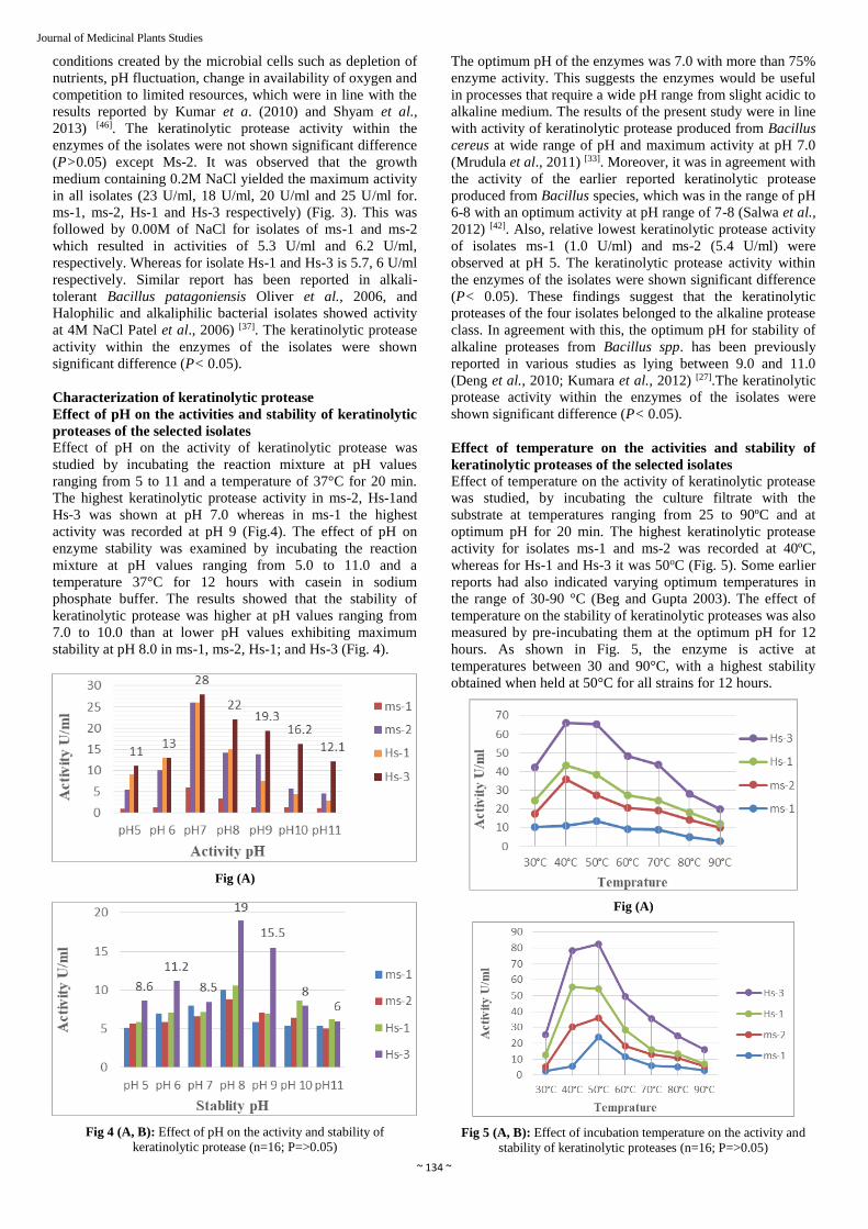
Effect of pH on the activity of keratinolytic protease was
studied by incubating the reaction mixture at pH values
ranging from 5 to 11 and a temperature of 37°C for 20 min.
The highest keratinolytic protease activity in ms-2, Hs-1and
Hs-3 was shown at pH 7.0 whereas in ms-1 the highest
activity was recorded at pH 9 (Fig.4). The effect of pH on
enzyme stability was examined by incubating the reaction
mixture at pH values ranging from 5.0 to 11.0 and a
temperature 37°C for 12 hours with casein in sodium
phosphate buffer. The results showed that the stability of
keratinolytic protease was higher at pH values ranging from
7.0 to 10.0 than at lower pH values exhibiting maximum
stability at pH 8.0 in ms-1, ms-2, Hs-1; and Hs-3 (Fig. 4).
Fig (A)
Fig 4 (A, B): Effect of pH on the activity and stability of
keratinolytic protease (n=16; P=>0.05)
The optimum pH of the enzymes was 7.0 with more than 75%
enzyme activity. This suggests the enzymes would be useful
in processes that require a wide pH range from slight acidic to
alkaline medium. The results of the present study were in line
with activity of keratinolytic protease produced from Bacillus
cereus at wide range of pH and maximum activity at pH 7.0
(Mrudula et al., 2011) [33]. Moreover, it was in agreement with
the activity of the earlier reported keratinolytic protease
produced from Bacillus species, which was in the range of pH
6-8 with an optimum activity at pH range of 7-8 (Salwa et al.,
2012) [42]. Also, relative lowest keratinolytic protease activity
of isolates ms-1 (1.0 U/ml) and ms-2 (5.4 U/ml) were
observed at pH 5. The keratinolytic protease activity within
the enzymes of the isolates were shown significant difference
(P< 0.05). These findings suggest that the keratinolytic
proteases of the four isolates belonged to the alkaline protease
class. In agreement with this, the optimum pH for stability of
alkaline proteases from Bacillus spp. has been previously
reported in various studies as lying between 9.0 and 11.0
(Deng et al., 2010; Kumara et al., 2012) [27].The keratinolytic
protease activity within the enzymes of the isolates were
shown significant difference (P< 0.05).
Effect of temperature on the activities and stability of
keratinolytic proteases of the selected isolates
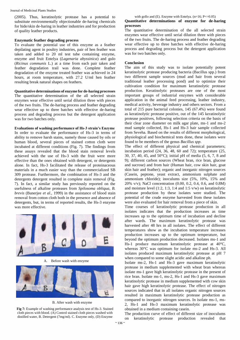
Effect of temperature on the activity of keratinolytic protease
was studied, by incubating the culture filtrate with the
substrate at temperatures ranging from 25 to 90ºC and at
optimum pH for 20 min. The highest keratinolytic protease
activity for isolates ms-1 and ms-2 was recorded at 40ºC,
whereas for Hs-1 and Hs-3 it was 50oC (Fig. 5). Some earlier
reports had also indicated varying optimum temperatures in
the range of 30-90 °C (Beg and Gupta 2003). The effect of
temperature on the stability of keratinolytic proteases was also
measured by pre-incubating them at the optimum pH for 12
hours. As shown in Fig. 5, the enzyme is active at
temperatures between 30 and 90°C, with a highest stability
obtained when held at 50°C for all strains for 12 hours.
Fig (A)
Fig 5 (A, B): Effect of incubation temperature on the activity and
stability of keratinolytic proteases (n=16; P=>0.05)

~ 135 ~
Journal of Medicinal Plants Studies
The results of this study clearly indicate that the optimum
temperature of proteolytic activity is in the range of the
optimum temperature of enzyme production. The
keratinolytic protease activity within the enzymes of the
isolates were shown significant difference (P< 0.05). From
the previous reports in stability of enzymes (Al-Shehri and
Mostafa, 2004) [6-15], the protease activity was relatively stable
at temperatures ranging from 50-65°C and 85.2% of the
activity was retained after incubation at 60°C. The stability of
protease enzyme could be due to the organisms’ genetic
adaptability to carry out their biological activities at higher
temperatures (Brock, 2012). The keratinolytic protease
activity within the enzymes of the isolates were shown
significant difference (P< 0.05).
Partial purification of crude enzymes for hair removal
The crude enzymes produced from the selected bacterial
isolates were partially purified by ammonium sulfate
precipitation at 80% saturation level and dialysis using
phosphate buffer. After precipitating the crude enzymes of
isolates ms-1, ms-2, Hs-1 and Hs-3 by adding ammonium
sulfate, purity of the enzymes were increased by 2.4, 2.6 and
2.5 folds, respectively. Moreover, after dialysis, purity of the
enzymes of the isolates ms-1, ms-2, Hs-1 and Hs-3 were
increased by 2.8, 2.9, and 3.0 folds, respectively (Table 2).
Table 2: Partial purification of keratinolytic Protease produced from selected bacterial isolates (n=16; P=>0.05)
Bacterial Isolates Purification steps Total volume (ml) Enzyme Activity (U/ml) Final Purification (folds)
ms-1 Crude 50 7.8 1.0
(NH3)2SO4 Precipitation 20 14.8 2.0
ms-2
Dialysis 9 20.5 2.7
Crude 50 12.2 1.0
(NH3)2SO4 Precipitation 21 29.5 2.42
Hs-1 Dialysis 8 33.4 2.7
Crude 50 7.5 1.0
(NH3)2SO4 Precipitation 18 18.5 2.5
Dialysis 11 36.5 4.9
Hs-3
Crude 50 8.3 1.0
(NH3)2SO4 Precipitation 25 15.8 1.9
Dialysis 10 42.2 5.1
Molecular weight determination of partially purified
keratinolytic protease using SDS PAGE
Molecular weights of the partially purified Keratinolytic
protease produced from the bacterial isolates were determined
using SDS PAGE. In this study, 6X Tries protein (10-
100kDA) was used as reference marker protein. The partially
purified enzymes had a single protein band on the SDS
PAGE. In relation to migration of the reference marker
protein, the approximate molecular weights of the partially
purified enzymes were found to be 80 kDa (Figure14).
Enzymatic evaluations of cow hide de-haring
To evaluate the potential use of this enzyme as a hide
depilating agent in leather industries, pair of cow hide was
taken and added to 250 ml flask containing enzyme, enzyme
and fruit Entelya (Lagenaria abyssinica) and gulo (Ricinus
communis L.) At a time from each pair taken and hair removal
trail was done. As shown in Fig. 6 below, complete de-haring
of the enzyme treated skin was achieved in 24 hours, at room
temperature, with 27.2 U/ml cow hide resulting pelt (hide) of
natural pore (grain) on dehaired surface.
(A)Control (B).Enzyme
(C) Both fruit with Enzyme (D) Total of removed hair
Fig 6: Example of cow hair de-haring performance analysis test of
Hs-3. (A) Control with distilled water, B. Enzyme only, C. Enzyme
with Entelya. (n=16; P=>0.05)
Results of enzymatic cow hide de-haring showed successful
use of the enzyme as a de-hairing agent. Complete de-hairing
of hide with fruit was achieved at 12 hours. Because of
specificity to hydrolysen on collagen protein part at hair roots
in hide, keratinolytic proteases are very important in
shortening hide de-haring time and in production of high
quality full gain leather having natural hair pores on the
surface (Siva Subramanian et al., 2008) [47]. Cow hide usually
treated with de-hairing chemicals in adrum for 24 hours
(Thanikaivelan et al., 2004) [50]. Shortening of de-haring time
has been also reported, 20 hours for Aspergilus flavus
protease by Malathi & Chakraborty, (1991) [30], and 9 hours
for keratinases of Bacillus subtilis S14 by Macedo et al.,
~ 136 ~
Journal of Medicinal Plants Studies
(2005). Thus, keratinolytic protease has a potential to
substitute environmentally objectionable de-haring chemicals
for hide/skin de-haring in leather industries and for production
of quality leather products.
Enzymatic feather degrading process
To evaluate the potential use of this enzyme as a feather
depilating agent in poultry industries, pair of hen feather was
taken and added to 20 ml test tube containing enzyme,
enzyme and fruit Entelya (Lagenaria abyssinica) and gulo
(Ricinus communis L.) at a time from each pair taken and
feather degradation trail was done. Complete feather
degradation of the enzyme treated feather was achieved in 24
hours, at room temperature, with 27.2 U/ml hen feather
resulting break natural shapes on feathers.
Quantitative determinations of enzyme for de-haring processes
The quantitative determination of the all selected strain
enzymes wear effective until serial dilution three with pieces
of the two fruits. The de-haring process and feather degrading
wear effective up to three batches with effective de-haring
process and degrading process but the detergent application
was for two batches only.
Evaluations of washing performance of Hs-3 strain’s Enzyme
In order to evaluate the performance of Hs-3 in terms of
ability to remove harsh stains, namely those caused by oils or
human blood, several pieces of stained cotton cloth were
incubated at different conditions (Fig. 7). The findings from
these assays revealed that the blood stain removal levels
achieved with the use of Hs-3 with the fruit were more
effective than the ones obtained with detergent, or detergents
alone. In fact, Hs-3 facilitated the release of proteinaceous
materials in a much easier way than the commercialized SB
309 protease. Furthermore, the combination of Hs-3 and the
detergents detergent resulted in complete stain removal (Fig.
7). In fact, a similar study has previously reported on the
usefulness of alkaline proteases from Spilosoma obliqua, B.
brevis (Banerjee et al., 1999) in the assistance of blood stain
removal from cotton cloth both in the presence and absence of
detergents, but, in terms of reported results, the Hs-3 enzyme
was more effective.
A. Before wash with enzyme
B. After wash with enzyme
Fig 7: Example of washing performance analysis test of Hs-3. Stained
cloth pieces with blood. (A) Control stained cloth pieces washed with
distilled water, B. Detergent (7mg/ml). C. Enzyme only, (D) Enzyme
with gollo and (E). Enzyme with Entelya. (n=16; P=>0.05)
Quantitative determinations of enzyme for de-haring
processes
The quantitative determination of the all selected strain
enzymes wear effective until serial dilution three with pieces
of the two fruits. The de-haring process and feather degrading
wear effective up to three batches with effective de-haring
process and degrading process but the detergent application
was for two batches only.
Conclusion
The aim of this study was to isolate potentially potent
keratinolytic protease producing bacteria (Bacillus spp.) from
two different sample sources (mud and hair from several
traditional leather processing pond) and to optimize their
cultivation condition for maximum keratinolytic protease
production. Keratinolytic proteases are one of the most
important groups of industrial enzymes with considerable
application in the animal feed processing, leather industry,
medical activity, beverage industry and others sectors. From a
total of 215 pure bacterial colonies, 145 (67.4%) were found
as keratinolytic protease positive, out of the 145 keratinolytic
protease positives, following selection criteria on the basis of
their clear zone diameter on milk agar plate, ms-1 and ms-2
mud sample collected, Hs-1 and Hs-3 hair sample collected
from Seveha. Based on the results of different morphological,
physiological and biochemical tests done, these isolates were
found to be members of the genus Bacillus spp.
The effect of different physical and chemical parameters;
incubation period (24, 36, 48, 60 and 72); temperature (25,
30, 37, 40, 45, and 50°C); initial pH of media (5, 6, 7, 8 and
9); different carbon sources (Wheat bran, rice bran, glucose
and sucrose) and from hair (Human hair, cow skin hair, goat
skin hair and feather); organic and inorganic nitrogen sources
(Casein, peptone, yeast extract, ammonium sulphate and
ammonium chloride); inoculums size (5%, 10%, 15% and
20% v/v); NaCl concentration (0.00, 0.2, 0.4, 0.6, and 0.8M)
and moisture level (1:2, 1:3, 1:4 and 1:5 v/w) on keratinolytic
protease production by these isolates were studied. The
potential of the crude enzyme harvested from these isolates
were also evaluated for hair removal from a piece of skin.
Time courses of keratinolytic protease production in all
isolates indicates that the production increases as time
increases up to the optimum time of incubation and decline
after wards. The maximum keratinolytic protease was
harvested after 48 hrs in all isolates. The effect of different
temperatures show as the incubation temperature increases
production increases up to the optimum temperature, but
beyond the optimum production decreased. Isolates ms-1 and
Hs-1 produce maximum keratinolytic protease at 40°C,
whereas 30°C was optimum for isolate ms-2 and Hs-3. All
isolates produced maximum keratinolytic protease at pH 7
when compared to some slight acidic and alkaline ph.
Isolate ms-2, Hs-1 and Hs-3 gave maximum keratinolytic
protease in medium supplemented with wheat bran whereas
isolate ms-1 gave high keratinolytic protease in the present of
rice bran. Isolate ms-1, ms-2, Hs-1 and Hs-3 gave maximum
keratinolytic protease in medium supplemented with cow skin
hair gave high keratinolytic protease. The effect of nitrogen
sources indicated that in all isolates organic nitrogen sources
resulted in maximum keratinolytic protease production as
compared to inorganic nitrogen sources. In isolate ms-1, ms-
2, Hs-1 and Hs-3 maximum keratinolytic protease was
obtained in a medium containing casein.
The production curve of effect of different size of inoculums
on keratinolytic protease production revealed that

~ 137 ~
Journal of Medicinal Plants Studies
keratinolytic protease production increased when the percent
of inoculums increased up to the optimum and decreased
beyond the optimum size. In all isolates maximum
keratinolytic protease were harvested in 10% v/v inoculums.
The effect of moisture level on keratinolytic protease
production indicated that keratinolytic protease production
increased with increased bran to moistening agent till
optimum decreased beyond the optimum and all isolates give
maximum protease at 1:3 v/w bran to moisture ratio. On the
other hand, production of keratinolytic protease is also
influenced by the concentration of NaCl on the growth media.
The optimum NaCl concentration was found to be 0.2 M for
all four isolates.
Although many potent isolates are on market for enzyme
production, scientists prefer studying new isolates because
they could be alternative for commercial use in many aspects.
Many studies showed that researches will continue to isolate
alternative strains for production of enzymes as well as
keratinolytic proteases. The isolated new source of
keratinolytic protease producing bacteria, from the soil and
water samples that are collected from traditional leather
processing ponds might be an alternative source for the
potential industrial applications.
References
1. Aiba S, Kitai K, Imanaka T. Cloning and Expression of
Thermostable protease Gene from Bacillus spp. Appl.
Environ. Microbiol. 1983; 46:1059-1065.
2. Akcan N. Production of extracellular protease in
submerged fermentation by Bacillus licheniformis ATCC
12759. African Journal of Biotechnology. 2012;
11(7):1729-1735.
3. Akpomie OO, Akponah E, Okorawhe P. Amylase
production potential of bacterial isolates obtained from
cassava root peels. Agricultural Science Research
Journals. 2012; 2(2):95-99.
4. Agrawal R, Singh R, Verma A, Panwar P, Verma AK. et
al. Partial Purification and Characterization of Alkaline
Protease from Bacillus sp. Isolated from Soil. World
Journal of Agricultural Sciences. 2012; 8(1):129-133.
5. Ali S. et al. ‘An Enzyme with Mutiple Industrial
Applications (Review), European journal of
pharmaceutics and medical research. 2017; 4(7):63-70.
6. Al-Shehri A, Mostafa M, Yasser S. Production and some
properties of protease produced by Bacillus licheniformis
isolated from Tihamet Asser, Saudi Arabia. Pak. J Biol.
Sci. 2004; 7:1631-1635.
7. Annamalai N, Thavasi R, Vijayalakshmi S,
Balasubramanian T. Extraction, purification, and
characterization of thermostable, alkaline tolerant
amylase from Bacillus cereus. Indian J Microbiol. 2011;
51(4):424-429.
8. Ashwini K, Kumar G, Karthik L, Rao BKV.
Optimization, production and partial purification of
extracellular protease from Bacillus spp. Marini.
Archives of applied Science Research. 2011; 3(1):33-42.
9. Bhalla TC, Kumar D, Gajju H, Agrawal
HO. Thermophilic bacterial proteases. J. Punjab Acad.
Sci. 1999; 1:77-91.
10. Benjamin, S, Smitha RB, Jisha VN, Pradeep S, Sajith S,
Sreedevi S. et al. A monograph on protease from Bacillus
spp. Advances in Bioscience and Biotechnology. 2013;
4:227-241.
11. Deng A, Wua J, Zhang Y, Zhang G, Wena T. Purification
and characterization of a surfactant-stable high-alkaline
protease from Bacillus sp. B001 Bioresource
Technology. 2010; 101:7100-7106.
12. Duncan F. Applied Microbiology Laboratory Manual. 4th,
2005, 1-70.
13. Durham DR. Utility of GX as a detergent additve. J.
Appl. Bacteriol. 1987; 63:381-386.
14. Enshasy EH, Abuol-Enein A, Helmy S, Azaly E.
Optimization of The industrial production of alkaline
protease by Bacillus licheniformis in different production
scales. Australian Journal of Basic and Applied Sciences.
2008; 2:583-593.
15. Folin O, Ciocalteu V, Enzymatic Assay of Protease
Casein as a Substrate J. Biol. Chem. 73:627Al-Shehri, A.;
Mostafa M, Yasser S. Production and some properties of
protease produced by Bacillus licheniformis isolated from
Tihamet Asser, Saudi Arabia. Pak. J. Biol. Sci. 2004;
7:1631-1635.
16. Jisha VN, et al. Versatility of microbial proteases,
Advances in Enzyme Research. 2013; 1(3):39-51.
17. Gajju H, Bhalla TC, Agarwal HO. Thermostable alkaline
protease from thermophilic Bacillus coagulans PB-77.
Indian J. Microbiol. 1996; 36:153-155.
18. Gangadharan D, Sivaramakrishnan S, Namboothiri KM,
Pandey A. Solid culturing of Bacillus amyloliquefaciens
for α-amylase production. Food Technol. Bio techno.
2006; 44:269-274.
19. Gessesse A, Hatti-Kaul R, Gashe BA, Mattiasson B.
Novel alkaline proteases from alkaliphilic bacteria grown
on chicken feather. Enzyme and Microbial Technology.
2003; 32(5):519-524.
20. Gessesse AF, Mula SL, Lyantagaye L. Nyine-Wamwize
Mattisson B. et al. Industrial Enzyme for sustainable Bio-
economy: Large scale production of application in
environment and agriculture in Eastern Africa, Nairobi,
LRI, 2011.
21. Gessesse A, Gashe BA. Production of alkaline protease
by an alkaliphilic bacteria isolated from an alkaline soda
lake. Biotechnology Letters. 1997; 19(5):479-481.
22. Ghaemi FS, Tabandeh F, Yakhchali B, Eftekhar F.
Enhancement of alkaline protease production by Bacillus
clausii using Taguchi experimental design. African
Journal of Biotechnology. 2007; 6(22):2559-2564.
23. Harley JP, Prescott LM. Laboratory Exercise in
Microbiology 5th ed, The McGraw−Hill Companies,
2002, 466.
24. Hema TA, Shiny M. Production of Protease Enzyme rom
Bacillus Clausii Sm3. IOSR Journal of Pharmacy and
Biological Sciences. 2012; 1:37-40.
25. Holt JG, Krieg NR, Sneath PHA, Staley JT. Bergey’s
Manual of Determinative Bacteriology. Nineteenth
edition, Williams and Wilkins company, Baltimore MD.
USA, 1994, 255-273.
26. Josephine S, Ramya V, Devi N, Ganapa B,
Siddalingeshwara KG, Venugopal. N. et al. Isolation,
production and characterization of protease from Bacillus
Sp isolated from soil sample. J Microbiol. Biotech. Res.
2012; 2(1):163-168.
27. Kumar DJM, Venkatachalam P, Govindarajan N,
Balakumaran MD, Kalaichelvan PT. Production and
Purification of Alkaline Protease from Bacillus sp.
MPTK 712 Isolated from Dairy Sludge. Global
Veterinaria. 2012; 8(5):433-439.
28. Kumar R, Vats R. Protease Production by Bacillus
subtilis Immobilized on Different Matrices. New York
Science Journal. 2010; 3(7):20-24.

~ 138 ~
Journal of Medicinal Plants Studies
29. Macedo AJ, Silva WOB, Termignoni C. Properties of a
non collagendegrading Bacillus subtilis keratinase. Can. J
Microbiol. 2008; 54:180-188.
30. Malathi S, Chakraborty R. Production of alkaline
protease by a new Aspergillus flavus isolate under solid
substrate fermentation conditions for use as a depilation
agent. Appl Environ Microbiol. 1991; 57:712-16.
31. Meenakshi C, Narender K, Vikrant A, Karupothula S,
Shobhana B. Sushma S. et al. Isolation and optimization
of various conditions of growth. Research Journal of
Biotechnology. 2009; 4(1):50-56.
32. Moon SY, Oh TK, Rho HM. Purification and
characterization of an extra cellular alkaline protease
from Bacillus subtilis RM 615. Korean Biochem. J. 1994;
27:323-329.
33. Mrudula S, Gopal R, Seenayya G. Effect of substrate and
culture conditions on the produuction of amylase and
pullulanase by thermophilic Clostridium
thermosulfurogenes SVM17 in solid state fermentation.
Malaysian J Microbiol. 2011; 7(1):15-21.
34. Muthu P, Christudhas W. Purification and
characterization of neutral protease enzyme from Bacillus
Subtilis. J Microbiol. Biotech. Res. 2012; 2(4):612-618.
35. Niadu KSB, Devi KL. Optimization thermostable
alkaline protease production from species Bacillus using
rice bran. Afr. J Biotechnol. 2005; 4:724-726.
36. Pandey A, Nigam P, Soccol R, Soccol T, Singh D,
Mohan R. et al. Advances in microbial amylases.
Biotechnol. Appl. Biochem. 2000; 31(2):135-152.
37. Patel RK, Dodia MS, Joshi RH, Singh SP. Production of
extracellular halo-alkaline protease from newly isolated
Halophilic Bacillus sp. isolated from sea water in
Western India. WJ Microbiol. Biotechnol. 2006; 22:375-
382.
38. Qadar SAU, Shireen E, Iqbal S, Anwar A. Optimization
of Protease production from newly isolated strains of
Bacillus sp. PCSIR EA-3. Indian Journal of
Biotechnology. 2009; 8:286-290.
39. Oliveira AN, Oliveira LA, Andrade S. Production and
Some Properties of Crude Alkaline Proteases of
Indigenous Central Amazonian Rhizobia Strains.
Brazilian Archives of Biology and Technology. 2010;
53:1185-1195.
40. Olivera N, Sequeiros C, Sineriz F, Breccia J.
Characterization of alkaline proteases from novel alkali-
tolerant bacterium Bacillus patagoniensis.
World J Microbiol. Biotechnol. 2006; 22:737-743.
41. Ogbonnaya N, Odiase A. Influence of media composition
on the production of alkaline protease from Bacillus
subtilis CB-18. Acta Sci. Pol. Technol. Aliment. 2012;
11(3):231-238.
42. Salwa EI, Hassan BE, Elmutaz NH, Elhadi S. Amylase
production on solid-state fermentation by Bacillus
Species. Food publi0c health. 2012; 2(1):30-35.
43. Saxena R, Singh R. Amylase production by solid-state
fermentation of agro industrial wastes using Bacillus
species. Brazil. J Microbiol. 2011; 42:10334-1342.
44. Serin B, Akcan N, Uyar F. Production and optimization
of α-amylase from Bacillus, circulans ATCC 4516 with
solid-state fermentation. Hacettepe J Biol. Chem. 2012;
40(4):393-400.
45. Sharma K. Manual of Microbiology: Tools and
Techniques. 2nd ed. Ane Books India, New Delhi. ISBN.
2007; (10):81-8052-143-5.
46. Shyam SA, Sonia SS, Lal G. Amylase activity of a starch
degrading bacteria isolated from soil. Archives Appl. Sc.
R. 2013; 5(1):15-24.
47. Siva Subramanian S, Murali Manohar B, Puvanakrishnan
R. Mechanism of enzymatic dehairing of skins using a
bacterial alkaline protease. Chemosphere. 2008; 70:1025-
1034.
48. Sumantha A, Larroche C, Pandey A. Microbiology and
Industrial Biotechnology of Food-Grade Proteases: A
Perspective. Food Technol. Biotechnol. 2006; 44(2):211-
220.
49. Tambekar DH, Tambekar SD. Partial Characterization
and optimization of alkaline protease production of
bacillus pseudofirmus from Lonar Lake. International
journal of advance pharmaceutical and biological
sciences. 2012; 2(1):130-138.
50. Thanikaivelan P, Rao JR, Nair BU, Ramasami T.
Progress and recent trends in biotechnological methods
for leather processing. Trends in Biotechnology. 2004;
22:181-188.
51. Vigneshwaran C, Shanmugam S, Kumar TS. Screening
and characterization of keratinase from Bacillus
licheniformis isolated from namakkal poultry farm.
Researcher. 2010; 2:89-96.
52. Sevinc N, Demirkan E. Production of Protease by
Bacillus sp. N-40 Isolated from Soil and Its Enzymatic
Properties. J Biol. Environ. Sci. 2011; 5(14):95-103.