Embed Size (px)
Citation preview
Sizing for endovascular aneurysm repair: clinical
evaluation of a new automated three-dimensional
software.
Adrien Kaladji, Antoine Lucas, Gaelle Kervio, Pascal Haigron, Alain Cardon
To cite this version:
Adrien Kaladji, Antoine Lucas, Gaelle Kervio, Pascal Haigron, Alain Cardon. Siz-ing for endovascular aneurysm repair: clinical evaluation of a new automated three-dimensional software.. Annals of Vascular Surgery, Elsevier Masson, 2010, 24 (7), pp.912-20.<10.1016/j.avsg.2010.03.018>. <inserm-00522933>
HAL Id: inserm-00522933
http://www.hal.inserm.fr/inserm-00522933
Submitted on 17 Nov 2010
HAL is a multi-disciplinary open accessarchive for the deposit and dissemination of sci-entific research documents, whether they are pub-lished or not. The documents may come fromteaching and research institutions in France orabroad, or from public or private research centers.
L’archive ouverte pluridisciplinaire HAL, estdestinee au depot et a la diffusion de documentsscientifiques de niveau recherche, publies ou non,emanant des etablissements d’enseignement et derecherche francais ou etrangers, des laboratoirespublics ou prives.
Sizing for EVAR: clinical evaluation of
a new automated three-dimensional
software
Kaladji A1, 2, Lucas A1, 2, 3, 4, Kervio G2, Haigron P3,4, and Cardon A1, 2
(1) Vascular surgery unit, Pontchaillou Hospital, Rennes, France
(2) Center for Clinical Investigation and Technological Innovation, Inserm U642, Rennes,
France
(3) Rennes 1 University, LTSI, Rennes F-35042, France
(4) INSERM U642, Rennes F-35000, France
Corresponding author:
Adrien Kaladji, Service de Chirurgie Vasculaire, CHU Hôpital Pontchaillou, 2 rue Henri Le
Guilloux, 35033 Rennes cedex 9, France
Email: [email protected]

Abstract
Purpose: To assess the reproducibility and accuracy of the sizing procedure prior to aortic
endograft implantation using new sizing automated software compared to standard
radiological procedures.
Methods: Based on original spiral-computed tomography images, the sizing of 32 patients
with abdominal aortic aneurysm (AAA) treated by endovascular aneurysm repair (EVAR)
was retrospectively compared. The first sizing was performed by a radiologist using a
standard workstation (General electrics) and software (Advanced vessel analysis). The second
was performed twice by two surgeons using a personal computer with automatic three-
dimensional sizing software (Endosize, Therenva). All diameters and lengths required prior to
EVAR were measured (17 items). Additionally, thirteen qualitative criteria regarding EVAR
feasibility, including neck length, were compared. Intra- and interobserver variability with
Endosize, as well as the variability between the two measurement methods were analyzed
using the intraclass correlation coefficient (ICC) and Bland and Altman’s method. Qualitative
variables were analyzed using Fischer’s exact test and kappa coefficient.
Results: Intra-observer variability with Endosize proved to be efficient. None of the ICCs
were lower than 0.9, and more than 90% of the absolute differences between two
measurements were less than 2mm. Inter-observer variability with Endosize was assessed in a
similar manner. Measurement variability of vessel diameters was less marked than that of
vessel lengths. This trend was observed for all data sets. Comparison of the two measurement
methods demonstrated a good correlation (minimum ICC=0.697; maximum ICC=0.974),
though less so than that observed using Endosize. Mean time consumption using Endosize
was 13.1+/-4.53 minutes (range: 7.2-32.7). Analysis of the alarm sets demonstrated a high
agreement between observers (kappa coefficient=0.81).

Conclusion: Sizing using the Endosize software is as reliable as conventional radiological
procedures. Sizing by surgeons using an automated, user-friendly, and mobile tool appears to
be reproducible.
Keys words: Sizing, EVAR, endovascular intervention, computed tomography angiography,
workstation, post-processing image treatment, preoperative measurements
INTRODUCTION
The sizing, which is the first step of endovascular aneurysm repair (EVAR), is essential for a
successful procedure. Several sizing methods1,2 have been assessed, using highly
sophisticated and expensive radiological workstations and software. Surgeons must be able to
control this first step using reliable software, with results that are as accurate as those obtained
at radiological workstations. For surgeons using this software, preoperative navigation within
the vessels and accurate measurements are the primary objectives, enabling them to
accurately plan an EVAR. To our knowledge, there is little data on automated software testing
in a clinical evaluation context. This study aimed to assess whether the sizing procedure
using automated three-dimensional (3D) sizing software, which had been developed in our
clinical investigation and technological innovation center, was as accurate and reproducible as
that performed at a radiological workstation.
METHODS
In total, 32 patients (29 men, 3 women; mean age: 74.9 +/-9.4) with abdominal aortic
aneurysm (AAA) and treated by endovascular AAA repair (EVAR) were studied
retrospectively. They were randomly assigned to EVAR procedure between 2006 and 2007.
Patients were selected for EVAR based on clinical and morphological criteria (Table 1).
Measurements
All patients were evaluated using spiral computed tomography angiography (CTA) prior to
EVAR. All imaging examinations were performed on a multislice CT scanner (General
Electric Medical Systems, Milwaukee, Wisconsin, LightSpeed16). Parameters for the
acquisitions were 1.25mm slice thickness, 120 kVp, and 215-360 mA tube current. Imaging
was initiated after administering 120mL of low-osmolar iodinated contrast agent (Hexabrix,
iodine concentration 320mg/ml). Soft tissue window settings with a width of 400HU and a
center of 40HU were applied.
Quantitative variables studied (Fig. 1) included the largest and smallest diameter on CT slices
on the first slice distal to the lowermost renal artery (D1a), 15mm below this landmark (D1b),
healthy neck end (D1c), as well as left (D2) and right (D3) distal primitive iliac arteries.
Maximal AAA diameter (DAAA) and smallest diameter of left (Lmin) and right (Rmin)
external iliac arteries were also measured. Length measurements included proximal aortic
neck (NL), length between the lowermost renal artery and aortic bifurcation (L1), as well as
left (L2) and right (L3) common iliac arteries.
Qualitative variables, referred to as "alarms", were defined as morphological features of the
aneurysm which could change the therapeutic strategy or draw the surgeon’s attention to
potential technical difficulties during the procedure. These variables were NL !15mm (V1),
D1a !18 mm or "32 mm (V2), reverse taper neck (or difference between D1a and D1c "25%)
(V3), bony-sharp neck "60° (V4), aortic bifurcation (D4) ! 20 mm (V5), L2 !10 mm (V6),
L3 !10 mm (V7), Lmin !7.5 mm (V8), Rmin !7.5 mm (V9), D2 "20 mm (V10), D3 "20 mm
(V11), left iliac tortuosity (V12), and right iliac tortuosity (V13). These data sets were
characterized as present or absent.
In addition, stent graft type (aorto-uni-iliac or aorto-bi-iliac) and predicted complementary
procedures or complications which arose during EVAR were reported.
Image Analysis
The first sizing was performed by an interventional radiologist. Original contrast-enhanced
CT images were electronically transferred to a General Electrics workstation, and
measurements of aorto-iliac lengths and diameters were carried out using advanced vessel
analysis (AVA) software. These measurements were considered as references for the
procedure planning. Thereafter, a vascular surgeon, who was blinded to all radiological
results, carried out the sizing of the endograft after importing the same scan data. This sizing
was performed using Endosize (Therenva, Rennes), a 3D sizing software tool that had been
optimized to run on a conventional personal computer (PC). A 2.4 GHz processor and 2 Go
random access memory (RAM) were components of the surgeon’s PC. Measurements with
Endosize were repeated twice (Sizing 1 and 2) by two vascular surgeons (Surgeon 1 and 2),
separated by a 2-week time interval. Each sizing was recorded. Endosize operated in four
steps:
1. CT data loading and visualization: Two-dimensional slice views and smooth volume
rendering view were obtained.
2. Data processing: The vessel lumen and centerline extraction required viewing the aortic
structure in 3D. The image analysis process was based on a combination of boundary-based
and region-based segmenting algorithms including morphological operations to automatically
remove connection between the vasculature of interest (aorto-iliac structure) and bone
structures (such as vertebra), as well as to determine the centerlines of vascular branches3,4,5.
Moreover, a powerful and optimized volume-rendering process exploiting graphics
processing unit computation performances was implemented in order to visualize three-
dimensional vascular geometry. In addition to this, 3D vessel description scheme, as well as
reformatted CT slices and contours of the vessel lumen were computed along the vessels’
curvilinear axes. Only one parameter could intuitively be adjusted during the data processing
step, which determined the discrete volume (voxels) contained in the vessel lumen. The
computation time for data processing and rendering, consecutive to a new adjustment of this
parameter, took approximately 1 second, which was compatible with using interactive
software. Moreover, as the default value of this analysis parameter had been settled for AAA
CTA observations, in most cases no modifications were needed. The graphics user interface
thus allowed for linking the anatomical features of the aorto-iliac structure (geometry and
parietal quality) to those of the stent graft, while the user (vascular surgeon or interventional
radiologist) remained in control of the decision-making process.
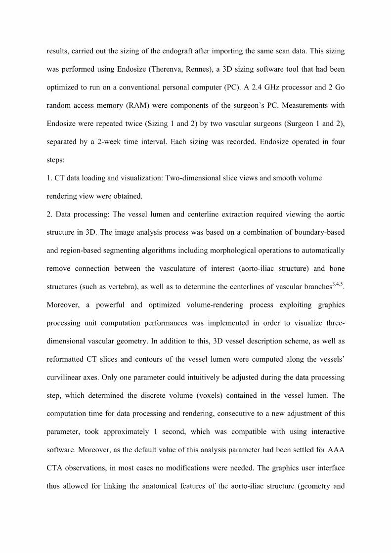
3. Semi-automatic measuring (Fig. 2): Useful lengths and diameters taken on the vessel
centerlines were automatically obtained after a simple interactive step consisting in a three-
dimensional point picking sequence. To this end, eight points were required, corresponding to
the suprarenal aorta, infrarenal fixation site, healthy neck end, aortic bifurcation, left iliac
bifurcation, right iliac bifurcation, left external distal site, and right external distal site. In the
next step, while the length and diameter measurements were proposed by the software, the
user had to control the measurements’ accuracy on a separate window showing the slice
(perpendicular to the centerline) used by the software to assess the diameters. The software
proposed inner-to-inner diameter measurements. Surgeons were given the option of including
a thrombus in the measurements, with an adjustable parameter for an enlarged area of
measurement of interest. In the case of disagreement, the user could easily adjust the
measurement.
4. Sizing report (Fig.3): The feasibility of the procedure was defined by the aforementioned
alarms.
Statistical Analysis
Quantitative and qualitative variables were analyzed separately by several methods. For
quantitative variables, correlation was assessed using the intraclass coefficient correlation
(ICC), and variation between the data sets was compared by calculating the mean pair
difference for each data point and averaging them for the dataset comparison. According to
the method described by Bland and Altman, limits of agreement were calculated (for each
point: mean difference between observers +/- the standard deviation multiplied by 1.96).
Mean time consumption for sizing was compared using the non-parametric Wilcoxon signed
rank test (intra- and inter-observer). Qualitative variables were analyzed using Fischer’s exact
test and kappa coefficient. P ‹0.05 was considered significant. Complete agreement was
defined as 1.0.
RESULTS
Quantitative Data
Intra-observer: All ICCs were above 0.9, except for and mean differences were less than
1mm for all diameters measured (Table 2, Fig. 4). Differences were more pronounced for
length measurements, though still less than 2mm, with similar trends observed for absolute
differences. For all diameters, at least 95% of absolute differences were less than 2mm, and in
no case did they exceed 5mm. Absolute differences in lengths were more pronounced,
exceeding 5mm in some of the cases. Mean time consumption for sizing (Table 5) did not
exceed 15 minutes. There was no statistical difference in time consumption for the two sizing
by each surgeon (p value from Wilcoxon test for surgeon 1 for the comparison between sizing
1 and 2 was 0.215 for surgeon 1, and 0.473 for surgeon 2).
Inter-observer: All ICCs were above 0.9 except for D1a max (0.891) (table 4, Fig. 4). In
addition, mean differences were more pronounced for lengths than diameters. The maximum
mean difference was 2.051mm for L1 (length between renal arteries and aortic bifurcation),
with 92.2% of absolute differences in diameters being lower than 2mm, and 100% lower than
5mm. Absolute differences for lengths were also more marked (in the worst scenario, 68.8%
were lower than 2mm for L1). For the first sizing, there was no statistical difference between
surgeons 1 and 2 (p=0.401), in contrast to the second sizing, where a significant difference
was observed (p=0.001).
Radiological workstation/Endosize: Six items presented an ICC!0.9, seven items an ICC
between 0.8 and 0.9, and three items an ICC between 0.7 and 0.8 (minimum for D1a min,
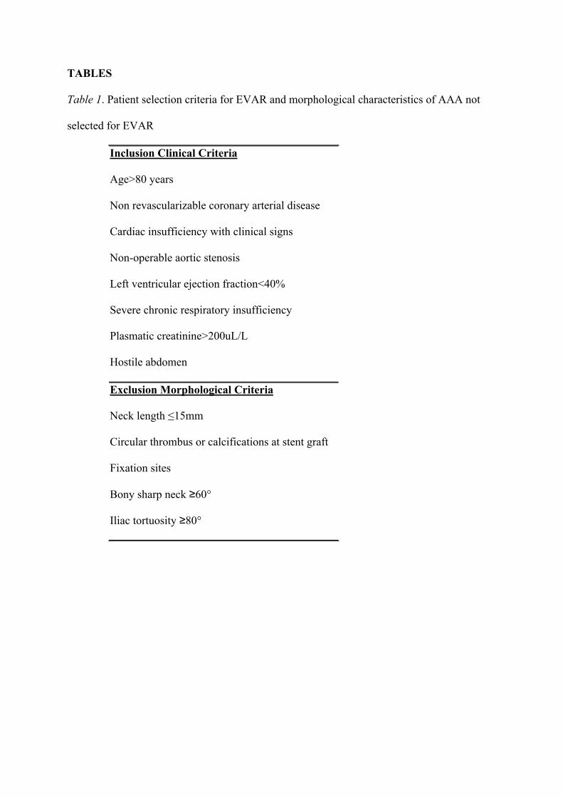
ICC=0.697) (Table 4,Fig.4, Fig. 5 a-c). The maximum mean difference was 3.974mm (L1).
For more than 50% of length measurements, absolute differences were above 2mm (versus
70.2% for diameters). Mean time consumption for sizing during the study was 13.1+/-4.53
minutes (minimum 7.2; maximum 32.7).
Qualitative Data
Regarding EVAR alarms, the radiologist emitted 41 alarms and the surgeon 37 alarms.
Fischer’s exact test revealed no significant difference between the alarms emitted by the
radiologist and surgeon. Overall, 87% of the alarms emitted by the radiologist were the same
as those emitted by the surgeon, while 94% of the alarms emitted by the surgeon were the
same as those emitted by the radiologist. Analysis of the alarm sets reveals a high agreement
between observers (kappa coefficient=0.81).
Type of stent graft
Five patients, three with an aortic bifurcation stenosis and two with AAA and iliac aneurysm,
were treated using an aorto-uni-iliac stent graft with femoro-femoral cross bypass (Table 6).
For three patients (A, B, C), V5 alarm (aortic bifurcation! 20mm) was emitted by the
radiologist and surgeon. Patients A and B had their endograft implanted on the right side, and
patient C on the left side. For patients A and B, no other alarm was emitted by either the
radiologist or surgeon (regarding iliac status). For patient C however, both of them had
emitted the V8 and the V9 alarms (left and right external iliac stenosis), and an angioplasty
was performed on the right side prior to EVAR. For patients D and E, no aortic bifurcation
stenosis was detected by either the radiologist or surgeon. For patient D, V9 alarm (right
external iliac stenosis) was emitted by both the radiologist and surgeon, while V11 alarm
(right primitive iliac aneurysm) was emitted by radiologist alone. The endograft was
implanted on the left side with a ligation of the right primitive iliac artery (diameter of the
right primitive iliac artery by the surgeon was 13 and 22mm. For the last patient, only the V10
alarm (left primitive iliac aneurysm) was emitted by both the radiologist and surgeon (about
iliac level).
EVAR procedure
Predicted complementary procedures during EVAR included two hypogastric coverages: one
on the right side where a right distal primitive iliac aneurysm was identified by both
observers, and the other on the left side where a bilateral distal primitive iliac aneurysm was
identified. Non-predicted complementary procedures included an iliac stent graft extender
module where an iliac tortuosity was detected; Type 1a endoleak treated with a Palmaz stent,
where any alarms about the neck were emitted; a right external iliac dissection, treated using a
bypass, following an aorto-uni-iliac stent graft implanted on the right side, where no stenosis
along the artery was detected by either the radiologist or surgeon.
DISCUSSION
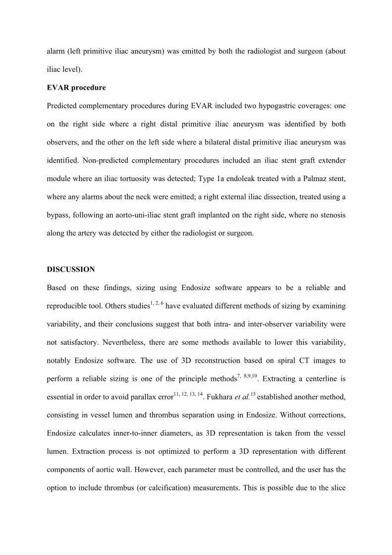
Based on these findings, sizing using Endosize software appears to be a reliable and
reproducible tool. Others studies1, 2, 6 have evaluated different methods of sizing by examining
variability, and their conclusions suggest that both intra- and inter-observer variability were
not satisfactory. Nevertheless, there are some methods available to lower this variability,
notably Endosize software. The use of 3D reconstruction based on spiral CT images to
perform a reliable sizing is one of the principle methods7, 8,9,10. Extracting a centerline is
essential in order to avoid parallax error11, 12, 13, 14. Fukhara et al.15 established another method,
consisting in vessel lumen and thrombus separation using in Endosize. Without corrections,
Endosize calculates inner-to-inner diameters, as 3D representation is taken from the vessel
lumen. Extraction process is not optimized to perform a 3D representation with different
components of aortic wall. However, each parameter must be controlled, and the user has the
option to include thrombus (or calcification) measurements. This is possible due to the slice
which is perpendicular to centerline, and a tool adjusting the area of interest in measurement,
which detects lumen, thrombi, and calcifications. Moreover, in this study, there was no case
of severe thrombus in the aortic neck or at iliac fixation sites, as these are considered to be
contraindications to EVAR (exclusion criteria). Thrombus within the aneurysm does not
influence the planning of EVAR. We believe that when detecting a thrombus, it is essential to
schedule navigation within the vessel during the procedure, and that this is accurately
assessed with multiplanar reformatted slices.
Regarding the intra-observer variability, reproducibility proved efficient. Automatic tracking
appears to provide the same measurements as long as the eight points placed in aorto-iliac
representation are at the same location. The variability noted between observers is due to
discrepancies in the placement of these points, which explains why ICCs and variations were
more pronounced for lengths.
Differences between the two measurement methods were more pronounced, particularly for
lengths, but still appear to be widely acceptable. Indeed, the point defining aortic bifurcation
is a slightly higher when using Endosize than AVA. This point corresponded to the
intersection with the centerline of iliac arteries, while in the radiologist’s measurements, it
was located on the real point in the arterial wall of the aortic bifurcation. One limitation of the
present study was that the same observer’s variability when using the two methods could not
be studied due to the retrospective design of the study.
The feasibility of EVAR, as represented by the study’s qualitative variables, was similarly
evaluated by both the radiologist and surgeon. For the five aorto-uni-iliac endografts
implanted, an appropriate alarm was emitted each time by the Endosize software except in the
case of patient D. The radiologist identified an iliac aneurysm, whereas the Endosize software
did not emit the appropriate alarm as the alarm cut-off was 20mm. However, for this patient,
iliac artery measurements of 13mm and 22mm (average 17.5mm) did reveal an iliac ectasia.

Predicted complementary procedures were identified by Endosize as well as the radiologist.
Nonetheless, observers did not anticipate all complications, suggesting that CTA analysis
prior to EVAR should not be limited to sizing the AAA, but also requires special attention
directed at the quality of the aortic wall (calcifications, thrombus…).
While several sizing software for EVAR are used by radiologists and surgeons, only a few
studies have reported on the assessment of software aimed specifically at surgeons, and which
is as reliable as conventional radiological software. Neri et al.16 reported on an analysis
system using a remote web server with similar methods, but did not report on time
consumption (the mean time necessary for segmentation 1h, and more in the case of
calcifications). In our study, the time required for sizing, including segmentation and
measurements, proved more appropriate for surgeons’ expectations. Lee et al.17 have recently
proposed the TeraRecon Aquarius workstation, using the same reconstructions as described
above, but without clinical evaluation. Currently, most of the semi-automatic software used
for sizing requires powerful hardware, and thus cannot be used by all vascular surgeons
performing EVAR. Endosize software is aimed at fulfilling the surgeons’ specific
expectations, allowing for total autonomy in the forward planning of EVAR. This is achieved
by using a mobile and user-friendly tool, which is able to run on a personal computer, unlike
other software. Moreover, this tool has a quick learning curve and requires little time, which
is the landmark of semi-automatic software18.
Our data suggests that the accuracy of the different lengths and diameters determined using
Endosize prior to EVAR may be as reliable as that obtained with conventional radiological
equipment. To plan EVAR, surgeons have a powerful new tool, which is more suitable than
software currently used in clinical evaluations. Preoperative assessment prior to EVAR
appears reliable, though peroperative complications may occur with no warning. Further
studies are necessary to assess the peroperative value of an angionavigation computer system

in order to secure the placement of aortic stent grafts in 3D, and for more precise control over
the deployment of different branches.
Acknowledgments
The authors thank Dr. JF Heautot, Dr. A. Larralde and Dr. A. Roux from the radiology unit
for their contributions.
This work was partially supported by French institutional grants (French National Research
Agency (ANR) Through TecSan program (project ANGIOVISION n°ANR-09-TECS-003)).
REFERENCES
1. Diehm N, Baumgartner I, Silvestro A, et al. Automated software supported versus manual
aorto-iliac diameter measurements in CT angiography of patients with abdominal aortic
aneurysms: assessment of inter- and intraobserver variation. Vasa. 2005;34:255-261.
2. Boll DT, Lewin JS, Duerk JL, et al. Assessment of automatic vessel tracking techniques in
preoperative planning of transluminal aortic stent graft implantation. J Comput Assist
Tomogr. 2004;28:278-285.
3. Haigron P, Bellemare ME, Acosta O, et al. Depth-map-based scene analysis for active
navigation in virtual angioscopy. IEEE Transactions on Medical Imaging. 2004;23:1380-90.
4 Fleureau J, Garreau M, Simon A, et al. Assessment of global cardiac function in MSCT
imaging using fuzzy connectedness segmentation. Computers In Cardiology 2008
5. Boldak C, Rolland Y,Toumoulin C, et al. An improved model-based vessel tracking
algorithm with application to computed tomography angiography. Journal of Biocybernetics
and Biomedical Engineering.2003;23: 41-63.
6. Singh K, Jacobsen BK, Solberg S, et al. Intra- and interobserver variability in the
measurements of abdominal aortic and common iliac artery diameter with computed
tomography. The Tromsø study. Eur J Vasc Endovasc Surg. 2003;25:399-407.
7. Dillavou ED, Buck DG, Muluk SC,et al. Two-dimensional versus
three-dimensional CT scan for aortic measurement. J Endovasc Ther. 2003;10:531-8.
8. Shin CK, Rodino W, Kirwin JD, et al. Can preoperative spiral CT scans alone determine
the feasibility of endovascular AAA repair? A comparison to angiographic measurements. J
Endovasc Ther. 2000;7:177-183
9. Parker MV, O'Donnell SD, Chang AS, et al. What imaging studies are necessary for
abdominal aortic endograft sizing? A prospective blinded study using conventional computed
tomography, aortography, and three-dimensional computed tomography. J Vasc Surg.
2005;41:199-205.
10. Sprouse LR, Meier GH, Parent FN, et al. Is three-dimensional computed tomography
reconstruction justified before endovascular aortic aneurysm repair? J Vasc Surg. 2004;
40:443-447.
11. Velazquez OC, Woo EY, Carpenter JP, et al. Decreased use of iliac extensions and
reduced graft junctions with software-assisted centerline measurements in selection of
endograft components for endovascular aneurysm repair. J Vasc Surg. 2004;40:222-227.
12. Cayne NS, Veith FJ, Lipsitz EC, et al.Variability of maximal aortic aneurysm diameter
measurements on CT scan: significance and methods to minimize. J Vasc Surg. 2004;39:811-
815.
13. Isokangas JM, Hietala R, Perälä J, et al. Accuracy of computer-aided measurements in
endovascular stent-graft planning: an experimental study with two phantoms. Invest Radiol.
2003;38:164-170
14. Aziz I, Lee J, Lee JT, et al. Accuracy of three-dimensional simulation in the sizing of
aortic endoluminal devices. Ann Vasc Surg. 2003;17:129-136.
15. Fukuhara R, Ishiguchi T, Ikeda M, et al. Evaluation of abdominal aortic aneurysm for
endovascular stent-grafting with volume-rendered CT images of vessel lumen and thrombus.
Radiat Med. 2004;22:332-341
16. Neri E, Bargellini I, Rieger M, et al. Abdominal aortic aneurysms: virtual imaging and
analysis through a remote web server. Eur Radiol. 2005;15:348-352.
17. Lee WA. Endovascular abdominal aortic aneurysm sizing and case planning using the
TeraRecon Aquarius workstation. Vasc Endovascular Surg. 2007;41:61-67

18. Boll DT, Lewin JS, Duerk JL, et al. Assessment of automatic vessel tracking techniques in
preoperative planning of transluminal aortic stent graft implantation. J Comput Assist
Tomogr. 2004;28:278-285
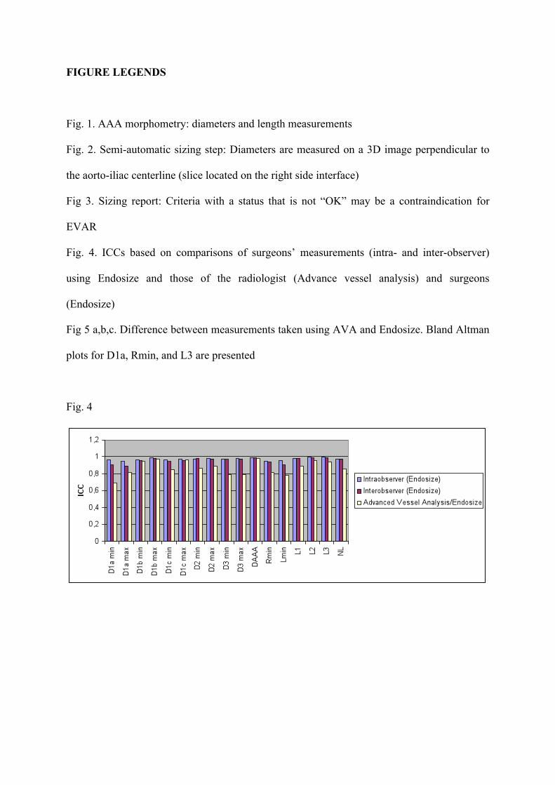
FIGURE LEGENDS
Fig. 1. AAA morphometry: diameters and length measurements
Fig. 2. Semi-automatic sizing step: Diameters are measured on a 3D image perpendicular to
the aorto-iliac centerline (slice located on the right side interface)
Fig 3. Sizing report: Criteria with a status that is not “OK” may be a contraindication for
EVAR
Fig. 4. ICCs based on comparisons of surgeons’ measurements (intra- and inter-observer)
using Endosize and those of the radiologist (Advance vessel analysis) and surgeons
(Endosize)
Fig 5 a,b,c. Difference between measurements taken using AVA and Endosize. Bland Altman
plots for D1a, Rmin, and L3 are presented
Fig. 4
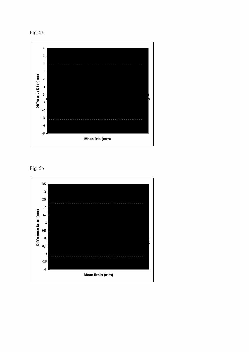
Fig. 5a
Fig. 5b
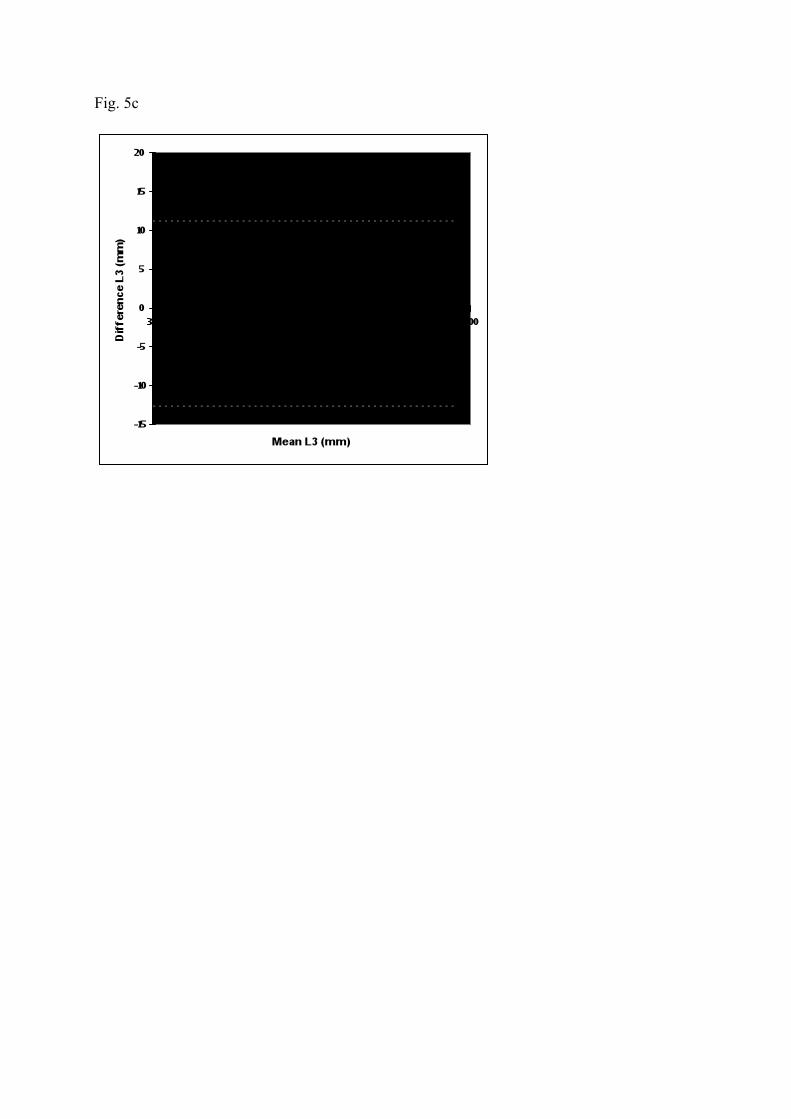
Fig. 5c
TABLES Table 1. Patient selection criteria for EVAR and morphological characteristics of AAA not
selected for EVAR
Inclusion Clinical Criteria
Age>80 years
Non revascularizable coronary arterial disease
Cardiac insufficiency with clinical signs
Non-operable aortic stenosis
Left ventricular ejection fraction<40%
Severe chronic respiratory insufficiency
Plasmatic creatinine>200uL/L
Hostile abdomen
Exclusion Morphological Criteria
Neck length !15mm
Circular thrombus or calcifications at stent graft
Fixation sites
Bony sharp neck !60°
Iliac tortuosity !80°
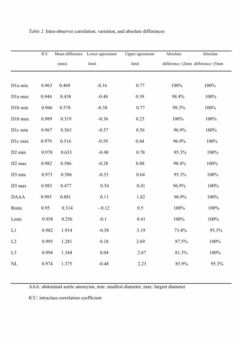
Table 2. Intra-observer correlation, variation, and absolute differences
ICC Mean difference Lower agreement Upper agreement Absolute Absolute
(mm) limit limit difference !2mm difference !5mm
D1a min 0.963 0.469 -0.16 0.77 100% 100%
D1a max 0.944 0.438 -0.48 0.39 98.4% 100%
D1b min 0.966 0.578 -0.38 0.77 98.3% 100%
D1b max 0.989 0.319 -0.36 0.23 100% 100%
D1c min 0.967 0.563 -0.57 0.56 96.9% 100%
D1c max 0.979 0.516 -0.59 0.44 96.9% 100%
D2 min 0.978 0.633 -0.48 0.78 95.3% 100%
D2 max 0.982 0.586 -0.28 0.88 98.4% 100%
D3 min 0.973 0.586 -0.53 0.64 95.3% 100%
D3 max 0.983 0.477 0.54 0.41 96.9% 100%
DAAA 0.993 0.881 0.11 1.82 96.9% 100%
Rmin 0.95 0.314 - 0.12 0.5 100% 100%
Lmin 0.958 0.256 -0.1 0.41 100% 100%
L1 0.982 1.914 -0.58 3.19 73.4% 95.3%
L2 0.995 1.281 0.18 2.69 87.5% 100%
L3 0.994 1.344 0.04 2.67 81.3% 100%
NL 0.974 1.375 -0.48 2.23 85.9% 95.3%
AAA: abdominal aortic aneurysm, min: smallest diameter, max: largest diameter
ICC: intraclass correlation coefficient
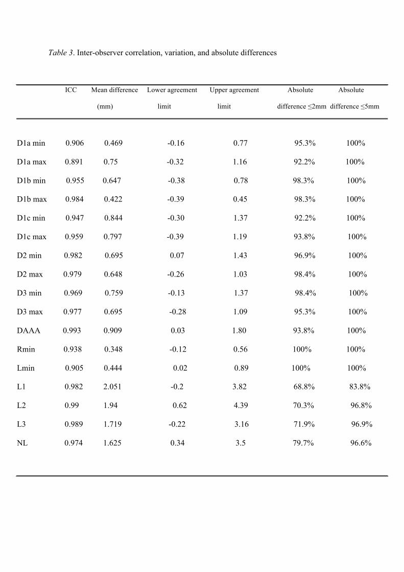
Table 3. Inter-observer correlation, variation, and absolute differences
ICC Mean difference Lower agreement Upper agreement Absolute Absolute
(mm) limit limit difference !2mm difference !5mm
D1a min 0.906 0.469 -0.16 0.77 95.3% 100%
D1a max 0.891 0.75 -0.32 1.16 92.2% 100%
D1b min 0.955 0.647 -0.38 0.78 98.3% 100%
D1b max 0.984 0.422 -0.39 0.45 98.3% 100%
D1c min 0.947 0.844 -0.30 1.37 92.2% 100%
D1c max 0.959 0.797 -0.39 1.19 93.8% 100%
D2 min 0.982 0.695 0.07 1.43 96.9% 100%
D2 max 0.979 0.648 -0.26 1.03 98.4% 100%
D3 min 0.969 0.759 -0.13 1.37 98.4% 100%
D3 max 0.977 0.695 -0.28 1.09 95.3% 100%
DAAA 0.993 0.909 0.03 1.80 93.8% 100%
Rmin 0.938 0.348 -0.12 0.56 100% 100%
Lmin 0.905 0.444 0.02 0.89 100% 100%
L1 0.982 2.051 -0.2 3.82 68.8% 83.8%
L2 0.99 1.94 0.62 4.39 70.3% 96.8%
L3 0.989 1.719 -0.22 3.16 71.9% 96.9%
NL 0.974 1.625 0.34 3.5 79.7% 96.6%
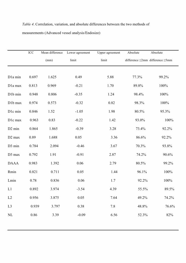
Table 4. Correlation, variation, and absolute differences between the two methods of
measurements (Advanced vessel analysis/Endosize)
ICC Mean difference Lower agreement Upper agreement Absolute Absolute
(mm) limit limit difference !2mm difference !5mm
D1a min 0.697 1.625 0.49 5.88 77.3% 99.2%
D1a max 0.813 0.969 -0.21 1.70 89.8% 100%
D1b min 0.948 0.806 -0.35 1.24 98.4% 100%
D1b max 0.974 0.573 -0.32 0.82 98.3% 100%
D1c min 0.846 1.52 -1.05 1.98 80.5% 95.3%
D1c max 0.963 0.83 -0.22 1.42 93.0% 100%
D2 min 0.864 1.865 -0.39 3.28 73.4% 92.2%
D2 max 0.89 1.688 0.05 3.36 86.6% 92.2%
D3 min 0.784 2.094 -0.46 3.67 70.3% 93.8%
D3 max 0.792 1.91 -0.91 2.87 74.2% 90.6%
DAAA 0.983 1.392 0.06 2.79 80.5% 99.2%
Rmin 0.821 0.711 0.05 1.44 96.1% 100%
Lmin 0.78 0.836 0.06 1.7 92.2% 100%
L1 0.892 3.974 -3.54 4.39 55.5% 89.5%
L2 0.956 3.875 0.05 7.64 49.2% 74.2%
L3 0.939 3.797 0.38 7.8 48.8% 76.6%
NL 0.86 3.39 -0.09 6.56 52.3% 82%
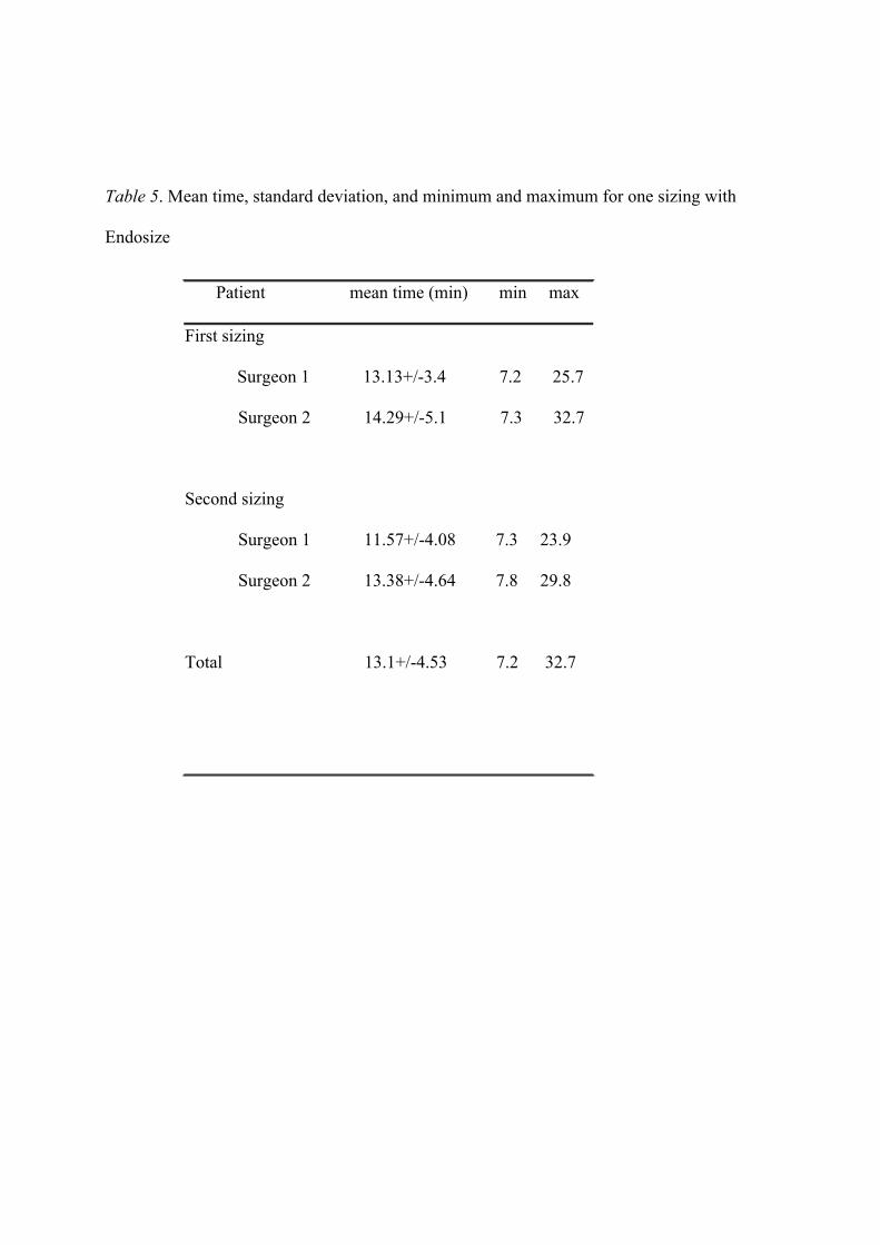
Table 5. Mean time, standard deviation, and minimum and maximum for one sizing with
Endosize
Patient mean time (min) min max
First sizing
Surgeon 1 13.13+/-3.4 7.2 25.7
Surgeon 2 14.29+/-5.1 7.3 32.7
Second sizing
Surgeon 1 11.57+/-4.08 7.3 23.9
Surgeon 2 13.38+/-4.64 7.8 29.8
Total 13.1+/-4.53 7.2 32.7
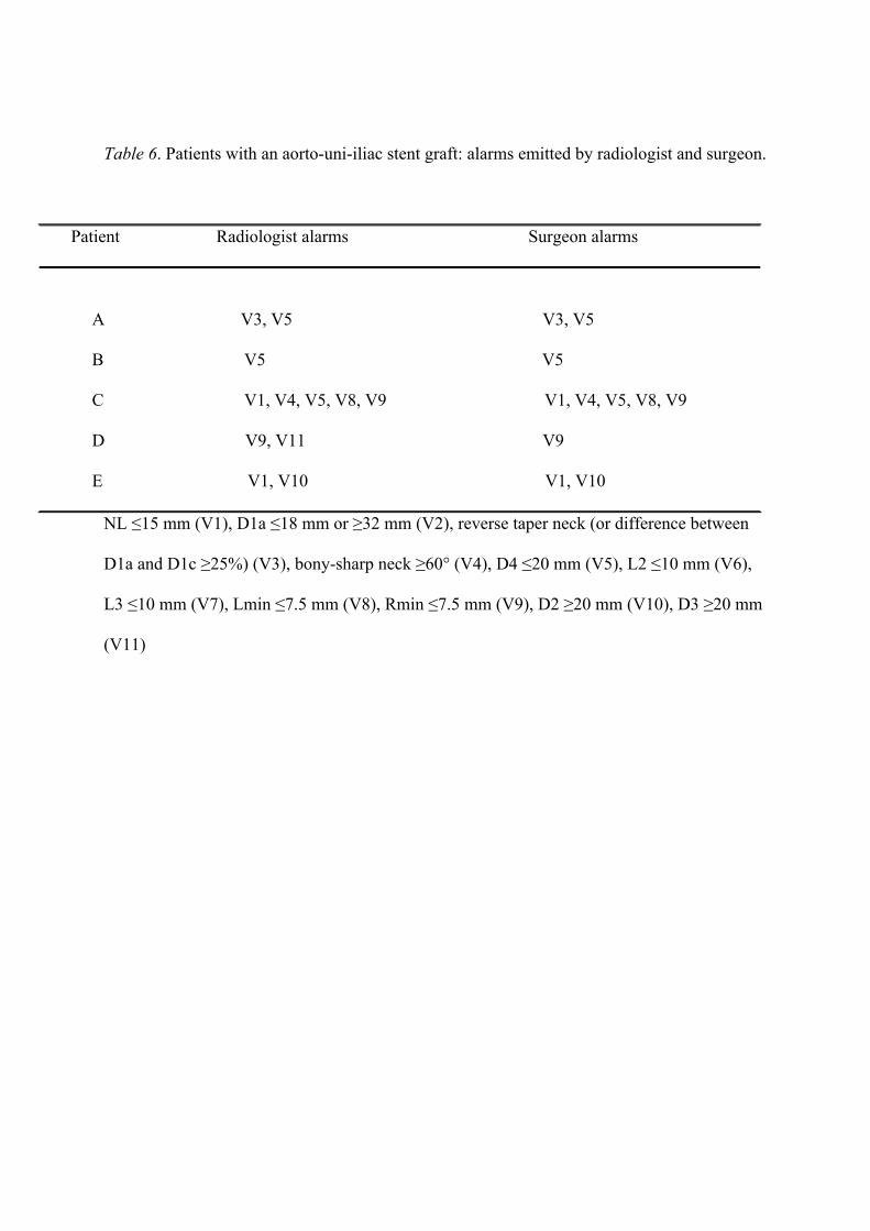
Table 6. Patients with an aorto-uni-iliac stent graft: alarms emitted by radiologist and surgeon.
Patient Radiologist alarms Surgeon alarms
A V3, V5 V3, V5
B V5 V5
C V1, V4, V5, V8, V9 V1, V4, V5, V8, V9
D V9, V11 V9
E V1, V10 V1, V10
NL !15 mm (V1), D1a !18 mm or "32 mm (V2), reverse taper neck (or difference between
D1a and D1c "25%) (V3), bony-sharp neck "60° (V4), D4 !20 mm (V5), L2 !10 mm (V6),
L3 !10 mm (V7), Lmin !7.5 mm (V8), Rmin !7.5 mm (V9), D2 "20 mm (V10), D3 "20 mm
(V11)